SOLITARY FIBROUS TUMOR OF THE PARANASAL SINUS...
4
Solitary fibrous tumours (SFTs) are unusual mesenchymal tumours that were first described as primary spindle-cell neoplasms of the pleura. These tumours have been described in many other locations, including the urogenital system, orbit, mediastinum, and upper respiratory tract. These tumours are generally benign in nature, however some of them can be locally invasive and have the potential to be malignant. Although about 12%-15% of them occur in the head and neck area, SFT of the nasal cavity and paranasal sinuses are extremely rare. We present a case of a solitary fibrous tumour arising from the right maxillary sinus in a 50 year old Chinese man. Keywords: Solitary fibrous tumours (SFTs), Mesenchymal tumours, Paranasal sinuses 1,2 Mansharan Kaur Chainchel Singh SOLITARY FIBROUS TUMOR OF THE PARANASAL SINUS PRESENTING AS AN ANTROCHOANAL POLYP Corresponding Author's Email: [email protected] 1 Institute of Pathology, Laboratory and Forensic Medicine (I-P Per ForM), Malaysia 2 Department of Radiology, Universiti Teknologi MARA (UiTM) Faculty of Medicine, Universiti Teknologi MARA (UiTM), Selangor Darul Ehsan, Malaysia ABSTRACT INTRODUCTION Solitary fibrous tumour (SFT) also known as benign fibrous mesothelioma and submesothelial fibroma was first described as primary spindle cell tumour of the pleura by Klemperer and Rabin (Alobid et al.,2003). This tumour usually arises in the pleura but can also arise in the head and neck, including the sinonasal cavity, orbit, nasopharynx and thyroid, however SFT of the paranasal sinuses are extremely rare and only 33 cases have been reported worldwide (Yang et al., 2013; Zielińska-Kaźmierska et al., 2015). Initially the tumour presents as an asymptomatic, well- circumscribed soft tissue mass, later nasal obstruction, epistaxis, rhinorrhoea and exophthalmos may occur. SFT is diagnosed by clinical examination, radiological imaging and immunohistochemistry. Histomorphologically, they are composed of spindle cells arranged in a “patternless” pattern, around a “haemangiopericytoma” like vascular network and are CD 34 positive. While SFT is usually benign, it may occasionally behave in an aggressive manner (Van Roggen and Hogendoorn, 2004; Zeitler et al. , 2017). Case History: A 50 year old Chinese man initially presented to a general practitioner with history of nasal obstruction and noisy breathing for one year associated with loss of weight and appetite. On examination, there was a mass in the post nasal space. A diagnosis of nasopharnygeal carcinoma was made and the patient was referred to the otorhinolarnygology clinic in University Malaya Medical Centre for further evaluation. At the clinic, on examination, there was a huge pale looking, non tender mass in the right nostril extending into the nasopharynx and oropharynx pushing the uvula forward. Thus the airway though patent was narrowed. An x-ray of the paranasal sinus showed a mass in the right maxillary sinus and a diagnosis of right antrochoanal polyp was made (refer Figure 1). Patient was subjected to a CT scan of the paranasal sinuses which revealed a large mass extending from the right maxillary sinus into the right side of the nasal cavity and posteriorly into the nasopharynx and oropharynx. This mass measured approximately 8.0 cm x 4.0 cm x 8.5 cm. There was also a dense focus within the lesion which raised the possibility of calcification (refer Figure 2 and 3). The surrounding bone did not show any evidence of erosion. During the operation, a huge right antrochoanal polyp measuring 6 cm x 11 cm in diameter was removed in two pieces and sent for histopathological examination. Malaysian Journal of Medical Research | Vol. 2 (2) APRIL 2018 | 91 doi: 10.31674/mjmr.2018.v02i02.014
-
Upload
nguyentruc -
Category
Documents
-
view
216 -
download
0
Transcript of SOLITARY FIBROUS TUMOR OF THE PARANASAL SINUS...

Solitary fibrous tumours (SFTs) are unusual mesenchymal tumours that were first described as primary spindle-cell neoplasms of the pleura. These tumours have been described in many other locations, including the urogenital system, orbit, mediastinum, and upper respiratory tract. These tumours are generally benign in nature, however some of them can be locally invasive and have the potential to be malignant. Although about 12%-15% of them occur in the head and neck area, SFT of the nasal cavity and paranasal sinuses are extremely rare. We present a case of a solitary fibrous tumour arising from the right maxillary sinus in a 50 year old Chinese man.
Keywords: Solitary fibrous tumours (SFTs), Mesenchymal tumours, Paranasal sinuses
1,2Mansharan Kaur Chainchel Singh
SOLITARY FIBROUS TUMOR OF THE PARANASAL SINUSPRESENTING AS AN ANTROCHOANAL POLYP
Corresponding Author's Email: [email protected]
1Institute of Pathology, Laboratory and Forensic Medicine (I-P Per ForM), Malaysia
2Department of Radiology, Universiti Teknologi MARA (UiTM) Faculty of Medicine, Universiti Teknologi MARA (UiTM), Selangor Darul Ehsan, Malaysia
ABSTRACT
INTRODUCTIONSolitary fibrous tumour (SFT) also known as benign fibrous mesothelioma and submesothelial fibroma was first described as primary spindle cell tumour of the pleura by Klemperer and Rabin (Alobid et al.,2003). This tumour usually arises in the pleura but can also arise in the head and neck, including the sinonasal cavity, orbit, nasopharynx and thyroid, however SFT of the paranasal sinuses are extremely rare and only 33 cases have been reported worldwide (Yang et al., 2013; Zielińska-Kaźmierska et al., 2015).
Initially the tumour presents as an asymptomatic, well-circumscribed soft tissue mass, later nasal obstruction, epistaxis, rhinorrhoea and exophthalmos may occur.
SFT is diagnosed by clinical examination, radiological imaging and immunohistochemistry. Histomorphologically, they are composed of spindle cells arranged in a “patternless” pattern, around a “haemangiopericytoma” like vascular network and are CD 34 positive. While SFT is usually benign, it may occasionally behave in an aggressive manner (Van Roggen and Hogendoorn, 2004; Zeitler et al., 2017).
Case History:
A 50 year old Chinese man initially presented to a general practitioner with history of nasal obstruction
and noisy breathing for one year associated with loss of weight and appetite. On examination, there was a mass in the post nasal space. A diagnosis of nasopharnygeal carcinoma was made and the patient was referred to the otorhinolarnygology clinic in University Malaya Medical Centre for further evaluation.
At the clinic, on examination, there was a huge pale looking, non tender mass in the right nostril extending into the nasopharynx and oropharynx pushing the uvula forward. Thus the airway though patent was narrowed.
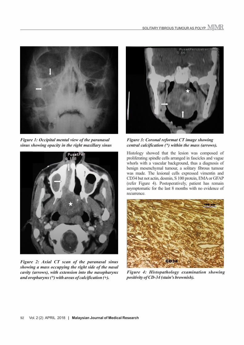
An x-ray of the paranasal sinus showed a mass in the right maxillary sinus and a diagnosis of right antrochoanal polyp was made (refer Figure 1). Patient was subjected to a CT scan of the paranasal sinuses which revealed a large mass extending from the right maxillary sinus into the right side of the nasal cavity and posteriorly into the nasopharynx and oropharynx. This mass measured approximately 8.0 cm x 4.0 cm x 8.5 cm. There was also a dense focus within the lesion which raised the possibility of calcification (refer Figure 2 and 3). The surrounding bone did not show any evidence of erosion. During the operation, a huge right antrochoanal polyp measuring 6 cm x 11 cm in diameter was removed in two pieces and sent for histopathological examination.
Malaysian Journal of Medical Research | Vol. 2 (2) APRIL 2018 | 91
doi: 10.31674/mjmr.2018.v02i02.014
Vol. 2 (2) APRIL 2018 | Malaysian Journal of Medical Research92
Figure 1: Occipital mental view of the paranasal sinus showing opacity in the right maxillary sinus
Figure 2: Axial CT scan of the paranasal sinus showing a mass occupying the right side of the nasal cavity (arrows), with extension into the nasopharynx and oropharynx (*) with areas of calcification (+).
Figure 3: Coronal reformat CT image showing central calcification (*) within the mass (arrows).
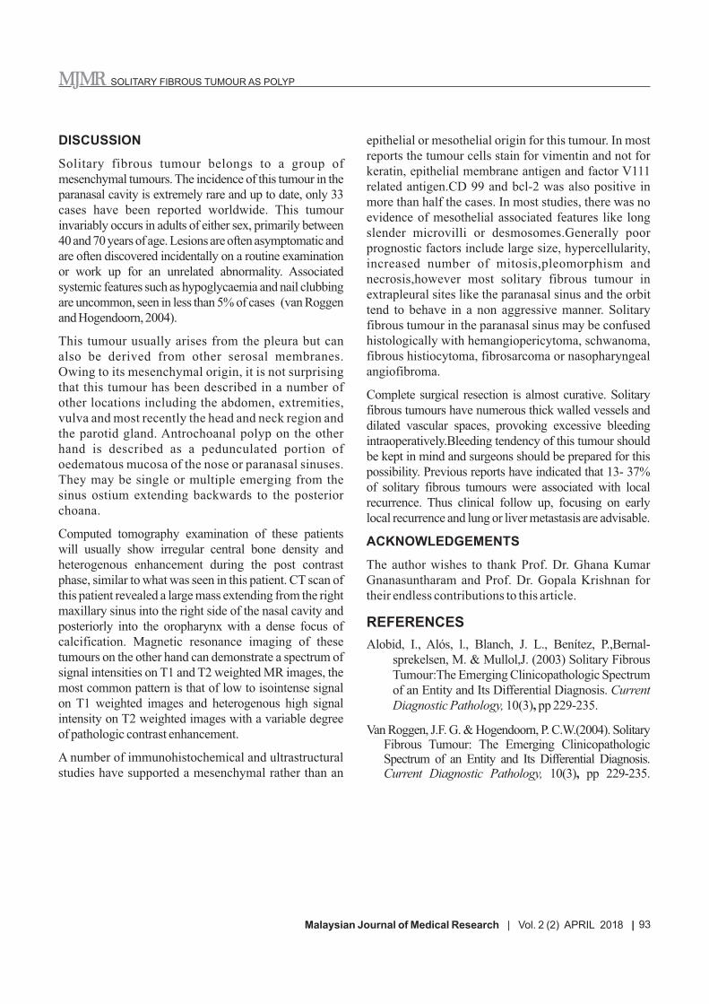
Histology showed that the lesion was composed of proliferating spindle cells arranged in fascicles and vague whorls with a vascular background, thus a diagnosis of benign mesenchymal tumour, a solitary fibrous tumour was made. The lesional cells expressed vimentin and CD34 but not actin, desmin, S 100 protein, EMA or GFAP (refer Figure 4). Postoperatively, patient has remain asymptomatic for the last 8 months with no evidence of recurrence.
Figure 4: Histopathology examination showing positivity of CD-34 (stain's brownish).
SOLITARY FIBROUS TUMOUR AS POLYP
DISCUSSIONSolitary fibrous tumour belongs to a group of mesenchymal tumours. The incidence of this tumour in the paranasal cavity is extremely rare and up to date, only 33 cases have been reported worldwide. This tumour invariably occurs in adults of either sex, primarily between 40 and 70 years of age. Lesions are often asymptomatic and are often discovered incidentally on a routine examination or work up for an unrelated abnormality. Associated systemic features such as hypoglycaemia and nail clubbing are uncommon, seen in less than 5% of cases (van Roggen and Hogendoorn, 2004).
This tumour usually arises from the pleura but can also be derived from other serosal membranes. Owing to its mesenchymal origin, it is not surprising that this tumour has been described in a number of other locations including the abdomen, extremities, vulva and most recently the head and neck region and the parotid gland. Antrochoanal polyp on the other hand is described as a pedunculated portion of oedematous mucosa of the nose or paranasal sinuses. They may be single or multiple emerging from the sinus ostium extending backwards to the posterior choana.
Computed tomography examination of these patients will usually show irregular central bone density and heterogenous enhancement during the post contrast phase, similar to what was seen in this patient. CT scan of this patient revealed a large mass extending from the right maxillary sinus into the right side of the nasal cavity and posteriorly into the oropharynx with a dense focus of calcification. Magnetic resonance imaging of these tumours on the other hand can demonstrate a spectrum of signal intensities on T1 and T2 weighted MR images, the most common pattern is that of low to isointense signal on T1 weighted images and heterogenous high signal intensity on T2 weighted images with a variable degree of pathologic contrast enhancement.
A number of immunohistochemical and ultrastructural studies have supported a mesenchymal rather than an
epithelial or mesothelial origin for this tumour. In most reports the tumour cells stain for vimentin and not for keratin, epithelial membrane antigen and factor V111 related antigen.CD 99 and bcl-2 was also positive in more than half the cases. In most studies, there was no evidence of mesothelial associated features like long slender microvilli or desmosomes.Generally poor prognostic factors include large size, hypercellularity, increased number of mitosis,pleomorphism and necrosis,however most solitary fibrous tumour in extrapleural sites like the paranasal sinus and the orbit tend to behave in a non aggressive manner. Solitary fibrous tumour in the paranasal sinus may be confused histologically with hemangiopericytoma, schwanoma, fibrous histiocytoma, fibrosarcoma or nasopharyngeal angiofibroma.
Complete surgical resection is almost curative. Solitary fibrous tumours have numerous thick walled vessels and dilated vascular spaces, provoking excessive bleeding intraoperatively.Bleeding tendency of this tumour should be kept in mind and surgeons should be prepared for this possibility. Previous reports have indicated that 13- 37% of solitary fibrous tumours were associated with local recurrence. Thus clinical follow up, focusing on early local recurrence and lung or liver metastasis are advisable.
ACKNOWLEDGEMENTSThe author wishes to thank Prof. Dr. Ghana Kumar Gnanasuntharam and Prof. Dr. Gopala Krishnan for their endless contributions to this article.
REFERENCESAlobid, I., Alós, l., Blanch, J. L., Benítez, P.,Bernal-
sprekelsen, M. & Mullol,J. (2003) Solitary Fibrous Tumour:The Emerging Clinicopathologic Spectrum of an Entity and Its Differential Diagnosis. Current Diagnostic Pathology, 10(3), pp 229-235.
Van Roggen, J.F. G. & Hogendoorn, P. C.W.(2004). Solitary Fibrous Tumour: The Emerging Clinicopathologic Spectrum of an Entity and Its Differential Diagnosis. Current Diagnostic Pathology, 10(3), pp 229-235.
SOLITARY FIBROUS TUMOUR AS POLYP
Malaysian Journal of Medical Research | Vol. 2 (2) APRIL 2018 | 93

Yang, B.T., Song, Z.L., Wang, Y.Z., Dong, J.Y. & Wang, Z. C.(2013).Solitary Fibrous Tumor of the Sinonasal Cavity: CT and MR Imaging Findings. American Journal of Neuroradiology, 34(6), pp 1248-1251.
Zeitler, D. M., Kanowitz, S. J.& Har-El, G. (2007).Malignant Solitary Fibrous Tumor of the Nasal Cavity. Skull Base,
17(4), pp 239-246.
Zielińska-Kaźmierska, B., Grodecka, J. & SzyszkowskI, A. (2015).Solitary Fibrous Tumor of the Nasal Cavity and Paranasal Sinuses: A case Report. Journal of Oral Biology and Craniofacial Research, 5(2), pp 112-116.
SOLITARY FIBROUS TUMOUR AS POLYP
94 Vol. 2 (2) APRIL 2018 I Malaysian Journal of Medical Research