SOFT TISSUE CALCIFICATION AND OSSIFICATION
71
SOFT TISSUE CALCIFICATION AND OSSIFICATION Dr Sangeeta Malik Professor Department of Oral Medicine and Radiology Subharti Dental College SVSU, Meerut
Transcript of SOFT TISSUE CALCIFICATION AND OSSIFICATION

SOFT TISSUE CALCIFICATION AND OSSIFICATION
Dr Sangeeta Malik
Professor
Department of Oral Medicine and Radiology
Subharti Dental College
SVSU, Meerut

HETROTROPHIC CALCIFICATION
Deposition of calcium salts in unorganized fashion in soft tissues.
Heterotopic calcifications may be divided into three
categories:
• Dystrophic calcification
• Idiopathic calcification
• Metastatic calcification
HETROTROPHIC OSSIFICATION
Minerals deposited in bone as organized, well formed bone.
Posttraumatic ossification
Progressive myositis ossificans
Ankylosing spondylitis
Dystrophic calcification
• Calcification that occurs in degenerating, diseased and dead tissue despite normal serum calcium and phosphate levels.

• Dystrophic calcification
• Long standing cysts
• Calcified lymph nodes
• Calcifications in tonsils
• Cysticercosis
• Arterial calcification
- Monckebergs medial calcinosis (arteriosclerosis)
- Calcified atherosclerotic plaque

General dystrophic calcifications of the oral lesions
Clinical features-
• Site-gingiva, tongue, lymph nodes and cheek
• No signs or symptoms
• Occasional enlargement and ulceration of overlying soft tissues and
• calcium salts might be palpated

Radiographic features
• Barely perceptible, fine grains of radiopacities to……….larger ,irregular radiopaque particles which rarely exceed 0.5cm in diameter.
• Calcification- homogenous or may contain punctate areas.
• Outline- irregular or indistinct

Dystrophic calcification
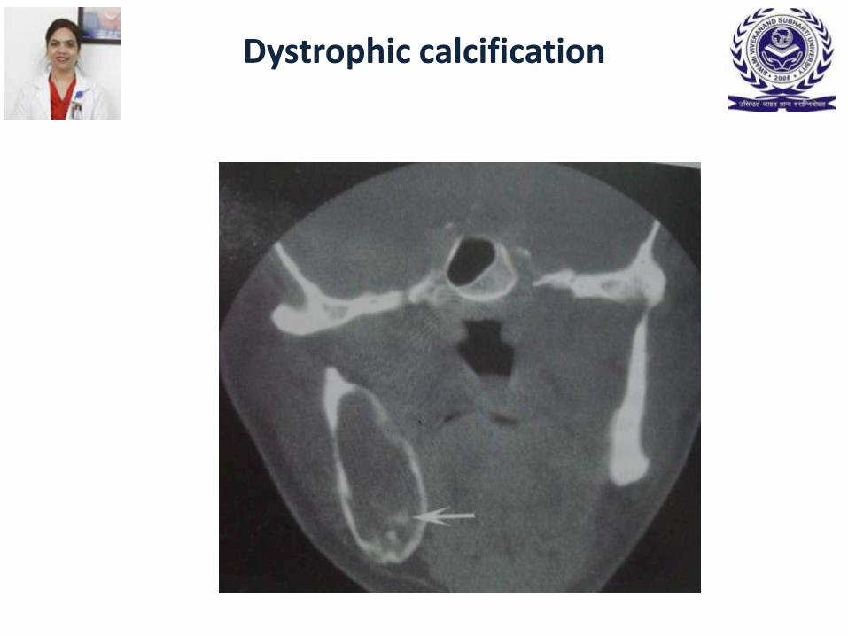
Dystrophic calcification

Dystrophic calcification
Calcified lymph nodes
Diseases causing calcified lymph nodes- • TB
• Sarcoidosis
• Cat-scratch disease
• Lymphoma previously treated with radiotherapy
• Fungal infections
• Metastates from distant calcifying neoplasm

Calcified lymph nodes
Clinical features-
• Mostly incidental findings
• Submandibular and cervical lymph nodes commonly involved
• Hard round and oblong masses

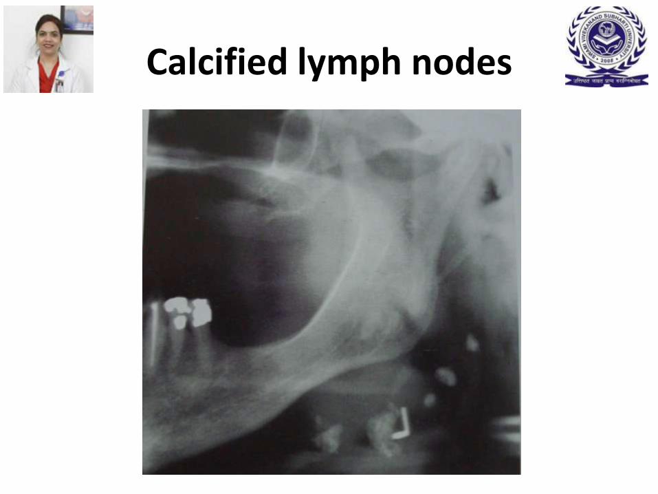
Calcified lymph nodes
Radiographic features
Location-
Submandibular region-at or below inferior border of mandible near
angle
Between posterior border of ramus and cervical spine
Single node or series of nodes may be affected-lymph node chaining

Calcified lymph nodes
Radiographic features…
• Periphery- Well –defined, irregular, lobulated
• Internal structure- vary in degree of radiopacity
• Differential diagnosis-Sialolith
Phlebolith
Management- No treatment required

Calcified lymph nodes

Calcified lymph nodes
Calcified lymph nodes

Dystrophic calcification in the tonsils
• Tonsillar calcification forms when repeated bouts of inflammation
enlarge the tonsillar crypts .
• Incomplete resolution of dead bacteria and pus serve as nidus for dystrophic calcification.

Dystrophic calcification in the tonsils
Clinical features-
• 20-68 yrs age group
• Hard , round ,white or yellow objects projecting from tonsillar crypts.
• Pain
• Swelling
• Fetor orisdysphagia
• Foreign body sensation on swallowing

Radiographic features-
• Location -Single or multiple radiopacities
-Overlap the midportion of the mandibular ramus
-in the region where the image of the dorsal surface of
the tongue crosses the ramus in the palatoglossal or
glossopharyngeal air spaces
• Periphery- Cluster of multiple ill-defined radiolucencies
• Internal structure- Slightly more radiopaque than cancellous bone
and approximately same as cortical bone
Dystrophic calcification in the tonsils

Dystrophic calcification in the tonsils
• Differential diagnosis Clinical Differential diagnosis- Calcified granulomatous disease, syphillis, mycosis or lymphoma Radiographic Differential diagnosis- Dense bone islands • Treatment- No treatment is required for most tonsillar calcifications. However, large calcifications with associated symptoms are removed surgically

Dystrophic calcification in the tonsils

Dystrophic calcification in the tonsils

CYSTICERCOSIS
• Eggs from taenia solium ingested
• Eggs digested in stomach
• Larval form-cystecercus cellulose hatches
• Penetrate the mucosa , enter the blood vessels and lymphatics and distributed in tissues all over the body
• Brain ,heart, muscle and skin, oral and peri-oral region

CYSTICERCOSIS
Clinical features
• GI- mild GI upset, nausea, vomitting
• Brain- seizure, headache, visual disturbances, acute obstructive hydrocephalus, Irritability and loss of consciousness
• Head and neck- palpable, soft fluctuant swellings resembling
mucocele
-multiple small nodules in masseter, suprahyoid
muscles, buccal mucosa, lip

CYSTICERCOSIS
Radiographic features
• Location- muscles of mastication,and facial expression,the suprahyoid muscle and the posterior cervical musculature
• Periphery- multiple well-defined ,elliptical radiopacities resembling grains of rice
• Internal structure- homogenous and radiopaque
CYSTICERCOSIS
• Differential diagnosis-
Sialolith
• Management-
Prevention-Antihelminthic
Once calcified -harmless

Arterial calcifications
• Arteriosclerosis
• Calcified atherosclerotic plaque

Arteriosclerosis
• Fragmentation, degeneration and eventual loss of elastic fibres
followed by the deposition of calcium within the medial coat of the vessel.

Arteriosclerosis
• Clinical features-cutaneous gangrene
- peripheral vascular disease
- myositis due to vascular
insuficiency
• Sturge weber syndrome-intracranial calcifications

Arteriosclerosis
Radiographic features
• Location- facial artery ,carotid artery
• Periphery and shape -pipe-stem or tram –track appearance

Arteriosclerosis

Calcified Atherosclerotic plaque
Location- Develops at arterial bifurcations -greater cornu of the hyoid bone and the cervical vertebrae C3, C4, or the intervertebral space between them
Periphery and shape-multiple - irregular in shape - sharply defined -vertical linear distribution.
Internal structure-heterogeneous radiopacity

Calcified Atherosclerotic plaque
• Differential Diagnosis- Calcified triticeous cartilage
• Management- should refer to their physician for cerebrovascular
and cardiovascular workup.
IDIOPATHIC CALCIFICATIONS
Idiopathic calcification (or calcinosis) results from deposition of
calcium in normal tissue despite normal serum calcium and
phosphate levels.

Sialolith
Clinical Features-
• Age-middle and later years
• Sex-male
• Gland –submandibular gland ( swelling in floor of mouth)

Sialolith
Radiographic features
• Location-submandibular gland-83-94%
parotid gland-4-10%
sublingual gland-1-7%
Submandibular-50% in distal portion of whartons duct
20%-proximal
30%-in the gland
Periphery and shape- cylindric
Internal structure- homogenously radiopaque or multiple layers of
calcification

• Applied Radiology-
- IOPA
- OCCLUSAL
- Lateral oblique and OPG
• DD-lymph nodes-asymptomatic
• Management-
- milked out by bimanual palpation
- Piezoelectric extracorporeal shock wave
lithotripsy
-Surgical removal
Sialolith

Sialolith

Sialolith

Sialolith

Phleboliths
C/F- swollen, throbbing, discoloured -diascopy positive R/F-location- common in hemangiomas Periphery and shape- In cross-section round or oval-upto6mm in diameter viewed from side-straight or slightly curved sausage Internal structure- homogenous or bulls eye DD- sialolith

Phleboliths

Laryngeal Cartilage Calcifications
The small, paired triticeous cartilages are found within the lateral thyrohyoid ligaments. Both the thyroid and triticeous cartilages consist of hyaline cartilage, which has a tendency
to calcify or ossify with advancing age.
Clinical Features-incidental radiographic finding with no clinical features.
Radiographic Features Location. within the pharyngeal airspace inferior to the greater cornu of the hyoid bone and adjacent to the superior border of C4.

Calcified triticeous cartilage
Radiographic Features cont..
• Periphery and shape. The word triticeous means "grain of wheat," and the cartilage measures 7 to 9mm inlength and 2 to 4 mm in width.
The periphery is well-defined and smooth.
• Internal structure.‘homogeneous radiopacity
Differential Diagnosis
Calcified atheromatous plaque in the carotid bifurcation
- Solitary nature and extremely uniform size and shape of calcified triticeous cartilage should be discriminatory.

Rhinolith/Antrolith

Rhinolith/Antrolith
• Pain
• Congestion
• Ulceration
• Purulent rhinorrhea
• Sinusitis
• Headache
• Epistaxis
• Anosmia
• Fetor
• Fever
Clinical features-

Rhinolith/Antrolith
Radiographic Features
• Location. Rhinoliths develop in the nose whereas antroliths develop in the
antrum of the maxillary sinus
• Periphery and shape. These stones have a variety of shapes and sizes.
• Internal structure. They may present as homogeneous or heterogeneous radiopacities, depending on the nature of the nidus and sometimes have laminations. Occasionally the density will exceed the surrounding bone.

Metastatic calcifications
Metastatic calcification results when minerals precipitate into normal tissue as a result of higher than normal serum levels of calcium
• Hyperparathyroidism
• Hypercalcemia of malignancy

Hyperparathyroidism
• Pathologic calcifications in soft tissues have a punctate or nodular appearance and occur in the kidneys and joints.
Heterotopic Bone .
Ossification of Stylohyoid ligament
• C/F- can be palpated over the tonsil as a hard ,pointed structure.
• Vague ,nagging to intense pain in the pharynx on swallowing, turning the head, or opening the mouth,especially on yawning. • Eagle’s syndrome
• Stylohyoid syndrome
• Otalgia, tinnitus, temporal headache and vertigo or transient syncope
The term heterotopic indicates that bone has formed in an abnormal (extraskeletal) location

Ossification of Stylohyoid ligament
Radiographic Features-
• Incidental feature on panaromic
• Location-mastoid process-posterio-inferior aspect of the ramus towards the hyoid bone.
• Shape-long, tapering,thin,radiopaque process -thicker at it’s base projects downwards and forwards -0.5-2.5cm in length. • Internal structure-homogenously radiopaque

Ossification of Stylohyoid ligament

Ossification of Stylohyoid ligament

Osteoma Cutis
• Rare soft tissue ossification in the skin.
• C/F- face, tongue
- occasionally appears yellowish white
otherwise no visible change
-multiple miliary osteoma cutis
• R/F- location-cheek and lip region
-may get superimposed over tooth
root or alveolar process-dense bone
area
-periphery and shape-smoothly outlined, radiopaque,washer
shaped (0-1-5cm in size)

Osteoma Cutis

Myositis Ossificans
• Fibrous tissue and heterotopic bone form within the interstitial tissue of the muscle and associated tendons and ligaments.
• Secondary destruction and atrophy of the muscle occur as this fibrous tissue and bone interdigitate and separate the muscle fibres.
• There are two principal forms- localized and progressive

LOCALIZED (TRAUMATIC) MYOSITIS OSSIFICANS
Etiology
• Acute or chronic trauma
• Heavy muscular strain caused by certain occupations and sports.
• Muscle injury from multiple injections (occasionally from dental anesthetic)
The injury leads to considerable hemorrhage into the muscle or associated tendons or fascia.
This hemorrhage organizes and undergoes progressive scarring.
During the healing process, heterotopic bone and in some cases cartilage is formed.

LOCALIZED (TRAUMATIC) MYOSITIS OSSIFICANS
Clinical Features-
• Age- Can occur in any age
• Sex- often in young men who engage in vigorous activity.
• The site -swollen, tender, and painful and the overlying skin may be red and inflamed
• If a muscle of mastication is involved, opening the jaws may be difficult.
• After about 2 or 3 weeks, the area of ossification becomes apparent in the tissues; a firm, intramuscular mass can be palpated.
• The localized lesion may enlarge slowly, but eventually it stops growing.
• The lesion may appear fixed, or it may be freely movable on palpation.

LOCALIZED (TRAUMATIC) MYOSITIS OSSIFICANS
Radiographic Features Location. The most commonly involved muscles of the head and neck are the masseter and sternocleidomastoid. A radiolucent band can be seen between the area of ossification and adjacent bone, and the heterotopic bone may lie along the long axis of the muscle Periphery and shape. The periphery is more radiopaque than the internal structure. Variation in shape from irregular, oval to linear streaks (pseudo trabeculae) running in the same direction as the normal muscle fibers.

LOCALIZED (TRAUMATIC) MYOSITIS OSSIFICANS
Radiographic Features... Internal structure
The internal structure varies with time.
Within the third or fourth week after injury- faintly homogeneous radiopacity.
This organizes further, and by 2 months- a delicate lacy or feathery radiopaque internal structure
These changes indicate the formation of bone; however, this bone does not have a normal appearing
trabecular pattern.
Gradually the image becomes denser and better defined, maturing fully in about 5 to 6 months.
After this period the lesion may shrink.

LOCALIZED (TRAUMATIC) MYOSITIS OSSIFICANS
Differential Diagnosis
• Stylohyoid ligament and other soft tissue calcifications
• Bone-forming tumors such as osteogenic sarcoma-can form a linear bone pattern but the tumor is contiguous with the adjacent bone, and signs of bone destruction often are present.

LOCALIZED (TRAUMATIC) MYOSITIS OSSIFICANS
Management
• Rest and limitation of use are recommended to diminish the extent of the calcific deposit.
• Early surgical removal of the lesion usually stimulates rapid (within 1 month) and extensive recurrence (from origin to insertion of the affected muscle).
• Recurrence is not likely if removal of the involved area of muscle is postponed until the process has become stationary

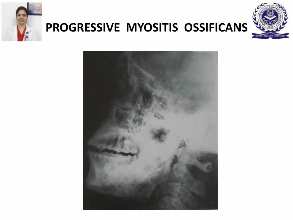
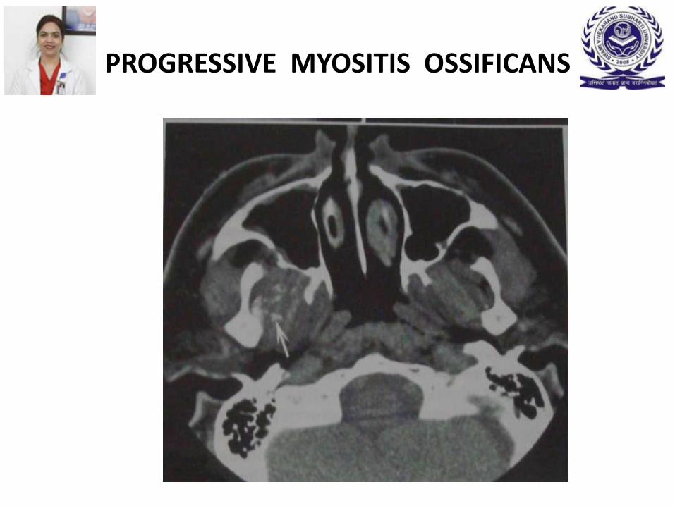
PROGRESSIVE MYOSITIS OSSIFICANS
• Progressive myositis ossificans is a rare disease
• This condition may be inherited or may be a spontaneous mutation affecting the mesenchyma.
Clinical features
• Affects children before 6years of age, occasionally as early as infancy.
• More common in males.

PROGRESSIVE MYOSITIS OSSIFICANS
• Starts in the muscles of the neck and upper back and moves to the extremities.
• Soft tissue swelling that is tender and painful and may show redness and heat
• The acute symptoms subside, and a firm mass remains in the tissues.
• In some cases the spread of ossification is limited; in others it becomes extensive, affecting almost all the large muscles of the body.
• This condition may affect any of the striated muscles, including the heart and diaphragm.
• Stiffness and limitation of motion of the neck, chest, back, and extremities (especially the shoulders) gradually increase.

PROGRESSIVE MYOSITIS OSSIFICANS
• Advanced stages of the disease result in the "petrified man" condition.
• During the third decade the process may spontaneously arrest;
• However, most patients die during the third or fourth decade.
• Premature death usually results from respiratory embarrassment or from inanition through the involvement of the muscles of mastication.

PROGRESSIVE MYOSITIS OSSIFICANS
Radiographic Features • The heterotopic bone more commonly is oriented along the long axis of the
involved muscle
Differential Diagnosis • Rheumatoid arthritis -may be difficult to distinguish between the two
• Calcinosis-the deposits of amorphous calcium salts frequently resorb, but
in progressive myositis ossificans, the bone never disappears.
Management
• No effective treatment exists for progressive myositis ossificans.

PROGRESSIVE MYOSITIS OSSIFICANS
PROGRESSIVE MYOSITIS OSSIFICANS
PROGRESSIVE MYOSITIS OSSIFICANS

Conclusion
• Soft tissue opacities are fairly common, present on about 4% of panoramic radiographs.
• In most cases the goal is to identify the calcification correctly to determine
whether treatment or further investigation is required.
• Some soft tissue calcifications require no intervention or long-term surveillance, whereas others may be life-threatening and the underlying cause requires treatment.
• When the soft tissue calcification is adjacent to bone, it sometimes is difficult to determine whether the calcification is within bone or soft tissue.
• In such cases another radiographic view at right angles is useful.


![Transcriptional Network Controlling Endochondral Ossification · branous ossification and endochondral ossification.[1] During intramembranous ossification, osteoblasts produce type](https://static.fdocuments.in/doc/165x107/5e8cf0c24763783dcf0d78ef/transcriptional-network-controlling-endochondral-ossification-branous-ossification.jpg)