Smartphone-based Fundus Photography for Screening ... - Ophthalmology … · Smartphone-based...
14
Smartphone-based Fundus Photography for Screening of Plus-disease Retinopathy of Prematurity October 25, 2018
Transcript of Smartphone-based Fundus Photography for Screening ... - Ophthalmology … · Smartphone-based...

Smartphone-based Fundus Photography for Screening of Plus-disease Retinopathy of
PrematurityOctober 25, 2018

FINANCIAL DISCLOSURES:
T.N. Kim, T. Margolis, D.A. Fletcher are co-inventors on US patent application 20160296112, “Retinal CellScope Apparatus”
T.N. Kim, Y.M. Paulus are co-inventors on a US provisional patent through the University of Michigan, “RetinaScope Apparatus”
01

MY ROLE IN THIS RESEARCH:
• Conception and design of the project
• Successful application as PI of Knights Templar Eye Foundation Career-Starter Research Grant
• Acquisition of data
• Analysis and interpretation of data
• Creation and critical review of the presentation
02
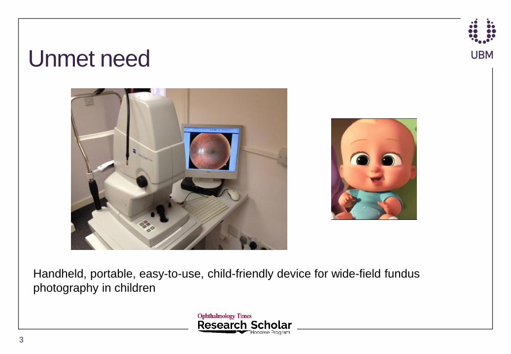
Unmet need
3
Handheld, portable, easy-to-use, child-friendly device for wide-field fundus
photography in children

Retinopathy of prematurity screening
• Despite advances in treatment strategies, ROP continues to be a leading cause of childhood blindness due to inadequate screening.
• Barriers to inadequate screening are numerous and include shortage of ophthalmologists trained and willing to screen for ROP and lack of patient access to trained ophthalmologists in resource-poor communities.
• Telemedicine has great potential to improve screening for treatment-warranted ROP; SUNDROP and eROP studies have clearly demonstrated the accuracy of telemedicine as a screening tool.
4

RetinaScope
5Kim et al. A smartphone-based tool for rapid, portable, and automated wide-field retinal imaging. TVST 2018

Fundus photography in children
6

Purpose
• To evaluate the feasibility and accuracy of screening for clinically significant posterior pole vascular dilation and tortuosity in premature infants (plus disease) using RetinaScope.
7
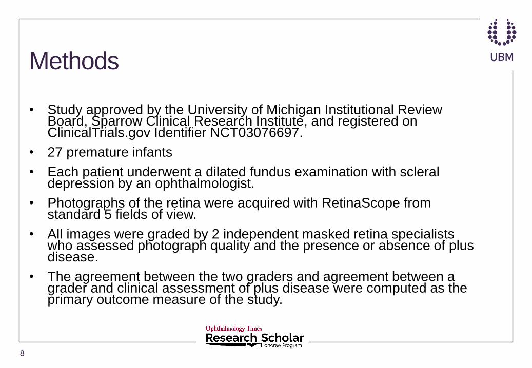
Methods
• Study approved by the University of Michigan Institutional Review Board, Sparrow Clinical Research Institute, and registered on ClinicalTrials.gov Identifier NCT03076697.
• 27 premature infants
• Each patient underwent a dilated fundus examination with scleral depression by an ophthalmologist.
• Photographs of the retina were acquired with RetinaScope from standard 5 fields of view.
• All images were graded by 2 independent masked retina specialists who assessed photograph quality and the presence or absence of plus disease.
• The agreement between the two graders and agreement between a grader and clinical assessment of plus disease were computed as the primary outcome measure of the study.
8
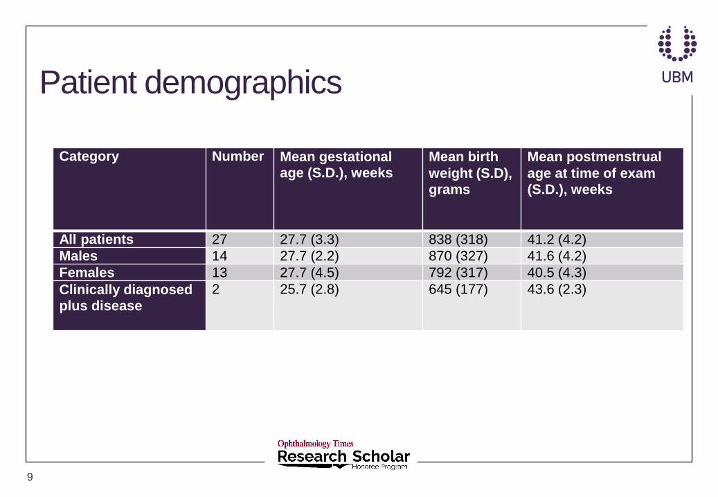
Patient demographics
Category Number Mean gestational age (S.D.), weeks
Mean birth
weight (S.D), grams
Mean postmenstrual
age at time of exam (S.D.), weeks
All patients 27 27.7 (3.3) 838 (318) 41.2 (4.2)
Males 14 27.7 (2.2) 870 (327) 41.6 (4.2)
Females 13 27.7 (4.5) 792 (317) 40.5 (4.3)
Clinically diagnosed plus disease
2 25.7 (2.8) 645 (177) 43.6 (2.3)
9

Representative images
10
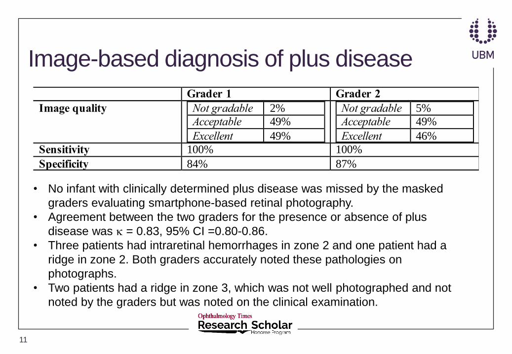
Image-based diagnosis of plus disease
11
Grader 1 Grader 2
Image quality Not gradable 2%
Acceptable 49%
Excellent 49%
Not gradable 5%
Acceptable 49%
Excellent 46%
Sensitivity 100% 100%
Specificity 84% 87%
• No infant with clinically determined plus disease was missed by the masked
graders evaluating smartphone-based retinal photography.
• Agreement between the two graders for the presence or absence of plus
disease was = 0.83, 95% CI =0.80-0.86.
• Three patients had intraretinal hemorrhages in zone 2 and one patient had a
ridge in zone 2. Both graders accurately noted these pathologies on
photographs.
• Two patients had a ridge in zone 3, which was not well photographed and not
noted by the graders but was noted on the clinical examination.
Discussion
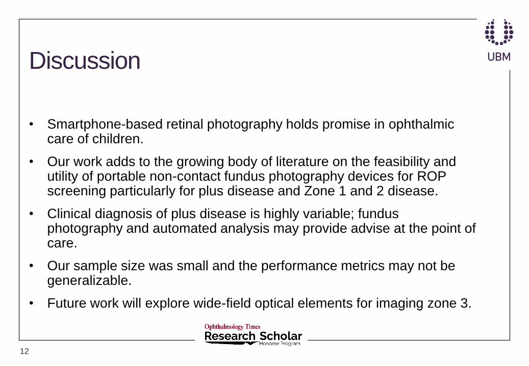
• Smartphone-based retinal photography holds promise in ophthalmic care of children.
• Our work adds to the growing body of literature on the feasibility and utility of portable non-contact fundus photography devices for ROP screening particularly for plus disease and Zone 1 and 2 disease.
• Clinical diagnosis of plus disease is highly variable; fundus photography and automated analysis may provide advise at the point of care.
• Our sample size was small and the performance metrics may not be generalizable.
• Future work will explore wide-field optical elements for imaging zone 3.
12

Acknowledgments
Yannis Paulus, MD, FACS (PI)
Tyson Kim, MD, PhD (co-PI)
Philip Lieu, MD
Vaidehi Dedania, MD
Cynthia Qian, MD
Cagri Besirli, MD, PhD
Todd Margolis, MD, PhD
Daniel Fletcher, PhD
13