
Small molecule activation at uranium coordination ... - b513755c.pdf · Small molecule activation...
16
Small molecule activation at uranium coordination complexes: control of reactivity via molecular architecture Ingrid Castro-Rodrı ´guez a and Karsten Meyer{* b Received (in Cambridge, UK) 27th September 2005, Accepted 15th November 2005 First published as an Advance Article on the web 24th January 2006 DOI: 10.1039/b513755c Electron-rich uranium coordination complexes display a pronounced reactivity toward small molecules. In this Feature article, the exciting chemistry of trivalent uranium ions coordinated to classic Werner-type ligand environments is reviewed. Three fundamentally important reactions of the [(( R ArO) 3 tacn)U]-system are presented that result in alkane coordination, CO/CO 2 activation, and nitrogen atom-transfer chemistry. Introduction From a synthetic chemist’s perspective, it is rather remarkable that after fifty years of synthetic organometallic actinide research, much is still unknown about the non-aqueous inorganic coordination chemistry of low-valent uranium. 1,2 Historically, this is not surprising considering that until fairly recently, synthetic access to uranium(III) coordination compounds was restricted due to lack of suitable starting materials. With the exception of homoleptic [( i-Pr ArO) 3 U] 3 and its derivatives, 4 it was not until Clark and Sattelberger’s synthesis of the solvated trivalent [UI 3 L 4 ] (L = THF and DME) and the solvent-free [((Me 3 Si) 2 N) 3 U] 5 complexes reported in a 1997 issue of Inorganic Synthesis that coordina- tion chemists finally had a synthetic protocol. 6–9 This provided a convenient and highly reproducible entry into the exciting world of trivalent uranium chemistry. In the literature of the following years, there is an increasing number of articles reporting classical inorganic coordination complexes of uranium, which employ traditional inorganic ligands such as a Department of Chemistry, University of California, Latimer Hall, Berkeley, California, 94720, USA. E-mail: [email protected]; Tel: +1 510 642 2516 b Department of Chemistry and Biochemistry, University of California @ San Diego, 9500 Gilman Drive MC 0358, La Jolla, California, 92093- 0358, USA { Present address: Friedrich-Alexander-University Nuremberg- Erlangen, Institute of Inorganic Chemistry, Egerlandstr. 1, 91058 Erlangen, Germany. E-mail: [email protected], Fax: +49 (0)9131 8527367, Tel: +49 (0)9131 8527360 Ingrid Castro-Rodrı ´guez was born in 1977 in Rı ´o Piedras, Puerto Rico. She received her Bachelor of Science degree in chemistry at the University of Puerto Rico (Rı´o Piedras Campus) where she worked with Professor Reginald Morales on synthetic methods to isolate snake venoms. In fall 2000, Ingrid started her studies in the Department of Chemistry and Biochemistry at the University of California, San Diego, where she joined Karsten’s laboratory in January 2001. Ingrid’s research focused on the activation and functionalization of small molecules employing low-valent coordinatively unsaturated uranium complexes in sterically encumbering ligand environments. For her research accom- plishments she received UCSD’s Teddy Traylor award and a Carl Storm fellowship from the Gordon Research Conference. After receiving her PhD in inorganic chemistry in summer 2005, Ingrid was awarded a Glenn T. Seaborg postdoctoral fellowship and is currently working under the guidance of Professor Kenneth Raymond at the Lawrence Berkeley National Laboratory and University of California, Berkeley. Karsten Meyer was born in 1968 in Herne, Germany. In May 1995, he received his diploma in chemistry from the Ruhr-University Bochum. He then began his graduate education under the direction of Professor Karl Wieghardt at the Max-Planck-Institute for Bioinorganic Chemistry in Mu ¨lheim/Ruhr. Karsten’s thesis work involved the synth- esis and spectroscopic investi- gation of transition metal nitrido complexes. He received his doctoral degree in January 1998, which was awarded with distinction. Supported by a DFG postdoctoral fellowship, he continued his education by joining the laboratory of Professor Christopher C. Cummins at the Massachusetts Institute of Technology (Cambridge, MA) where he developed his passion for uranium chemistry. In January 2001 he was appointed to the faculty of the University of California, San Diego and was named an Alfred P. Sloan Fellow in summer 2004. Karsten has recently accepted a chair of inorganic chemistry at the University of Erlangen-Nuremberg where he will continue to pursue his research interests involving redox- active d-block and actinide metal complexes. The Meyer group specializes in manipulating complex reactivity by employing their understanding of molecular and electronic structure interplay. Ingrid Castro-Rodrı ´guez Karsten Meyer FEATURE ARTICLE www.rsc.org/chemcomm | ChemComm This journal is ß The Royal Society of Chemistry 2006 Chem. Commun., 2006, 1353–1368 | 1353
Transcript of Small molecule activation at uranium coordination ... - b513755c.pdf · Small molecule activation...

Small molecule activation at uranium coordination complexes: controlof reactivity via molecular architecture
Ingrid Castro-Rodrıgueza and Karsten Meyer{*b
Received (in Cambridge, UK) 27th September 2005, Accepted 15th November 2005
First published as an Advance Article on the web 24th January 2006
DOI: 10.1039/b513755c
Electron-rich uranium coordination complexes display a pronounced reactivity toward small
molecules. In this Feature article, the exciting chemistry of trivalent uranium ions coordinated to
classic Werner-type ligand environments is reviewed. Three fundamentally important reactions of
the [((RArO)3tacn)U]-system are presented that result in alkane coordination, CO/CO2 activation,
and nitrogen atom-transfer chemistry.
Introduction
From a synthetic chemist’s perspective, it is rather remarkable
that after fifty years of synthetic organometallic actinide
research, much is still unknown about the non-aqueous
inorganic coordination chemistry of low-valent uranium.1,2
Historically, this is not surprising considering that until
fairly recently, synthetic access to uranium(III) coordination
compounds was restricted due to lack of suitable starting
materials. With the exception of homoleptic [(i-PrArO)3U]3 and
its derivatives,4 it was not until Clark and Sattelberger’s
synthesis of the solvated trivalent [UI3L4] (L = THF and
DME) and the solvent-free [((Me3Si)2N)3U]5 complexes
reported in a 1997 issue of Inorganic Synthesis that coordina-
tion chemists finally had a synthetic protocol.6–9 This provided
a convenient and highly reproducible entry into the exciting
world of trivalent uranium chemistry. In the literature of the
following years, there is an increasing number of articles
reporting classical inorganic coordination complexes of
uranium, which employ traditional inorganic ligands such as
aDepartment of Chemistry, University of California, Latimer Hall,Berkeley, California, 94720, USA. E-mail: [email protected];Tel: +1 510 642 2516bDepartment of Chemistry and Biochemistry, University of California @San Diego, 9500 Gilman Drive MC 0358, La Jolla, California, 92093-0358, USA{ Present address: Friedrich-Alexander-University Nuremberg-Erlangen, Institute of Inorganic Chemistry, Egerlandstr. 1, 91058Erlangen, Germany. E-mail: [email protected], Fax:+49 (0)9131 8527367, Tel: +49 (0)9131 8527360
Ingrid Castro-Rodrıguez wasborn in 1977 in Rıo Piedras,Puerto Rico. She received herBachelor of Science degree inchemistry at the University ofPuerto Rico (Rıo PiedrasCampus) where she workedwith Professor ReginaldMorales on synthetic methodsto isolate snake venoms. Infall 2000, Ingrid started herstudies in the Department ofChemistry and Biochemistry atthe University of California,San Diego, where she joinedKars ten ’ s laboratory in
January 2001. Ingrid’s research focused on the activation andfunctionalization of small molecules employing low-valentcoordinatively unsaturated uranium complexes in stericallyencumbering ligand environments. For her research accom-plishments she received UCSD’s Teddy Traylor award and aCarl Storm fellowship from the Gordon Research Conference.After receiving her PhD in inorganic chemistry in summer 2005,Ingrid was awarded a Glenn T. Seaborg postdoctoral fellowshipand is currently working under the guidance of Professor KennethRaymond at the Lawrence Berkeley National Laboratory andUniversity of California, Berkeley.
Karsten Meyer was born in1968 in Herne, Germany. InMay 1995, he received hisdiploma in chemistry fromthe Ruhr-University Bochum.He then began his graduateeducation under the directionof Professor Karl Wieghardtat the Max-Planck-Institutefor Bioinorganic Chemistry inMu lheim/Ruhr. Karsten’sthesis work involved the synth-esis and spectroscopic investi-gation of transition metalnitrido complexes. He receivedhis doctoral degree in January
1998, which was awarded with distinction. Supported by a DFGpostdoctoral fellowship, he continued his education by joining thelaboratory of Professor Christopher C. Cummins at theMassachusetts Institute of Technology (Cambridge, MA) wherehe developed his passion for uranium chemistry. In January 2001he was appointed to the faculty of the University of California,San Diego and was named an Alfred P. Sloan Fellow in summer2004. Karsten has recently accepted a chair of inorganicchemistry at the University of Erlangen-Nuremberg where hewill continue to pursue his research interests involving redox-active d-block and actinide metal complexes. The Meyer groupspecializes in manipulating complex reactivity by employing theirunderstanding of molecular and electronic structure interplay.
Ingrid Castro-Rodrıguez Karsten Meyer
FEATURE ARTICLE www.rsc.org/chemcomm | ChemComm
This journal is � The Royal Society of Chemistry 2006 Chem. Commun., 2006, 1353–1368 | 1353
the chelating tris(2-pyridyl) and tris(2-pyrazinyl)methylamine
(tpa and tpza),10,11 hydro-tris(pyrazolyl)borate (Tp2),12 tris-
(amido)amine (RN3N32),13 macrocyclic calix[4]tetrapyrrole
tetraanion14 (Chart 1), as well as monodentate bis-
(trimethylsilyl)amide and anilide (Ar(R)N2) systems.15
The electron-rich trivalent complexes [(N3N)U],13
[(calix[4]pyrrole)U(dme)][K(dme)],14 [((Me3Si)2N)3U]9 and
[(N(R)Ar)3U(THF)]16 exhibit an unusual and interesting
reactivity towards the activation of small inert molecules such
as the first example of dinitrogen activation by an actinide
compound. The reactivity of these new, more classical,
coordination complexes exceeds, in some regards, that of the
thoroughly explored archetypal [(Cp)3U]-system and its
derivatives.
The first example of dinitrogen fixation in actinide chemistry
was reported by Scott and co-workers in 1998.17 The trivalent
uranium complex [(N3N)U] (obtained via reduction of
[(N3N)UCl] and vacuum sublimation of the dinuclear pre-
cursor [{(N3N)U}2(m-Cl)], where N3N = N(CH2CH2NSi-
(t-Bu)Me2)3) reversibly binds dinitrogen side-on forming the
complex [{(N3N)U}2(m:g2,g2-N2)] (Fig. 1). This uranium
complex also reacts with trimethylsilyl azide and trimethylsilyl
diazomethane to form the corresponding imido and hydrazido
complexes.13,18
Shortly after the first diuranium dinitrogen complex was
reported, Cummins and co-workers published the first stable
heterodimetallic dinitrogen complex involving uranium.16 The
mononuclear trivalent [(Ar(t-Bu)N)3U(THF)]?3THF complex,
which by itself is unreactive towards N2, was prepared
employing the monodentate anilide ligand [Ar(t-Bu)N2].
This trivalent uranium complex reacts with dinitrogen in the
presence of the structurally very similar and exceedingly
reactive [(Ar(Ad)N)3Mo] to form the hetero-bimetallic bridged
dinitrogen complex [(Ar(Ad)N)3Mo](m:g1,g1-N2)[U(N(t-
Bu)Ar)3] with an end-on coordinated N2 ligand. It was
suggested that this complex forms via nucleophilic attack of
an intermediate molybdenum–dinitrogen complex
[(Ar(Ad)N)3Mo(N2)] on the uranium(III) fragment.16
Another interesting reaction involving this class of com-
pounds is the reaction of [(Ar(R)N)3U(I)] (R = C(CH3)3, Ar =
3,5-C6H3Me2) with toluene under strongly reducing condi-
tions, which leads to the formation of
[{(Ar(R)N)2U}2(m-C7H8)] (Fig. 2).19,20 Following conventional
electron-counting rules, the U centers in this dinuclear complex
are assigned the formal oxidation state of +II. As expected for
such a low-valent metal ion, this complex can be engaged in
remarkable four-electron redox reactions with, e.g., azoben-
zene and diphenyl disulfide compounds, yielding the
uranium(IV/IV) species [{(N(R)Ar)2U}2(m-NPh)2] and
[{(N(R)Ar)2U(SPh)}2(m-SPh)2], respectively.19 However, the
+II oxidation state has never been observed in U chemistry and
thus, it was suggested that in this complex strong d-back-
bonding reduces and activates the bridging arene. The uranium
center’s ability to form strong covalent d-bonds in the context
of arene binding therefore provides access to a previously
unknown divalent uranium synthon.
More recently, pyrrole-based polyanions have emerged as
another class of versatile ligands for f-block metals. These
supporting ligands have proven to substantially increase the
reactivity of their low-valent metal complexes. This is
especially evident for the chelation of trivalent uranium by
the calix[4]tetrapyrrole tetraanion, which leads to binding and,
most remarkably, cleavage of a dinitrogen ligand with
formation of the unique m-nitrido mixed-valent UV/UIV
complex [{K(dme)(calix[4]tetrapyrrole)U}2(m-NK)2][K(dme)4]
reported by Gambarotta in 2002.21 The highly reactive
uranium(III) complex was also shown to participate in solvent
deoxygenation and polysilanol depolymerization processes.21
The above-mentioned discoveries by Scott, Cummins,
Gambarotta and others have opened exciting new perspectives
on actinide chemistry and evoked trust in the great potential
offered by trivalent uranium complexes for unprecedented
chemical transformations. Despite this increased interest in new
modes of uranium reactivity, the coordination chemistry of
uranium complexes with coordinated macrocyclic polyamine
ligands remained largely unexplored. With the exception of
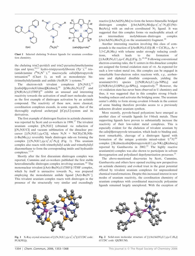
Chart 1 Selected chelating N-donor ligands for uranium coordina-
tion chemistry.
Fig. 1 X-Ray crystal structure of [{(N3N)U}2(m:g2,g2)] (CCDC code:
PUKPEQ).
Fig. 2 Solid-state molecular structure of [{(Ar(Ad)N)2U}2(m-C7H8)]
(CCDC code: QIZRUM).
1354 | Chem. Commun., 2006, 1353–1368 This journal is � The Royal Society of Chemistry 2006

our initial results in 200222 and an isolated report on a
uranium complex of a tris-amido derivatized triazacyclono-
nane in 2003 (Chart 2, right),23 systematic studies on the
molecular and electronic structure as well as the reactivity of
uranium complexes supported by coordinated macrocyclic
polyamine ligands24 were missing from the literature. This is in
contrast to the increasing amount of interesting data resulting
from the thoroughly investigated organometallic chemistry of
uranium with cyclopentadienyl ligands and their derivatives, as
well as the more recently developed amido chemistry of
uranium.
In our quest to identify and isolate uranium complexes with
enhanced reactivity relevant to binding, activation, and
functionalization of small molecules, we are currently inves-
tigating the coordination chemistry of uranium centers with
classic Werner-type polyamine chelators. Our research was
encouraged by the most recent exciting accomplishments in
uranium coordination chemistry as outlined above, as well as
by the well known benefits of macrocyclic chelators for
stabilization of reactive transition metal ions.
This review presents the synthesis of complexes of uranium
coordinated by polyamine macrocycle 1,4,7-tris(3,5-alkyl-2-
hydroxybenzylate)-1,4,7-triazacyclononane.25 Due to the small
ring size of the neutral triazacyclononane macrocycle (tacn),
this ligand by itself is obviously not a good chelator for the
large hard uranium ion. Instead, the macrocyclic polyamine
tacn was chosen to serve as an anchoring unit for stronger
additional pendent arm ligands. This anchoring unit has
proven to exhibit several distinct advantages for chelation of
highly reactive uranium complexes. For instance, the poly-
amine ligand is a weak ligand for uranium ions, and
consequently, the metal orbitals do not participate in strong
metal–ligand interactions to the tacn fragment. Instead, the
macrocycle is merely shielding one side of the ion while
simultaneously serving as an anchor for strongly binding
aryloxide pendant arms. As a result of the uranium ion’s size
and preference to bind hard ligands in a trigonal plane,
aryloxides bind strongly to the central ion in a distorted
trigonal planar fashion. This coordination geometry places
aliphatic groups (R) ortho to the aryloxide in a manner that
provides a protective cavity at the uranium ion’s open and
reactive coordination site. The molecular architecture of the
axial binding site is greatly influenced by the alkyl-substituent
R. Consequently, strategic choices of ligand substituents R
allow for control of complex reactivity via molecular
architecture. Herein it is shown that electron-rich uranium
complexes supported by an aryloxide-functionalized triazacy-
clononane ligand provide a unique platform for enhanced
reactivity at a single uranium center. Specifically, we show that
the introduction of hexadentate tris-anionic 1,4,7-tris(3,5-
alkyl-2-hydroxybenzylate)-1,4,7-triazacyclononane derivatives
((RArO)3tacn32 with R = tert-butyl (t-Bu)25 and 1-adamantyl
(Ad)26) to redox-active uranium centers results in formation of
stable, coordinatively unsaturated core complexes. This leaves
a single axial coordination site (L) available for ligand binding,
substitution reactions and redox events associated with small
molecule activation and functionalization.
In addition, the unprecedented series of uranium III, IV, V,
and VI complexes has provided a unique opportunity to
study the electronic structure and bonding of analogous
uranium complexes with identical core structures but
differing formal oxidation states. A combination of synthetic,
structural, spectroscopic (VT-VF SQUID magnetization,
VT X-band EPR, UV/vis/NIR, and XANES spectroscopy),
and computational investigations (DFT) of these complexes
has provided fundamental insight into the nature and
reactivity of the chemical bond in uranium coordination
complexes.
1. Synthesis and characterization of low valent
uranium(III) species and insights into their reactivity
1.1 Reactive uranium(III) precursor complexes of (RArO)3tacn32
The first successful isolation of a mononuclear uranium-tacn
complex was realized by treatment of the free 1,4,7-tris(3,5-di-tert-
butyl-2-hydroxybenzyl)-1,4,7-triazacyclononane ((t-BuArOH)3-
tacn)25 with one equivalent of [U(N(SiMe3)2)3]8,9 in cold
pentane solution. This protocol yields the six-coordinate
uranium(III) complex [((t-BuArO)3tacn)U] (1, Scheme 1) as a
microcrystalline precipitate on a multigram scale.22 The 1HChart 2 Tris-aryloxide (left) and tris-amido (right) triazacyclononane
ligands for uranium coordination chemistry.
This journal is � The Royal Society of Chemistry 2006 Chem. Commun., 2006, 1353–1368 | 1355

NMR spectrum of trivalent 1 (an f3 species) recorded in
benzene-d6 at 20 uC displays ten paramagnetically shifted
and broadened resonances between 222 and +13 ppm. The
signal pattern is in agreement with an idealized C3
symmetry, with the three pendent arms arranged in a
twisted propeller-like arrangement. This splits the signals
of the tacn backbone into four, and each methylene linkage
into two diastereotopic hydrogens.22
Complex 1 can be prepared on a multi-gram scale, is stable
in a dry N2-atmosphere, and very soluble in hydrocarbon
solvents. Yet, high quality single-crystals suitable for an X-ray
diffraction study could not be obtained from pure solutions of
pentane, hexane, benzene or toluene, nor mixtures thereof.
Every attempt to re-crystallize 1 from ethereal solvents, such as
Et2O and THF at room or low temperature, yielded mono-
and dinuclear seven-coordinate uranium(IV) complexes,
namely [((t-BuArO)3tacn)U(OAr)] (2) and [{((t-BuArO)3-
tacn)U}2(m-O)] (3).22 Formation of 2 likely occurs via the
sp2–sp3 bond cleavage by complex 1 on one of the methylene-
linked phenolate pendent arms of the tris-aryloxide functio-
nalized tacn ligand coordinated to another U(III) center.
Similarly, dinuclear 3 forms almost quantitatively through C–
O bond activation and oxygen atom abstraction of 1 from
Et2O and THF solvents. This observation demonstrates the
underlying enhanced reactivity of monomeric, trivalent 1. The
solid-state molecular structures of 2 and 3 suggest that
precursor complex 1 is indeed a six-coordinate mononuclear
species with an unprotected and coordinatively unsaturated
reactive site.
The X-ray diffraction analyses of single crystals of seven-
coordinate 2 and 3 show an [N3O4]-ligand environment, with
the fourth oxygen atom provided either by the g1-bound
aryloxide or a bridging oxo ligand, respectively. The
average U–N(tacn) bond distances were determined to be
2.703 in 2 and 2.746 A in 3. The uranium–aryloxide interaction
is strong, resulting in U–O(g1-ArO) bond lengths of
2.195 A ((ArO)3tacn) and 2.165 A (g1-OAr) in 2 and
2.225 A in 3.22 It will be shown below that the central
uranium ion is displaced from an idealized plane formed by
the three aryloxide oxygen atoms and toward the triazacyclo-
nonane macrocycle. Such as displacement is a distinct
structural feature present in all complexes of this series.
These out-of-plane shifts of the uranium centers in seven-
coordinate 2 and 3 were determined to be 20.2 A and 20.08 A,
respectively.
Careful recrystallization of 1 from acetonitrile at low-
temperatures (240 uC) yielded the purple crystalline compound
[((t-BuArO3)tacn)U(NCCH3)] (4).27 The 1H NMR spectrum of 4
in C6D6 is very similar to 1, with resonances between 225 and
20 ppm. A resonance that could be unambiguously assigned to
the acetonitrile methyl group was not observed. This can be
attributed to the close proximity of the acetonitrile group to the
paramagnetic U ion which broadens the acetonitrile signal to
baseline levels. However, an X-ray diffraction study on single
crystals of 4 revealed the axial acetonitrile molecule, completing
the coordination sphere of the seven-coordinate trivalent
uranium center. It is interesting to note that Cummins recently
published a bimetallic radical cross coupling reaction of a
benzonitrile adduct of the strongly reducing [(Ar(t-Bu)N)3Mo]
with a titanium(III) species in the presence of carbon dioxide.28
The UV/vis and SQUID spectroscopic data of complex 4,
however, show that a reduction of the bound acetonitrile ligand
in 4 does not occur and thus, a similar type of C–C bond or
nitrile coupling was not observed for this complex. Similar to
the coordination polyhedron of 2 and 3, 4 can be described as
distorted trigonal prismatic with an off-centered uranium ion
shifted toward the trigonal plane formed by the three aryloxide
oxygen atoms. The average U–O, U–N(CH3CN), and U–
N(tacn) bond distances were determined to be 2.26 A, 2.66 A,
and 2.70 A, respectively.27
The out of plane shift of the U ion with respect to the tris-
aryloxide plane was determined to be 20.442 A below the
trigonal plane, deviating significantly from the displacement of
the uranium(IV) ion in the tetravalent complexes 2 and 3. It
therefore appears that this structural parameter varies
significantly with the strength (covalency) of the axial ligand
and the uranium ion’s oxidation state.
Considering the weak tacn-coordination and the metal ion’s
position with respect to the hard aryloxide ligands, the
coordination geometries in complexes 1–4 might actually be
considered trigonal (1) or trigonal-pyramidal (2–4), and
therefore are similar to structures of the four-coordinate tris-
aryloxide,3 trimethylsilyl amido9 and anilide systems.16 The
macrocyclic tacn chelator merely serves as an anchor for the
potent aryloxide ligators, while also providing efficient
protection of the U ion’s ‘‘backside’’.
1.2 Metal–alkane coordination
Aware of the enhanced reactivity of trivalent [(t-BuArO)3-
tacn)U] (1) towards ‘‘non-innocent’’ solvents, we decided to
Scheme 1 Scheme for synthesis of complex [((t-BuArO)3tacn)U] (1).
1356 | Chem. Commun., 2006, 1353–1368 This journal is � The Royal Society of Chemistry 2006
challenge its reactivity and expose the complex to more inert
solvents like alkanes. Remarkably, recrystallization of 1 from
pentane solutions containing various cycloalkanes, i.e. cyclo-
hexane, afforded the coordination of one cycloalkane molecule
to the electron-rich U center (Scheme 2).29
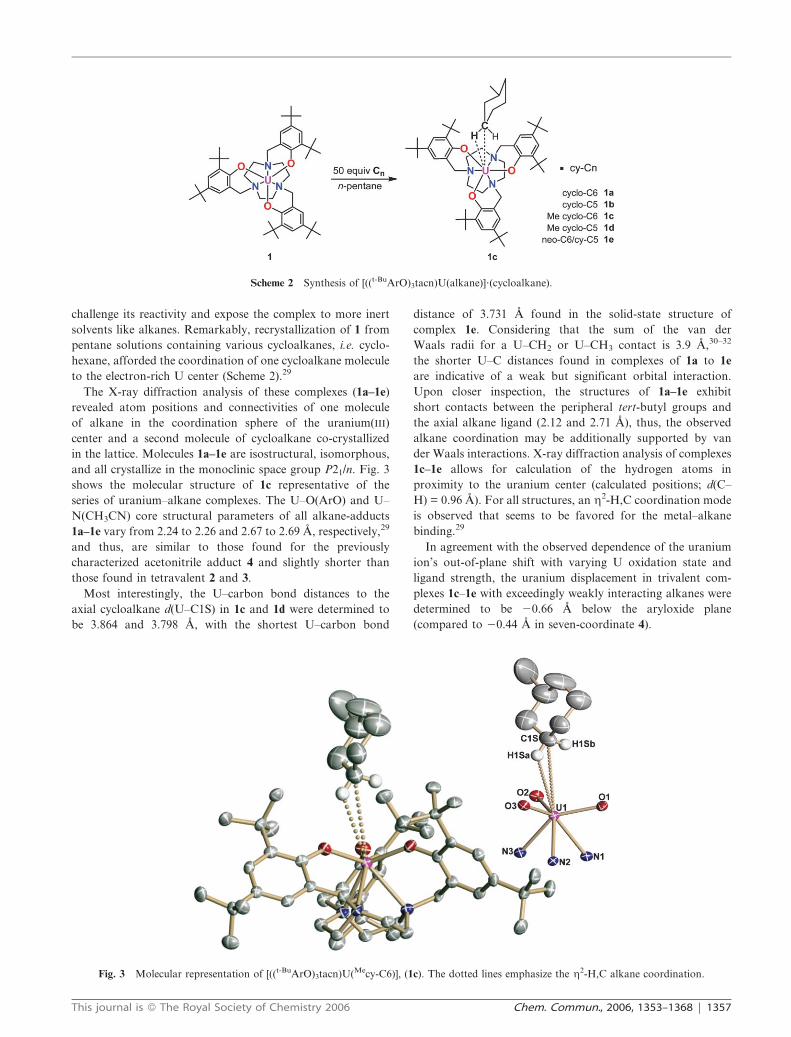
The X-ray diffraction analysis of these complexes (1a–1e)
revealed atom positions and connectivities of one molecule
of alkane in the coordination sphere of the uranium(III)
center and a second molecule of cycloalkane co-crystallized
in the lattice. Molecules 1a–1e are isostructural, isomorphous,
and all crystallize in the monoclinic space group P21/n. Fig. 3
shows the molecular structure of 1c representative of the
series of uranium–alkane complexes. The U–O(ArO) and U–
N(CH3CN) core structural parameters of all alkane-adducts
1a–1e vary from 2.24 to 2.26 and 2.67 to 2.69 A, respectively,29
and thus, are similar to those found for the previously
characterized acetonitrile adduct 4 and slightly shorter than
those found in tetravalent 2 and 3.
Most interestingly, the U–carbon bond distances to the
axial cycloalkane d(U–C1S) in 1c and 1d were determined to
be 3.864 and 3.798 A, with the shortest U–carbon bond
distance of 3.731 A found in the solid-state structure of
complex 1e. Considering that the sum of the van der
Waals radii for a U–CH2 or U–CH3 contact is 3.9 A,30–32
the shorter U–C distances found in complexes of 1a to 1e
are indicative of a weak but significant orbital interaction.
Upon closer inspection, the structures of 1a–1e exhibit
short contacts between the peripheral tert-butyl groups and
the axial alkane ligand (2.12 and 2.71 A), thus, the observed
alkane coordination may be additionally supported by van
der Waals interactions. X-ray diffraction analysis of complexes
1c–1e allows for calculation of the hydrogen atoms in
proximity to the uranium center (calculated positions; d(C–
H) = 0.96 A). For all structures, an g2-H,C coordination mode
is observed that seems to be favored for the metal–alkane
binding.29
In agreement with the observed dependence of the uranium
ion’s out-of-plane shift with varying U oxidation state and
ligand strength, the uranium displacement in trivalent com-
plexes 1c–1e with exceedingly weakly interacting alkanes were
determined to be 20.66 A below the aryloxide plane
(compared to 20.44 A in seven-coordinate 4).
Scheme 2 Synthesis of [((t-BuArO)3tacn)U(alkane)]?(cycloalkane).
Fig. 3 Molecular representation of [((t-BuArO)3tacn)U(Mecy-C6)], (1c). The dotted lines emphasize the g2-H,C alkane coordination.
This journal is � The Royal Society of Chemistry 2006 Chem. Commun., 2006, 1353–1368 | 1357

2. C1 coordination and activation chemistry
Due to the vastness of its supply, the greenhouse gas CO2
represents a valuable and renewable C1 source for the
production of fine chemicals and fuels. Therefore, the interest
in metal-mediated multi-electron reduction of carbon dioxide
remains high. The continued interest in uranium carbonyl
coordination is documented by the [(Cp)3U(CO)] success
story. First synthesized in 1986 by Andersen et al.,
[(Cp)3U(CO)]33 derivatives of the parent system were crystal-
lographically characterized in 199534 and 2003.35 Encouraged
by the remarkable reactivity of our complexes, we set out to
exploit this reactivity for CO2 and CO activation and
reduction.
2.1 Carbon monoxide activation assisted by [((t-BuArO)3tacn)U]
A pentane solution of coordinatively unsaturated, trivalent 1
cleanly reacts with carbon monoxide by binding and activating
the CO, resulting in rapid and quantitative formation of m-CO
bridged diuranium species [{((t-BuArO)3tacn)U}2(m-CO)] (5,
Scheme 3).36 Infra-red vibrational spectra of this material
reproducibly show a band at 2092 cm21 (in Nujol) suggestive
of a CO ligand. However, this frequency appears to be rather
high for a coordinated and activated CO molecule. Despite
numerous attempts, CO isotopomers of 5 could not be
synthesized. This puzzling lack of success in isotopomer
synthesis is likely due to impurities in commercially available
sources of CO isotopes that, among other impurities, contain
up to 20 ppm CO2 and O2. Both impurities lead to rapid
formation of m-oxo bridged 3, the only isolable product of all
labeling attempts (see below). To circumvent this problem, in
situ generation of 13CO will be attempted in the near future.
The molecular structure of 5 was characterized crystal-
lographically and revealed an iso-carbonyl bonding motif,
which is unique in actinide chemistry.36 A representative
structure [{((t-BuArO)3tacn)U}2(m-CO)] in crystals of 5?3C6H6,
(Fig. 4) was modeled by employing an asymmetrical U–CO–U
entity, with one short U–C bond and a longer U–O
isocarbonyl interaction, disordered on two positions at the
inversion center (rhombohedral space group R3). The mole-
cular structure of 5 exhibits two staggered [((t-BuArO)3tacn)U]-
fragments linked via a linearly bridged CO ligand in a m:g1,g1
fashion. The resolution of the data is limited and, therefore, no
reliable bond distances between the CO ligand and the U
center can be provided. Considering the unusual frequency of
the n(CO) stretch and the crystallographic disorder, it should
be emphasized that the corresponding dinitrogen-bridged
species could not be synthesized, neither under y1 atm nor
an overpressure (80 psi) of N2 gas, rendering the assignment of
the disordered bridging CO-atoms unambiguous.
The U–O(ArO) and U–N(tacn) distances in 5 were
determined to be 2.185(5) and 2.676(4) A. This U–N(tacn)
bond distance is very similar to that found in the [((t-BuArO)3-
tacn)U] fragments of [((t-BuArO)3tacn)U(alkane)] (d(U–
N(tacn)) = 2.676(6) A)29 and [((t-BuArO)3tacn)U(NCCH3)]
(d(U–N(tacn)) = 2.699(6) A).27 In contrast, the average
U-O(ArO) bond distance in 5 is significantly shorter than
those found in structurally related trivalent [((t-BuArO)3-
tacn)U] complexes.
The displacement of the uranium ion out of the idealized
trigonal aryloxide plane towards the coordinated triazacyclo-
nonane polyamine chelator in 5 was determined to be only
20.377 A. A diagram delineating this structural parameter for
known complexes of the [((t-BuArO)3tacn)U(Lax)] type clearly
illustrates a linear correlation of higher oxidation states with
smaller out-of-plane shifts (Fig. 5).27,36,37 Based on this
Scheme 3 Synthesis of [{((t-BuArO)3tacn)U}2(m-O)] (3), [{((t-BuArO)3-
tacn)U}2(m-CO)] (5), and [((t-BuArO)3tacn)U(m-N3)] (6).
Fig. 4 Molecular representation of [{((t-BuArO)3tacn)U}2(m-CO)] (5).
Fig. 5 Plot of the uranium ion’s out-of-plane shift vs. the formal U
oxidation state (see section 3 for [((t-BuArO)3tacn)U(Lax)] with Lax =
N32 and Me3SiN22).
1358 | Chem. Commun., 2006, 1353–1368 This journal is � The Royal Society of Chemistry 2006
correlation and in agreement with the crystallographic
disorder, 5 can be assigned an average oxidation state of
+3.5, suggestive of a mixed-valent uranium(III/IV) species. We
suggested that 5 forms via a charge-separated U(IV)–CO?2
intermediate, which reacts with excess 1 to yield the formally
mixed-valent U(IV)–CO–U(III) species 5.
In this context, the m-azido-bridged U(III/IV) species
[{(L)U}2(m-N3)] (6) was synthesized by reacting [(L)UIV(N3)]
with [LUIII] to serve as an isostructural (and isomorphous,
rhombohedral space group R3) analogue of triatomic-bridged
intermediate IM (see below) as well as an electronic model for
mixed-valent 5 (Scheme 3). The out-of-plane shift found in
mixed-valent 6 (20.368 A) is virtually identical to 5 (Fig. 5)
and thus supports the charge-separation proposed for mixed-
valent 5.
2.2 Carbon dioxide coordination and activation assisted by
[((RArO)3tacn)U]
Addition of CO2-saturated pentane to the deeply colored
solution of red-brown 1 in pentane affords a colorless solution
and subsequent formation of a pale-blue solution and CO-gas.
The pale-blue material was identified as the known m-oxo
bridged diuranium(IV/IV) complex [{((t-BuArO)3tacn)U}2-
(m-O)] (3, see above).36 The driving force for this remarkable
2e2 cleavage reaction of the thermodynamically stable CO2
molecule likely is the concerted two-ion U(III) to U(IV)
oxidation, the most stable oxidation state in this system.
Additionally, this reaction is sterically facilitated by the ligand
environment. Attempts to isolate this colorless intermediate via
solvent evaporation in vacuum resulted in recovery of 1
(Scheme 3). We suggest a dinuclear CO2-bridged diuranium
species IM as a possible intermediate. The reaction of 1 with
CO2 is reminiscent of the reductive cleavage of COS by
[(Cp9)3U] (Cp9 = MeC5H4), proceeding via a COS-bridged
intermediate.38,39 Accordingly, complete 2e2 reduction of CO2
to yield CO and 3 likely proceeds stepwise via a fleeting CO2-
bridged intermediate, colorless IM, that is in equilibrium with
1 and CO2.
After consideration of the dimerization products formed by
complex 1, it seemed likely that the tert-butyl derivatized
ligand employed did not provide sufficient steric bulk to
obstruct a complete 2e2 reduction of CO2 to CO. In order to
further study this unique CO2 activation at the electron-rich
uranium center, a ligand was needed that could provide greater
steric bulk and allow for better protection of the open uranium
coordination site. This steric bulk was provided in the
next generation of our aryloxide-functionalized ligand system
and the corresponding uranium complex, [((AdArO)3tacn)U]
(1-Ad).
2.3 The need for a sterically more demanding ligand
Our initial results on the tert-butyl derivatized [((t-BuArO)3-
tacn)U]-system have suggested that undesired side-reactions
trans to the reactive site are effectively eliminated due to
shielding by the triazacyclononane fragment. However, it is
also evident that the ortho-functionalized tert-butyl groups of
the three aryloxide pendant arms do not form a protective
cavity at the reactive, electron-rich uranium center. Instead,
the tilted aryloxides force the tert-butyl groups to form a bowl-
shaped cavity (Fig. 6) with little protection to prevent
decomposition reactions, such as ligand and solvent degrada-
tion as well as formation of dinuclear complexes. It seemed
clear that if reactive intermediates could be better protected, a
uranium(III) complex of the general [((ArO)3tacn)U]-type
would provide a powerful platform for reactivity studies at
the apical position. In order to prevent dimerization, a
sterically more encumbering derivative was required.
Accordingly, protocols for the synthesis of the adamantane-
functionalized ligand 1,4,7-tris(3-adamantyl-5-tert-butyl-2-
hydroxybenzyl)1,4,7-triazacyclononane, (AdArOH)3tacn, and
its corresponding U(III) precursor complex [((AdArO)3tacn)U]
(1-Ad) were developed.26
Similar to the preparation of 1, reaction of (AdArOH)3tacn
with one equivalent of [U(N(SiMe3)2)3] in benzene yielded the
six-coordinate U(III) complex [((AdArO)3tacn)U] (1-Ad) as a
red-brown powder in multigram quantities.26 In striking
contrast to 1, complex 1-Ad is stable in chlorinated and
ethereal solutions and thus can be re-crystallized from
mixtures of Et2O/CH2Cl2. Single-crystals of 1-Ad were
studied by X-ray crystallography26 and its molecular
core structure was compared to its parent complex as found
in [((t-BuArO)3tacn)U(Mecy-C6)] (1c, bound alkane omitted for
clarity).29
A comparison of the core molecular structures in 1 and 1-Ad
show comparable U–O(ArO) and U–N(tacn) bond distances
(see Table 1). The most striking difference in these structures is
the displacement of the U ion from the trigonal aryloxide
plane. While the out-of-plane shift in 1 is 20.66 A, the
uranium ion in 1-Ad was found to be 20.88 A below the
aryloxide plane. The U ion displacement from the aryloxide
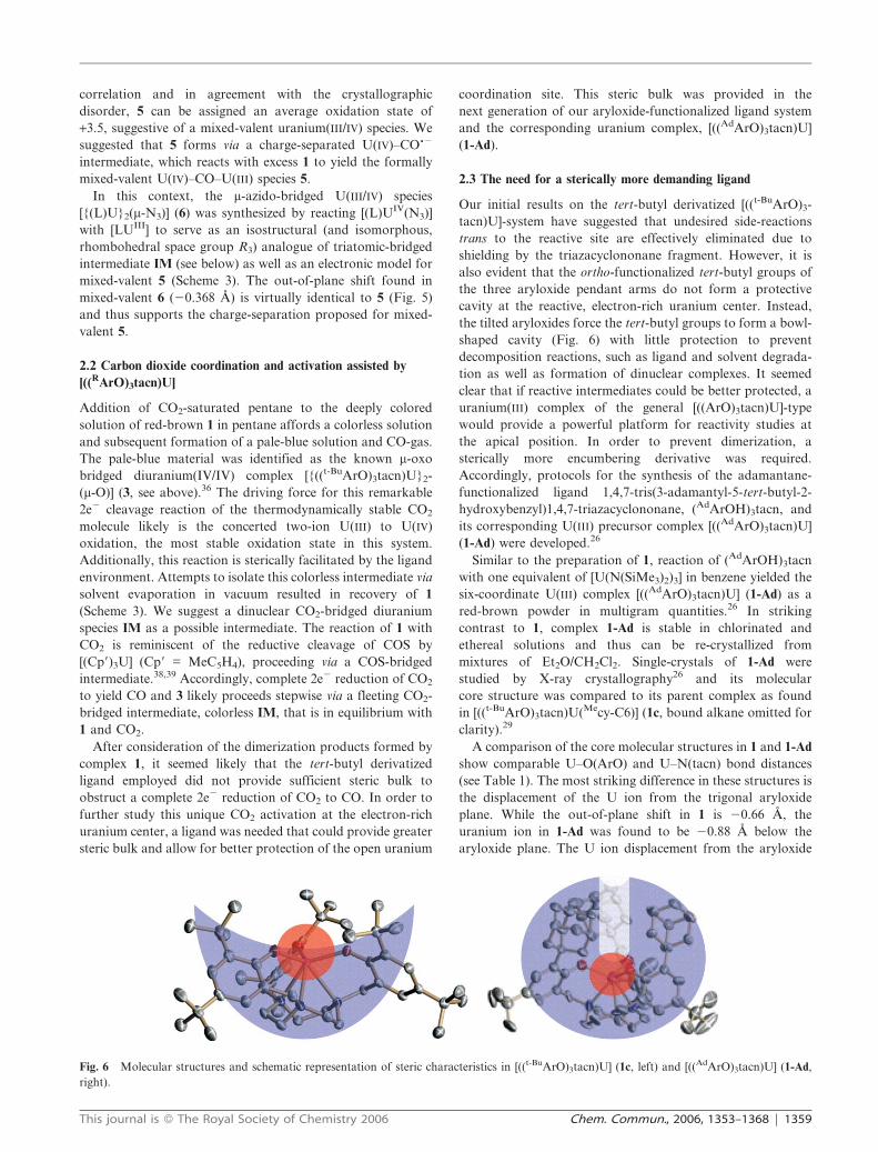
Fig. 6 Molecular structures and schematic representation of steric characteristics in [((t-BuArO)3tacn)U] (1c, left) and [((AdArO)3tacn)U] (1-Ad,
right).
This journal is � The Royal Society of Chemistry 2006 Chem. Commun., 2006, 1353–1368 | 1359
plane in addition to the increased steric bulk provided by the
adamantane substituents of 1-Ad lead to a narrow and
approximately 4.7 A deep cylindrical cavity. This cavity
provides restricted access of an incoming ligand to the uranium
ion, thereby protecting the uranium center from bimolecular
decomposition reactions (Fig. 6).
Accordingly, exposure of intensely colored
[((AdArO)3tacn)U] (1-Ad) in toluene or solid-state to CO2 gas
(1 atm) results in instantaneous discoloration of the samples.37
Colorless crystals of [((AdArO)3tacn)U(CO2)] (7) can be
isolated from a saturated CH2Cl2/Et2O solution. The infrared
spectrum in nujol exhibits a distinct vibrational band centered
at 2188 cm21, indicative of a coordinated and activated CO2
ligand. The 12C/13C isotopic ratio R(2188/2128) of 1.0282 is
close to that of free CO2 gas (R 2349/2284 = 1.0284),
suggestive of a molecule that has the same linear geometry
as that of free CO2 as well as the same carbon motion in the n3
(nas(OCO)) vibrational mode. While the assignment of this
vibrational band is unambiguous, we note that the nas(OCO)
found in 7 is significantly higher than frequencies observed for
other mononuclear M–CO2 complexes with carbon- (g1-CO2)
and carbon–oxygen-bound (g2-OCO) bent CO2 ligands, in
which nas(OCO) were reportedly found between 1550 and
1750 cm21 (Scheme 4).40
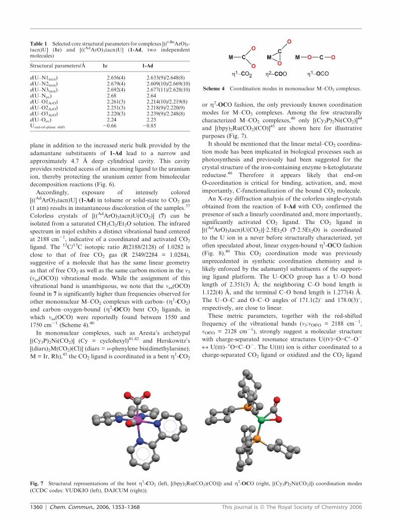
In mononuclear complexes, such as Aresta’s archetypal
[(Cy3P)2Ni(CO2)] (Cy = cyclohexyl)41,42 and Herskowitz’s
[(diars)2M(CO2)(Cl)] (diars = o-phenylene bis(dimethylarsine);
M = Ir, Rh),43 the CO2 ligand is coordinated in a bent g1-CO2
or g2-OCO fashion, the only previously known coordination
modes for M–CO2 complexes. Among the few structurally
characterized M–CO2 complexes,40 only [(Cy3P)2Ni(CO2)]44
and [(bpy)2Ru(CO2)(CO)]45 are shown here for illustrative
purposes (Fig. 7).
It should be mentioned that the linear metal–CO2 coordina-
tion mode has been implicated in biological processes such as
photosynthesis and previously had been suggested for the
crystal structure of the iron-containing enzyme a-ketoglutarate
reductase.46 Therefore it appears likely that end-on
O-coordination is critical for binding, activation, and, most
importantly, C-functionalization of the bound CO2 molecule.
An X-ray diffraction analysis of the colorless single-crystals
obtained from the reaction of 1-Ad with CO2 confirmed the
presence of such a linearly coordinated and, more importantly,
significantly activated CO2 ligand. The CO2 ligand in
[((AdArO)3tacn)U(CO2)]?2.5Et2O (7?2.5Et2O) is coordinated
to the U ion in a never before structurally characterized, yet
often speculated about, linear oxygen-bound g1-OCO fashion
(Fig. 8).40 This CO2 coordination mode was previously
unprecedented in synthetic coordination chemistry and is
likely enforced by the adamantyl substituents of the support-
ing ligand platform. The U–OCO group has a U–O bond
length of 2.351(3) A; the neighboring C–O bond length is
1.122(4) A, and the terminal C–O bond length is 1.277(4) A.
The U–O–C and O–C–O angles of 171.1(2)u and 178.0(3)u,respectively, are close to linear.
These metric parameters, together with the red-shifted
frequency of the vibrational bands (n3:nO12CO = 2188 cm21,
nO13CO = 2128 cm21), strongly suggest a molecular structure
with charge-separated resonance structures U(IV)LOLC?–O2
« U(III)–+OMC–O2. The U(III) ion is either coordinated to a
charge-separated CO2 ligand or oxidized and the CO2 ligand
Table 1 Selected core structural parameters for complexes [((t-BuArO)3-tacn)U] (1c) and [((AdArO)3tacn)U] (1-Ad, two independentmolecules)
Structural parameters/A 1c 1-Ad
d(U–N1tacn) 2.656(4) 2.633(9)/2.648(8)d(U–N2tacn) 2.679(4) 2.609(10)/2.669(10)d(U–N3tacn) 2.692(4) 2.677(11)/2.628(10)d(U–Nav) 2.68 2.64d(U–O1ArO) 2.261(3) 2.214(10)/2.219(8)d(U–O2ArO) 2.251(3) 2.218(9)/2.220(9)d(U–O3ArO) 2.220(3) 2.239(9)/2.248(8)d(U–Oav) 2.24 2.23Uout-of-plane shift 20.66 20.85
Scheme 4 Coordination modes in mononuclear M–CO2 complexes.
Fig. 7 Structural representations of the bent g1-CO2 (left, [(bpy)2Ru(CO2)(CO)]) and g2-OCO (right, [(Cy3P)2Ni(CO2)]) coordination modes
(CCDC codes: VUDKIO (left), DAJCUM (right)).
1360 | Chem. Commun., 2006, 1353–1368 This journal is � The Royal Society of Chemistry 2006

reduced by one electron. This reduction results in activation of
the inert CLO double-bond and is expected to increase
reactivity of the thermodynamically stable CO2 molecule.
The discrepancy between the significant degree of activation
(as judged from the bond distances) and the relatively
small red-shift of nas(OCO) is currently the subject of a
computational study.
3. Nitrogen atom transfer chemistry employinguranium complexes
Nitrogen atom transfer chemistry is of considerable interest to
inorganic47,48 and organic49–53 chemists. While inorganic
coordination chemists are fascinated by the reactivity of
terminal nitrido ligands,54 organic chemists employ these
reagents for the synthesis of aziridines, highly strained three-
membered rings systems that undergo ring-opening to yield the
corresponding amino functionality.53,55 The formal metal-
nitrido triple-bond is one of the strongest metal–ligand
interactions known to coordination chemists56,57 and yet,
these species can undergo facile and complete inter-metal 2e2
and 3e2 nitrogen atom transfer reactions.58 However, for
aziridination, the insertion of the nitrido nitrogen into CLC
double-bonds, nitrido ligand activation (e.g. with TFAA) is
often indispensable due to the highly covalent character of the
dp–pp interaction.59,60 In contrast, the valence f-orbitals of
uranium complexes do not participate in strong covalent
bonding. As a result, the uranium–nitrogen moiety is more
ionic U(d+)–N(d2) and thus, we expected uranium imido and
nitrido complexes to be more reactive toward electrophilic
substrates.
The enhanced reactivity of 1 and 1-Ad was ample impetus
for us to explore the synthesis of high-valent uranium imido
and nitrido complexes and probe them for their application in
nitrogen atom- and group transfer chemistry.
In our attempts to synthesize high-valent uranium com-
plexes with multiple-bonded N ligands, the trivalent uranium
starting complexes 1 and 1-Ad were treated with various
organic azides following reported protocols. We found that
reaction of 1 with one equivalent of trimethylsilyl azide in
hexane yielded the expected uranium(V) imido [((t-BuArO)3-
tacn)U(NSiMe3)] (8) as well as a uranium(IV) azido species
[((t-BuArO)3tacn)U(N3)] (9).27 Reaction of trivalent acetonitrile
complex 4 with trimethylsilyl azide yielded complex 9
exclusively. In accordance with literature reports, we suggest
that pentavalent complex 8 forms through coordination of the
azide’s Na atom (R–Na–Nb–Nc), in a second step dinitrogen is
expelled, and lastly, the formally electron-deficient trimethyl-
silyl nitrene oxidizes the trivalent uranium ion by two units to
yield a U(V) imido complex. Formation of an azido complex,
such as 9, is without precedent and could be explained by
coordination of the azides’ terminal Nc atom with subsequent
homolytic Si–Na bond cleavage that leads to a Me3Si? and N3?
radical. While the Me3Si? radical recombines to form Me6Si2,
the azide radical oxidizes the U(III) complex to form the U(IV)
azido complex 9. Steric considerations in seven-coordinate 4
do not allow for metal-coordination of the crowded Na azide
nitrogen. As a result, coordination of the unhindered terminal
Nc atom is enforced, followed by radical elimination and 1e2
oxidation. This suggested mechanism is supported by the
U(III)/U(IV) oxidative driving force and would be facilitated by
an organic azide that permits homolytic Na–R bond cleavage.
This hypothesis was tested by employing organic azides with
different Na–C bonds. While trityl azide (Ph3C–N3) will
readily cleave its C–Na bond (forming Gomberg’s dimer),
the homolytic bond cleavage in adamantyl azide (Ad–N3) is
energetically not favorable; formation of the U(V) imido
species should thus be preferred. As shown in Scheme 5,
compound 9 can be obtained reproducibly by treating 1 with
trityl azide. In addition, the imido species [((t-BuArO)3-
tacn)U(N(CPh3))] (8b) is formed in 40% yield as a by-product.
Fig. 8 Molecular representation of [((AdArO)3tacn)U(CO2)] (7, left) with core structure and geometrical parameters (right) in A and degrees.
Scheme 5 Reaction of trivalent 1 (R = t-Bu) and 1-Ad (R = Ad) with
various organic azides.
This journal is � The Royal Society of Chemistry 2006 Chem. Commun., 2006, 1353–1368 | 1361

In contrast, treatment of 1 with 1-adamantyl azide produces
the uranium(V) imido species [((t-BuArO)3tacn)U(N(Ad))] (8c),
exclusively.27
Like transition metal imido complexes, high-valent
uranium(V) and (VI) imido species typically exhibit short,
formal UMN(imido) triple bonds with bond distances ranging
from 1.85 to 2.01 A and /(U–N–R) bond angles varying from
slightly bent to linear (163.33–180.0u).27,61–68 Accordingly, the
structural parameters of imido complexes 8 and 8b (d(U–
N(imido)) = 1.989(5) and 1.992(4) A and /(U–N–R) =
173.7(3) and 177.7(3)u) are similar to those reported for other
metal imido complexes in the literature.27 As a result of strong
bonding to the axial imido ligand, the U ion in 8 and 8b moves
closer to the trigonal plane formed by the three aryloxide
oxygens and is found to be 20.151 (8) and 20.148 A (8b)
below the plane.
In contrast to the large number of transition metal
azido complexes, only few uranium azido species have been
reported in the literature.69,70 The molecular structure of
the uranium(IV) azide complex in crystals of 9 resembles
those of typical metal azido complexes. The linear N32 ligand
(/Na–Nb–Nc = 178.2(14)u) is bound to the metal ion in a
bent fashion with an U–Na–Nb angle of 145.9(9)u. The
uranium–azide bond distance was determined to be
2.564(12) A and the weakly bound azide ligand in 9 leads to
an average out-of-plane shift of the uranium(IV) ion of
20.307 A.
Neither of the above described azido and imido uranium
complexes [((t-BuArO)3tacn)U(L)] (L = N32 (9) and RN22 (R
= SiMe3 (8), CPh3 (8b))),27 however, could be transformed to a
high-valent uranium nitrido species (via thermolysis, photo-
lysis or Si–N bond cleavage) nor did they exhibit the desired
nitrogen atom nucleophilicity and resulting atom and/or group
transfer chemistry. However, steric pressure introduced by a
bulkier chelator was expected to increase the complexes’
reactivity. Accordingly, the sterically encumbering
[((AdArO)3tacn)U] (1-Ad) was employed in the reaction with
organic azides.
3.1 Reactivity induced by steric pressure in
[((AdArO)3tacn)U(L)] complexes
Similar to 1, complex 1-Ad reacts with one equivalent of
trimethylsilyl azide to yield the uranium(IV) azido complex
[((AdArO)3tacn)U(N3)] (10, via Me3Si radical elimination and
formation of Me6Si2) and the uranium(V) imido species
[((AdArO)3tacn)U(NSiMe3)] (11, with evolution of N2).71
The X-ray diffraction analysis of 10 and 11 clearly
demonstrated the influence of the sterically more demanding
adamantyl groups in these complexes. A comparison of
selected structural parameters found in complexes of 10 and
11 with those in the sterically unhindered 8 and 9 is given in
Table 2; structural representations of imido complexes 8 and
11 are depicted in Fig. 9. The most remarkable difference
between azido complexes 9 and 10 is the linearly coordinated
azido ligand in 10 (/(U–Na–Nb) = 145.6(9)u (9) vs. /(U–Na–
Nb) = 175.6(3)u (10)). This linear coordination leads to an
increased M–L orbital overlap, resulting in significantly
shorter U–N3 bond distances d(U–N3) = 2.564 (9) vs.
2. 372(3) A (10).
The structural parameters of imido complex 8 are also
strongly affected by the adamantyl-derivatized ligand. The U–
N(imido) bond distance found in 11 is the longest ever
reported for a metal imido complex and deviates significantly
from linearity (d(U–N(imido)) = 2.1219(18) A and /(U–N–R)
= 162.55(12)u). Additionally, the out-of-plane shift in 11 was
found to be 20.188 A in comparison to 20.151 and 20.148 A
Table 2 Selected structural parameters for complexes [((t-BuArO)3-tacn)U(NSiMe3)] (8, two independent molecules), [((t-BuArO)3-tacn)U(N3)] (9), [((AdArO)3tacn)U(N3)] (10), and [((AdArO)3-tacn)U(NSiMe3)] (11), (Bond distances in A, bond angles in u)
Structuralparameters 8 11 9 10
d(U–N1tacn) 2.719(5)/2.791(4) 2.675(2) 2.825(9) 2.667(3)d(U–N2tacn) 2.737(5)/2.724(4) 2.729(2) 2.758(9) 2.661(3)d(U–N3tacn) 2.660(5)/2.735(4) 2.683(2) 2.886(9) 2.649(3)d(U–Nav) 2.70/2.75 2.70 2.83 2.66d(U–O1ArO) 2.196(4)/2.161(4) 2.2028(17) 2.294(8) 2.171(2)d(U–O2ArO) 2.203(4)/2.185(4) 2.2179(17) 2.295(8) 2.152(2)d(U–O3ArO) 2.209(4)/2.222(4) 2.2109(17) 2.286(8) 2.143(2)d(U–Oav) 2.20/2.19 2.21 2.29 2.16d(U–Nazido) — — 2.564(12) 2.372(3)d(U–Nimido) 1.985(5)/1.992(4) 2.1219(18) — —Uout-of-plane shift 0.151 0.188 0.308 0.292/(U–Nimido–Si) 178.5(3)/168.9(3) 162.55(12) — —/(U–Na–Nb) — — 145.6(9) 176.9(8)
Fig. 9 Comparison of molecular structures of [((RArO)3tacn)U(NSiMe3)] with R = t-Bu (8, left) and Ad (11, right).
1362 | Chem. Commun., 2006, 1353–1368 This journal is � The Royal Society of Chemistry 2006

in sterically unhindered 8 and 8b, respectively. These unusual
structural features of 11 are likely due to the steric pressure
brought about by the sterically encumbering adamantane
groups that form a narrow cylindrical cavity and prevent the
Me3SiN22 from optimal binding. Accordingly, the imido
nitrogen p-orbitals cannot participate in efficient M–L
p-bonding, which results in the observed structural parameters
of 11.
It is expected that the peculiar structural features observed
in complexes 10 and 11 (compared to 8 and 9 as well as other
known azido and imido species) will result in an increased and
atypical reactivity of the axial ligand.
3.2 Nitrogen atom transfer via multiple bond metathesis
While imido complex 8 was unreactive towards p-acids, we
found that complex 11 reacts cleanly with CO (1 atm) and
CH3NC (1 eq.) to form the uranium(IV) isocyanate
complex [((AdArO)3tacn)U(NCO)] (12) and carbodiimide
complex [((AdArO)3tacn)U(NCNMe)] (13) with concomitant
formation of Me3Si? which immediately recombines to
produce Me6Si2.71 The IR spectra of 12 and 13 exhibit one
strong vibrational band centered at 2185 and 2101 cm21 that
can be assigned to the g1-coordinate isocyanate (12)
and carbodiimide (13) ligands. Elemental analysis (C, H,
N) and 1H NMR spectroscopy suggest that complexes 12 and
13 are isoelectronic and isostructural to the previously
prepared uranium(IV) heterocumulene complexes
[((AdArO)3tacn)U(g1-OCO)] (7) and [((AdArO)3tacn)U(g1-
N3)] (10).37
The X-ray crystallographic analysis of 12 and 13 confirmed
formation of nearly linear, axial g1-bound isocyanate and
carbodiimide ligands in these complexes (Fig. 10 and 11).
The U–N4 bond distances and /(U–N4–C70) angles
were determined to be 2.389(6) A and 171.2(6)u in 12 and
2.327(3) A and 161.9(3)u in 13 and are very similar to the
corresponding parameters found in 7 and 10. Likewise, the
inner N–C–O and N–C–NMe angles of 178.2(9)u and
174.3(4)u, respectively, are also close to linear. The out-of-
plane shifts of the uranium ion with respect to the tris-
aryloxide plane are 20.301 and 20.318 A for 12 and 13.
As mentioned earlier, these out-of-plane shifts are
generally very sensitive to the formal oxidation state of
the uranium center. The shifts of the central U ion of
complexes 12 and 13 fall between the values found for
the analogous U(V) and U(III) complexes and therefore
appear to indicate a formal U(IV) oxidation state for 12 and
13.
Fig. 10 Molecular representation of [((AdArO)3tacn)U(NCO)] (12).
Fig. 11 Molecular representation of [((AdArO)3tacn)U(NCNCH3)] (13).
This journal is � The Royal Society of Chemistry 2006 Chem. Commun., 2006, 1353–1368 | 1363

Interestingly, the isocyanate and carbodiimide ligands of 12
and 13 are reactive and can be transferred to organic
molecules. For instance, the carbodiimide ligand in 13 reacts
with CH3I or CH2Cl2 to release the functionalized organic
carbodiimides, CH3NCNCH2Cl and CH3NCNCH3, yielding
the corresponding halide complexes [((AdArO)3tacn)U(X)] (X
= Cl (14a), I (14b); Scheme 6, 11 A 14a).71 Furthermore, these
halide complexes can be regenerated to the uranium(III)
starting complex 1-Ad via sodium/amalgam reduction. This
series of reactions represents a synthetic cycle 1-Ad A 11A 13
A 14a A 1-Ad, in which the imido nitrogen atom (or
intermediate nitrido nitrogen) is transferred from the uranium
complex and incorporated into an organic substrate via CMO
and R9NMC/UMNR multiple-bond metathesis in successive
one-electron events. A close examination of the calculated
frontier orbitals in 11 suggests that the remarkable reactivity of
the uranium imido complex 11 originates from a high degree of
ionic character within the U5+–NR22 moiety. This bond is very
different from imido and nitrido bonds of group 6 transition
metal complexes, which typically exhibit very strong covalent
multiple bonds.
4. Electronic structure of low and high-valent
uranium complexes
In contrast to light transition metal complexes, magnetic
susceptibility data for actinide complexes do not allow for
simple interpretations and thus, do not provide instant
information on the number of unpaired electrons and the
complexes’ formal oxidation state. Due to large spin–orbit
coupling constants (j) and relatively small interelectronic
repulsion interactions (e2/r) in addition to electric field terms
(V) that often are comparable in magnitude to j and e2/r, the
Russell–Saunders (L–S) coupling formalism cannot be applied
nor can it be replaced by jj-coupling.72 Consequently, relatively
few magnetic studies of actinide coordination compounds are
reported in the literature,73 barring the mere report of room-
temperature magnetic moments as determined by the Evans’
method. Despite these difficulties, we believe that the
quantitative comparison of temperature-dependent magnetiza-
tion data of a series of complexes can provide valuable
information. The following is a descriptive chapter, a
collection of data rather than a magnetization study on a
microscopic level, which is under way and will be reported on
in due course. Regardless, the complexes presented above
provide a unique opportunity to study the electronic properties
of a series of [((RArO)3tacn)U(L)] uranium coordination
complexes in which the [((RArO)3tacn)U]–core structure
remains unperturbed while the axial ligand L (CH3CN, N32,
OCN2, CH3NCN2, CO2?2, RN22) varies with the complexes’
formal oxidation state (+III to +VI).
The magnetic moments, meff, of solid samples of trivalent 1,
1-Ad, and 4 are strongly temperature dependent, varying from
1.77, 1.74 and 1.66 mB at 5 K to 2.92, 2.83 and 2.90 mB at 300 K,
respectively (Fig. 12, right). The experimentally determined
effective magnetic moments meff at room temperature are
considerably lower than that calculated for a mononuclear f3
uranium species with a 4I9/2 ground state. The theoretical
magnetic moment for an ion with an 5f3 configuration is
calculated to be meff(calcd) = gJ(J(J + 1))1/2 = 3.69 mB.74 The
observed reduced magnetic moments of 1-Ad and 4 are likely
due to the strong ligand field, introduced by the equatorial
aryloxide oxygen ligands, which splits the J = 9/2 ground state
in U(III) ions. It is suggested that the splitting of the lowest J
manifold is such that the all of the Jz states are not equally
populated at room temperature. Consequently, the experimen-
tally observed moments are smaller than the free-ion moment.
Minor covalent contributions in U(III) complexes 1 and 4 may
further reduce the observed magnetic moment via orbital
Scheme 6 Synthesis of complexes and nitrogen-atom transfer chemi-
stry in successive one-electron steps.
Fig. 12 X-band EPR spectrum of 1-Ad (left) recorded in frozen benzene solution at T = 14 K. Experimental spectrum (magenta): frequency,
9.4666 GHz; power, 0.63 mW; modulation amplitude, 10 G. Simulation (in black): g = 2.005, WFWHM = 400 G and temperature dependent SQUID
magnetization data for 1, 1-Ad, and 4 (right).
1364 | Chem. Commun., 2006, 1353–1368 This journal is � The Royal Society of Chemistry 2006

reduction. In contrast, the experimentally determined magnetic
moments of U(IV) (f2) complexes at room temperature are
generally similar but, surprisingly, sometimes even higher
(meff(expt) # 3–3.5 mB) than the analogous moments of the
U(III) f3 ions of the [((RArO)3tacn)U(L)]-system.
Note that the theoretically expected moment 3.58 mB for a
U(IV) ion with an f2 electron configuration and 3H4 ground
state is only y0.1 mB lower than 3.69 mB expected for an U(III)
ion with three unpaired f-electrons.
Accordingly, room-temperature magnetic moments often do
not permit for an unambiguous assignment of the +3 and +4
oxidation state in molecular uranium compounds.73 However,
the temperature dependence of meff in the range 4–300 K and
especially the low-temperature behavior below 75 K, often
allows for a clear assignment of U(III) and U(IV) oxidation
states. Generally U(IV) complexes possess a singlet ground
state that exhibits temperature-independent paramagnetism
(TIP) at low temperatures, resulting in magnetic moments of
ca. 0.5–0.8 mB at approx. 4 K (Fig. 13).73
In contrast, an isolated f3 ion cannot be an orbital singlet
and thus, the doublet ground state in mononuclear trivalent
uranium complexes gives rise to higher magnetic moments
at low temperature; in case of 1, 1-Ad, and 4, moments of
y1.7 mB are observed at 4 K. Notably, we found that frozen
solutions of trivalent uranium complexes 1-Ad, 4, and
[U(N(SiMe3)2)3] are EPR active at temperatures below 20 K.
X-band EPR spectra of 4 and [U(N(SiMe3)2)3] show broad
and unsymmetrical signals centered at g = 2.016 and 2.50,
respectively. The spectrum of 1-Ad, recorded in frozen
benzene solution at 14 K, exhibits a metal-centered isotropic
signal at g = 2.005 (Fig. 12, left), which is in excellent
agreement with its low-temperature magnetic moment of meff =
1.73mB = K(3g2)1/2.
Despite the difficulties in understanding the magnetism of
complexed actinide ions, the most remarkable spectroscopic
difference between trivalent and tetravalent uranium com-
plexes of the [((RArO)3tacn)U(L)]-type is their characteristic
color. In contrast to their deeply colored red–brown to purple
trivalent analogues, uranium(IV) complexes appear very pale
aquamarine/blue–green in the solid state and almost colorless
in solution. Accordingly, electronic absorption spectra of
all U(IV) complexes [((RArO)3tacn)UIV(L)] show very
similar spectra with various sharp, low intensity bands (e =
5–80 M21 cm21) between 350–2100 nm. These bands originate
from Laporte-forbidden f–f transitions. In addition to these
characteristic low-intensity f–f transitions in the visible and
near-infrared region between 500 and 2200 nm, U(III)
complexes often show intense, color-giving d–f transitions in
the visible part of the absorption spectrum.75
The temperature dependence of mB of molecular complexes
of uranium(V) (f1) is clearly distinguishable from their f2 and f3
analogues. For example, derivatives of pentavalent imido
complexes 8 and 11 show temperature-dependent magnetic
moments that vary from y1.5 mB at 5 K to y2–2.4 mB at 300 K
(Fig. 14). These observed moments are reduced significantly
below the theoretical value of 2.54 mB, calculated for a free
ion in the L–S coupling scheme,72 and are always lower
than their corresponding f2 and f3 counterparts in the
[((RArO)3tacn)U(L)]-system. Boudreaux and Mulay72 have
attributed this phenomenon to covalency effects in high-valent
uranium complexes, in which the high-oxidation state is often
stabilized by strongly p-donating ligands, such as terminal oxo
or, as in 8 and 11, strongly bound imido ligands. In both cases,
the metal–ligand interactions can be best described as formal
MML triple bonds.
Like the trivalent complexes with the (RArO)3tacn ligand,
the uranium(V) imido species of this ligand system are
intensely colored. Derivatives of 8 and complex 11 are deep-
green in color and show intense ligand-to-metal charge-
transfer bands below 500 nm. In addition, their spectra also
show numerous weak but sharp absorption bands in the visible
and near infrared region between 500 and 2200 nm (e = 20–
100 M21cm21), characteristic for f–f transitions.
5. Is the CO2 ligand in [((AdArO)3tacn)U(CO2)]
activated?
The large number of isostructural and isoelectronic complexes
that have been obtained allows for a systematic study of their
Fig. 13 Temperature-dependent SQUID magnetization data for
[((AdArO)3tacn)U(N3)] (9) and closely related U(IV) halide complexes
[((AdArO)3tacn)U(Cl)], [((AdArO)3tacn)U(Br)], and [((AdArO)3tacn)U(I)].
Fig. 14 Temperature dependent SQUID magnetization data for the
U(V) complex [((AdArO)3tacn)U(NSi(CH3)3)] (11).
This journal is � The Royal Society of Chemistry 2006 Chem. Commun., 2006, 1353–1368 | 1365

molecular and electronic structures. It is interesting to
compare structural and spectroscopic features of the fascinat-
ing and unique U–CO2 complex (7) to analogous complexes.
In the following section, we will compare 7 to the series of
complexes [((AdArO)3tacn)Un(L)]m+ (n = III, IV, V, VI; m = 0,
1; and L = CH3CN, N32, CH3NCN2, OCN2, and RN22) and
discuss whether or not the bound CO2 ligand in
[((AdArO)3tacn)U(CO2)] is ‘‘activated’’ or ‘‘reduced’’ and if
so, to what degree.
A coordination chemist is trained to observe color changes
during the course of a chemical reaction. While this certainly is
by no means ‘‘high-tech’’, it is worth mentioning that the
chemist who synthesized the colorless [((AdArO)3tacn)U(CO2)]
immediately ‘‘knew’’ that the deeply-colored trivalent starting
complex [((AdArO)3tacn)U] was oxidized to uranium(IV) upon
reaction with CO2. Why? Because of the observed color
change! It was emphasized earlier that, while U(III) and U(V)
complexes are red and green colored, respectively, all U(IV)
complexes [((AdArO)3tacn)UIV(L)] are colorless. Exposure of
toluene solutions or even solid samples of deeply red-colored
1-Ad to CO2 gas resulted in instantaneous discoloration and,
eventually, colorless crystals were obtained. Its solution UV/
vis/NIR electronic absorption spectrum is strikingly similar to
all other U(IV) complexes synthesized in this study. All other
spectroscopic evidence, including advanced techniques, such as
single-crystal diffraction, X-ray absorption and SQUID
magnetization studies, as well as the standard laboratory
spectroscopy techniques that were accumulated so far suggest
that the U ion in 7 is oxidized by 1e2 and thus, the CO2 ligand
is reduced to a CO22? radical anion.
The molecular structure of 7 already revealed bond distances
of the coordinated CO2 ligand that were quite different from
those of the symmetrical free CO2 and thus, suggested a
significant degree of ligand reduction. The uranium ion’s
displacement from the idealized trigonal plane of the three
aryloxide ligators further implies the U ions’ oxidation
upon CO2 binding. While the out-of-plane shift in
precursor 1-Ad was determined to be 20.88 A, the U ion in
7 and all other U(IV) heterocumulene complexes of
the [((AdArO)3tacn)UIV(L)] system is displaced only 0.29–
0.32 A below the plane (Fig. 15). In fact, an extrapolation
of all available out-of-plane shifts vs. oxidation state places
the two complexes with ambiguous oxidation states,
[((AdArO)3tacn)U(CO2)] and [{((t-BuArO)3tacn)U}2(m-CO)],
correctly at +4 and +3.5.
Spectroscopic data further support an intramolecular redox-
reaction upon CO2 coordination to 1-Ad. The vibrational
spectrum of 7 exhibits a band at 2188 cm21 that shifts to
2128 cm21 upon 13C isotope labeling. Although this band can
be assigned unambiguously to the asymmetric stretching
vibration of the coordinated CO2 ligand, a significantly higher
red-shift is expected for a 1e2 reduced CO2 ligand.
Accordingly, upon initial observation, a comparison of CO2
stretching frequencies to those of known M2CO2 complexes,
which feature signals n(CO2) between 1600 and 1750 cm21,
suggests that the activation found in 7 cannot be a ‘‘complete’’
one-electron reduction. However, considering the linear g1-
OCO coordination mode in 7, which is unprecedented, a
comparison of vibrational frequencies with complexes that
possess bent C-bound (g1-COO) or C,O-bound (g2-OCO)
CO2 ligands may not be valid.
SQUID magnetization measurements of 7 were recorded
and compared to the large number of similar complexes
(Fig. 16). The magnetic moment meff of 7 was determined to
be 2.89 mB at 300 K and 1.51 mB at 5 K. Although the
Fig. 15 Summary of out-of-plane shifts vs. oxidation state for complexes [((RArO)3tacn)U(Lax)].
Fig. 16 Temperature dependent SQUID magnetization data for the
U(III) and U(IV) complexes [((AdArO)3tacn)U] (1-Ad),
[((AdArO)3tacn)U(N3)] (9), and [((AdArO)3tacn)U(CO2)] (7).
1366 | Chem. Commun., 2006, 1353–1368 This journal is � The Royal Society of Chemistry 2006

room-temperature moment of 7 is close to the magnetic
moment found for the azide complex 10, the low-temperature
value is similar to that of the U(III) (f3) starting material 1-Ad
(1.73 mB at 5 K), which has a doublet ground state at low
temperatures. As mentioned above, the magnetic moments of
U(III) (f3) and U(IV) (f2) complexes at room temperature are
generally very similar and often do not allow for an
unambiguous assignment of the complexes’ oxidation state.
The temperature dependence of mB in the range 4–300 K,
however, shows a curvature reminiscent of data obtained for
all closely-related U(IV) complexes of this type. Although
U(IV) complexes possess a singlet ground state, which typically
results in magnetic moments of ca. 0.5–0.8 mB, the magnetic
moment of 7 at low temperatures is significantly higher,
suggesting that the open-shell CO2?2, unlike the closed-shell
N32 ligand, likely contributes to the observed increased
magnetic moment of 7 at low temperatures. The temperature
dependence and low temperature value of 7 are in agreement
with the description of the CO2 ligand as a one-electron
reduced CO2?2 radical anion coordinated to a U(IV) ion.
Finally, in order to unambiguously determine the uranium
ion’s +IV oxidation state in 7, UL3 edge energy XANES
measurements of the isostructural complexes [((AdArO)3-
tacn)Um(L)]n+ (with L = CH3CN, N32, and Me3SiN22, m =
III, IV, V, and VI, and n = 0,1) were performed and compared to 7.
Details of this study will be published elsewhere. However,
preliminary data analysis shows the UL3 edge energy for
[((AdArO)3tacn)U(CO2)] is virtually identical to that measured
for the uranium IV complex [((AdArO)3tacn)U(N3)]. This
observation confirms the +IV oxidation state in
[((AdArO)3tacn)UIV(g1-CO2?2)], which implies that the coordi-
nated carbon dioxide ligand is in fact reduced by one electron.
Future computational studies will attempt to shed light on the
peculiar electronic structure and spectroscopic features, such as
the complexes relatively low nas(CO2) red-shift.
5. Concluding remarks
Our laboratory has shown that an aryloxide-functionalized
triazacyclononane ligand can be an impressive effector for
unique binding and small-molecule activation at low-valent
uranium centers, resulting in potentially effective agents for
functionalization of otherwise inert molecules. The series of
complexes described herein are distinctive in the respect that
they represent a set of isostructural complexes possessing a
range of oxidation states, as well as differing electronic and
magnetic behaviors. This presents a distinct benefit for the
understanding of fundamental actinide chemistry in general
and uranium in particular. Topics such as the nature of
covalency and the role of f-orbitals in bonding can be
advanced. After several years of uranium research we are still
very excited about this unique class of actinide compounds and
are certain that more unexpected and novel reactivity is still to
be discovered in the future.
Acknowledgements
This research was supported by the U.S. Department of
Energy (DE–FG02-04ER15537), the Alfred P. Sloan
Foundation (fellowship to K.M.), and an ACS-PRF Type G
grant (40019-G3). We thank NIH for a fellowship to I.C.-R.
(3 T32 DK07233-2651) and Drs Hidetaka Nakai (synthesis,
UCSD), Kristian Olsen and Xile Hu (computation, UCSD) as
well as Dr Wayne Lukens (electronic structure, Lawrence
Berkeley National Laboratory) and Drs Steven Conradson
and David Clark (XANES, Los Alamos National Laboratory)
for their contributions to this article. We wish to thank Ryan
L. Holland and Oanh P. Lam (UCSD) for their assistance in
finalizing this manuscript (O. P. L.) and prepaing the cover
picture (R. L. H.).
References
1 D. Seyferth, Organometallics, 2004, 23, 3562–3583.2 I. Korobkov and S. Gambarotta, Prog. Inorg. Chem., 2005, 54,
321–348.3 W. G. Vandersluys, C. J. Burns, J. C. Huffman and
A. P. Sattelberger, J. Am. Chem. Soc., 1988, 110, 5924–5925.4 L. R. Avens, D. M. Barnhart, C. J. Burns, S. D. McKee and
W. H. Smith, Inorg. Chem., 1994, 33, 4245–4254.5 R. A. Andersen, Inorg. Chem., 1979, 18, 1507–1509.6 D. L. Clark, A. P. Sattelberger, S. G. Bott and R. N. Vrtis, Inorg.
Chem., 1989, 28, 1771–1773.7 L. R. Avens, S. G. Bott, D. L. Clark, A. P. Sattelberger,
J. G. Watkin and B. D. Zwick, Inorg. Chem., 1994, 33, 2248–2256.8 D. L. Clark, A. P. Sattelberger and R. A. Andersen, Inorg. Synth.,
1997, 31, 307–315.9 J. L. Stewart and R. A. Andersen, Polyhedron, 1998, 17, 953–958.
10 M. Mazzanti, R. L. Wietzke, J. Pecaut, J. M. Latour, P. Maldiviand M. Remy, Inorg. Chem., 2002, 41, 2389–2399.
11 R. Wietzke, M. Mazzanti, J. M. Latour and J. Pecaut, J. Chem.Soc., Dalton Trans., 1998, 4087–4088.
12 A. J. Amoroso, J. C. Jeffery, P. L. Jones, J. A. McCleverty, L. Rees,A. L. Rheingold, Y. M. Sun, J. Takats, S. Trofimenko, M. D. Wardand G. P. A. Yap, J. Chem. Soc., Chem. Commun., 1995,1881–1882.
13 P. Roussel, P. B. Hitchcock, N. Tinker and P. Scott, Chem.Commun., 1996, 2053–2054.
14 I. Korobkov, S. Gambarotta, G. P. A. Yap, L. Thompson andP. J. Hay, Organometallics, 2001, 20, 5440–5445.
15 C. C. Cummins, Prog. Inorg. Chem., 1998, 47, 685–836.16 A. L. Odom, P. L. Arnold and C. C. Cummins, J. Am. Chem. Soc.,
1998, 120, 5836–5837.17 P. Roussel and P. Scott, J. Am. Chem. Soc., 1998, 120, 1070–1071.18 P. Roussel, R. Boaretto, A. J. Kingsley, N. W. Alcock and P. Scott,
J. Chem. Soc., Dalton Trans., 2002, 1423–1428.19 P. L. Diaconescu, P. L. Arnold, T. A. Baker, D. J. Mindiola and
C. C. Cummins, J. Am. Chem. Soc., 2000, 122, 6108–6109.20 P. L. Diaconescu and C. C. Cummins, J. Am. Chem. Soc., 2002,
124, 7660–7661.21 I. Korobkov, S. Gambarotta and G. P. A. Yap, Angew. Chem., Int.
Ed., 2002, 41, 3433–3436.22 I. Castro-Rodriguez, K. Olsen, P. Gantzel and K. Meyer, Chem.
Commun., 2002, 2764–2765.23 B. Monteiro, D. Roitershtein, H. Ferreira, J. R. Ascenso,
A. M. Martins, A. Domingos and N. Marques, Inorg. Chem.,2003, 42, 4223–4231.
24 M. Nierlich, J. M. Sabattie, N. Keller, M. Lance and J. D. Vigner,Acta Crystallogr., Sect. C, 1994, C50, 52–54.
25 P. Chaudhuri and K. Wieghardt, Prog. Inorg. Chem., 2001, 50,151–216.
26 H. Nakai, X. Hu, L. N. Zakharov, A. L. Rheingold and K. Meyer,Inorg. Chem., 2004, 43, 855–857.
27 I. Castro-Rodriguez, K. Olsen, P. Gantzel and K. Meyer, J. Am.Chem. Soc., 2003, 125, 4565–4571.
28 A. Mendiratta and C. C. Cummins, Inorg. Chem., 2005, 44,7319–7321.
29 I. Castro-Rodriguez, H. Nakai, P. Gantzel, L. N. Zakharov,A. L. Rheingold and K. Meyer, J. Am. Chem. Soc., 2003, 125,15734–15735.
This journal is � The Royal Society of Chemistry 2006 Chem. Commun., 2006, 1353–1368 | 1367
30 The methyl group, CH3, as a whole is assigned the van der Waalsradius 2.0 A. According to Pauling, the methylene group, CH2, canbe assigned the same value. The van der Waals radius for uraniumwas taken as 1.9 A.
31 L. Pauling, The Nature of the Chemical Bond, Cornell UniversityPress, Ithaca, New York, 3rd edn, 1960.
32 J. E. Huheey, E. A. Keiter and R. L. Keiter, Inorganic Chemistry:Principles of Structure and Reactivity, HarperCollins, New York,4th edn, 1993.
33 J. G. Brennan, R. A. Andersen and J. L. Robbins, J. Am. Chem.Soc., 1986, 108, 335–336.
34 J. Parry, E. Carmona, S. Coles and M. Hursthouse, J. Am. Chem.Soc., 1995, 117, 2649–2650.
35 W. J. Evans, S. A. Kozimor, G. W. Nyce and J. W. Ziller, J. Am.Chem. Soc., 2003, 125, 13831–13835.
36 I. Castro-Rodriguez and K. Meyer, J. Am. Chem. Soc., 2005, 127,11242–11243.
37 I. Castro-Rodriguez, H. Nakai, L. N. Zakharov, A. L. Rheingoldand K. Meyer, Science, 2004, 305, 1757–1759.
38 J. G. Brennan, R. A. Andersen and A. Zalkin, Inorg. Chem., 1986,25, 1756–1760.
39 J. G. Brennan, R. A. Andersen and A. Zalkin, Inorg. Chem., 1986,25, 1761–1765.
40 D. H. Gibson, Chem. Rev., 1996, 96, 2063–2095.41 M. Aresta and C. F. Nobile, J. Chem. Soc., Dalton Trans., 1977,
708–711.42 M. Aresta, C. F. Nobile, V. G. Albano, E. Forni and
M. Manassero, J. Chem. Soc., Chem. Commun., 1975, 636–637.43 T. Herskovitz, J. Am. Chem. Soc., 1977, 99, 2391–2392.44 A. Dohring, P. W. Jolly, C. Kruger and M. J. Romao,
Z.Naturforsch., B, 1985, 40b, 484–488.45 H. Tanaka, H. Nagao, S. M. Peng and K. Tanaka, Organo-
metallics, 1992, 11, 1450–1451.46 H. J. Lee, M. D. Lloyd, K. Harlos, I. J. Clifton, J. E. Baldwin and
C. J. Schofield, J. Mol. Biol., 2001, 308, 937–948.47 T. Birk and J. Bendix, Inorg. Chem., 2003, 42, 7608–7615.48 J. Bendix, J. Am. Chem. Soc., 2003, 125, 13348–13349.49 D. A. Evans, M. M. Faul and M. T. Bilodeau, J. Org. Chem., 1991,
56, 6744–6746.50 D. A. Evans, M. M. Faul, M. T. Bilodeau, B. A. Anderson and
D. M. Barnes, J. Am. Chem. Soc., 1993, 115, 5328–5329.51 Z. Li, K. R. Conser and E. N. Jacobsen, J. Am. Chem. Soc., 1993,
115, 5326–5327.52 Z. Li, R. W. Quan and E. N. Jacobsen, J. Am. Chem. Soc., 1995,
117, 5889–5890.53 D. E. N. Jacquot, M. Zollinger and T. Lindel, Angew. Chem., Int.
Ed., 2005, 44, 2295–2298.
54 J. K. Brask, V. Dura-Vila, P. L. Diaconescu and C. C. Cummins,Chem. Commun., 2002, 902–903.
55 J. DuBois, C. S. Tomooka, J. Hong and E. M. Carreira, J. Am.Chem. Soc., 1997, 119, 3179–3180.
56 J. Bendix, R. J. Deeth, T. Weyhermuller, E. Bill and K. Wieghardt,Inorg. Chem., 2000, 39, 930–938.
57 J. P. F. Cherry, A. R. Johnson, L. M. Baraldo, Y. C. Tsai,C. C. Cummins, S. V. Kryatov, E. V. Rybak-Akimova,K. B. Capps, C. D. Hoff, C. M. Haar and S. P. Nolan, J. Am.Chem. Soc., 2001, 123, 7271–7286.
58 L. K. Woo, Chem. Rev., 1993, 93, 1125–1136.59 J. DuBois, C. S. Tomooka, J. Hong, E. M. Carreira and M. W. Day,
Angew. Chem., Int. Ed., 1997, 36, 1645–1647.60 J. DuBois, C. S. Tomooka, J. Hong and E. M. Carreira, Acc. Chem.
Res., 1997, 30, 364–372.61 R. E. Cramer, K. Panchanatheswaran and J. W. Gilje, Angew.
Chem., Int. Ed. Engl., 1984, 23, 912–913.62 J. G. Brennan and R. A. Andersen, J. Am. Chem. Soc., 1985, 107,
514–516.63 A. Zalkin, J. G. Brennan and R. A. Andersen, Acta Crystallogr.,
Sect. C, 1988, C44, 1553–1554.64 C. J. Burns, W. H. Smith, J. C. Huffman and A. P. Sattelberger,
J. Am. Chem. Soc., 1990, 112, 3237–3239.65 D. S. J. Arney and C. J. Burns, J. Am. Chem. Soc., 1993, 115,
9840–9841.66 D. S. J. Arney and C. J. Burns, J. Am. Chem. Soc., 1995, 117,
9448–9460.67 B. P. Warner, B. L. Scott and C. J. Burns, Angew. Chem., Int. Ed.,
1998, 37, 959–960.68 J. L. Kiplinger, D. E. Morris, B. L. Scott and C. J. Burns, Chem.
Commun., 2002, 30–31.69 P. Charpin, M. Lance, M. Nierlich, D. Vigner, J. Livet and
C. Musikas, Acta Crystallogr., Sect. C, 1986, C42, 1691–1694.70 L. Prasad, E. J. Gabe, B. Glavincevski and S. Brownstein, Acta
Crystallogr., Sect. C, 1983, C39, 181–184.71 I. Castro-Rodriguez and K. Meyer, Angew. Chem., Int. Ed., 2006,
in press.72 E. A. Boudreaux and L. N. Mulay, Theory and Applications of
Molecular Paramagnetism, John Wiley & Sons, New York, 1976.73 J. L. Stewart and R. A. Andersen, New J. Chem., 1995, 19,
587–595.74 meff(calc) = gJJ(J + 1)1/2, with the intermediate-coupling Lande
splitting factor for U3 + gJ = 0.743, for 4I9/2, J is 9/2. E. R. Jones,M. E. Hendricks, J. A. Stone and D. G. Karraker, J. Chem. Phys.,1974, 60, 2088.
75 J. Katz, L. R. Morss and G. T. Seaborg, The Chemistry of theActinide Elements, Chapman Hall, New York, 1980.
1368 | Chem. Commun., 2006, 1353–1368 This journal is � The Royal Society of Chemistry 2006


![Western Uranium Corporation [Type text]western-uranium.com/media/Western Uranium Corp...2015, Western Uranium acquired Black Range Minerals Ltd to acquire additional uranium assets](https://static.fdocuments.in/doc/165x107/5e9e2fdc39245c320521c248/western-uranium-corporation-type-textwestern-uranium-corp-2015-western-uranium.jpg)


