Slipknotting upon native-like loop formation in a trefoil ... · Slipknotting upon native-like loop...
6
Slipknotting upon native-like loop formation in a trefoil knot protein Jeffrey K. Noel, Joanna I. Sulkowska, and José N. Onuchic 1 Center for Theoretical Biological Physics, University of California at San Diego, Gilman Drive 9500, La Jolla, CA 92037 Contributed by José N. Onuchic, July 1, 2010 (sent for review May 26, 2010) Protein knots and slipknots, mostly regarded as intriguing oddities, are gradually being recognized as significant structural motifs. Recent experimental results show that knotting, starting from a fully extended polypeptide, has not yet been observed. Under- standing the nucleation process of folding knots is thus a natural challenge for both experimental and theoretical investigation. In this study, we employ energy landscape theory and molecular dynamics to elucidate the entire folding mechanism. The full free energy landscape of a knotted protein is mapped using an all-atom structure-based protein model. Results show that, due to the topological constraint, the protein folds through a three-state mechanism that contains (i) a precise nucleation site that creates a correctly twisted native loop (first barrier) and (ii) a rate-limiting free energy barrier that is traversed by two parallel knot-forming routes. The main route corresponds to a slipknot conformation, a collapsed configuration where the C-terminal helix adopts a hair- pin-like configuration while threading, and the minor route to an entropically limited plug motion, where the extended terminus is threaded as through a needle. Knot formation is a late transition state process and results show that random (nonspecific) knots are a very rare and unstable set of configurations both at and below folding temperature. Our study shows that a native-biased landscape is sufficient to fold complex topologies and presents a folding mechanism generalizable to all known knotted protein topologies: knotting via threading a native-like loop in a preor- dered intermediate. free energy landscape ∣ knotted protein kinetics ∣ nontrivial protein topology ∣ protein folding ∣ structure-based model P rotein structures have been observed with several complex folding motifs including knots and slipknots. These include nontrivial topologies containing 3 1 , 4 1 , 5 2 , and 6 1 knots (1–5). While the mechanism by which these proteins manage to reliably fold from a disordered linear polypeptide into complicated topol- ogies is still largely a mystery, energy landscape theory is starting to provide us the key to resolve this challenge. In a minimally frustrated, funnel-like energy landscape, one expects that native contacts are on average favorable and dominate over nonfavor- able nonnative ones (6–8). Topological constraints imposed by the existence of a native knot radically alters the funneled land- scape. Many routes are barred from reaching the native state due to the obstacle imposed by the knot. Forming a knot requires in- tricate crossings of the polypeptide; any one made incorrectly leads to an unknotted protein or a wrong chirality. Therefore at first sight the problem of folding knots appears perplexing, but there is no reason to doubt that clues will be found in the native structure itself. Here it is shown how an all-atom struc- ture-based model, which is dominated by native attractive inter- actions, is sufficient to uncover the energy landscape and folding routes of the smallest knotted protein. Recent experiments have shown that a knotted protein can fold from preknotted denatured states (9). These experiments monitored the kinetic refolding of homodimeric α/β-knot methyl- transferase, YibK, which contains a 3 1 knot, and showed that mutations in the native-knotted region slowed the early stages of refolding of the denatured, but still knotted, protein (9, 10). There is still no experimental observation of a knotting process starting from an unfolded protein that does not already contain a knot. Theoretical investigations by Sulkowska et al. showed that the native state of YibK is kinetically accessible with a native- biased coarse-grained model through a knotting mechanism where the protein has significant native structure when the knot is created (11). In this scenario one of the termini threads a native-like loop through a slipknot intermediate, a collapsed con- figuration where the terminal polypeptide makes native contacts and adopts a hairpin-like configuration while threading (for detailed description of slipknot topology see also ref. 12). An alternative knotting mechanism is a plug motion, an extended configuration analogous to threading a needle. This scenario is seen during coarse-grained kinetic simulations of YibK by Wallin et al. but required introduction of attractive nonnative interac- tions around the knotted region (13). Here we adopt a different Fig. 1. Structure of the smallest knotted protein. (Top) Stereo projection of a cartoon view of the crystal structure (PDB ID code 2efv). The coloring corre- sponds to the schematic view. (Bottom Left) Van der Waals sphere represen- tation showing the geometry of the all-atom protein model. (Bottom Right) Schematic view showing the crossings leading to the 3 1 topology. β 2 forms a β-sheet with its image in the homodimer. Only the monomer is shown for simplicity. Author contributions: J.K.N., J.I.S., and J.N.O. designed research; J.K.N. and J.I.S. performed research; J.K.N., J.I.S., and J.N.O. analyzed data; and J.K.N., J.I.S., and J.N.O. wrote the paper. The authors declare no conflict of interest. 1 To whom correspondence should be addressed. E-mail: [email protected]. This article contains supporting information online at www.pnas.org/lookup/suppl/ doi:10.1073/pnas.1009522107/-/DCSupplemental. www.pnas.org/cgi/doi/10.1073/pnas.1009522107 PNAS ∣ August 31, 2010 ∣ vol. 107 ∣ no. 35 ∣ 15403–15408 BIOPHYSICS AND COMPUTATIONAL BIOLOGY PHYSICS Downloaded by guest on June 23, 2020
Transcript of Slipknotting upon native-like loop formation in a trefoil ... · Slipknotting upon native-like loop...

Slipknotting upon native-like loopformation in a trefoil knot proteinJeffrey K. Noel, Joanna I. Sułkowska, and José N. Onuchic1
Center for Theoretical Biological Physics, University of California at San Diego, Gilman Drive 9500, La Jolla, CA 92037
Contributed by José N. Onuchic, July 1, 2010 (sent for review May 26, 2010)
Protein knots and slipknots, mostly regarded as intriguing oddities,are gradually being recognized as significant structural motifs.Recent experimental results show that knotting, starting from afully extended polypeptide, has not yet been observed. Under-standing the nucleation process of folding knots is thus a naturalchallenge for both experimental and theoretical investigation. Inthis study, we employ energy landscape theory and moleculardynamics to elucidate the entire folding mechanism. The full freeenergy landscape of a knotted protein is mapped using an all-atomstructure-based protein model. Results show that, due to thetopological constraint, the protein folds through a three-statemechanism that contains (i) a precise nucleation site that createsa correctly twisted native loop (first barrier) and (ii) a rate-limitingfree energy barrier that is traversed by two parallel knot-formingroutes. The main route corresponds to a slipknot conformation, acollapsed configuration where the C-terminal helix adopts a hair-pin-like configuration while threading, and the minor route toan entropically limited plug motion, where the extended terminusis threaded as through a needle. Knot formation is a late transitionstate process and results show that random (nonspecific) knotsare a very rare and unstable set of configurations both at andbelow folding temperature. Our study shows that a native-biasedlandscape is sufficient to fold complex topologies and presents afolding mechanism generalizable to all known knotted proteintopologies: knotting via threading a native-like loop in a preor-dered intermediate.
free energy landscape ∣ knotted protein kinetics ∣ nontrivial proteintopology ∣ protein folding ∣ structure-based model
Protein structures have been observed with several complexfolding motifs including knots and slipknots. These include
nontrivial topologies containing 31, 41, 52, and 61 knots (1–5).While the mechanism by which these proteins manage to reliablyfold from a disordered linear polypeptide into complicated topol-ogies is still largely a mystery, energy landscape theory is startingto provide us the key to resolve this challenge. In a minimallyfrustrated, funnel-like energy landscape, one expects that nativecontacts are on average favorable and dominate over nonfavor-able nonnative ones (6–8). Topological constraints imposed bythe existence of a native knot radically alters the funneled land-scape. Many routes are barred from reaching the native state dueto the obstacle imposed by the knot. Forming a knot requires in-tricate crossings of the polypeptide; any one made incorrectlyleads to an unknotted protein or a wrong chirality. Thereforeat first sight the problem of folding knots appears perplexing,but there is no reason to doubt that clues will be found in thenative structure itself. Here it is shown how an all-atom struc-ture-based model, which is dominated by native attractive inter-actions, is sufficient to uncover the energy landscape and foldingroutes of the smallest knotted protein.
Recent experiments have shown that a knotted protein canfold from preknotted denatured states (9). These experimentsmonitored the kinetic refolding of homodimeric α/β-knot methyl-transferase, YibK, which contains a 31 knot, and showed thatmutations in the native-knotted region slowed the early stagesof refolding of the denatured, but still knotted, protein (9, 10).
There is still no experimental observation of a knotting processstarting from an unfolded protein that does not already contain aknot. Theoretical investigations by Sulkowska et al. showed thatthe native state of YibK is kinetically accessible with a native-biased coarse-grained model through a knotting mechanismwhere the protein has significant native structure when the knotis created (11). In this scenario one of the termini threads anative-like loop through a slipknot intermediate, a collapsed con-figuration where the terminal polypeptide makes native contactsand adopts a hairpin-like configuration while threading (fordetailed description of slipknot topology see also ref. 12). Analternative knotting mechanism is a plug motion, an extendedconfiguration analogous to threading a needle. This scenario isseen during coarse-grained kinetic simulations of YibK by Wallinet al. but required introduction of attractive nonnative interac-tions around the knotted region (13). Here we adopt a different
Fig. 1. Structure of the smallest knotted protein. (Top) Stereo projection of acartoon view of the crystal structure (PDB ID code 2efv). The coloring corre-sponds to the schematic view. (Bottom Left) Van der Waals sphere represen-tation showing the geometry of the all-atom protein model. (Bottom Right)Schematic view showing the crossings leading to the 31 topology. β2 forms aβ-sheet with its image in the homodimer. Only the monomer is shown forsimplicity.
Author contributions: J.K.N., J.I.S., and J.N.O. designed research; J.K.N. and J.I.S. performedresearch; J.K.N., J.I.S., and J.N.O. analyzed data; and J.K.N., J.I.S., and J.N.O. wrotethe paper.
The authors declare no conflict of interest.1To whom correspondence should be addressed. E-mail: [email protected].
This article contains supporting information online at www.pnas.org/lookup/suppl/doi:10.1073/pnas.1009522107/-/DCSupplemental.
www.pnas.org/cgi/doi/10.1073/pnas.1009522107 PNAS ∣ August 31, 2010 ∣ vol. 107 ∣ no. 35 ∣ 15403–15408
BIOPH
YSICSAND
COMPU
TATIONALBIOLO
GY
PHYS
ICS
Dow
nloa
ded
by g
uest
on
June
23,
202
0
approach from these two previous studies. By applying an all-atom model to a smaller knotted protein, the thermodynamicsof folding knots can be studied rather than only the kinetics.
Beyond tying knots, these proteins must also be able to reliablyavoid topological traps, kinetic traps on the landscape whosesolution would require chain interpenetration or “chain cross-ing.” Chain crossing is not allowed, thus the connectivity imposesa topological constraint. A simple solution is to evolve a suffi-ciently sequential or polarized folding route that orders thetopological crossings correctly. Typical small and intermediatesize proteins fold by a collection of multiple converging pathwaystoward the native state, a folding funnel. Deviations from thisideal picture can arise from constraints imposed by geometricproblems related to steric collision or by entropic effects relatedto chain connectivity (14–18). Such constraints cause the foldingmechanism to be dominated by just a subset of the possible fold-ing pathways. The constraints on the folding funnel becomesevere in the case of knotted proteins, where topological issuesbecome important. The covalently connected nature of the poly-peptide backbone makes some of the folding pathways for aknotted protein sterically impossible. Even under this morerestricted situation, it has been shown that, just as with traditionalproteins, a funnel-like landscape dominated by native interac-tions manages to fold topologically complex proteins (11). Theinherent geometric constraints in the structure are able to guidethe necessary preordering of chain crossings. This earlier work,however, was qualitative. The kinetic folding simulations had alow success rate of reaching the native knot (<5%), insteadbecoming trapped in misfolded states in most trajectories. Herewe employ an all-atom model to gain a more quantitative under-standing of the topological effects. While various kinds of geome-trical bottlenecks have been popularly referred to in the proteinfield as topological constraints, in this paper, we limit the useof the term “topological” to the stricter mathematical meaningrelated to crossings of the polypeptide chain (19).
A natural choice for this study is the smallest knotted protein,MJ0366, fromMethanocaldococcus jannaschii (5, 20). The proteincrystallized as a homodimer and its trefoil knot structure is shownin Fig. 1. The existence of a knot in a small globular protein gives aunique opportunity to investigate the full process of folding. Anall-atom structure-based protein model is employed (18) toprovide an in-depth characterization of the folding mechanismand free energy landscape of a knotted protein. Despite thatMJ0366 is a small globular protein and might therefore beexpected to show simple two-state folding behavior, the thermo-dynamic and kinetic data show a three-state system: unfolded,native-like loop formed, native-knotted structure. The correctknotting follows after native loop formation andnever as a random
knotting event. This polarized folding pathway is robust to evenhigh concentrations of monomer. The slipknot and plug knottingroutes are both populated at folding temperature (Fig. 2). Thecomparison between a coarse-grained model and an all-atommodel bridges previous work with current results. The comparisonilluminates the topological constraints imposed by the knot andshows how the protein avoids topological trapping.
Results and DiscussionThermodynamically Meta-Stable State Precedes Knotting. Theknotted α/β protein MJ0366 has 82 residues and creates a 31(trefoil) knot (Fig. 1). We characterize the knot position by mon-itoring its depth (11), distance along the sequence KN , KC to theknot, respectively, from the N terminal and C terminal. In thecase of a slipknot we additionally monitor depth of a slipknotloop (12), which is located between KN and KC. In the crystalstructure the knot begins at Asn15 and ends at Ala70, hence,K0
N ¼ 15 and K0C ¼ 12 where the superscript denotes the native
value. The knot covers 82 − K0N − K0
C ¼ 55 residues, where N isthe total number of residues. Helices α1 and α2 and their linkerscreate the loop through which the C terminal threads. The loop istwisted and its ends are glued by the β-strands. In its native statethe knotted domain forms a dense hydrophobic core, largely com-posed of α3-α4 packing with β1.
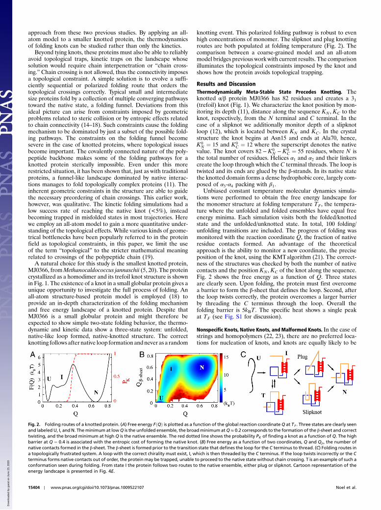
Unbiased constant temperature molecular dynamics simula-tions were performed to obtain the free energy landscape forthe monomer structure at folding temperature TF , the tempera-ture where the unfolded and folded ensembles have equal freeenergy minima. Each simulation visits both the folded/knottedstate and the unfolded/unknotted state. In total, 100 folding/unfolding transitions are included. The progress of folding wasmonitored with the reaction coordinate Q, the fraction of nativeresidue contacts formed. An advantage of the theoreticalapproach is the ability to monitor a new coordinate, the preciseposition of the knot, using the KMTalgorithm (21). The correct-ness of the structures was checked by both the number of nativecontacts and the position KN , KC of the knot along the sequence.Fig. 2 shows the free energy as a function of Q. Three statesare clearly seen. Upon folding, the protein must first overcomea barrier to form the β-sheet that defines the loop. Second, afterthe loop twists correctly, the protein overcomes a larger barrierby threading the C terminus through the loop. Overall thefolding barrier is 5kBT. The specific heat shows a single peakat TF (see Fig. S1 for discussion).
Nonspecific Knots, Native Knots, and Malformed Knots. In the case ofstrings and homopolymers (22, 23), there are no preferred loca-tions for nucleation of knots, and knots are equally likely to be
Fig. 2. Folding routes of a knotted protein. (A) Free energy FðQÞ is plotted as a function of the global reaction coordinateQ at TF . Three states are clearly seenand labeled U, I, and N. Theminimum at lowQ is the unfolded ensemble, the broadminimum atQ ≈ 0.2 corresponds to the formation of the β-sheet and correcttwisting, and the broad minimum at high Q is the native ensemble. The red dotted line shows the probability PK of finding a knot as a function of Q. The highbarrier at Q ¼ 0.4 is associated with the entropic cost of forming the native knot. (B) Free energy as a function of two coordinates, Q and Qβ , the number ofnative contacts formed in the β-sheet. The β-sheet is formed prior to the transition state that defines the loop for the C terminus to thread. (C) Folding routes ina topologically frustrated system. A loop with the correct chirality must exist, I, which is then threaded by the C terminus. If the loop twists incorrectly or the Cterminus forms native contacts out of order, the protein may be trapped, unable to proceed to the native state without chain crossing. T is an example of such aconformation seen during folding. From state I the protein follows two routes to the native ensemble, either plug or slipknot. Cartoon representation of theenergy landscape is presented in Fig. 4E.
15404 ∣ www.pnas.org/cgi/doi/10.1073/pnas.1009522107 Noel et al.
Dow
nloa
ded
by g
uest
on
June
23,
202
0

found anywhere along the sequence. The existence of nativestructure differentiates a protein from these traditional modelsystems where knotting is considered. On a funneled landscape,a protein progressively forms native structure, which implies thata protein is more likely to nucleate a knot in a location containinga loop in the native structure. A knot formed by threading a loopconsisting of native structure is called a native knot, whereas aknot threading a nonnative loop is called a nonspecific (random)knot.
A reasonable criterion to distinguish between these two casesis to define a native knot as when (1) at least one knot crossingdiffers from the native value by less than 10 residues, for example,K0
C − 10 < KC < K0C þ 10 for the C-terminal crossing, and (2)
projection of the knot in the plane gives the native chirality(see SI Appendix for subtleties of the 41 knot). This definitionincludes the following examples of nonspecific knotting: (i) shal-low knots, which could easily appear in a long protein with a deepnative knot; (ii) knots tighter or deeper than native; (iii) knotslocated on the opposite side of the sequence relative to the nativeposition; (iv) an incorrect knot (e.g., of the wrong chirality), whichwould have to untie prior to correct folding. In the unfoldedensemble of MJ0366 we find a nonspecific knot less than 0.1%of the time and never find that a nonspecific knot nucleatesfolding. Most of the nonspecific knotted configurations wereof types i and iii; however, one iv case was also found. This isconsistent with theoretical evidence that folding nucleation bynonspecific knots is entropically unlikely in proteins (11, 13). Thisprocess should have a barrier with a large entropic contributionbecause there is little energetic stabilization until the nativeenvironment forms around the knot.
Kinetic traps on the folding landscape, whose solution wouldbe a chain crossing, are called topological traps. Two types oftopological traps can be defined: (a) a nonspecific knot of typeii, iii, and iv; or (b) a malformed topology with some correct cross-ings but at least one incorrect crossing. These malformed topol-ogies would include cases where the knot is missing. In type a,nonspecific knots of type ii and iii must jump along the sequenceto find the correct native position, but this process might beprohibitively slow (12, 24, 25–26). Nonspecific knots of type ivmust backtrack completely. Backtracking is the process of break-ing a subset of native contacts in order to fall further down thefolding funnel (16). Traps of type bmake subtle crossing mistakes,and these errors can persist to structures with high Q (Fig. 4).Because chain crossing is forbidden, large backtracking excur-sions are required to correct the crossings. Traps of type a weretransient at TF and were not observed during kinetic folding.Traps of type b are also transient at TF but become prevalentat lower temperatures. The specific trapped structures andhow side-chain packing affects their population is discussed indetail in later sections.
Folding Mechanism of a Knotted Domain. To fold a protein with a 31knot there are three distinct folding routes (11): a native-like knotwhere either the N-terminal protein forms a native-like loop forthe C terminus to thread, the C-terminal protein forms a native-
like loop for the N terminus to thread, or a nonspecific knot withlittle native structure that then coalesces to the native knot.Fig. 2C diagrams the folding mechanism at TF . The protein isnever seen to form a knot outside of state I that continues tothe native state. Nonspecific knots of type ii are never seenand those of type i are exceedingly rare. Types iii and iv aretrapped configurations that have to unfold before proceedingto the native state. Because types i and ii are not observed to foldnative knots, the route to the native state must be through nativeloop formation. There are two possible loops to form: loops tobe threaded by either the C terminus (C loop) or N terminus(N loop). The C loop is defined by contacts between Asn15and Tyr49, a loop length ΔL ¼ 34 residues, and is anchored bythe β-sheet. The N-loop can be defined by Ala42 and Val79,ΔL ¼ 37, or Leu28 and Leu74, ΔL ¼ 46. The N loop is notanchored by any secondary structure but is stabilized by thepacking of the helices. Because the loop lengths are approxi-mately the same, the choice of which loop is formed first andsubsequently threaded is likely determined by energetics. TheC loop is stabilized by ∼90 contacts whereas either N loop isstabilized by only ∼60 contacts. Fig. 2B clearly shows that theintra-β sheet contacts required to form the native C-loop struc-ture occur before the transition state. C-loop formation leads toan unstable intermediate with a free energy barrier of 2kBT.
Simply forming the β-sheet is not enough to define the native Cloop; the loop must be twisted correctly. It is possible to twistthe β-sheet 360 ° and arrive at a nearly native configuration thatdiffers only in the crossing near Asn15 and Tyr49 (Fig. 4C). This
Fig. 3. Two possible native loops for threading the trefoil knot. K0N and K0
Cdenote the length of the native threaded N terminal and C terminal, respec-tively, and dotted lines show their extension. The dominant route depends onthe relative values of K0
N and K0C . (Center) Native structure with the two loops
highlighted in red and blue. (Left) Folding via C loop. (Right) Folding via Nloop.
Fig. 4. Energy landscape of a knotted protein and possible topological trapsfor a 31 knot. (A) The native fold. (B–D) Examples of topologically trappedstructures. Backtracking must occur for these configurations to reach thenative knot. The arrows denote the incorrect crossings. Configuration B isthe most prevalent all-atom trap while C and D are most prevalent in Cα.Configuration D is not observed in all-atom. Configuration C is the transitionstate for the less-accessible slipknot via N-loop route (see Fig. 3). (E) Funneledlandscape. The protein is biased to a preordered intermediate that has therelevant polypeptide crossings correct and contains a loop in the native posi-tion. α4 threads the loop via parallel pathways. The empty space emphasizesthe bifurcation in the landscape. Due to threading, not all routes are allowed.Topologically trapped states exist on the landscape and can differ by only asingle crossing. Thus, these traps can be very deep in energy. Since chain cross-ing is not allowed, they are disconnected from the native state and mustbacktrack to fold correctly. The higher energy traps correspond to panel Cand the symmetric traps close to the native structure correspond to panelB. In the structure-based model these traps are transient at TF but canbecome kinetic traps at lower temperature.
Noel et al. PNAS ∣ August 31, 2010 ∣ vol. 107 ∣ no. 35 ∣ 15405
BIOPH
YSICSAND
COMPU
TATIONALBIOLO
GY
PHYS
ICS
Dow
nloa
ded
by g
uest
on
June
23,
202
0
minor structural difference gives a topologically incorrect andpotentially trapped structure lacking the knot. Forming the twisttakes the protein from the meta-stable intermediate at Q ∼ 0.2 tothe plateau atQ ∼ 0.25. It is essentially a barrierless process, but itmust occur before the transition state.
After the C loop is formed and correctly twisted, the C termi-nus must overcome both an entropic barrier and a topologicalbarrier to reach the knotted fold. The entropic barrier arisesas the C terminus trades its conformational freedom for the for-mation of the hydrophobic core, and the topological barrier arisesfrom the excluded volume of the loop and the need to threadthe C terminus through it. This topological barrier manifestsas an increased entropic barrier, because the number of routesto the native state are limited by the constraint. Forming thehydrophobic core could be a driving force toward forming theknot because it consists mostly of contacts between β1 and thethreaded α4. The two possible mechanisms for threading areeither a plug or a slipknot intermediate (see Fig. 2C). The plug-ging mechanism appears when the C terminus is the first part ofthe protein chain to thread the C loop. Native contacts arenot formed until the C terminus reaches its native position. Thismechanism happens through random fluctuations of the C termi-nus impinging on the C loop. The slipknotting mechanism occurswhen part of the protein chain (near the C terminus) threads theC loop but doubles back so the protein chain stays unknotted, ahairpin-like configuration (see Movie S1). The slipknot is stabi-lized by forming native hydrophobic core contacts between α3-α4and β1, between Phe10 and Ile63, for example. As the slipknottedintermediate is stabilized by native contacts, the C terminus hastime to thread the loop. The C loop’s ability to accommodate thisbulky configuration is facilitated by the flexible five residue chainβ2 and the melting of the C loop helices. At TF the protein foldsby the plugging mechanism 55% and by the slipknot mechanism45%. The coexistence of these two pathways was also seen in thefolding of YibK (13). The equilibrium between these twomechanisms, though, is highly dependent on the length of thethreaded C terminus and is discussed in the next section.
Slipknotting Is a General Knotting Mechanism. To investigate howthe knotting mechanism is affected by the depth of the knot, fold-ing of the MJ0366 structure with an extended C-terminal helixwas studied and is summarized in Table 1. Because the actualsequence of MJ0366 used for crystallization has five additionalresidues at the C terminus, it is known that MJ0366 is able toknot with a longerC-terminal tail. The simulated C-terminal helixwas extended using the five additional residues indicated in thecrystal data, increasing K0
C to 17. The only native contacts addedwere local helix contacts in the extended region; no additionalcontacts with the rest of the protein were added. When kineticfolding is performed at 0.96TF , the extended structure foldsvia the slipknot route 99% of the time. The plugging mechanismis dependent on putting the C-terminal chain into a preciseconfiguration to slide across the loop, which becomes less likely
as the entropy of the C-terminal chain increases with additionalresidues. At the same time, the additional entropy of the ex-tended C terminus stabilizes the native contacts that supportthe slipknot intermediate.
A comparison between a coarse-grained (Cα) model that has asingle bead-per-residue with the all-atom (AA) model allowsmore direct connection with previous simulations on knottedproteins (5, 11, 13). Two of these studies (5, 11) suggested, usinga completely funneled Cα folding model, that slipknotting is thelikely folding route for a more deeply knotted protein. The AAsimulations of the extended C-terminal tail MJ0366 corroboratethis claim. For completeness, kinetic folding simulations of aCα model at 0.96TF of both the PDB structure and an extendedC-terminal structure were performed. The observed foldingroutes are shown in Table 1. The folding of the unextendedstructure is reminiscent of the unextended AA model where theprotein folds via either slipknotting or plugging through the Cloop. Interestingly, a novel route is seen in the extended structure,slipknotting via the N loop (Fig. 3). As the C terminus is ex-tended, the difficulty of threading it through the C loop increasesrelative to theN-terminal threading theN loop, opening up a newkinetically accessible route. This route is not seen in the AAmod-el, likely because the addition of side chains makes the N termi-nus too bulky to fit through the much tighter N loop. Although,upon extending the C terminus beyond 10 additional residues,this route may become accessible in the AA model. In summary,the deeper knots follow the same slipknot mechanism, though theprotein may switch the threading terminus depending on thedepth of the knot at the two termini. This is possible in this pro-tein due to the approximate symmetry between the N loop andC loop.
Topological Traps on the Folding Landscape.As explained previously,malformed knots with subtle crossing mistakes are topologicaltraps. These traps are regions of configuration space that canbe close to native-like states in energy but are topologicallydistant because they can only be directly connected to the nativestate through chain crossing. Trapped configurations are forcedto backtrack in order to reach the native fold.
The folding landscape at TF is smooth, because the thermody-namic data showed no evidence of long-lived trapped states.Because physiological temperatures are below TF , new featuresin the landscape can arise from the altered competition betweenenergy and entropy. Collapsed states become more favorable,which impacts topologically frustrated systems that must be ableto easily backtrack from the frustrating conformations for effi-cient folding. As the temperature is lowered, the time for escapefrom these conformations, the backtracking time, is greater.Investigating folding below TF can ascertain which topologicaltraps might become important at lower temperatures.
Kinetic folding simulations of MJ0366 were performed, start-ing from random unfolded conformations and quenched totemperatures 0.96TF , 0.91TF , 0.86TF . They are summarized inFig. 5 C and D. As the temperature is decreased the folding timealso decreases as the competition between energy and entropyfavors an increasingly compact ensemble. If the temperature isdecreased far enough, the mean first passage time to reachthe native ensemble τmfpt begins to increase as the protein spendsmore time in topological traps. At 0.96TF the time spent in trapsis negligible compared to τmfpt, while at 0.91TF 3% of the trajec-tories visit a trap and at 0.86TF 14% of the trajectories visit atrap. The average time spent in the topological traps increasesbecause the barriers to backtracking are increasing. The topologyof the most common trap is shown in Fig. 4B. The C terminusmakes native hydrophobic core contacts without threading theloop. A second topological trap is seen at 0.86TF , the β-sheetforms with the incorrect chirality for the loop. The C terminuscan thread the incorrectly twisted loop and make most of its
Table 1. Relative population of folding routes (in %) for theAA and Cα model
AA model Cα model
Route PDB PDB+5 PDB PDB+5
slipC 68 99.3 38 16slipN 2 0.35 1.5 72plugC 28 0.35 57.5 3plugN - - - 1.5nonspecific - - 1.5 1.5
Results for the PDB structure and the five residue extended structureare shown. Subscripts N and C denote slipknotting or pluggingknotting route via the N loop and C loop, respectively. Forexample, slip slipC denotes slipknotting by threading the C loop.
15406 ∣ www.pnas.org/cgi/doi/10.1073/pnas.1009522107 Noel et al.
Dow
nloa
ded
by g
uest
on
June
23,
202
0

native contacts, even though the overall topology is trivial. Thisconfiguration is shown in Fig. 4C. These traps, along with others,exist at TF . They simply have much shorter lifetimes.
Addition of Side Chains Reduces Topological Trapping. A closer lookat the comparison between the Cα model and the AAmodel high-lights the role of the geometry in discriminating folding routes.The results show the Cα model is more prone to topologicallytrapped structures than AA. To quantify the ability of the AAmodel to avoid topological traps, kinetic folding of the Cα modeland the AA model are compared. Fig. 5D shows that the Cαtrajectories fall into traps more often than the AA. All of thetrapped Cα structures, but only ≈20% of the trapped AA struc-tures, were of the types in Fig. 4 C and D. Of the few trapped AAstructures, most are of the type shown in Fig. 4B. The additionof side chains serves to break the symmetry in the Cα geometry,for example, in the β-sheet (Fig. 4 C and D).
A clear difference between the two models is captured inFig. 5A by comparing route measure RðQÞ (27, 28) along thefolding pathway. RðQÞ quantifies the amount of available config-uration space that is actually accessed during folding (seeSI Appendix). A larger route measure signifies a smaller numberof routes traversed during folding. Knot formation is the stage offolding where avoiding incorrect crossings is critical. TheCα mod-el is seen to have a more diverse set of routes leading to the tran-sition state. Also the smaller slope of the knot probability versusQ shows knot formation is less cooperative in the Cα model. Theincreased persistence length coupled to more precise atomicpacking in the AA model imposes an energetic penalty on routescontaining improper chain crossings and therefore reduces topo-logical trapping. Due to the importance of correct packing,knotted proteins may be particularly sensitive to mutations inthe crossing regions.
Dimerization Occurs After Knotting. Studying the process of dimer-ization is important because it could have an effect on the foldingof the knotted structures. The question is whether the topologyforces native-like monomers to fold before dimerization orwhether the dimerization step could be coupled to the foldingprocess as in so-called obligatory dimers (29). YibK, a 31 knottedprotein, has been shown experimentally to first fold to a native-like monomeric state before a slow dimerization step (10).MJ0366 has a homodimeric interface similar to that of YibK:Both interfaces include the C-terminal helix directly involvedin the knotted structure. See SI Appendix for the structure ofthe dimer. MJ0366 has a shallower knot than YibK and a higher
proportion of dimeric contacts than YibK, both of which couldcause the dimerization to more greatly impact the folding ofMJ0366.
The dimerization process was investigated by performing fold-ing simulations with two monomers present, starting with bothunfolded. Contacts between the monomers in the crystal struc-ture were included with the same strength as intramonomercontacts. Results show that knot formation is unaffected bythe presence of the dimer (see Figs. S2 and S3 for details).The transition-state ensemble is nearly identical whether foldingin isolation or in the presence of another dimer. A correlationcoefficient of 0.995 is seen between the transition state of anisolated monomer and two monomers held in close proximityby a harmonic spring constant k ¼ 4ϵ∕ nm2. Also, contactsbetween monomers are rarely formed in the transition statesof the monomers, Qdimer < 0.05 over a broad range of effectivemonomer concentrations. This remarkable result emphasizes therobustness of the proposed monomeric folding mechanism.
ConclusionsThis study maps the full thermodynamic energy landscape of aknotted protein, and we find that the folding is a thermodyna-mically three-state system: unfolded, loop formation, native-knotted structure. Below TF , kinetic folding also follows thesame three-state mechanism along with increased prevalenceof topological traps. An earlier Cα model for folding was shownto overestimate the importance of trapping. At folding tempera-ture two parallel knotting mechanisms are observed: slipknot andplug. At lower temperatures and with an extended C-terminaltail, the mechanism switches exclusively to slipknotting, as theentropically limited plug pathway is suppressed. Further supportfor the slipknot pathway comes from the observation of slipknotsin native protein structures (4). This folding route is consistentwith previous work on YibK (11), so it will be instructive to applythe all-atom model to YibK and other larger knotted proteins.
Our results suggest some general features of folding knots inproteins. More deeply knotted proteins should tend toward creat-ing knots through slipknotting. The viability of the slipknot routeassumes that there is some preordered native structure in theknotted domain to provide both scaffolding to anchor the slip-knot intermediate and a native loop for the terminus to thread.The inherent geometric constraints in the knotted domain mustbe sufficient to ensure the correct ordering of crossings. It hasbeen shown that more complicated “closed” knots, 41, 52, 61,can be unknotted by switching a single crossing (3, 5). This isequivalent to the “open” knots being able to be tied through a
Fig. 5. Side-chain packing reduces topological frustration. (A) (Top) Free energy as a function of global parameter QCA are compared between the all-atom(AA) model (dashed) and a one-bead-per-residue (Cα) model (solid). The Cα model has a much less defined shoulder around QCA ¼ 0.25 and has an unfoldedbasin corresponding to a formed β-sheet. The probability PK of a knot is shown in red. The knot is formed more gradually alongQCA in the Cα model. Also notethat the folded basin is more likely to be unknotted in the Cα model. (bottom). A comparison of the route measure RðQÞ. The AA model peaks at the transitionstate where the knot is formed while the Cα model peaks near the unfolded state. The protein configurations leading to the transition state in the AA modelare fewer, leading to fewer topological traps. (B) An example kinetic folding trajectory with the AAmodel at T ¼ 0.91TF . The protein spends time in the loopedintermediate state before falling into a topological trap T. The proteinmust backtrack before reaching the native state. KN and KC denote theN and C-terminaldepths of the knot, respectively. The knot only forms upon folding to the native state. (C) Mean first passage times τmfpt are shown for four differenttemperatures 0.86TF , 0.91TF , 0.96TF , and TF for the AA model (black) and Cα model (red). τmfpt is split between the trajectories that spend a significant timein a topological trap (squares) and those that do not (triangles). The overall τmfpt is shown (crosses). τmfpt decreases as temperature is lowered from TF andreaches a minimum near 0.91TF. Due to trapping the overall τmfpt is greater at 0.86TF than 0.91TF . (D) The percentage of trajectories that become trapped Ptrap
for the AA model (black) and Cα model (red) at different temperatures.
Noel et al. PNAS ∣ August 31, 2010 ∣ vol. 107 ∣ no. 35 ∣ 15407
BIOPH
YSICSAND
COMPU
TATIONALBIOLO
GY
PHYS
ICS
Dow
nloa
ded
by g
uest
on
June
23,
202
0

single loop crossing event. These more complicated protein knotscan therefore fit naturally into the mechanism of a preordereddomain coupled to a final native loop threading that createsthe nontrivial topology. This would extend the folding pathwayfor the smallest knotted protein to all knotted proteins.
This scenario of preordered native structure preceding knotformation is in direct contrast to folding through a random, non-specific knot that then coalesces into the native knot. The resultsfrom the extended Cα model show an underlying plasticity in thefolding landscape because the protein can switch the threadingterminus if the native geometry is perturbed, obviating a kineti-cally accessible nonspecific knotting route. There are infrequentinstances of nonspecific knots forming in the unfolded ensemblein our simulations, but these events do not nucleate folding.Instead, the nonspecific knots always backtrack. This result issomewhat surprising because the early formation of a knot wouldseem to surpass the topological barrier. It shows there are stillsignificant barriers to jumping the position of a random knotto the native position. This is in contrast to the behavior of knotsin flexible random polymers.
These observations raise the important question of the dy-namics of knots in denatured proteins. Suppose a knotted proteinis rapidly denatured in an experiment before the knot can untie.Are the dynamics of the knot on the denatured polypeptide“polymer-like,” where a knot is able to become either tightenedor slide along the sequence, or are the dynamics “protein-like,”where there exist large barriers (12, 24) to changing the knot’sposition? The answer to this question is crucial for interpretingexperimental refolding data of knotted proteins and is currentlyunder investigation.
Materials and MethodsAll-Atom Model. The all-atom model was described previously (18) and isavailable on a web server (30). The model used in this work is identical except
that we use a Gaussian-type contact potential (31). In the all-atom, structure-based model of the protein, only heavy (non-hydrogen) atoms are included.Each atom is represented as a single bead of unit mass. Bond lengths, bondangles, improper dihedrals, and planar dihedrals are maintained by harmonicpotentials. Nonbonded atom pairs that are in contact in the native state aregiven an attractive Gaussian well potential, while all other nonlocal interac-tions are repulsive. All attractive potentials are given values taken from thenative state. GROMACS 3.3.3 was used for the molecular dynamics (32). Forcomparison, a standard Cα coarse-grained protein model was used and isdescribed in refs. 15 and 30. The Cα contact map is constructed from theall-atom contact map by including all residue pairs that have at least oneatom–atom contact between them. Thermodynamics data was obtainedfrom constant temperature molecular dynamics, and histograms from multi-ple temperatures were combined using the Weighted Histogram AnalysisMethod (33). All structures were visualized using VMD (34).
Reaction Coordinates. Q is defined as the fraction of native residues in contact(18). A residue contact is formed if any of their native atomic contacts areformed. A contact between atoms i and j is formed if rij < 1.2r0ij , where r0ijis the pair distance in the native state. QCA is a coarse-grained version of Qthat defines a residue contact as formed if the Cα positions of residues i
and j satisfy rij < 1.2r0ij. Qβ comprises the contacts between residues 7–12and 49–54.
Identification of the Knot in the Protein, KMT Algorithm. The positions KN∕KC during folding were determined in the same way as the native knot(21), applying the procedure at each simulation snapshot as described inref. 24. The slipknot conformationwas detected as described before in ref. 12.
ACKNOWLEDGMENTS. This work was supported by the Center for TheoreticalBiological Physics sponsored by the National Science Foundation (NSF)(Grant PHY-0822283) with additional support from NSF-MCB-0543906. J.K.N.acknowledges support from the National Institutes of Health MolecularBiophysics Training Program, Grant T32GM08326.
1. Mansfield ML (1994) Are there knots in proteins? Nat Struct Biol 1:213–214.2. Virnau P, Mirny LA, Kardar M (2006) Intricate knots in proteins: Function and
evolution. PLOS Comput Biol 2:e122 doi: 10.1371/journal.pcbi.0020122.3. Taylor WR (2007) Protein knots and fold complexity: Some new twists. Comput Biol
Chem 31:151–162.4. King NP, Yeates EO, Yeates TO (2007) Identification of rare slipknots in proteins and
their implications for stability and folding. J Mol Biol 373:153–166.5. Bolinger D, et al. (2010) A Stevedore’s protein knot. PLoS Comput Biol 6:e1000731.6. Bryngelson JD, Wolynes PG (1987) Spin glasses and the statistical mechanics of protein
folding. Proc Natl Acad Sci USA 84:7524–7528.7. Leopold PE, Montal M, Onuchic JN (1992) Protein folding funnels—A kinetic approach
to the sequence structure relationship. Proc Natl Acad Sci USA 89:8721–8725.8. Onuchic JN, Wolynes PG (2004) Theory of protein folding. Curr Opin Struct Biol
14:70–75.9. Mallam AL, Rogers JM, Jackson SE (2010) Experimental detection of knotted confor-
mations in denatured proteins. Proc Natl Acad Sci USA 107:8189–8194.10. Mallam AL, Morris ER, Jackson SE (2008) Exploring knotting mechanisms in protein
folding. Proc Natl Acad Sci USA 105:18740–18745.11. Sulkowska JI, Sulkowski P, Onuchic JN (2009) Dodging the crisis of folding proteins
with knots. Proc Natl Acad Sci USA 106:3119–3124.12. Sulkowska JI, Sulkowski P, Onuchic JN (2009) Jamming proteins with slipknots and
their free energy landscape. Phys Rev Lett 103:268103.13. Wallin S, Zeldovich KB, Shakhnovich EI (2007) The folding mechanics of a knotted
protein. J Mol Biol 368:884–893.14. Shea J, Onuchic JN, Brooks CL (1999) Exploring the origins of topological frustration:
Design of a minimally frustrated model of fragment B of protein A. Proc Natl Acad SciUSA 96:12512–12517.
15. Clementi C, Nymeyer H, Onuchic JN (2000) Topological and energetic factors:What determines the structural details of the transition state ensemble and“on-route” intermediates for protein folding? An investigation for small globularproteins. J Mol Biol 298:937–953.
16. Gosavi S, Chavez L, Jennings P, Onuchic JN (2006) Topological frustration and thefolding of interleukin-1 beta. J Mol Biol 357:986–996.
17. Klimov DK, Thirumalai D (1999) Deciphering the timescales andmechanisms of proteinfolding using minimal off-lattice models. Curr Opin Struct Biol 9:197–207.
18. Whitford PW, Noel JK, Gosavi S, Schug A, Onuchic JN (2009) An all-atom structure-based potential for proteins: Bridging minimal models with empirical forcefields.Proteins 75:430–441.
19. Norcross T, Yeates TO (2006) A framework for describing topological frustration inmodels of protein folding. J Mol Biol 362:605–621.
20. Bult CJ, et al. (1996) Complete genome sequence of the methanogenic archaeon,Methanococcus jannaschii. Science 273:1058–1073.
21. Koniaris K, Muthukumar M (1991) Knottedness in ring polymers. Phys Rev Lett66:2211–2214.
22. Virnau P, Mirny LA, Kardar M (2005) Knots in globule and coil phases of a modelpolyethylene. J Am Chem Soc 27:15102–15106.
23. Raymer DM, Smith DE (2007) Spontaneous knotting of an agitated string. Proc NatlAcad Sci USA 104:16432–16437.
24. Sulkowska JI, Sulkowski P, Szymczak P, Cieplak M (2008) Tightening of knots in theproteins. Phys Rev Lett 100:058106.
25. Huang L, Makarov DE (2008) Translocation of a knotted polypeptide through a pore.J Chem Phys 129:121107.
26. Bornschlogl T, et al. (2009) Tightening the knot in phytochrome by single-moleculeatomic force microscopy. Biophys J 96:1508–1514.
27. Plotkin SS, Onuchic JN (2002) Understanding protein folding with energy landscapetheory—Part I: Basic concepts. Q Rev Biophys 35:111–167.
28. Clementi C, Garcia AE, Onuchic JN (2003) Interplay among tertiary contacts, secondarystructure formation and side-chain packing in the protein folding mechanism:All-atom representation study of protein L. J Mol Biol 326:933–954.
29. Levy Y, Wolynes PG, Onuchic JN (2004) Protein topology determines binding mecha-nism. Proc Natl Acad Sci USA 101:511–516.
30. Noel JK, Whitford PC, Sanbonmatsu KY, Onuchic JN (2010) SMOG@ctbp: Simplifieddeployment of structure-based models in GROMACS. Nucleic Acids Res doi:10.1093/nar/gkq498.
31. Lammert H, Schug A, Onuchic JN (2009) Robustness and generalization of structure-based models for protein folding and function. Proteins 77:881–891.
32. Hess B, Kutzner C, van der Spoel D, Lindahl E (2008) GROMACS 4: Algorithms for highlyefficient, load-balanced, and scalable molecular simulation. J Chem Theory Comput4:435–447.
33. Kumar S, et al. (1992) The weighted histogram analysis method for free-energycalculations on biomolecules. I. The method. J Comput Chem 13:1011–1021.
34. Humphrey W, Dalke A, Schulten K (1996) VMD—Visual molecular dynamics. J MolGraphics 14:33–38.
15408 ∣ www.pnas.org/cgi/doi/10.1073/pnas.1009522107 Noel et al.
Dow
nloa
ded
by g
uest
on
June
23,
202
0