Slides - Regional Geriatric Program of Eastern Ontario
75
Champlain LHIN Falls Prevention Strategy: Pilot Algorithm – Box 5 Assessment Dr. Frank Molnar Medical Director, Regional Geriatric Program of Eastern Ontario ( www.rgpeo.com ) Ottawa Geriatric Assessment Outreach Teams (GAOT) Champlain Geriatric Emergency Management (GEM) Program Staff Physician, Geriatric Medicine - The Ottawa Hospital - The Winchester District Memorial Hospital - The Cornwall Community Hospital
Transcript of Slides - Regional Geriatric Program of Eastern Ontario
Champlain LHIN Falls Prevention
Strategy: Pilot Algorithm
– Box 5 Assessment
Dr. Frank Molnar
Medical Director,
Regional Geriatric Program of Eastern Ontario ( www.rgpeo.com )
Ottawa Geriatric Assessment Outreach Teams (GAOT)
Champlain Geriatric Emergency Management (GEM) Program
Staff Physician, Geriatric Medicine
- The Ottawa Hospital
- The Winchester District Memorial Hospital
- The Cornwall Community Hospital

•It is estimated that one in three seniors are likely to fall
at least once per year
(World Health Organization (WHO 2007)
•Falls are one of the most common cause of injury and
the sixth leading cause of death for seniors. Senior Care Canada
• Every 10 minutes in Ontario, at least one older adult visits an ED
due to a fall
• Ontario Injury Prevention Statistics, 2007-2008
• Every 30 minutes in Ontario, at least one older adult is admitted to
hospital due to a fall
• Ontario Injury Prevention Statistics, 2007-2008
• Falls are the leading cause of overall injury costs in Canada and
account for $6.2 billion or 31% of total costs of all injuries
• Smartrisk, 2009

The Good News !!!
The literature suggests that as many
as 1/3 of falls- related adverse
outcomes are preventable
Division of Aging and Seniors 2006
The Vital sign concept (opportunity to help)
The sudden onset of falls in someone who
previously did not have a falling tendency most
likely represents underlying illness
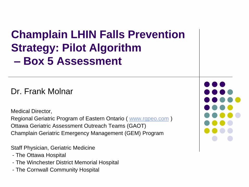
Why are FALLS so difficult to assess?
No other mammals spend their day standing upright - upright balance requires smooth functioning and integration of complex neurological and cardiovascular systems.
Therefore FALLS can be caused by multiple problems: vertigo, strokes, cardiac and neurological diseases, neck disorders, physical deconditioning, and medications that do not fall under a single organ-specific specialty.
The Assessment of FALLS is not a major focus of the medical curriculum.


In Office Assessment
Box 5 based on American Geriatrics Society and
British Geriatrics Society Clinical Practice Guideline
Priority influenced by prevalence of problems seen
by Geriatric Assessment Outreach Teams (GAOT
see 1800 seniors per year)
Most modifiable first
This pilot is an ongoing iterative process. Your input
throughout the study will help refine what is and is
not realistic in Primary Care setting – you will drive
refinements of the algorithm

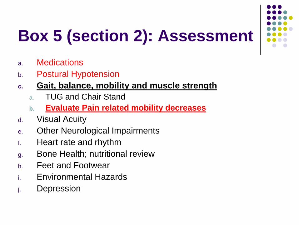
Box 5 (section 2): Assessment
a. Medications
b. Postural Hypotension
c. Gait, balance, mobility and muscle strength
a. TUG and Chair Stand
b. Evaluate Pain related mobility decreases
d. Visual Acuity
e. Other Neurological Impairments
f. Heart rate and rhythm
g. Bone Health; nutritional review
h. Feet and Footwear
i. Environmental Hazards
j. Depression

Box 5 – 2a.
Medications
1. Slow reaction time (drugs that cause delirium and slow mentation) Narcotics, benzodiazepines, ETOH, Anticholinergics (e.g.
Ditropan, Detrol, Tricyclic antidepressants ).
2. Decrease cerebral perfusion (see Postural hypotension – 6 ANTIs
Anti-hypertensives,Anti-anginals,Anti-parkinsonian medications (e.g. sinemet),Anti-depressants (e.g., Anti-cholinergic tricyclics),Anti-psychotics (Anti-cholinergic effect),Anti-BPH (e.g. Hytrin, Flomax)
3. Cause parkinsonism Antipsychotics
GI – Stemetil, Maxeran
4. Vestibular Toxicity Aminoglycosides, High dose loop diretics
5. SSRIs Evidence is building that SSRIs increase fall risk

Decrease or stop drugs that
can cause Delirium
Is the patient on delirium inducing drugs :
Benzodiazepines
Narcotics
Alcohol
Antihistamines
Neuroleptics (Antipsychotics) Haldol, Respiridone, Olanzepine
Anticonvulsants (Seizure medications)
Dilantin (Phenytoin), Gabapentin, Pregabalin
Keep dilantin level at level that previously controlled seizures – if this info not available then try to keep level < 60)
Anticholinergics (see next slides)

How can one sort through this daunting
list of medications?
Look for a time-based relationship Falls or confusion
worsened after starting this medication (or increasing the dose).
Ask Pharmacist to review drugs that may be contributing to falls and/or impairing cognition (theoretical perspective) and then apply a practical lens based on personal knowledge of patient to develop a tailored personalized plan for medication adjustments (“strategic deprescribing”).

Box 5 (section 2): Assessment
a. Medications
b. Postural Hypotension
c. Gait, balance, mobility and muscle strength
a. TUG and Chair Stand
b. Evaluate Pain related mobility decreases
d. Visual Acuity
e. Other Neurological Impairments
f. Heart rate and rhythm
g. Bone Health; nutritional review
h. Feet and Footwear
i. Environmental Hazards
j. Depression

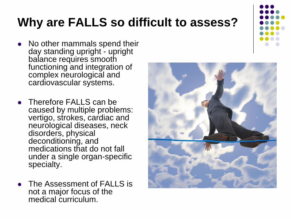
Box 5 – 2b.
Postural Hypotension
Lightheadedness 1-3 min after sitting or standing Perspiration, nausea, weakness, dizziness
Measure BP and Pulse after the person has been lying for at least 3-5 minutes and 1 - 3 minutes after standing
A decline of >20 mm Hg in systolic BP and/or >10 mm Hg in diastolic BP on the assumption of an upright posture with or without an increase in PR American Academy of Neurology
High incidence (as high as 30%) among older people (due to age-related changes in the CV & nervous systems & medication use)
Box 5 – 2b.
Postural Hypotension
4D-AID acronym Causes associated with a compensatory tachycardia – 3Ds
Deconditioning
Dysfunctional Heart Myocardium (very low Left Ventricular Ejection Fraction)
Aortic Stenosis
Dehydration Disease
Dialysis (post dialysis dry weight too low)
Drugs
Diuretics
Anorexic Drugs – narcotics, digoxin, antibiotics, cholinesterase inhibitors
Drugs – 6 ANTIs Anti-hypertensives
Anti-anginals
Anti-parkinsonian medications (e.g. sinemet)
Anti-depressants (e.g., Anti-cholinergic tricyclics)
Anti-psychotics (Anti-cholinergic effect)
Anti-BPH (e.g. Hytrin, Flomax)
Causes that present with lack of compensatory tachycardia - AID
Autonomic Dysfunction Diabetic autonomic neuropathy (consider if patient has peripheral neuropathy)
Low B12
Hypothyroidism
ETOH abuse
Parkinsonism (Parkinson’s disease, Progressive Supranuclear Palsy, Multisystem Atrophy (e.g. Shy Drager))
Idiopathic (Bradbury-Eggleston) Depletion of Norepinephrine from sympathetic nerve terminals
Drugs Beta-Blockers
Box 5 (section 2): Assessment
a. Medications
b. Postural Hypotension
c. Gait, balance, mobility and muscle strength
a. TUG and Chair Stand
b. Evaluate Pain related mobility decreases
d. Visual Acuity
e. Other Neurological Impairments
f. Heart rate and rhythm
g. Bone Health; nutritional review
h. Feet and Footwear
i. Environmental Hazards
j. Depression

Box 5 – 2c.
Gait, balance, mobility and muscle strength
Romberg
Get Up and Go
Timed Up and Go
30 second Chair Stand Test
Question?
Which, if any, of these do you feel are useful in
clinical practice?

Romberg's Test is NOT a test of cerebellar function
It is a test of the proprioception receptors and
pathways function.

Romberg’s Test
What is Being Tested in the Romberg Test?
With the eyes open, three sensory systems provide
input to the cerebellum to maintain truncal stability.
These are vision, proprioception, and vestibular
sense.
Proprioception-The brain's awareness of a joint's or
limb's position in relation to the rest of the body
Vestibular Sense- Equilibrium

EYES CLOSED
If there is a mild lesion in the Vestibular or
Proprioception systems, one is usually able
to compensate with the eyes open. With the
eyes closed, however, visual input is
removed and instability can be brought out.
Increased swaying with eyes closed
would indicate postural position sense is
affected, posterior column disease or a
peripheral neuropathy.

When you do the Romberg maneuver, you need to stand close to the patient and be ready to catch them in case they fall
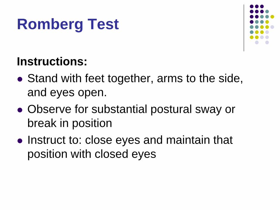
Romberg Test
Instructions:
Stand with feet together, arms to the side,
and eyes open.
Observe for substantial postural sway or
break in position
Instruct to: close eyes and maintain that
position with closed eyes
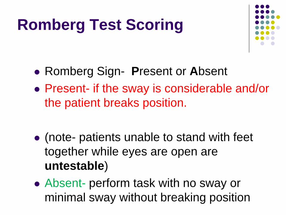
Romberg Test Scoring
Romberg Sign- Present or Absent
Present- if the sway is considerable and/or
the patient breaks position.
(note- patients unable to stand with feet
together while eyes are open are
untestable)
Absent- perform task with no sway or
minimal sway without breaking position

Get Up and Go Test
The "Get Up and Go" test was developed by
Mathias, Nayak, and Issacs in 1986.
A general physical performance test
used to assess mobility, balance and
locomotor performance in elderly people
with balance disturbances. More
specifically, it assesses the ability to
perform sequential motor tasks relative
to walking and turning
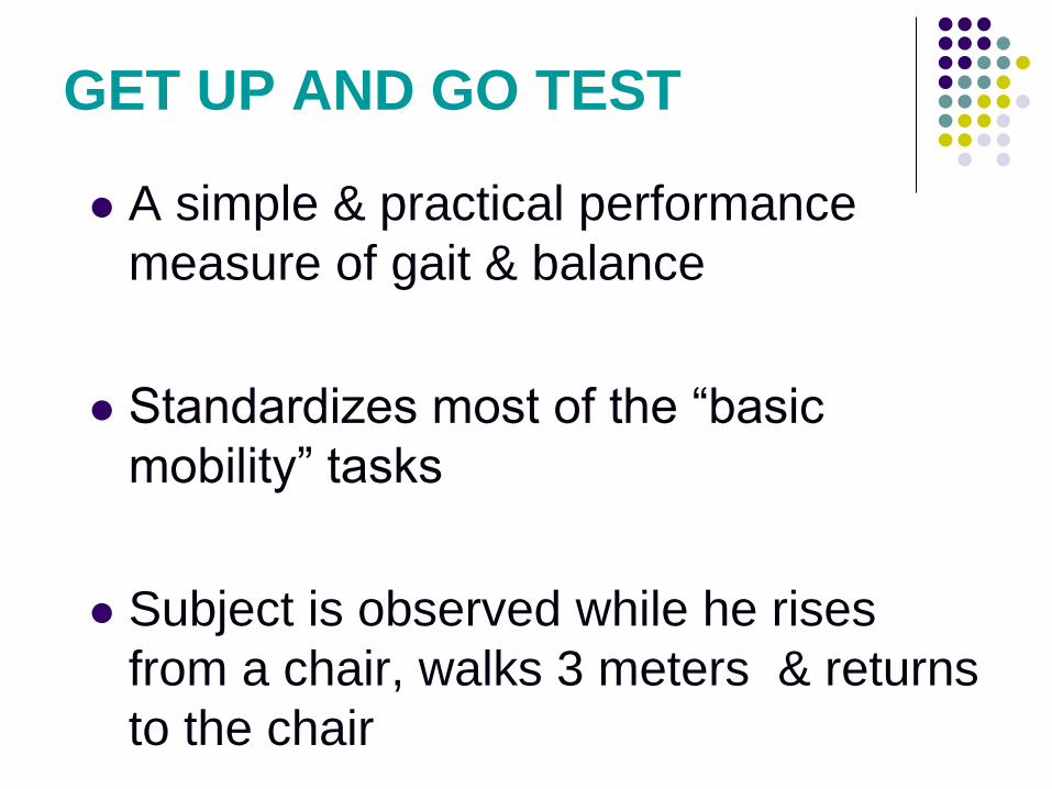
GET UP AND GO TEST
A simple & practical performance
measure of gait & balance
Standardizes most of the “basic
mobility” tasks
Subject is observed while he rises
from a chair, walks 3 meters & returns
to the chair

PROCEDURE
Place a straight-back chair 3 meters from and facing the wall (preferably one that does not have a seat which slants back)
1. Ask senior to rise from chair, without using arms for support & stand still for a moment
2. Walk towards the wall
3.Turn without touching the wall & walk back to the chair& sit down

Get Up and Go
Observe rising from chair-watch the speed of rising, do they need assistance or a boost, watch their shoulders to see if they lean forward on rising. Are you worried they might fall.?

Get Up and Go
Standing- what is their stance?, do they lean
to one side?, do they sway?, do they have
any balance problems?, are you worried they
might fall?
Check for postural abnormalities
Do they complain of pain standing still?

Get Up and Go
Walking-watch the height and width of their steps, are their steps irregular?, can they maintain their balance while walking?, are you worried they might fall? Look for asymmetric arm swing, abnormal arm and hand postures, and instability of the trunk. Hesitancy might suggest Parkinson's.
Decreased step height might suggest CNS disease, multiple Sensory deficits, Fear of Falling, Parkinson's, NPH, Habit. Path deviation might suggest Cerebellar disease, multiple Sensory deficits, sensory or motor Ataxia

Get Up and Go
Turning-watch the speed of turning
steadiness and number of foot placements
needed to complete the turn.
Are you worried they might fall?
Unsteadiness may suggest Parkinson's,
multiple Sensory deficits, Cerebellar disease,
Hemiparesis, loss of Visual Field, Ataxia
Get Up and Go
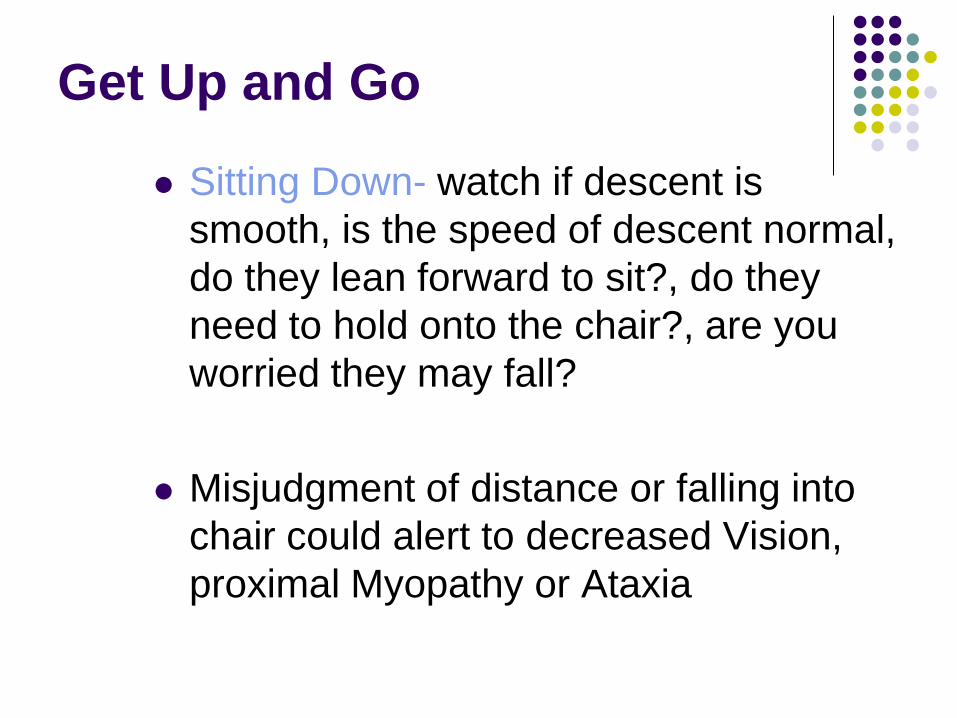
Sitting Down- watch if descent is
smooth, is the speed of descent normal,
do they lean forward to sit?, do they
need to hold onto the chair?, are you
worried they may fall?
Misjudgment of distance or falling into
chair could alert to decreased Vision,
proximal Myopathy or Ataxia

Uses chair to sit, does
not control descent,
nearly misses chair
Slow descent,
hesitates or pauses
during descent
Smooth decent, does
not use chair for
support
Sitting down
More than 6 foot
placements to turn or
cannot safely execute
turn, staggers
Slowness, hesitation,
4-5 foot placements to
turn
No hesitation, takes
2-3 foot
placements to turn
Turning
Severe trunk sway (5-
10 degrees), reaches
out hand to balance,
staggers
Wide stance, irregular
posture
No signs of
instabilityStanding
Uses assist
throughout rising,
leans forward
Uses assist to begin
rising
No slowness (<
4sec) or hesitancyRising from
chair
Mod/Severely
Abnormal
Mild
Abnormalities
NormalManeuver
Get Up and Go Check List

Scoring of Get up and Go
It can be scored qualitatively
Normal or Abnormal Or on a scale from 1- 5
l (normal )
2 (slightly abnormal)
3 (mildly abnormal)
4 (moderately abnormal)
5 (severely abnormal)

TUG Timed Up and Go Test
The TUG was published by Podsiadlo
and Richardson in 1991 to address the
issues of poor inter-rater reliability
observed with intermediate scores in the
"Get Up and Go". The TUG incorporates
time as the measuring component to
assess general balance and function.
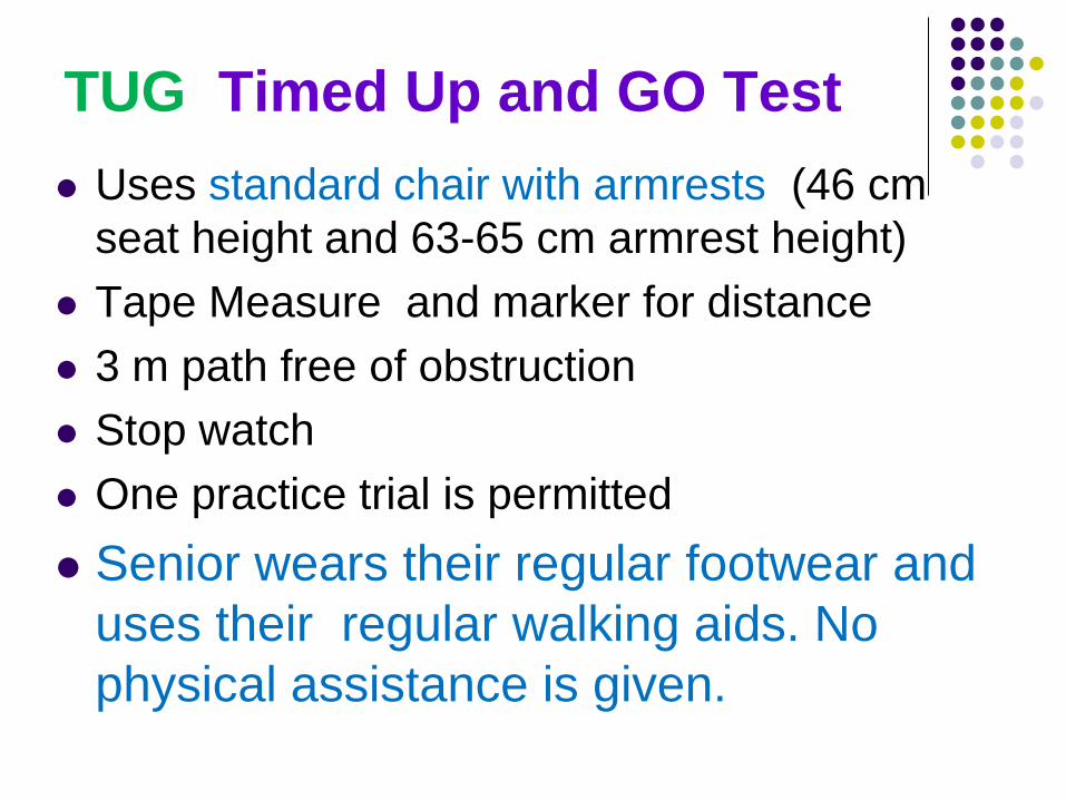
TUG Timed Up and GO Test
Uses standard chair with armrests (46 cm
seat height and 63-65 cm armrest height)
Tape Measure and marker for distance
3 m path free of obstruction
Stop watch
One practice trial is permitted
Senior wears their regular footwear and
uses their regular walking aids. No
physical assistance is given.

TUG Directions
Begin with subject sitting correctly in
the chair, back resting against the
back of the chair.
“On the word GO you will stand up,
walk to the line on the floor, turn
around and walk back to the chair.
Walk at your regular pace”

Time
There is no time limit- may stop and rest
but not sit down
Healthy elderly usually complete the task in
10 seconds or less
Very frail or weak elderly with poor mobility
may take 2 minutes or more

TUG scoring
Steffen, Hacker and Mollinger (2002) reported that on average, healthy individuals between the ages of 60-80 years complete the TUG in 10 seconds or less.
Standardized cut-off scores to predict risk of falling -In one study, a cut-off score of ≥ 13.5 seconds was shown to predict falling in community-dwelling frail elders (Shumway-Cook et al., 2000).

The 30-second Chair Stand Test
Purpose: To assess leg strength and endurance.
Equipment: A chair with a straight back without arm rests (seat 17” high)
A stopwatch
Instructions to the patient: 1. Sit in the middle of the chair.
2. Place your hands on the opposite
shoulder crossed at the wrists.
3. Keep your feet flat on the floor.
4. Keep your back straight. 5. On “Go”, rise to a full standing position
and then sit back down again.
6. Repeat this for 30 seconds.
On “Go”, begin timing.
Count the number of times the patient comes to a full standing position in 30 seconds.
If the patient is over halfway to a standing position when 30 seconds have elapsed, count it as a stand.
Record the number of times the patient stands in 30 seconds.
A below average rating indicates a high risk for falls. See Algorithm sheet for interpretation

Box 5 (section 2): Assessment
a. Medications
b. Postural Hypotension
c. Gait, balance, mobility and muscle strength
a. TUG and Chair Stand
b. Evaluate Pain related mobility decreases
d. Visual Acuity
e. Other Neurological Impairments
f. Heart rate and rhythm
g. Bone Health; nutritional review
h. Feet and Footwear
i. Environmental Hazards
j. Depression

Box 5 – 2d.
Visual Acuity
Sudden vision changes with inadequate time to compensate
Cognitive problems interfering with inability to compensate for poor vision.
Severe vision problems beyond ability to compensate
DDX:
1. Glaucoma (lose peripheral vision – tunnel vision)
2. Cataracts
3. Age Related Macular Degeneration (ARMD)
– lose central color vision
– Sudden change in vision in patient with ARMD is an ophthalmologic emergency – call ophthalmologist ASAP to have them determine if patient has a growing retinal tear and needs laser treatment on an urgent basis.

Box 5 (section 2): Assessment
a. Medications
b. Postural Hypotension
c. Gait, balance, mobility and muscle strength
a. TUG and Chair Stand
b. Evaluate Pain related mobility decreases
d. Visual Acuity
e. Other Neurological Impairments
f. Heart rate and rhythm
g. Bone Health; nutritional review
h. Feet and Footwear
i. Environmental Hazards
j. Depression

Box 5 – 2e.
Other Neurological Impairments
3Ds - Dementia, Delirium, Depression
Apraxia, decreased compensation, slow mentation
Stroke, subdural hematoma, subarachnoid bleed, cerebellar disease, NPH
Spinal stenosis, Myasthenia Gravis, ALS
Peripheral or Autonomic neuropathy
ETOH, DM, B12 …
Parkinsonism (next slide)

Box 5 – 2e.
Other Neurological Impairments
DDx of Parkinsonism (Parkinson’s Plus)1. Parkinson’s Disease (idiopathic parkinsonism)
– TRAP: Resting Tremor, Cogwheel Rigidity, Akinesia / bradikinesia (slowness), Postural Instability (decreased balance, falls)
2. Vascular parkinsonism– TRAP, no response to Parkinson's meds, basal ganglia strokes
3. Drugs (antipsychotics, GI drugs [stemetil, maxeran])
4. Lewy Body disease– Dementia, Longstanding Hallucinations, Longstanding Fluctuation
5. Progressive Supranuclear Palsy (PSP)– Loss of downward gaze and then all eye movements, depression,
anxiety, psychosis, dementia
6. Late Alzheimer’s
7. Multisystem atrophies (MSA – multiple neurologic symptoms)1. Shy-dragger, OPCD, SND etc

Box 5 – 2e.
Other Neurological Impairments
Vertebrobasilar Insufficiency
Provoked by head or neck movement
Seconds to minutes
Other brainstem symptoms Diplopia
Dysarthria
Facial numbness
Ataxia
Reduced vertebral artery flow on doppler or angiography
Treatment: Behaviour modification

VERTIGO
Central
BPPV Meniere's Vestibular Neuronitis Labyrinthitis
Peripheral
Vertigo
Dysequilibrium
Presyncope
Medication
Non-specific dizziness
Non-vertigo
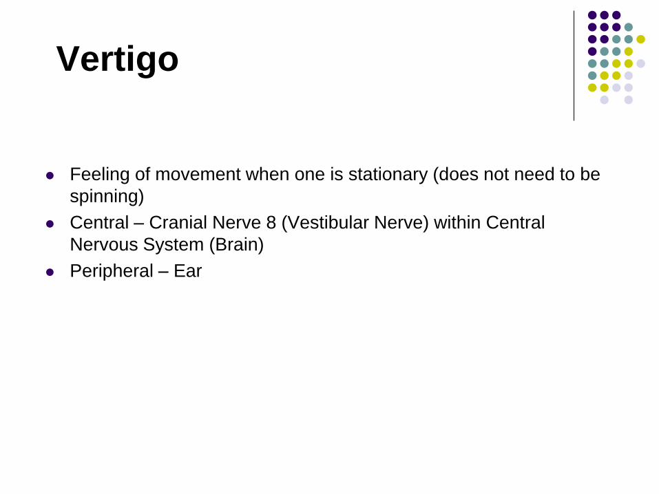
Vertigo
Feeling of movement when one is stationary (does not need to be
spinning)
Central – Cranial Nerve 8 (Vestibular Nerve) within Central
Nervous System (Brain)
Peripheral – Ear
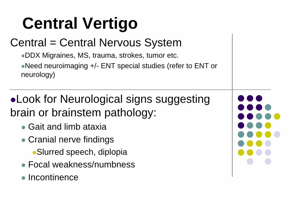
Central VertigoCentral = Central Nervous System
DDX Migraines, MS, trauma, strokes, tumor etc.
Need neuroimaging +/- ENT special studies (refer to ENT or
neurology)
Look for Neurological signs suggesting
brain or brainstem pathology:
Gait and limb ataxia
Cranial nerve findings
Slurred speech, diplopia
Focal weakness/numbness
Incontinence
Peripheral Vertigo
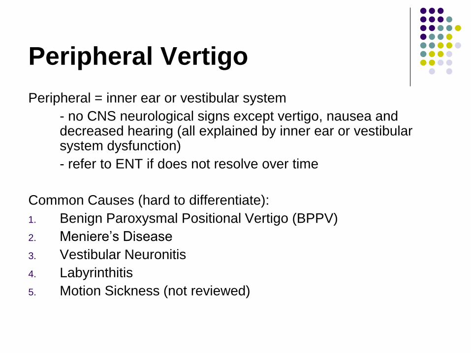
Peripheral = inner ear or vestibular system
- no CNS neurological signs except vertigo, nausea and decreased hearing (all explained by inner ear or vestibular system dysfunction)
- refer to ENT if does not resolve over time
Common Causes (hard to differentiate):
1. Benign Paroxysmal Positional Vertigo (BPPV)
2. Meniere’s Disease
3. Vestibular Neuronitis
4. Labyrinthitis
5. Motion Sickness (not reviewed)

• Commonest Cause of Chronic Vertigo in the Elderly. Sometimes associate with trauma.
• Cause: Calcium crystals dislodged and move to semi-circular canals
• Symptoms and signs: • Sudden onset vertigo lasting seconds to minutes,
episodic, brought on by changes in head position (rolling over, bending, looking upward)
• Nausea• Rotatory (torsional) Nystagmus where top of eye rotates
toward the affected ear in twitching fashion
1. BPPV or BPV:
BENIGN (Paroxysmal)
POSITIONAL VERTIGO
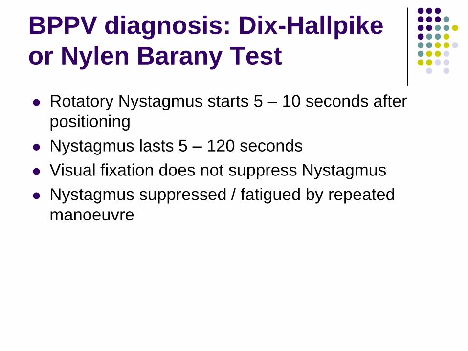
BPPV diagnosis: Dix-Hallpike
or Nylen Barany Test
Rotatory Nystagmus starts 5 – 10 seconds after
positioning
Nystagmus lasts 5 – 120 seconds
Visual fixation does not suppress Nystagmus
Nystagmus suppressed / fatigued by repeated
manoeuvre
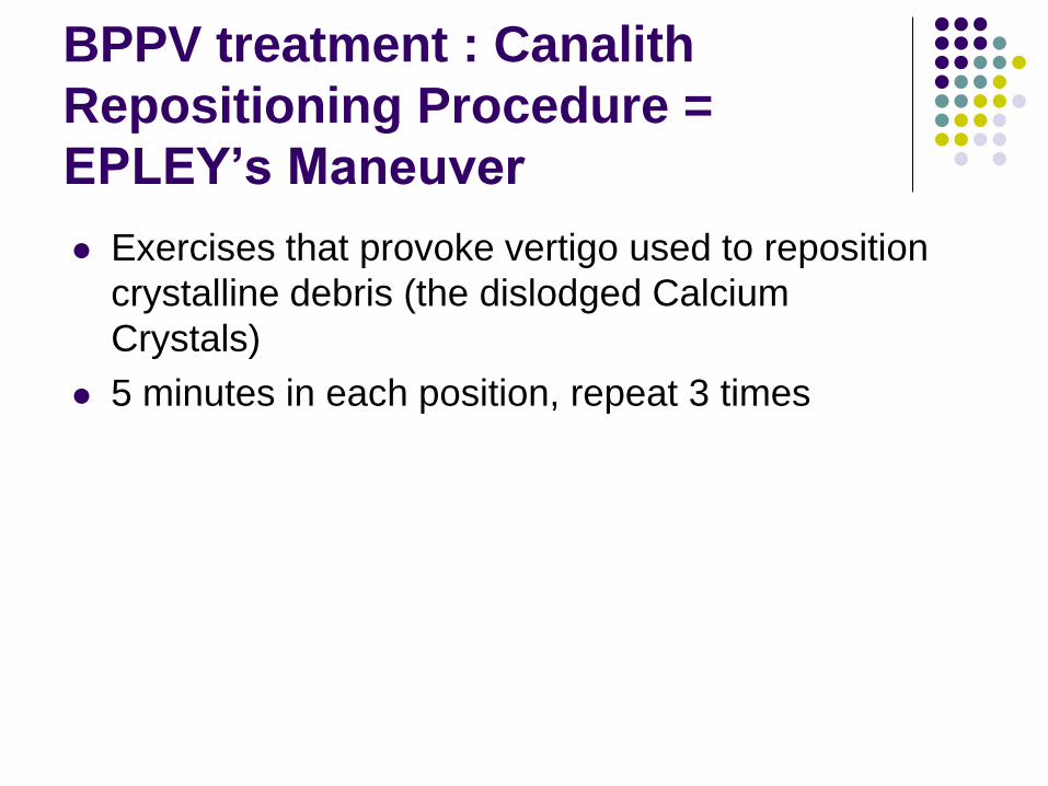
BPPV treatment : Canalith
Repositioning Procedure =
EPLEY’s Maneuver
Exercises that provoke vertigo used to reposition
crystalline debris (the dislodged Calcium
Crystals)
5 minutes in each position, repeat 3 times

2. Meniere’s Disease
Cause: Excess fluid in inner ear
Symptoms and Signs:
Attacks of Vertigo lasting minutes to hours (max 24 hours) – unexpected, not triggered by position
Fluctuating progressive hearing loss (one or both ears)
Unilateral or bilateral tinnitus
Sensation of fullness or pressure in ear.
Nausea, vomiting, sweating
Horizontal Nystagmus

3. Vestibular Neuronitis
Vestibular Neuronitis = inflammation of vestibular
nerve
Symptoms and Signs;
Vertigo + Nausea and Vomiting
unexpected, not triggered by position
+/- Nystagmus
Unlike labrynthitis (next topic) is NOT associated with
auditory symptoms (no tinnitus or decreased hearing)
May be associated with prior viral upper respiratory
tract infection

4. Labyrinthitis
Labyrinthitis – inflammation of inner ear.
Symptoms and Signs:
Acute onset of non-position dependent vertigo (often
severe)
+/- nausea and vomiting
+/- hearing loss and tinnitus
May occur after viral or bacteria infection (especially
upper respiratory tract infection), or head injury
Lasts 1 – 6 weeks but can have residual symptoms
for months or years

Box 5 (section 2): Assessment
a. Medications
b. Postural Hypotension
c. Gait, balance, mobility and muscle strength
a. TUG and Chair Stand
b. Evaluate Pain related mobility decreases
d. Visual Acuity
e. Other Neurological Impairments
f. Heart rate and rhythm
g. Bone Health; nutritional review
h. Feet and Footwear
i. Environmental Hazards
j. Depression

Box 5 – 2f.
Heart rate / rhythm + blood flow
Decreased Cardiac Output
1. Blockage of blood flow1. Valvular
1. aortic or mitral stenosis
2. Subaortic stenosis
3. Aortic dissection2. Pulmonary Embolus
2. Arrhythmia Tachycardia (inadequate time in diastole for heart to fill): VT, SVT,
WPW, VF, AFIB …
Bradycardia; SSS, conduction blocks (complete heart block) Can be precipitated by digoxin, beta-blocker (including Timoptic
/Timolol eye drops), Alzheimer medications (Cholinesterase Inhibitors), Ca Channel Blockers
Carotid Sinus Hypersensitivity
3. Very low Left Ventricular Ejection Fraction

Box 5 – 2f.
Heart rate / rhythm + blood flow
VASOVAGAL - Syncope Triggered by: Stress
Any painful or unpleasant stimuli, such as: Venepuncture
Hitting your funny bone
Experiencing medical procedures with local anesthesia
Post-surgical pain when standing up or moving too abruptly after the procedure
Giving or receiving a needle immunization
Watching someone give blood
Watching someone experience pain
Watching or experiencing medical procedures
Sight of blood
Occasions of slight discomfort, such as dental and eye examinations
Sudden onset of extreme emotions
Nausea or vomiting
Urination ('micturition syncope') or defecation, having a bowel movement ('defecation syncope')
Abdominal straining or 'bearing down'
Swallowing ('swallowing syncope') or coughing ('cough syncope')
Pressing upon certain places on the throat, sinuses, and eyes, also known as vagal reflex stimulation when performed clinically
etc

Box 5 (section 2): Assessment
a. Medications
b. Postural Hypotension
c. Gait, balance, mobility and muscle strength
a. TUG and Chair Stand
b. Evaluate Pain related mobility decreases
d. Visual Acuity
e. Other Neurological Impairments
f. Heart rate and rhythm
g. Bone Health; nutritional review
h. Feet and Footwear
i. Environmental Hazards
j. Depression

Box 5 – 2g.
Bone Health; nutritional review
Currently, Ottawa Public Health recommends daily
for adults 51 yrs and older:
3 or more servings of Milk and Alternatives
Adequate amounts of calcium and vitamin D rich foods
A vitamin D supplement of 400 IU
RCTs and meta-analyses have demonstrated a
beneficial effect of Vitamin D in fall prevention
distinct from its effect on bone health
Possibly through muscle strength and neuromuscular
function.

Ottawa Public Health
From food and \or
supplement:
Women
51-70 yrs : 1200 mg
71yrs + : 1200 mg
Men
51-70 yrs : 1000 mg
71yrs+ : 1200 mg
From food and
supplement, ( ♀ and ♂)
51-70 yrs : 600 IU
71yrs + : 800 IU
Recommendations
include a supplement
for all adults 50 yrs +
of 400 IU
Upper maximum intake:
4000 IU

Box 5 (section 2): Assessment
a. Medications
b. Postural Hypotension
c. Gait, balance, mobility and muscle strength
a. TUG and Chair Stand
b. Evaluate Pain related mobility decreases
d. Visual Acuity
e. Other Neurological Impairments
f. Heart rate and rhythm
g. Bone Health; nutritional review
h. Feet and Footwear
i. Environmental Hazards
j. Depression

Box 5 – 2h.
Feet and Footwear
Don’t forget to take off the socks and shoes to assess the feet. The feet reveal a great deal about a person. Neglected feet can be a marker of many things including inability to reach feet to care for them, depression, neglect, cognitive impairment…
Examine for Moderate or severe bunions
Toe / nail deformities
Ulcers
Loss of position sense (proprioception)
Filament test, vibration sensation may be more sensitive but less specific

Box 5 – 2h.
Feet and Footwear
Muscular: Myopathy / Myositis
Skeletal: Arthritis (foot, ankle, knee, hip, back)
Deformity altering biomechanics
Poor pain control start Tylenol Arthritis 650-1300mg TID straight
If still in pain and no CHF or renal dysfunction then consider NSAID
Later narcotics (watch for anorexia and weight loss, constipation, delirium)
CHF – pedal edema leading to loss of position sense and change in foot mechanics

Box 5 – 2h.
Feet and Footwear
Footwear
Ask to see shoes they were wearing when they fell (if possible) or at least get a description of the shoes. Look for:
Poor fit (foot moving in shoe)
Lack of support (not laced or buckled)
High heels
Note: some women develop Achilles tendon shortening with chronic high heel use and have difficulty transitioning to lower shoes
Small surface area contact with floor
Smooth slippery sole (lack of functional anti-slip surface by design or if worn out)

Box 5 (section 2): Assessment
a. Medications
b. Postural Hypotension
c. Gait, balance, mobility and muscle strength
a. TUG and Chair Stand
b. Evaluate Pain related mobility decreases
d. Visual Acuity
e. Other Neurological Impairments
f. Heart rate and rhythm
g. Bone Health; nutritional review
h. Feet and Footwear
i. Environmental Hazards
j. Depression

Box 5 – 2i.
Environmental hazards
Home hazards (kitchen, bathroom, bedroom)
Poorly lit stairs, ramps or doorways
Stairs with irregular step width or height
Stairs without handrails or marking on the edges
Slippery floors, throw rugs, loose carpets
Raised sills in door jams
Clutter
Low toilet seats
Lack of grab bars in bathrooms
Poorly maintained or improperly used mobility aids and equipment
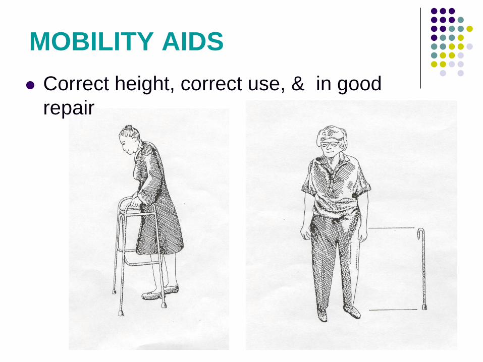
MOBILITY AIDS
Correct height, correct use, & in good
repair

Box 5 – 2i.
Environmental hazards
Outdoor hazards
Public/community hazards
Use of Assistive devices
Uneven sidewalks or cracks in sidewalks
Stairs without handrails or marking on the edges
Poor lighting
Objects on sidewalks or walkways such as garbage cans
Snow or ice on stairs or walkways
Unmarked curbs or corners without curb ramps
Long crosswalks without pedestrian islands

Box 5 – 2i.
Environmental hazards
Ask patient and family about;
Difficulty getting out of low bed or off low chair /
couch
Tripping over rugs, thresholds
Lighting
Stairs
Gives clues to risk reduction strategies
Falls Are all normally used walkways free of trip hazards?
Objects, low level furniture, pets, scatter rugs not properly secured, cords – encourage cordless phones,
How hazardous are the Floors?
Throw rugs not properly secured, slippery surfaces, thresholds
How hazardous are the Stairwells?
Two railings?, steep steps, steps in good repair, poor lighting
How hazardous are Transfers?
On/off of chairs (height, stability, arms), in/out of bathtub (any bathtub equipment or torn off the wall towel racks), on/off toilet (height), in/out of bed (height)
Is all equipment securely attached?
Other:
Accessibility of Phones in commonly used areas, by bed, living room, kitchen, basement
Commonly Used Items at reachable height – avoiding use of step stools
Proper Lighting (overhead lights and night lights)
Clothing – Proper footwear, proper length of clothing.

Box 5 (section 2): Assessment
a. Medications
b. Postural Hypotension
c. Gait, balance, mobility and muscle strength
a. TUG and Chair Stand
b. Evaluate Pain related mobility decreases
d. Visual Acuity
e. Other Neurological Impairments
f. Heart rate and rhythm
g. Bone Health; nutritional review
h. Feet and Footwear
i. Environmental Hazards
j. Depression
Box 5 – 2j.
Depression
MOOD
Anxiety/Panic disorders
Mood disorders (Depression)
with hyperventilation or emotional stress
Often associated with:
Somatic complaints
Insomnia/fatigue

TREATMENT & PREVENTION
Treatment
Goals of treatment
Prevent all falls (often unachievable ideal)
When that is not possible then decrease
frequency, severity and sequelae of falls
One sequelae is fractures so order a Bone Mineral
Density and consider aggressive treatment for
osteoporosis in all fallers if you feel their life
expectancy merits treatment

Comprehensive Falls Assessment
and Prevention Programs
Treatment
MD / RN Adjust medications (+/- Pharmacist)
Optimize control of medical problems
Bone Density (prevent fractures if does fall)
OT Compensatory Strategies
Assistive devices
PT Balance and Strength training
Ambulation Aides
SW Safe housing options + support services
Nutrition Improve oral intake
?

QUESTIONS ???