Slide 1 Lecture Notes: Cardiovascular...
8
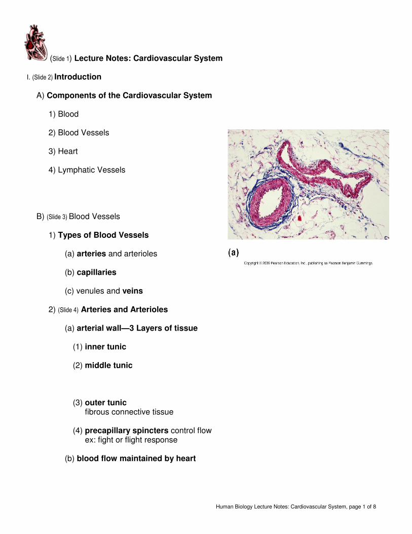
Human Biology Lecture Notes: Cardiovascular System, page 1 of 8 (Slide 1) Lecture Notes: Cardiovascular System I. (Slide 2) Introduction A) Components of the Cardiovascular System 1) Blood 2) Blood Vessels 3) Heart 4) Lymphatic Vessels B) (Slide 3) Blood Vessels 1) Types of Blood Vessels (a) arteries and arterioles (b) capillaries (c) venules and veins 2) (Slide 4) Arteries and Arterioles (a) arterial wall—3 Layers of tissue (1) inner tunic (2) middle tunic (3) outer tunic fibrous connective tissue (4) precapillary spincters control flow ex: fight or flight response (b) blood flow maintained by heart
Transcript of Slide 1 Lecture Notes: Cardiovascular...
Human Biology Lecture Notes: Cardiovascular System, page 1 of 8
(Slide 1) Lecture Notes: Cardiovascular System I. (Slide 2) Introduction
A) Components of the Cardiovascular System
1) Blood 2) Blood Vessels 3) Heart 4) Lymphatic Vessels
B) (Slide 3) Blood Vessels
1) Types of Blood Vessels
(a) arteries and arterioles (b) capillaries
(c) venules and veins
2) (Slide 4) Arteries and Arterioles
(a) arterial wall—3 Layers of tissue
(1) inner tunic (2) middle tunic
(3) outer tunic fibrous connective tissue
(4) precapillary spincters control flow ex: fight or flight response
(b) blood flow maintained by heart

Human Biology Lecture Notes: Cardiovascular System, page 2 of 8
3) (Slide 5) Capillaries (a) wall structure: (b) only small portion of capillaries are ‘open’—5%
blood volume within capillaries (c) Function:
4) (Slide 6) Veins (a) wall structure:
(1) inner tunic (2) middle tunic
(3) outer tunic-fibrous CT
(4)
valves
(b) function:
(1) return blood to heart (2) _
70% of blood volume in veins
(c) (Slide 7) Blood Flow in veins Maintained

Human Biology Lecture Notes: Cardiovascular System, page 3 of 8
C) (Slide 8) Heart
1) General (a) size fist (b) dorsal to sternum (c) tilts to the left (apex) (d) pericardial cavity (space within thoracic
cavity)
2) (Slide 9) External Heart Structures (a) 4 chambers
(1) right and left atria (2) right and left ventricles
(b) (Slide 10) Great Vessels (1) Aorta (arch) (2) Pulmonary Trunk (3) Superior vena Cava (4) Inferior vena cava (5) pulmonary veins (6) base
(c) (Slide 11) coronary blood vessels
(d) apex

Human Biology Lecture Notes: Cardiovascular System, page 4 of 8
3) (Slide 12) Heart Wall (a) Pericardium or Pericardial Sac
(1) outermost
(b) Myocardium (c) Endocardium :
(1) lines and covers internal heart chambers and structures
4) (Slide 13) Internal Heart Structures (a) Review:
(1) right atria (2) left atria (3) right ventricle (4) left ventricle (5) aorta (arch) (6) Pulmonary trunk (7) Superior Vena Cava (8) Inferior Vena Cava (9) Base (10) Apex
(b) myocardium (c) trabeculae carneae (d) (Slide 14) right atrioventricular valve (e) left
atrioventricular valve
(f) chordae tendineae
(g) papillary muscle
(h) (Slide 15) pulmonary semilunar valve
(i)
aortic semilunar valve
(j) interventricular septum

Human Biology Lecture Notes: Cardiovascular System, page 5 of 8
5) (Slide 15) Double Pump
(a) right atria (b) right ventricle
(c) left atria (d) left ventricle
6) (Slide 17) Valves
(a) two atrioventricular valves (1) lie between atria and ventricles (2) chordae tendineae attach valve flaps to
papillary muscle (3) papillary muscle-anchor
(b) right AV (tricuspid) (c) left AV (bicuspid or mitral) (d)
(Slide 18)Two Semilunar Valves:
(1) Location: flaps within arterial wall (2) Aortic Semilunar (3) Pulmonary Semilunar

Human Biology Lecture Notes: Cardiovascular System, page 6 of 8
7) (Slide 19) Blood Flow through the heart
(a) vena cava (from body) (b) R atria (c) R AV valve (d) R ventricle (e) Pulmonary semilunar (f) Pulmonary trunk (to lungs) (g) (Slide 20) pulmonary veins (from lungs) (h) L atria (i) L AV valve (j) L ventricle (k) Aortic semilunar (l) Aorta (to body)
II. (Slide 21) Cardiac Physiology A) Cardiac Cycle (heartbeat)
1) sequence (a) atra contract (b) ventricles contract (c) ensures blood flow in one direction (d) 70 beats per minute, approximately 0.85 seconds
2) Heart Sounds: (a) lub-AV valves closing (b) dup-semilunar valves closing
3) Pulse: vibration felt in arterial walls due to expansion of BV wall following contraction of ventricles (a) ‘pressure points’ (b) indicates rate of heart beat
4) (Slide 22) Contraction vs. Relaxation
(a) systole: (1) blood pressure highest in arteries
(b) diastole: (c) sphyngmomanometer: used to indirectly measure blood pressure
(1) systolic pressure (2) diastolic pressure (3) both decrease with distance from left ventricle

Human Biology Lecture Notes: Cardiovascular System, page 7 of 8
B)
(Slide 23)Regulation of Heart Beat
1) Intrinsic (internal) Control (a) sinoatrial node (nervous tissue) (b) impulse spreads/wringing heart
(1) atrioventricular node (2) atrioventricular bundle (3) purkinje fibers
(c) results in ventricular contraction
2) (Slide 24) Extrinsic (external) control
(a) cardiac control center (1) (brainstem) Medulla Oblongata (2) autonomic nervous system
(a) parasympathetic nervous system (b) sympathetic nervous system (fight or
flight)
(b) hormones
(1) adrenal glands secrete epinephrine/adrenaline
C) (Slide 25) Vascular Pathways
1) Pulmonary Circuit:

Human Biology Lecture Notes: Cardiovascular System, page 8 of 8
2) (Slide 26) Systemic Circuit: 3) (Slide 27) Coronary Arteries
(a) myocardial infaction (b) angina pectoris: chest pain due to brief lack of oxygen to heart muscle (warning)
(c) congestive heart failure