
Slide 1 Chest Radiology Interpretation: Findings of ... 1 Chest Radiology Interpretation: Findings...
51
Slide 1 Chest Radiology Interpretation: Findings of Tuberculosis ___________________________________ ___________________________________ ___________________________________ ___________________________________ ___________________________________ ___________________________________ ___________________________________ Slide 2 Case #1 ___________________________________ ___________________________________ ___________________________________ ___________________________________ ___________________________________ ___________________________________ ___________________________________ Slide 3 Reading the TB CXR Be systematic! Start centrally and work outwards Normal or abnormal Describe the finding(s) Consider the significance of the finding(s) ___________________________________ ___________________________________ ___________________________________ ___________________________________ ___________________________________ ___________________________________ ___________________________________
Transcript of Slide 1 Chest Radiology Interpretation: Findings of ... 1 Chest Radiology Interpretation: Findings...

Slide 1 Chest Radiology Interpretation: Findings of Tuberculosis
___________________________________
___________________________________
___________________________________
___________________________________
___________________________________
___________________________________
___________________________________
Slide 2 Case #1
___________________________________
___________________________________
___________________________________
___________________________________
___________________________________
___________________________________
___________________________________
Slide 3
Reading the TB CXR
Be systematic!
Start centrally and work outwards
Normal or abnormal
Describe the finding(s)
Consider the significance of the finding(s)
___________________________________
___________________________________
___________________________________
___________________________________
___________________________________
___________________________________
___________________________________

Slide 4 Mediastinum
___________________________________
___________________________________
___________________________________
___________________________________
___________________________________
___________________________________
___________________________________
Slide 5 Hila
___________________________________
___________________________________
___________________________________
___________________________________
___________________________________
___________________________________
___________________________________
Slide 6 Lungs
___________________________________
___________________________________
___________________________________
___________________________________
___________________________________
___________________________________
___________________________________

Slide 7 Pleura & Diaphragms
___________________________________
___________________________________
___________________________________
___________________________________
___________________________________
___________________________________
___________________________________
Slide 8 Pleura & Diaphragms
___________________________________
___________________________________
___________________________________
___________________________________
___________________________________
___________________________________
___________________________________
Slide 9 Pleura & Diaphragms
___________________________________
___________________________________
___________________________________
___________________________________
___________________________________
___________________________________
___________________________________
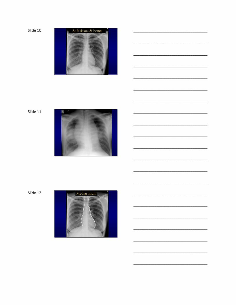
Slide 10 Soft tissue & bones
___________________________________
___________________________________
___________________________________
___________________________________
___________________________________
___________________________________
___________________________________
Slide 11
___________________________________
___________________________________
___________________________________
___________________________________
___________________________________
___________________________________
___________________________________
Slide 12 Mediastinum
___________________________________
___________________________________
___________________________________
___________________________________
___________________________________
___________________________________
___________________________________

Slide 13
Lymphoma
AbnormalNormal
___________________________________
___________________________________
___________________________________
___________________________________
___________________________________
___________________________________
___________________________________
Slide 14
Metastatic disease (unknown primary)
Normal Abnormal
___________________________________
___________________________________
___________________________________
___________________________________
___________________________________
___________________________________
___________________________________
Slide 15
Lung Cancer
Normal Abnormal
AO
PA
___________________________________
___________________________________
___________________________________
___________________________________
___________________________________
___________________________________
___________________________________

Slide 16 Heart
<55% thoracic diameter
Technique important
Larger in: AP film
Poor inspiration
Rotation
Children
True enlargement Chamber enlargement
Pericardial effusion
Mass
___________________________________
___________________________________
___________________________________
___________________________________
___________________________________
___________________________________
___________________________________
Slide 17 Artifactual cardiomegaly
___________________________________
___________________________________
___________________________________
___________________________________
___________________________________
___________________________________
___________________________________
Slide 18
___________________________________
___________________________________
___________________________________
___________________________________
___________________________________
___________________________________
___________________________________

Slide 19 End stage rheumatic heart disease
___________________________________
___________________________________
___________________________________
___________________________________
___________________________________
___________________________________
___________________________________
Slide 20 Pericarditis
___________________________________
___________________________________
___________________________________
___________________________________
___________________________________
___________________________________
___________________________________
Slide 21 Hila
___________________________________
___________________________________
___________________________________
___________________________________
___________________________________
___________________________________
___________________________________

Slide 22
___________________________________
___________________________________
___________________________________
___________________________________
___________________________________
___________________________________
___________________________________
Slide 23 Q1. Pathology in this patient is most
likely to show?
A. Caseating granulomas
B. Non-caseating granulomas
C. Atypical cells with high nuclear/cytoplasmic ratio
D. Fibrosis
___________________________________
___________________________________
___________________________________
___________________________________
___________________________________
___________________________________
___________________________________
Slide 24
Sarcoidosis
Normal Abnormal
___________________________________
___________________________________
___________________________________
___________________________________
___________________________________
___________________________________
___________________________________

Slide 25
Pulmonary Hypertension
Normal Abnormal
___________________________________
___________________________________
___________________________________
___________________________________
___________________________________
___________________________________
___________________________________
Slide 26 Lungs
___________________________________
___________________________________
___________________________________
___________________________________
___________________________________
___________________________________
___________________________________
Slide 27 Pleura & Diaphragms
___________________________________
___________________________________
___________________________________
___________________________________
___________________________________
___________________________________
___________________________________

Slide 28 Pleura & Diaphragms
___________________________________
___________________________________
___________________________________
___________________________________
___________________________________
___________________________________
___________________________________
Slide 29
___________________________________
___________________________________
___________________________________
___________________________________
___________________________________
___________________________________
___________________________________
Slide 30
___________________________________
___________________________________
___________________________________
___________________________________
___________________________________
___________________________________
___________________________________

Slide 31
___________________________________
___________________________________
___________________________________
___________________________________
___________________________________
___________________________________
___________________________________
Slide 32 Q2. Where is this lesion located?
A. Lung
B. Mediastinum
C. Pleura
D. Chest wall
___________________________________
___________________________________
___________________________________
___________________________________
___________________________________
___________________________________
___________________________________
Slide 33
___________________________________
___________________________________
___________________________________
___________________________________
___________________________________
___________________________________
___________________________________
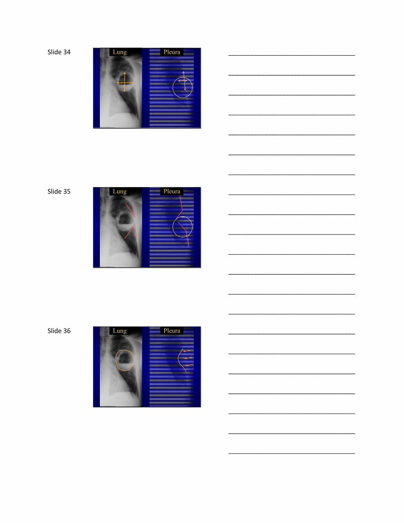
Slide 34 Lung Pleura
___________________________________
___________________________________
___________________________________
___________________________________
___________________________________
___________________________________
___________________________________
Slide 35 Lung Pleura
___________________________________
___________________________________
___________________________________
___________________________________
___________________________________
___________________________________
___________________________________
Slide 36 Lung Pleura
___________________________________
___________________________________
___________________________________
___________________________________
___________________________________
___________________________________
___________________________________

Slide 37 Lung Pleura
___________________________________
___________________________________
___________________________________
___________________________________
___________________________________
___________________________________
___________________________________
Slide 38 TB Empyema
___________________________________
___________________________________
___________________________________
___________________________________
___________________________________
___________________________________
___________________________________
Slide 39 Don’t forget about the bones
___________________________________
___________________________________
___________________________________
___________________________________
___________________________________
___________________________________
___________________________________
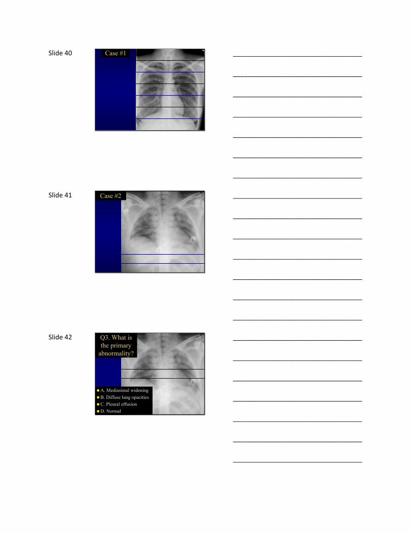
Slide 40 Case #1
___________________________________
___________________________________
___________________________________
___________________________________
___________________________________
___________________________________
___________________________________
Slide 41 Case #2
___________________________________
___________________________________
___________________________________
___________________________________
___________________________________
___________________________________
___________________________________
Slide 42 Q3. What is the primary
abnormality?
A. Mediastinal widening
B. Diffuse lung opacities
C. Pleural effusion
D. Normal
___________________________________
___________________________________
___________________________________
___________________________________
___________________________________
___________________________________
___________________________________

Slide 43 Inspiration: (≥10 posterior ribs)
___________________________________
___________________________________
___________________________________
___________________________________
___________________________________
___________________________________
___________________________________
Slide 44
___________________________________
___________________________________
___________________________________
___________________________________
___________________________________
___________________________________
___________________________________
Slide 45
1st rib
2nd rib3rd rib
___________________________________
___________________________________
___________________________________
___________________________________
___________________________________
___________________________________
___________________________________

Slide 46 2nd3rd
4th
5th
6th
7th
8th
9th
10th
1st
___________________________________
___________________________________
___________________________________
___________________________________
___________________________________
___________________________________
___________________________________
Slide 47 Poor inspiration
___________________________________
___________________________________
___________________________________
___________________________________
___________________________________
___________________________________
___________________________________
Slide 48 Good inspiration
___________________________________
___________________________________
___________________________________
___________________________________
___________________________________
___________________________________
___________________________________

Slide 49 Rotation
___________________________________
___________________________________
___________________________________
___________________________________
___________________________________
___________________________________
___________________________________
Slide 50
___________________________________
___________________________________
___________________________________
___________________________________
___________________________________
___________________________________
___________________________________
Slide 51
___________________________________
___________________________________
___________________________________
___________________________________
___________________________________
___________________________________
___________________________________

Slide 52 PenetrationIntervertebralDisks
___________________________________
___________________________________
___________________________________
___________________________________
___________________________________
___________________________________
___________________________________
Slide 53
Over-penetrated
___________________________________
___________________________________
___________________________________
___________________________________
___________________________________
___________________________________
___________________________________
Slide 54 Case #3
___________________________________
___________________________________
___________________________________
___________________________________
___________________________________
___________________________________
___________________________________

Slide 55 Q4. What is the most likely diagnosis?
A. Tuberculosis
B. Aspergillosis
C. Malignancy
D. Mycoplasma
___________________________________
___________________________________
___________________________________
___________________________________
___________________________________
___________________________________
___________________________________
Slide 56 Categories of lung opacities
1. Nodule(s) or mass(es)
2. Alveolar, airspace, consolidation
3. Interstitial (diffuse lines or nodules)
4. Airways (circular or tubular)
___________________________________
___________________________________
___________________________________
___________________________________
___________________________________
___________________________________
___________________________________
Slide 57 Nodule ≤ 3cm, Mass > 3 cm
2.7 cm3.4 cm
___________________________________
___________________________________
___________________________________
___________________________________
___________________________________
___________________________________
___________________________________
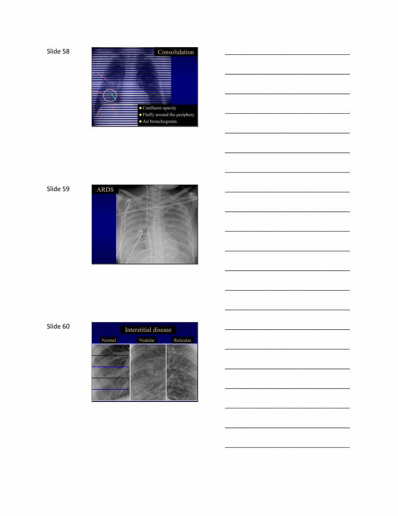
Slide 58 Consolidation
Confluent opacity
Fluffy around the periphery
Air bronchograms
___________________________________
___________________________________
___________________________________
___________________________________
___________________________________
___________________________________
___________________________________
Slide 59 ARDS
___________________________________
___________________________________
___________________________________
___________________________________
___________________________________
___________________________________
___________________________________
Slide 60
Normal Nodular Reticular
Interstitial disease
___________________________________
___________________________________
___________________________________
___________________________________
___________________________________
___________________________________
___________________________________
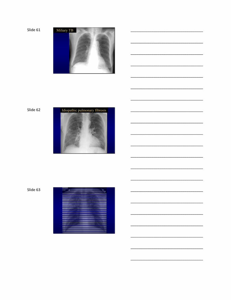
Slide 61 Miliary TB
___________________________________
___________________________________
___________________________________
___________________________________
___________________________________
___________________________________
___________________________________
Slide 62 Idiopathic pulmonary fibrosis
___________________________________
___________________________________
___________________________________
___________________________________
___________________________________
___________________________________
___________________________________
Slide 63
___________________________________
___________________________________
___________________________________
___________________________________
___________________________________
___________________________________
___________________________________

Slide 64 Airways disease
Circular
Tubular
___________________________________
___________________________________
___________________________________
___________________________________
___________________________________
___________________________________
___________________________________
Slide 65
___________________________________
___________________________________
___________________________________
___________________________________
___________________________________
___________________________________
___________________________________
Slide 66 Tuberculosis
___________________________________
___________________________________
___________________________________
___________________________________
___________________________________
___________________________________
___________________________________
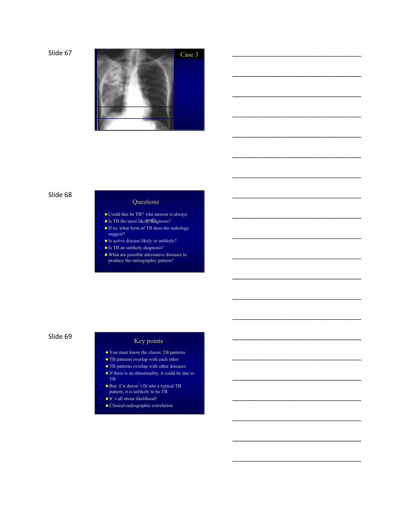
Slide 67 Case 3
___________________________________
___________________________________
___________________________________
___________________________________
___________________________________
___________________________________
___________________________________
Slide 68 Questions
Could this be TB?
Is TB the most likely diagnosis?
If so, what form of TB does the radiology suggest?
Is active disease likely or unlikely?
Is TB an unlikely diagnosis?
What are possible alternative diseases to produce the radiographic pattern?
(the answer is always yes!)
___________________________________
___________________________________
___________________________________
___________________________________
___________________________________
___________________________________
___________________________________
Slide 69 Key points
You must know the classic TB patterns
TB patterns overlap with each other
TB patterns overlap with other diseases
If there is an abnormality, it could be due to TB
But, if it doesn’t fit into a typical TB pattern, it is unlikely to be TB
It’s all about likelihood!
Clinical-radiographic correlation
___________________________________
___________________________________
___________________________________
___________________________________
___________________________________
___________________________________
___________________________________
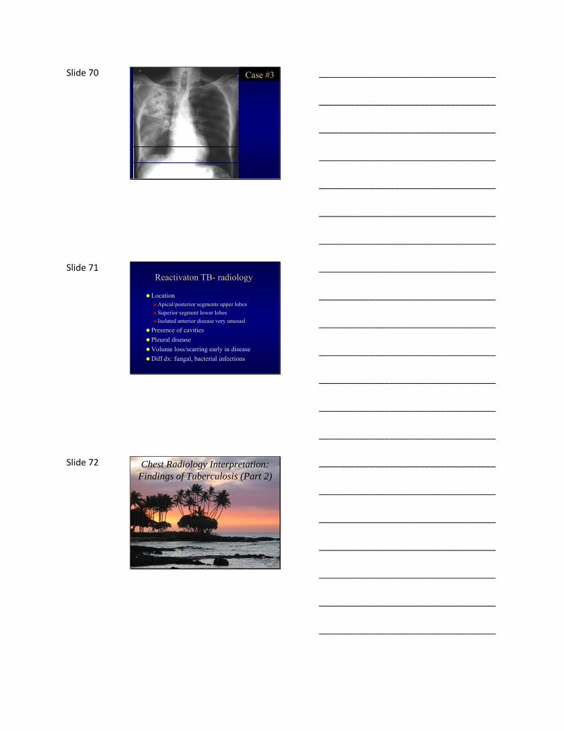
Slide 70 Case #3
___________________________________
___________________________________
___________________________________
___________________________________
___________________________________
___________________________________
___________________________________
Slide 71 Reactivaton TB- radiology
LocationApical/posterior segments upper lobes
Superior segment lower lobes
Isolated anterior disease very unusual
Presence of cavities
Pleural disease
Volume loss/scarring early in disease
Diff dx: fungal, bacterial infections
___________________________________
___________________________________
___________________________________
___________________________________
___________________________________
___________________________________
___________________________________
Slide 72 Chest Radiology Interpretation: Findings of Tuberculosis (Part 2)
___________________________________
___________________________________
___________________________________
___________________________________
___________________________________
___________________________________
___________________________________

Slide 73 Is this likely TB?
___________________________________
___________________________________
___________________________________
___________________________________
___________________________________
___________________________________
___________________________________
Slide 74 Q5. What lobe is involved?
A. Right upper lobe
B. Azygous lobe
C. Right middle lobe
D. Right lower lobe
___________________________________
___________________________________
___________________________________
___________________________________
___________________________________
___________________________________
___________________________________
Slide 75 Lobar anatomy
Left Lung
LLL
LUL
___________________________________
___________________________________
___________________________________
___________________________________
___________________________________
___________________________________
___________________________________

Slide 76
Right Lung
RLLRML
RUL
Lobar anatomy
___________________________________
___________________________________
___________________________________
___________________________________
___________________________________
___________________________________
___________________________________
Slide 77
Right Lung
RLLRML
RUL
Lobar anatomy
___________________________________
___________________________________
___________________________________
___________________________________
___________________________________
___________________________________
___________________________________
Slide 78 RUL Pneumonia
___________________________________
___________________________________
___________________________________
___________________________________
___________________________________
___________________________________
___________________________________

Slide 79
Right Lung
RLLRML
RUL
Lobar anatomy
___________________________________
___________________________________
___________________________________
___________________________________
___________________________________
___________________________________
___________________________________
Slide 80
Right Lung
RLLRML
RUL
Lobar anatomy
___________________________________
___________________________________
___________________________________
___________________________________
___________________________________
___________________________________
___________________________________
Slide 81
___________________________________
___________________________________
___________________________________
___________________________________
___________________________________
___________________________________
___________________________________
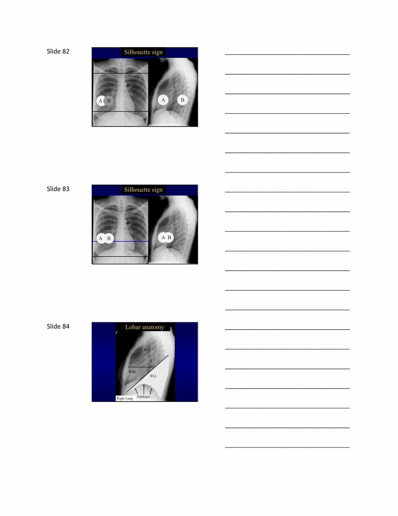
Slide 82 Silhouette sign
A B A B
___________________________________
___________________________________
___________________________________
___________________________________
___________________________________
___________________________________
___________________________________
Slide 83 Silhouette sign
A B A B
___________________________________
___________________________________
___________________________________
___________________________________
___________________________________
___________________________________
___________________________________
Slide 84
Right Lung
RLLRML
RUL
Lobar anatomy
Diaphragm
___________________________________
___________________________________
___________________________________
___________________________________
___________________________________
___________________________________
___________________________________

Slide 85
RLL
ObscuredDiaphragm
ClearHeartBorder
RLL pneumonia
___________________________________
___________________________________
___________________________________
___________________________________
___________________________________
___________________________________
___________________________________
Slide 86 ? Which lobe is involved
___________________________________
___________________________________
___________________________________
___________________________________
___________________________________
___________________________________
___________________________________
Slide 87
Right Lung
RLLRML
RUL
Lobar anatomy
___________________________________
___________________________________
___________________________________
___________________________________
___________________________________
___________________________________
___________________________________

Slide 88
RML
RML pneumonia
ClearDiaphragm
ObscuredHeartBorder
___________________________________
___________________________________
___________________________________
___________________________________
___________________________________
___________________________________
___________________________________
Slide 89 ? pneumonia
___________________________________
___________________________________
___________________________________
___________________________________
___________________________________
___________________________________
___________________________________
Slide 90 ? pneumonia
___________________________________
___________________________________
___________________________________
___________________________________
___________________________________
___________________________________
___________________________________

Slide 91
Anterior Posterior
Superior
Inferior
___________________________________
___________________________________
___________________________________
___________________________________
___________________________________
___________________________________
___________________________________
Slide 92 Lateral Viewof the Chest
Heart
___________________________________
___________________________________
___________________________________
___________________________________
___________________________________
___________________________________
___________________________________
Slide 93 Lateral Viewof the Chest
Spine
___________________________________
___________________________________
___________________________________
___________________________________
___________________________________
___________________________________
___________________________________

Slide 94 Lateral Viewof the Chest
Diaphragm
___________________________________
___________________________________
___________________________________
___________________________________
___________________________________
___________________________________
___________________________________
Slide 95 Lateral Viewof the Chest
Diaphragm
___________________________________
___________________________________
___________________________________
___________________________________
___________________________________
___________________________________
___________________________________
Slide 96 Normal LLL Pneumonia
___________________________________
___________________________________
___________________________________
___________________________________
___________________________________
___________________________________
___________________________________
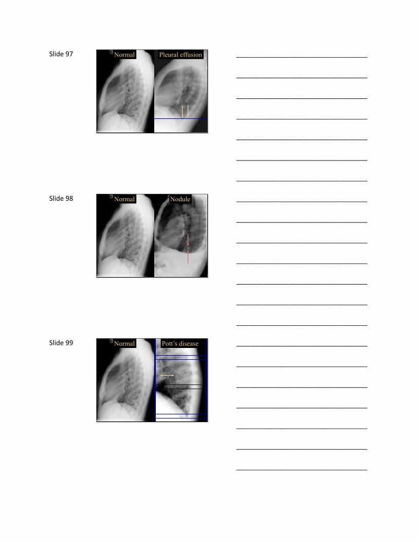
Slide 97 Normal Pleural effusion
___________________________________
___________________________________
___________________________________
___________________________________
___________________________________
___________________________________
___________________________________
Slide 98 Normal Nodule
___________________________________
___________________________________
___________________________________
___________________________________
___________________________________
___________________________________
___________________________________
Slide 99 Normal Pott’s disease
___________________________________
___________________________________
___________________________________
___________________________________
___________________________________
___________________________________
___________________________________

Slide 100 Case #4
___________________________________
___________________________________
___________________________________
___________________________________
___________________________________
___________________________________
___________________________________
Slide 101
Q6. What is the primary abnormality?1. Consolidation
2. Emphysema
3. Airway enlargement
4. Fibrosis
___________________________________
___________________________________
___________________________________
___________________________________
___________________________________
___________________________________
___________________________________
Slide 102 Abnormal Normal
___________________________________
___________________________________
___________________________________
___________________________________
___________________________________
___________________________________
___________________________________
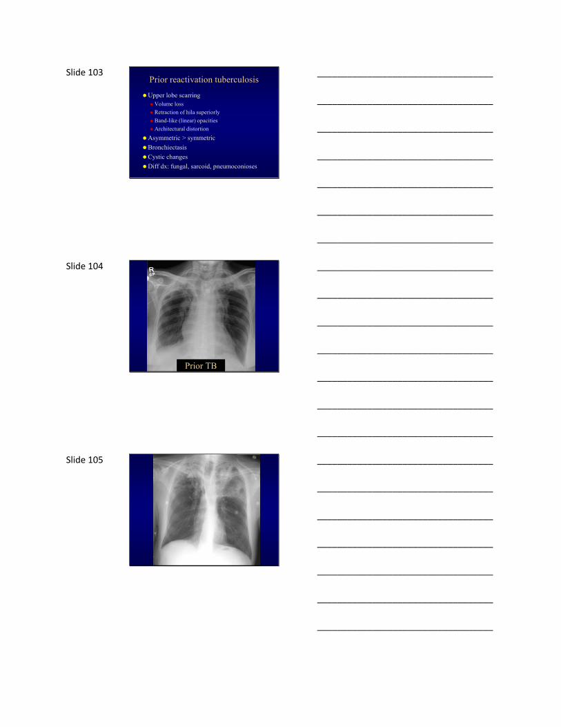
Slide 103 Prior reactivation tuberculosis
Upper lobe scarringVolume loss
Retraction of hila superiorly
Band-like (linear) opacities
Architectural distortion
Asymmetric > symmetric
Bronchiectasis
Cystic changes
Diff dx: fungal, sarcoid, pneumoconioses
___________________________________
___________________________________
___________________________________
___________________________________
___________________________________
___________________________________
___________________________________
Slide 104
Prior TB
___________________________________
___________________________________
___________________________________
___________________________________
___________________________________
___________________________________
___________________________________
Slide 105
___________________________________
___________________________________
___________________________________
___________________________________
___________________________________
___________________________________
___________________________________
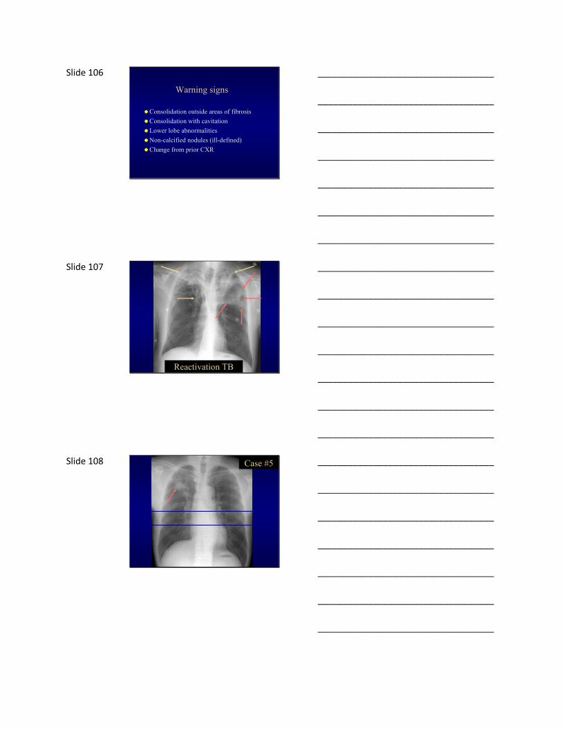
Slide 106
Warning signs
Consolidation outside areas of fibrosis
Consolidation with cavitation
Lower lobe abnormalities
Non-calcified nodules (ill-defined)
Change from prior CXR
___________________________________
___________________________________
___________________________________
___________________________________
___________________________________
___________________________________
___________________________________
Slide 107
Reactivation TB
___________________________________
___________________________________
___________________________________
___________________________________
___________________________________
___________________________________
___________________________________
Slide 108 Case #5
___________________________________
___________________________________
___________________________________
___________________________________
___________________________________
___________________________________
___________________________________

Slide 109 Q7. What is the likelihood of malignancy?
A. <5%
B. 5-10%
C. 10-20%
D. >20%
___________________________________
___________________________________
___________________________________
___________________________________
___________________________________
___________________________________
___________________________________
Slide 110
Solitary nodule/mass- the top 5
Granuloma
Hamartoma
Solitary metastasis
Bronchogenic carcinoma
Lots of others
___________________________________
___________________________________
___________________________________
___________________________________
___________________________________
___________________________________
___________________________________
Slide 111
So you see a nodule on CXR…
1. Look for old films
2. Is diffuse calcification present?
3. Get a CT scan
___________________________________
___________________________________
___________________________________
___________________________________
___________________________________
___________________________________
___________________________________
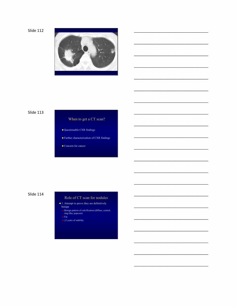
Slide 112
___________________________________
___________________________________
___________________________________
___________________________________
___________________________________
___________________________________
___________________________________
Slide 113
When to get a CT scan?
Questionable CXR findings
Further characterization of CXR findings
Concern for cancer
___________________________________
___________________________________
___________________________________
___________________________________
___________________________________
___________________________________
___________________________________
Slide 114 Role of CT scan for nodules
1. Attempt to prove they are definitively benignBenign pattern of calcification (diffuse, central,
ring-like, popcorn)
Fat
≥2 years of stability
___________________________________
___________________________________
___________________________________
___________________________________
___________________________________
___________________________________
___________________________________

Slide 115 Features of benign nodules include:
PopcornRing-like
CentralDiffuse Initial CT
24 monthfollow-up
Benign patterns of calcification
Presenceof fat
Long term stability
Hamartoma
___________________________________
___________________________________
___________________________________
___________________________________
___________________________________
___________________________________
___________________________________
Slide 116 Hamartoma
.
___________________________________
___________________________________
___________________________________
___________________________________
___________________________________
___________________________________
___________________________________
Slide 117 Irregular calcification: adenocarcinoma
___________________________________
___________________________________
___________________________________
___________________________________
___________________________________
___________________________________
___________________________________

Slide 118 Role of CT scan for nodules
1. Attempt to prove they are definitively benignBenign pattern of calcification (diffuse, central,
ring-like, popcorn)
Fat
≥2 years of stability
2. Determine likelihood of nodule being benign or malignant Low likelihood -> CT follow-up
High likelihood -> immediate action (e.g. biopsy)
___________________________________
___________________________________
___________________________________
___________________________________
___________________________________
___________________________________
___________________________________
Slide 119 Suspicious features of nodules include:
Initial CT
Follow-up
Large size Spiculatedborders
Growth
The size threshold above which malignancy is likely demonstrates geographic variability, depending upon the prevalence of endemic granulomatous infection.
___________________________________
___________________________________
___________________________________
___________________________________
___________________________________
___________________________________
___________________________________
Slide 120 Size and likelihood of cancer
Swensen. Radiology 2005; 235: 259
0% 1%
15%
81%
___________________________________
___________________________________
___________________________________
___________________________________
___________________________________
___________________________________
___________________________________

Slide 121 Follow-up recommendationsNodule size Low-risk patient High-risk patients
≤4 mm No follow-up 12 months
>4-6 mm 12 months 6-12 months
18-24 months
6-8 mm 6-12 months
18-24 months
3-6 months
9-12 months
24 months
>8 mm 3 months
9 months
24 months
3 months
9 months
24 months
Fleischner Guidelines. Radiology 2005; 237: 395.
___________________________________
___________________________________
___________________________________
___________________________________
___________________________________
___________________________________
___________________________________
Slide 122 Old tuberculosis
___________________________________
___________________________________
___________________________________
___________________________________
___________________________________
___________________________________
___________________________________
Slide 123 Bronchogenic carcinoma
___________________________________
___________________________________
___________________________________
___________________________________
___________________________________
___________________________________
___________________________________
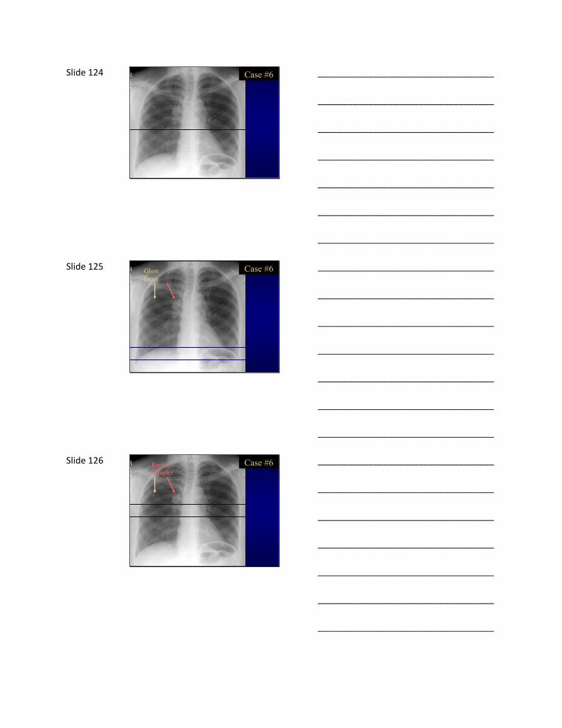
Slide 124 Case #6
___________________________________
___________________________________
___________________________________
___________________________________
___________________________________
___________________________________
___________________________________
Slide 125 Case #6Ghonfocus
___________________________________
___________________________________
___________________________________
___________________________________
___________________________________
___________________________________
___________________________________
Slide 126 Case #6Rankecomplex
___________________________________
___________________________________
___________________________________
___________________________________
___________________________________
___________________________________
___________________________________

Slide 127
Prior tuberculosis
Mid to lower lung predominance
Can be anywhere
Nodule: Ghon focus
Nodule + lymph node: Ranke complex
Calcification indicative of inactivity
___________________________________
___________________________________
___________________________________
___________________________________
___________________________________
___________________________________
___________________________________
Slide 128 Case #7
___________________________________
___________________________________
___________________________________
___________________________________
___________________________________
___________________________________
___________________________________
Slide 129 Q8. What is the most likely diagnosis?
A. Tuberculosis
B. Bacteria
C. Adenovirus
D. Mycoplasma
___________________________________
___________________________________
___________________________________
___________________________________
___________________________________
___________________________________
___________________________________

Slide 130 Primary tuberculosis
Difficult radiologic diagnosis
Mimics other diseases
FindingsNonspecific consolidation
Nodule
Lymphadenopathy
Cavitation unusual
LAD more common than with 2° TB (particularly kids + HIV)
___________________________________
___________________________________
___________________________________
___________________________________
___________________________________
___________________________________
___________________________________
Slide 131 Primary tuberculosis
___________________________________
___________________________________
___________________________________
___________________________________
___________________________________
___________________________________
___________________________________
Slide 132 Primary tuberculosis
___________________________________
___________________________________
___________________________________
___________________________________
___________________________________
___________________________________
___________________________________

Slide 133 Case #8
___________________________________
___________________________________
___________________________________
___________________________________
___________________________________
___________________________________
___________________________________
Slide 134 Q9. What is the LEAST likely diagnosis?
A. Tuberculosis
B. Hypersensitivity pneumonitis
C. Fungal infection
D. Sarcoidosis
___________________________________
___________________________________
___________________________________
___________________________________
___________________________________
___________________________________
___________________________________
Slide 135
Miliary pattern CXR
Miliary tuberculosis
Fungal infection (histo, cocci, blasto)
Metastases
Sarcoidosis
___________________________________
___________________________________
___________________________________
___________________________________
___________________________________
___________________________________
___________________________________

Slide 136
Miliary tuberculosis
___________________________________
___________________________________
___________________________________
___________________________________
___________________________________
___________________________________
___________________________________
Slide 137
Miliary TB
___________________________________
___________________________________
___________________________________
___________________________________
___________________________________
___________________________________
___________________________________
Slide 138
Sarcoidosis
___________________________________
___________________________________
___________________________________
___________________________________
___________________________________
___________________________________
___________________________________

Slide 139
Metastases
___________________________________
___________________________________
___________________________________
___________________________________
___________________________________
___________________________________
___________________________________
Slide 140 Case #10
___________________________________
___________________________________
___________________________________
___________________________________
___________________________________
___________________________________
___________________________________
Slide 141 Pleural + pericardial disease
Primary or secondary
May be only manifestation in 1° TB
Empyema more common in secondary
Adults >> kids
___________________________________
___________________________________
___________________________________
___________________________________
___________________________________
___________________________________
___________________________________
Slide 142 Suspected pleural effusion
___________________________________
___________________________________
___________________________________
___________________________________
___________________________________
___________________________________
___________________________________
Slide 143
___________________________________
___________________________________
___________________________________
___________________________________
___________________________________
___________________________________
___________________________________
Slide 144 Case #11
___________________________________
___________________________________
___________________________________
___________________________________
___________________________________
___________________________________
___________________________________
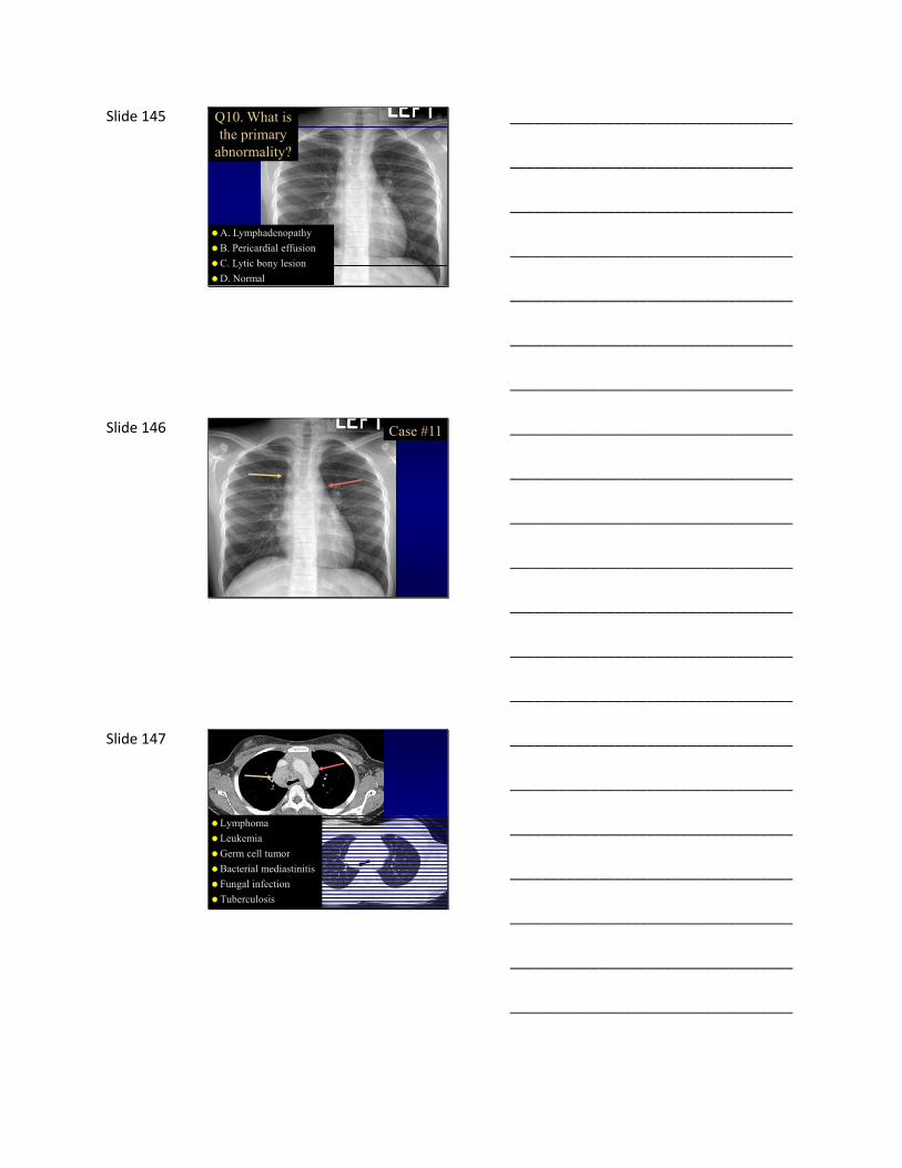
Slide 145 Q10. What is the primary
abnormality?
A. Lymphadenopathy
B. Pericardial effusion
C. Lytic bony lesion
D. Normal
___________________________________
___________________________________
___________________________________
___________________________________
___________________________________
___________________________________
___________________________________
Slide 146 Case #11
___________________________________
___________________________________
___________________________________
___________________________________
___________________________________
___________________________________
___________________________________
Slide 147
Lymphoma
Leukemia
Germ cell tumor
Bacterial mediastinitis
Fungal infection
Tuberculosis
___________________________________
___________________________________
___________________________________
___________________________________
___________________________________
___________________________________
___________________________________

Slide 148 Lymphadenopathy with TB
Kids >> adults
Primary >> secondary
Asymmetric (right > left)
Most common locationsHilar
Right paratracheal
Necrosis very common
___________________________________
___________________________________
___________________________________
___________________________________
___________________________________
___________________________________
___________________________________
Slide 149
TB lymphadenitis
___________________________________
___________________________________
___________________________________
___________________________________
___________________________________
___________________________________
___________________________________
Slide 150 Case #12
___________________________________
___________________________________
___________________________________
___________________________________
___________________________________
___________________________________
___________________________________

Slide 151
heart <65% thoracic diameter
thymus
___________________________________
___________________________________
___________________________________
___________________________________
___________________________________
___________________________________
___________________________________
Slide 152 Conclusions
Be systematic when reading CXR
Typical TB patterns
Mimics of TB
Get a CT scan when appropriate
___________________________________
___________________________________
___________________________________
___________________________________
___________________________________
___________________________________
___________________________________
Slide 153
___________________________________
___________________________________
___________________________________
___________________________________
___________________________________
___________________________________
___________________________________


