Sleep and Serotonin Modulate Paracapsular Nitric Oxide ... · PDF fileNeuronal Excitability...
18
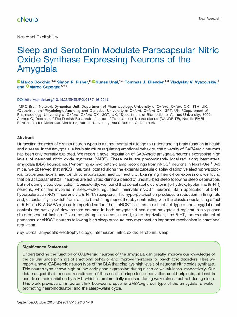
Neuronal Excitability Sleep and Serotonin Modulate Paracapsular Nitric Oxide Synthase Expressing Neurons of the Amygdala Marco Bocchio, 1,3 Simon P. Fisher, 2 Gunes Unal, 1,3 Tommas J. Ellender, 1,3 Vladyslav V. Vyazovskiy, 2 and Marco Capogna 1,4,5 DOI:http://dx.doi.org/10.1523/ENEURO.0177-16.2016 1 MRC Brain Network Dynamics Unit, Department of Pharmacology, University of Oxford, Oxford OX1 3TH, UK, 2 Department of Physiology, Anatomy and Genetics, University of Oxford, Oxford OX1 3PT, UK, 3 Department of Pharmacology, University of Oxford, Oxford OX1 3QT, UK, 4 Department of Biomedicine, Aarhus University, 8000 Aarhus C, Denmark, 5 The Danish Research Institute of Translational Neuroscience (DANDRITE), Nordic EMBL Partnership for Molecular Medicine, Aarhus University, 8000 Aarhus C, Denmark Abstract Unraveling the roles of distinct neuron types is a fundamental challenge to understanding brain function in health and disease. In the amygdala, a brain structure regulating emotional behavior, the diversity of GABAergic neurons has been only partially explored. We report a novel population of GABAergic amygdala neurons expressing high levels of neuronal nitric oxide synthase (nNOS). These cells are predominantly localized along basolateral amygdala (BLA) boundaries. Performing ex vivo patch-clamp recordings from nNOS neurons in Nos1-Cre ER ;Ai9 mice, we observed that nNOS neurons located along the external capsule display distinctive electrophysiolog- ical properties, axonal and dendritic arborization, and connectivity. Examining their c-Fos expression, we found that paracapsular nNOS neurons are activated during a period of undisturbed sleep following sleep deprivation, but not during sleep deprivation. Consistently, we found that dorsal raphe serotonin [5-hydroxytryptamine (5-HT)] neurons, which are involved in sleep–wake regulation, innervate nNOS neurons. Bath application of 5-HT hyperpolarizes nNOS neurons via 5-HT1A receptors. This hyperpolarization produces a reduction in firing rate and, occasionally, a switch from tonic to burst firing mode, thereby contrasting with the classic depolarizing effect of 5-HT on BLA GABAergic cells reported so far. Thus, nNOS cells are a distinct cell type of the amygdala that controls the activity of downstream neurons in both amygdaloid and extra-amygdaloid regions in a vigilance state-dependent fashion. Given the strong links among mood, sleep deprivation, and 5-HT, the recruitment of paracapsular nNOS neurons following high sleep pressure may represent an important mechanism in emotional regulation. Key words: amygdala; electrophysiology; interneuron; nitric oxide; serotonin; sleep Significance Statement Understanding the function of GABAergic neurons of the amygdala can greatly improve our knowledge of the cellular underpinnings of emotional behavior and improve therapies for psychiatric disorders. Here we report a novel GABAergic neuron type of the BLA that displays high levels of neuronal nitric oxide synthase. This neuron type shows high or low early gene expression during sleep or wakefulness, respectively. Our data suggest that reduced recruitment of these cells during sleep deprivation could originate, at least in part, from their inhibition by 5-HT, which is preferentially released during wakefulness but not during sleep. This work provides an important link between a specific GABAergic cell type of the amygdala, a wake- promoting neuromodulator, and the sleep–wake cycle. New Research September/October 2016, 3(5) e0177-16.2016 1–18
Transcript of Sleep and Serotonin Modulate Paracapsular Nitric Oxide ... · PDF fileNeuronal Excitability...
Neuronal Excitability
Sleep and Serotonin Modulate Paracapsular NitricOxide Synthase Expressing Neurons of theAmygdala
Marco Bocchio,1,3 Simon P. Fisher,2 Gunes Unal,1,3 Tommas J. Ellender,1,3 Vladyslav V. Vyazovskiy,2
and Marco Capogna1,4,5
DOI:http://dx.doi.org/10.1523/ENEURO.0177-16.2016
1MRC Brain Network Dynamics Unit, Department of Pharmacology, University of Oxford, Oxford OX1 3TH, UK,2Department of Physiology, Anatomy and Genetics, University of Oxford, Oxford OX1 3PT, UK, 3Department ofPharmacology, University of Oxford, Oxford OX1 3QT, UK, 4Department of Biomedicine, Aarhus University, 8000Aarhus C, Denmark, 5The Danish Research Institute of Translational Neuroscience (DANDRITE), Nordic EMBLPartnership for Molecular Medicine, Aarhus University, 8000 Aarhus C, Denmark
Abstract
Unraveling the roles of distinct neuron types is a fundamental challenge to understanding brain function in healthand disease. In the amygdala, a brain structure regulating emotional behavior, the diversity of GABAergic neuronshas been only partially explored. We report a novel population of GABAergic amygdala neurons expressing highlevels of neuronal nitric oxide synthase (nNOS). These cells are predominantly localized along basolateralamygdala (BLA) boundaries. Performing ex vivo patch-clamp recordings from nNOS� neurons in Nos1-CreER;Ai9mice, we observed that nNOS� neurons located along the external capsule display distinctive electrophysiolog-ical properties, axonal and dendritic arborization, and connectivity. Examining their c-Fos expression, we foundthat paracapsular nNOS� neurons are activated during a period of undisturbed sleep following sleep deprivation,but not during sleep deprivation. Consistently, we found that dorsal raphe serotonin [5-hydroxytryptamine (5-HT)]neurons, which are involved in sleep–wake regulation, innervate nNOS� neurons. Bath application of 5-HThyperpolarizes nNOS� neurons via 5-HT1A receptors. This hyperpolarization produces a reduction in firing rateand, occasionally, a switch from tonic to burst firing mode, thereby contrasting with the classic depolarizing effectof 5-HT on BLA GABAergic cells reported so far. Thus, nNOS� cells are a distinct cell type of the amygdala thatcontrols the activity of downstream neurons in both amygdaloid and extra-amygdaloid regions in a vigilancestate-dependent fashion. Given the strong links among mood, sleep deprivation, and 5-HT, the recruitment ofparacapsular nNOS� neurons following high sleep pressure may represent an important mechanism in emotionalregulation.
Key words: amygdala; electrophysiology; interneuron; nitric oxide; serotonin; sleep
Significance Statement
Understanding the function of GABAergic neurons of the amygdala can greatly improve our knowledge ofthe cellular underpinnings of emotional behavior and improve therapies for psychiatric disorders. Here wereport a novel GABAergic neuron type of the BLA that displays high levels of neuronal nitric oxide synthase.This neuron type shows high or low early gene expression during sleep or wakefulness, respectively. Ourdata suggest that reduced recruitment of these cells during sleep deprivation could originate, at least inpart, from their inhibition by 5-HT, which is preferentially released during wakefulness but not during sleep.This work provides an important link between a specific GABAergic cell type of the amygdala, a wake-promoting neuromodulator, and the sleep–wake cycle.
New Research
September/October 2016, 3(5) e0177-16.2016 1–18

IntroductionThe presence of functionally heterogeneous GABAergic
neurons equips the brain with unparalleled computationalpower (Klausberger and Somogyi, 2008; Hangya et al.,2014). Deciphering the operations carried out by distinctclasses of inhibitory cells is considered one of the majorneurobiological challenges (Lovett-Barron and Losonczy,2014).
The basolateral amygdala (BLA) is a cortical-like brainregion controlling emotional behavior (Duvarci and Pare,2014; Janak and Tye, 2015). Compared to hippocampusor neocortex, our knowledge of anatomy, physiology androle in behavior of specific GABAergic populations in therodent BLA is limited (Capogna, 2014). From a functionalperspective, two inhibitory neuron classes have receivedparticular attention so far: the parvalbumin (PV)-expressing and the somatostatin (SOM)-expressing in-terneurons (Rainnie et al., 2006; Woodruff and Sah,2007b; Wolff et al., 2014).
Many other GABAergic neuron types have been de-tected in the BLA of rodents (Spampanato et al., 2011),but our understanding of their functional roles is scant. Inaddition to the expression of PV and SOM, BLA interneu-rons express a variety of neurochemical markers, such ascalbindin (McDonald and Mascagni, 2001; Bienvenu et al.,2012), calretinin (McDonald and Mascagni, 2001), vaso-active intestinal peptide (Mascagni and McDonald, 2003),cholecystokinin (Jasnow et al., 2009; Vogel et al., 2016),and neuronal nitric oxide synthase (nNOS; McDonaldet al., 1993; Usunoff et al., 2006). Neurons of the BLAexpressing nNOS represent a particularly intriguing cellpopulations for several reasons. First, in areas with in-terneuron diversity similar to the BLA, such as hippocam-pus or neocortex, nNOS� neurons are as abundant, oreven denser, than PV� and SOM� cells (Fuentealba et al.,2008; Tricoire and Vitalis, 2012), suggesting a prominentimpact on both hippocampal and cortical circuits. Sec-ond, they are able to modulate neurons through a variety
of mechanisms, including slow inhibition (Capogna andPearce, 2011), retrograde release of nitric oxide (Li et al.,2014), and potentially via other neuropeptides (Fuent-ealba et al., 2008; Tricoire and Vitalis, 2012), suggestingthey might fulfill a function that is different from those ofother classic interneuron types. Third, neocortical nNOS�
neurons coexpressing SOM and NPY are thought to beatypical long-range GABAergic projection neurons (To-mioka et al., 2005; Tamamaki and Tomioka, 2010), andthis might also apply to the BLA (McDonald et al., 2012;McDonald and Zaric, 2015). Despite their prominence, thephysiological and behavioral roles of GABAergic nNOS�
neurons of the BLA remain elusive.The activity of BLA GABAergic neurons is eminently
controlled by subcortical neuromodulators released dur-ing arousal, such as serotonin [5-hydroxytryptamine (5-HT)], acetylcholine, and noradrenaline (Tully et al., 2007;Bocchio et al., 2015; Unal et al., 2015a). Among those,5-HT neurotransmission is compelling because it modu-lates emotional learning in the BLA (Bocchio et al., 2016),but it is also involved in the sleep–wake cycle (Portaset al., 2000; Gao et al., 2002). Specifically, extracellularforebrain 5-HT levels are low during non-rapid eye move-ment (NREM) and rapid eye movement (REM) sleep, andhigh during wakefulness (Portas et al., 1998; Bjorvatnet al., 2002). Consistently, electrophysiological experi-ments have shown that dorsal raphe nuclei (DRN) 5-HTneurons fire at higher rates during wakefulness and atlower rates during NREM sleep, and are virtually silentduring REM sleep (Sakai, 2011). Since the activity of BLAneurons also follows the sleep–wake cycle (Paré andGaudreau, 1996), 5-HT could play a crucial role in thevigilance state-dependent activity of BLA cells.
At a cellular level, the most commonly established ef-fect of 5-HT in the BLA is the depolarization of GABAergicinterneurons (Rainnie, 1999), and among those of PV�
interneurons via 5-HT2A receptors (Bocchio et al., 2015).However, recent in situ hybridization data have shownthat BLA NPY� cells, some of which are thought to benNOS� (McDonald et al., 1993), can also express inhibi-tory 5-HT1A receptors (Bonn et al., 2013), suggesting that5-HT could also hyperpolarize some GABAergic cells.Defining the diversity of 5-HT actions on BLA neurontypes is crucial if we are to understand the cellular dy-namics occurring in the BLA across different brain states.
In this study, we aimed to functionally characterizenNOS� neurons of the mouse BLA and to shed light ontheir behavioral role. Additionally, we wished to probewhether the 5-HT modulation of nNOS� neurons is in linewith the action of 5-HT on previously characterizedGABAergic neurons, and whether this modulation is con-sistent with the behavioral recruitment of nNOS� neurons.
Materials and MethodsAnimals
Since nNOS is broadly expressed during development(Bredt and Snyder, 1994), but its expression is morerestricted to particular cells following postnatal day 15(P15; Kubota et al., 2011; Taniguchi et al., 2011), aninducible Cre driver line (Nos1-CreER; B6;129S-
Received September 9, 2016; accepted September 12, 2016; First publishedSeptember 26, 2016.Authors declare no competing financial interests.Author contributions: M.B., V.V.V., and M.C. designed research; M.B.,
S.P.F., and G.U. performed research; T.J.E. contributed with reagents/analytictools; M.B., S.P.F., G.U., and V.V.V. analyzed data; M.B. and M.C. wrote thepaper.
This work was supported by Medical Research Council (UK) GrantU138197106 to M.C.; Medical Research Council (UK) Career DevelopmentAward MR/M009599/1 to T.J.E.; and Medical Research Council New Investi-gator Research Grant MR/L003635/1, John Fell OUP Research Fund Grant131/032, and Wellcome Trust Strategic Grant 098461/Z/12/Z to V.V.V.
Acknowledgments: We thank Katharine Whitworth, Ben Micklem, Jane Jan-son, and Liz Norman for excellent technical assistance. We also thank Profes-sor Trevor Sharp and Ayesha Sengupta for sharing and injecting the SERT-Cremice. In addition, we thank Professor Peter Somogyi, who assisted withlaboratory space and resources for the completion of this project.
Correspondence should be addressed to Marco Capogna, Department ofBiomedicine, Aarhus University, Wilhelm Meyers Allé 3, 8000 Aarhus C, Den-mark. E-mail: [email protected].
DOI:http://dx.doi.org/10.1523/ENEURO.0177-16.2016Copyright © 2016 Bocchio et al.This is an open-access article distributed under the terms of the CreativeCommons Attribution 4.0 International, which permits unrestricted use, distri-bution and reproduction in any medium provided that the original work isproperly attributed.
New Research 2 of 18
September/October 2016, 3(5) e0177-16.2016 eNeuro.org
Nos1tm1.1(cre/ERT2)Zjh/J; stock #014541, The Jackson Lab-oratory) was used to elicit Cre recombination postnatally.Nos1-CreER�/� mice were crossed with Ai9�/� reportermice (B6.Cg-Gt(ROSA)26Sortm9(CAG-tdTomato)Hze/J; stock#007909, The Jackson Laboratory) to generate Nos1-Cre;Ai9 offspring. To quantify the overlap of neurochemicalmarkers, WT C57BL/6J mice (Charles River Laboratories)were used. For anterograde tracing experiments, SERT-Cre�/� mice (MMRRC, B6.Cg-Tg(Slc6a4-cre)Et33Gsat;stock #031028-113, UC Davis) were used. Mice werehoused with their littermates with ad libitum access tofood and water in a dedicated housing room with a 12 hlight/dark cycle. To induce Cre recombinase and labelnNOS� neurons with tdTomato, Nos1-CreER;Ai9 mice(age range, postnatal day 20–45) received one to threeintraperitoneal injections of tamoxifen (10 mg/ml in cornoil, 10 �l/g body weight/d). For patch-clamp and anatom-ical experiments, mice (age range, postnatal day 27–60)were used at least 1 week after the first tamoxifen injec-tion.
For sleep experiments, adult male C57BL/6J mice (15weeks of age) were individually housed in custom-madeclear Plexiglas cages (20.3 � 32 � 35 cm) with freeaccess to a running wheel and ad libitum food and water.Cages were housed in ventilated, sound-attenuated Fara-day chambers (two cages per chamber; Campden Instru-ments) under a standard 12 h light/dark cycle [lights on8:00 A.M., zeitgeber time 0 (ZT0); light levels, �120–180lux]. Room temperature (RT) and relative humidity weremaintained at 22 � 1°C and 50 � 20%, respectively. Micewere habituated to both the cage and recording cables fora minimum of 16 d prior to recording.
All procedures involving experimental animals wereperformed in compliance with the Animals (Scientific Pro-cedures) Act, 1986 (UK) and associated regulations, un-der approved project licenses by Home Office UK (30/3061 and 70/7483) and with Society for NeurosciencePolicies on the Use of Animals in Neuroscience Research.
Ex vivo recordingsNos1-CreER;Ai9 mice (age range, postnatal day 27–60)
were decapitated under deep isoflurane anesthesia (4% inO2), and their brains were rapidly removed and placed inice-cold sucrose-containing artificial CSF (ACSF) cuttingsolution containing the following (in mM): 75 sucrose, 87NaCl, 25 NaHCO3, 2.5 KCl, 1.25 NaH2PO4, 0.5 CaCl2, 7MgCl2, and 25 glucose, saturated with 95% O2, 5% CO2,at pH 7.3–7.4. Slices (325 �m thickness), including theamygdala were cut (Microm HM 650 V, Thermo FisherScientific) and transferred onto a nylon mesh where theywere maintained in a chamber initially containing sucroseACSF cutting solution at 37°C for 30 min. During thisperiod, the cutting solution was gradually substituted (5ml/min) with normal ACSF consisting of the following (inmM): 130 NaCl, 24 NaHCO3, 3.5 KCl, 1.25 NaH2PO4, 2.5CaCl2, 1.5 MgSO4, and 10 glucose, saturated with 95%O2, 5% CO2, at pH 7.3.
Slices were transferred to a submerged recordingchamber and continuously perfused with oxygenatedACSF at a rate of �5 ml/min and at a mean temperature
of 34 � 1°C. Neurons were visualized with an uprightAxioskop microscope (Zeiss) using phase-contrast mi-croscopy under a LUMPlanFI 60� immersion objective(Olympus). A mercury vapor short-arc lamp (100 W; NHBC 103, Zeiss) was connected to the epifluorescencesystem to visualize the tdTomato� neurons. Micropipettes(5–6 M�) were pulled from borosilicate glass capillaries(1.2 mm; GC120F, Harvard Apparatus) with a DMZ puller(Zeitz-Instrumente). Somatic whole-cell patch-clamp re-cordings were performed from visually identified tdTo-mato� neurons. Electrodes were filled with an intracellularsolution composed of the following (in mM): 126K-gluconate, 4 KCl, 4 ATP-Mg, 0.3 GTP-Na2, 10 Na2-phosphocreatine, 10 HEPES, and 0.2-0.4% biocytin, withosmolarity of 270–280 mOsmol/L without biocytin, at pH7.3 adjusted with KOH.
For paired recordings, the presence of a connectionwas tested by evoking an action current (3-ms-long volt-age step from �60 to 0 mV) in paracapsular nNOS (pc-nNOS) cells. In some cases (n � 3), nearby cells wereloaded with the same intracellular solution mentionedabove (Cl- reversal potential, ECl, �91 mV) and held involtage-clamp mode at �40 mV. The remaining cells (n �16) were loaded with an intracellular solution with higherCl� to increase the driving force of IPSCs (ECl, �12 mV).This solution consisted of the following (in mM): 42K-gluconate, 84 KCl, 4 ATP-Mg, 0.3 GTP-Na2, 10 Na2-phosphocreatine, 10 HEPES, and 0.2–0.4% biocytin, withosmolarity of 270–280 mOsmol/L without biocytin, pH 7.3adjusted with KOH. In these cases, nearby cells were heldat �65 mV. Since this resulted in inward polarity of Cl�
currents, glutamatergic transmission was blocked witheither 3 mM kynurenic acid (or 10 �M NBQX and 50 �M
D-APV) to isolate GABAergic IPSCs. Action currents wereevoked at least 10 times, with 20 s interval betweensweeps. Principal neurons were distinguished from in-terneurons according to the following parameters: (1)smaller fast afterhyperpolarization (fAHP) amplitude andprominent medium AHP in an instantaneous firing rateprotocol; (2) adapting, 20 Hz maximum firing rates; (3)lower input resistance (Rin; 150 M�); and (4) longerspike half-width (�1 ms). Electrophysiological signalswere amplified using an EPC9/2 amplifier (HEKA Elec-tronik) and acquired using Patchmaster software (HEKAElectronik). Recordings were accepted only when theinitial seal resistance was 2 G�, the holding currentnecessary to clamp the cell at �60 mV was smaller than�50 pA, and the series resistance did not change by20% throughout the experiment. No correction wasmade for the liquid junction potential (16 mV) between thepipette and the ACSF.
Membrane potential (Vm) during 5-HT application wasmonitored while holding neurons in current clamp at �60� 2 mV. Hyperpolarizing and depolarizing current stepswere injected every 10 s to monitor Rin and firing, respec-tively. At the end of the recording, some slices containingbiocytin-filled cells were fixed overnight at 4°C in 4%paraformaldehyde (PFA) and 15% saturated picric acid in0.1 M PB. After 24 h, slices were embedded in gelatin and
New Research 3 of 18
September/October 2016, 3(5) e0177-16.2016 eNeuro.org
re-sectioned into 60- to 80-�m-thick sections with a VT-1000 vibrating microtome (Leica).
Analysis of ex vivo recordingsAnalysis of synaptic currents and intrinsic membrane
properties was performed using IGOR Pro (WaveMetrics)and MATLAB (MathWorks). The Rin was calculated fromthe slope of steady-state voltage responses to a series of8–10 subthreshold current injections (from �30 to �60pA) lasting 400 ms. The AHP (in mV) was determined fromthe first spike in response to a juxtathreshold positivecurrent injection. The spike duration of the action potentialwas measured as the width at half-amplitude between thethreshold potential and the peak of the action potential,which was evoked by a strong (800–1000 pA) and short(2–5 ms) depolarizing current pulse. The membrane timeconstant � was estimated from the monoexponentialcurve fitting of voltage responses to a �30 pA hyperpo-larizing pulse. The rheobase (in pA) was determined as a50 ms current injection, able to generate a spike in 50% ofthe cases in 10 trials. The instantaneous firing rate (in Hz)was defined as the number of action potentials evokedduring a 1 s depolarizing current pulse of twice the am-plitude of the rheobase current. The membrane capaci-tance was calculated as the ratio between the timeconstant and the Rin. The adaptation index (range, 0–1)was defined as the ratio between the first and last inter-spike intervals (ISIs; in ms) elicited by the same pulseused to measure the instantaneous firing rate. The restingVm was estimated by averaging a 20 s current-clamptrace recorded at a 0 pA holding current. Although manynNOS� neurons were spontaneously active, spikes didnot contaminate this estimate because firing rates were5 Hz and the average of the 20 s trace matched the Vm
sampled during ISIs.To minimize artificial changes in firing rate due to re-
cording conditions, cell-attached recordings were per-formed in loose-patch configuration (50 M� seal).Spikes were acquired in voltage clamp by setting thepipette potential to obtain 0 pA of membrane current(Alcami et al., 2012). Neurons were defined as “bursting”during 5-HT application if the peak of the ISI histogram (inLog scale) was 100 ms.
Electroencephalogram recordings and sleepdeprivationSurgical procedures and electrode implantation
Surgical procedures were performed using aseptictechniques under isoflurane anesthesia (3–5% induction,1–2% maintenance) and Metacam (1–2 mg/kg, s.c.;Boehringer Ingelheim) was administered preoperatively.During surgery, animals were head fixed using a stereo-taxic frame (David Kopf Instruments) and liquid gel (Vis-cotears, Alcon Laboratories) was applied to protect theeyes. In all animals, electroencephalogram (EEG) screwswere placed in the frontal [motor area: anteroposterior(AP), �2 mm; mediolateral (ML), �2 mm] and occipital(visual area, V1: AP, �3.5/�4 mm; ML, �2.5 mm) corticalregions using procedures previously described (Cui et al.,2014). A reference screw electrode was placed above thecerebellum, and an additional anchor screw was placed in
the left parietal hemisphere to ensure implant stability.EEG screws were soldered (prior to implantation) tocustom-made headmounts (Pinnacle Technology), and allscrews and wires were secured to the skull using dentalacrylic. Two single-stranded, stainless steel wires wereinserted on either side of the nuchal muscle to recordelectromyography (EMG). Saline (0.1 ml/20 g bodyweight, s.c.) was administered postoperatively and ani-mals were provided thermal support throughout and fol-lowing surgery. Metacam (1–2 mg/kg) was orallyadministered for at least 3 d after surgery. A minimum 2week recovery period was permitted prior to cabling theanimals.
Experimental designOn the experimental day, following a stable 24 h base-
line recording, mice were divided into the following twogroups: sleep deprivation (SD, n � 4); and SD plus recov-ery sleep (RS; n � 4). RS was defined as the sleepopportunity occurring immediately following SD, and waslimited to 1.5–2 h (Morairty et al., 2013). In both groups,SD was performed in the home cage of the animal for acontinuous 4 h period starting at light onset. During thistime, animals were spontaneously awake, and their be-havior as well as their EEG/EMG recordings were underconstant visual observation. Sleep was prevented by reg-ularly providing the animals with novel objects, an effec-tive method that mimics natural conditions ofwakefulness, is ethologically relevant, and does not ap-pear to stress the animals (Palchykova et al., 2006; Vya-zovskiy et al., 2007). All mice were well habituated to theexperimenter and to the exposure to novel objects prior tothe experiment. Novel objects included nesting and bed-ding material from other cages, wooden blocks, paperboxes, and tubes of different shape and color. SD wassuccessful with 97.98 � 2.51% of time spent awakeduring the 4 h procedure. After completion of the SDexperiment, animals in the SD group were injected with anoverdose of Euthatal (pentobarbitone sodium 200 mg/ml;0.3 ml, i.p.), and upon the loss of a response to toe pinchwere perfused transcardially with 30 ml of 0.9% PBSfollowed by 50 ml of 4% PFA in 0.1 M PB. All mice wereperfused within �30 min after the end of 4 h of SD, andwere kept awake continuously until the moment of injec-tion with Euthatal. The mice in the SD�RS group wereallowed to sleep undisturbed for a period of 1.5–2 h (anaverage of 1.81 � 0.15 h spent in NREM and REM sleep)and then were perfused according to the same procedure.Special care was taken to ensure that the animals in theSD�RS group were not awake for longer than a fewminutes prior to the injection of Euthanal. Brains wereremoved, postfixed in 4% PFA (in 0.1 M PB) overnight at4°C, then thoroughly washed in PBS and left in 0.1 M PBplus 0.05% sodium azide until further processing for c-Fos/nNOS immunohistochemical analysis (see below).c-Fos protein is a marker of neuronal activation that isproduced 30–60 min following stimulus/behavior onset(Sheng and Greenberg, 1990; Morgan and Curran, 1991).Several lines of evidence indicate that c-Fos levels canrapidly increase and decrease during both wake andsleep (Basheer et al., 1997; Cirelli and Tononi, 2000;
New Research 4 of 18
September/October 2016, 3(5) e0177-16.2016 eNeuro.org
Gerashchenko et al., 2008). These aspects render c-Fosstaining a convenient approach to investigate effects ofsleep and waking on neuronal activity (Cirelli and Tononi,2000).
EEG recordings and power spectra analysisData acquisition was performed using the Multichannel
Neurophysiology Recording System (TDT). Cortical EEGwas recorded from frontal and occipital derivations. EEG/EMG data were filtered between 0.1 and 100 Hz, amplified(PZ5 NeuroDigitizer Preamplifier, TDT) and stored on alocal computer at a sampling rate of 256.9 Hz. EEG/EMGdata were resampled off-line at a sampling rate of 256 Hz.Signal conversion was performed using custom-writtenMATLAB scripts and was then transformed into EuropeanData Format using open source Neurotraces software(www.neurotraces.com). For each recording, EEG powerspectra were computed by a fast Fourier transform rou-tine for 4 s epochs (using a Hanning window), with a 0.25Hz resolution (SleepSign Kissei Comtec Co.). Vigilancestates were scored off-line through manual visual inspec-tion of consecutive 4 s epochs (SleepSign, Kissei ComtecCo.). Two EEG channels (frontal and occipital) and EMGwere displayed simultaneously to aid vigilance state scor-ing. Vigilance states were classified as waking (low-voltage, high-frequency EEG with a high level of phasicEMG activity), NREM sleep (presence of slow waves, EEGsignal of a high amplitude and low frequency), or REMsleep (low-voltage, high-frequency EEG with a low level ofEMG activity). Great care was taken to eliminate epochscontaminated by eating, drinking, or gross movementsresulting in artifacts in at least one of the two EEG deri-vations.
Anterograde tracingTo selectively label dorsal raphe 5-HT axons, the viral
vector AAV2-EF1a-DIO EYFP (UNC Vector Core, Univer-sity of North Carolina, Chapel Hill, NC) was stereotaxicallyinjected (1 �l at 100 nl/min) into the dorsal raphe nuclei[coordinates according to bregma and the brain surface(in mm): AP, �4.1; dorsoventral, �2.5, �2.2, �1.9] ofSERT-Cre mice (age range, P30 to P75) anesthetizedusing 1–2% isoflurane in oxygen (2 L/min). On recoveryfrom surgery, mice were administered buprenorphine 0.3mg/kg, s.c., for postoperative analgesia. Three weekswere allowed for anterograde tracing before fixation byperfusion.
Histological proceduresImmunohistochemistry
For quantification of the overlap between neurochemi-cal markers, mice were transcardially perfused with salinefollowed by 4% PFA, 15% saturated picric acid in 0.1 M
PB. Brains were sectioned using a vibratome (VT 1000 S,Leica) into 60-�m-thick slices. Sections were stored in 0.1M PB containing 0.05% sodium azide until further usage.Resectioned slices (60–80 �m thickness) containing re-corded and biocytin-filled neurons were incubated over-night at 4°C in 1:2000 Alexa Fluor 488-conjugatedstreptavidin (Invitrogen). Following blocking with 10%normal donkey serum for 1 h at RT, sections were incu-
bated overnight at 4°C with the following primary antibod-ies: anti-5-HT raised in rabbit (1:2500; provided by H.Steinbusch, Maastricht University, Maastricht, The Neth-erlands); anti-c-Fos raised in rabbit (1:500; catalog#ab7963, Abcam); anti-GFP raised in chicken (1:1000;catalog #GFP-1020, Aves Labs); anti-nNOS raised in goat(1:500; catalog #ab1376, Abcam); anti-NK1 (substance Preceptor) raised in rabbit (1:1000; AB5060, Chemicon);anti-somatostatin raised in rat (1:250; catalog #MAB354,Chemicon); and anti-vesicular GABA transporter (VGAT)raised in rabbit (1:500; provided by M. Watanabe, FrontierInstitute Co. Ltd., Hokkaido, Japan; http://www.frontier-institute.com). Following 3� washes in PBS, immunore-activity was revealed with Alexa Fluor 488- (1:500),DyLight Cy3- (1:500), or Alexa Fluor 647-conjugated (1:250) secondary antibodies (all raised in donkey; JacksonImmunoResearch). For negative controls, the primary an-tibody was routinely omitted from the staining procedurewith no positive fluorescence signal detected. In somecases, each secondary antibody was omitted in turn toconfirm its specificity. Nissl staining was obtained viaincubation in NeuroTrace 640/660 Deep-Red FluorescentNissl Stain (1:200; catalog #N-21483, Thermo Fisher).
All reagents were diluted in PBS containing 0.3% TritonX-100. Immunoreactivity was visualized using an epifluo-rescence microscope (AxioImager M2, Zeiss) or a laser-scanning confocal microscope (LSM 510, Zeiss). Theboundaries between nuclei were determined with bright-field microscopy or Nissl staining.
Quantification of overlap between neurochemical mark-ers
Sections containing the BLA of SD and SD�RS micewere imaged with the epifluorescence microscope men-tioned above and StereoInvestigator software (MBF Bio-science). A region of interest delineating either the BLA orthe external capsule next to the lateral amygdala (LA) wasdefined using a bright-field microscope under a 5� 0.16numerical aperture (NA) objective lens. For quantificationof neurochemical markers expressed by nNOS cells, ste-reological sampling was performed in both hemispheresfrom one of three sections in the range �0.8 to �2.2 mmfrom bregma. Series of tiled stacked images were ac-quired using a 40� 1.3 NA oil-immersion objective and 1�m steps at a depth of 2–22 �m (“optical sections”) fromthe upper surface of each section. In order to minimizeartifacts arising from surface irregularities, the first 2 �mfrom the upper surface were defined as the “guard zone”and were not scanned. Counting was performed off-line inStereoInvestigator. A neuron was counted only if its im-munopositive nucleus came into focus in the optical sec-tion. Nuclei already in focus at the top optical sectionwere not counted (West, 1999). For quantification of theoverall percentage of c-Fos� cells in the paracapsulararea, stereological counting was performed by samplingthree evenly spaced sections in the range �0.8 to �2.2mm from bregma. To ensure the quantification of neuronalc-Fos, Nissl staining was used. Only nuclear c-Fos ex-pression in Nissl-stained cells with a diameter of 10 �mwas quantified. The experimenter was blind to behavioral
New Research 5 of 18
September/October 2016, 3(5) e0177-16.2016 eNeuro.org

testing conditions. Data were exported to Excel (Mi-crosoft) and pooled for further analysis.
Neurolucida reconstructionTwo-dimensional drawings were performed for two
biocytin-filled cells to reveal dendrites and axonal ar-borization present in the 325-�m-thick slice. The 60- to80-�m-thick sections were processed with DAB using apreviously published protocol (Unal et al., 2015b). Draw-ings were made using Neurolucida software (MBF Biosci-ence) under a light microscope (100� objective). Finaldrawings were corrected for tissue shrinkage caused byTriton X-100 processing. Dendritic and axonal lengthswere calculated using the same software.
Statistical testingData are presented as the means � SEM. Distributions
were compared using Student’s t tests or one-way ANO-VAs with Bonferroni post hoc correction. Statistical anal-ysis was performed with GraphPad Prism (GraphPadSoftware) and SigmaPlot (Systat Software Inc.), wherep 0.05 was considered to be statistically significant.
DrugsSerotonin hydrochloride, WAY 100635 maleate, NBQX,
D-APV, and SR95531 were purchased from Tocris Bio-science. Kynurenic acid and tamoxifen were purchasedfrom Sigma-Aldrich.
ResultsNeurochemical profile of nNOS� type I neurons ofthe BLA
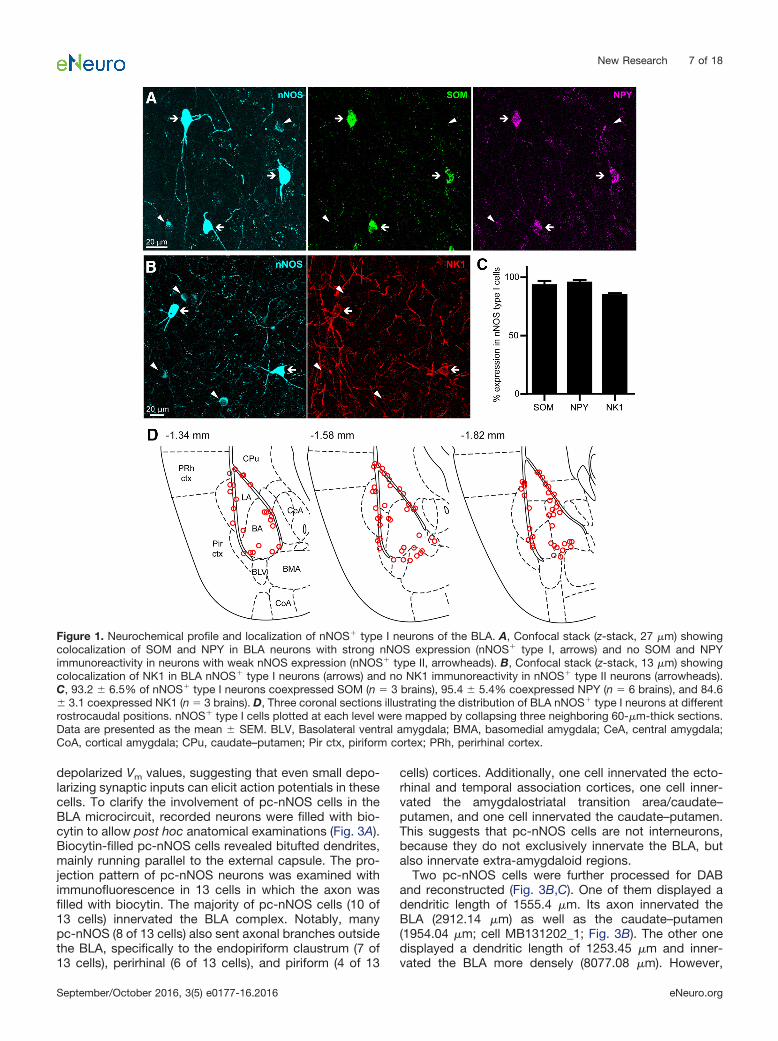
We aimed to uncover the anatomical and physiologicalfeatures of GABAergic nNOS� neurons of the BLA. First,we immunolabeled mouse coronal brain sections contain-ing the amygdala for nNOS. We detected neurons withstrong nNOS expression and others with light immunore-activity (Fig. 1A), suggesting that nNOS� neurons of theBLA can be classified according to the intensity of nNOSexpression, as in the case of neocortex (Yan et al., 1996;Smiley et al., 2000; Lee and Jeon, 2005). Following pre-viously used nomenclature (Yan et al., 1996; Perrenoudet al., 2012), we refer to neurons with strong nNOS ex-pression as “type I” nNOS� cells, and to neurons withweak nNOS expression as “type II” nNOS� cells. Type Ineurons displayed large ovoid somata and bitufted den-drites, whereas type II neurons had more heterogeneoussoma size and dendritic emissions.
In cortical areas, type I nNOS� cells often coexpressother SOM, neuropeptide Y (NPY), and neurokinin 1 (NK1)receptor. To investigate whether this coexpression pat-tern also applies to the BLA, we examined the proportionof type I and type II nNOS� cells expressing these threemarkers. We found that 93 � 4% of nNOS� type I cellscoexpressed SOM (n � 3 brains), 95 � 2% coexpressedNPY (n � 6 brains), and 85 � 2% coexpressed NK1 (n �3 brains; Fig. 1A–C). In contrast, nNOS� type II cells weremostly devoid of these neurochemical markers, with 2 �2% coexpressing SOM (n � 3 brains), 9 � 3% coexpress-ing NPY (n � 6 brains), and none coexpressing NK1 (n �3 brains; data not shown).
Thus, intense nNOS labeling identifies a neurochemi-cally homogeneous population of BLA neurons. Sinceobservations of neocortical nNOS� neurons have demon-strated that type II cells are more heterogeneous andcomprise several cell types (Tricoire and Vitalis, 2012),further investigations were focused on nNOS� type I neu-rons. The latter cells were localized primarily along BLAborders, namely adjacent to the external capsule, inter-mediate capsule, and the border between basal (BA) andbasomedial nuclei (Fig. 1D), which is in line with previousreports of nNOS� “border cells” (McDonald et al., 1993;Usunoff et al., 2006). Importantly, type I neurons repre-sented the great majority of cells expressing SOM andNPY (98.4 � 1.6%, n � 2 brains).
Intrinsic electrophysiological properties of pc-nNOSneurons
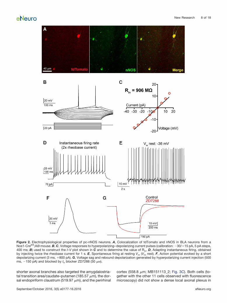
To study the physiology of nNOS type I neurons of theBLA, we crossed an inducible Cre driver mouse line(Nos1-CreER; Taniguchi et al., 2011) with an Ai9 reporterline. This enabled specific expression of tdTomato innNOS� neurons (97.9 � 0.7% nNOS/tdTomato overlap;n � 3 brains), because neurons that express nNOS onlytransiently during development were not labeled with td-Tomato. To selectively target nNOS� type I neurons, weprepared acute coronal brain slices and performed whole-cell patch-clamp recordings from tdTomato� cells lo-cated along the external capsule that separates the LAfrom the endopiriform claustrum/piriform cortex (in thefollowing, called pc-nNOS� neurons). In this region, typeI cells constitute 80 � 3% (n � 3 brains) of nNOS�
neurons and are easily distinguishable from type II cellsdue to their significantly larger ovoid somata (area, 128 �5 vs 88 � 2 �m2; p 0.0001; n � 25 cells from six brains).
We examined the intrinsic membrane properties dis-played by pc-nNOS neurons in brain slices from Nos1-CreER;Ai9 mice (Fig. 2; Table 1; n � 10 neurons). Thesecells were characterized by high Rin values (852.8 � 51.8M�), high membrane time constant (�, 27.2 � 2.0 ms),and high excitability (rheobase current, 28.3 � 4.0 pA),with even small positive current injections leading to sus-tained firing. Consistent with the general physiology ofBLA SOM� neurons (Wolff et al., 2014), pc-nNOS neuronswere not fast spiking (instantaneous firing rate, 23.2 � 1.8Hz) and showed relatively broad spikes (half-width, 0.75� 0.04 ms). Additionally, pc-nNOS cells exhibited verydepolarized resting Vm (�39.7 � 2.4 mV), often resultingin spontaneous firing (Fig. 2E). Finally, when hyperpolar-izing currents were injected, pc-nNOS neurons displayeda depolarizing sag and rebound depolarization (Fig. 2).Both responses were mediated by the hyperpolarization-activated cationic current (Ih) because they were abol-ished by the Ih blocker ZD7288 (30 �M; sag ratio: control,0.813 � 0.031; ZD7288, 1.076 � 0.081; rebound ampli-tude: control, 5.9 � 1 mV; ZD7288, �4.5 � 3.3 mV; bothp � 0.048, paired t test; n � 4; Fig. 2F).
Projection pattern and synaptic connectivity ofparacapsular type I nNOS� neurons
Thus, pc-nNOS neurons are highly excitable becausethey display high Rin values, low rheobase currents, and
New Research 6 of 18
September/October 2016, 3(5) e0177-16.2016 eNeuro.org
depolarized Vm values, suggesting that even small depo-larizing synaptic inputs can elicit action potentials in thesecells. To clarify the involvement of pc-nNOS cells in theBLA microcircuit, recorded neurons were filled with bio-cytin to allow post hoc anatomical examinations (Fig. 3A).Biocytin-filled pc-nNOS cells revealed bitufted dendrites,mainly running parallel to the external capsule. The pro-jection pattern of pc-nNOS neurons was examined withimmunofluorescence in 13 cells in which the axon wasfilled with biocytin. The majority of pc-nNOS cells (10 of13 cells) innervated the BLA complex. Notably, manypc-nNOS (8 of 13 cells) also sent axonal branches outsidethe BLA, specifically to the endopiriform claustrum (7 of13 cells), perirhinal (6 of 13 cells), and piriform (4 of 13
cells) cortices. Additionally, one cell innervated the ecto-rhinal and temporal association cortices, one cell inner-vated the amygdalostriatal transition area/caudate–putamen, and one cell innervated the caudate–putamen.This suggests that pc-nNOS cells are not interneurons,because they do not exclusively innervate the BLA, butalso innervate extra-amygdaloid regions.
Two pc-nNOS cells were further processed for DABand reconstructed (Fig. 3B,C). One of them displayed adendritic length of 1555.4 �m. Its axon innervated theBLA (2912.14 �m) as well as the caudate–putamen(1954.04 �m; cell MB131202_1; Fig. 3B). The other onedisplayed a dendritic length of 1253.45 �m and inner-vated the BLA more densely (8077.08 �m). However,
Figure 1. Neurochemical profile and localization of nNOS� type I neurons of the BLA. A, Confocal stack (z-stack, 27 �m) showingcolocalization of SOM and NPY in BLA neurons with strong nNOS expression (nNOS� type I, arrows) and no SOM and NPYimmunoreactivity in neurons with weak nNOS expression (nNOS� type II, arrowheads). B, Confocal stack (z-stack, 13 �m) showingcolocalization of NK1 in BLA nNOS� type I neurons (arrows) and no NK1 immunoreactivity in nNOS� type II neurons (arrowheads).C, 93.2 � 6.5% of nNOS� type I neurons coexpressed SOM (n � 3 brains), 95.4 � 5.4% coexpressed NPY (n � 6 brains), and 84.6� 3.1 coexpressed NK1 (n � 3 brains). D, Three coronal sections illustrating the distribution of BLA nNOS� type I neurons at differentrostrocaudal positions. nNOS� type I cells plotted at each level were mapped by collapsing three neighboring 60-�m-thick sections.Data are presented as the mean � SEM. BLV, Basolateral ventral amygdala; BMA, basomedial amygdala; CeA, central amygdala;CoA, cortical amygdala; CPu, caudate–putamen; Pir ctx, piriform cortex; PRh, perirhinal cortex.
New Research 7 of 18
September/October 2016, 3(5) e0177-16.2016 eNeuro.org
shorter axonal branches also targeted the amygdalostria-tal transition area/caudate–putamen (185.57 �m), the dor-sal endopiriform claustrum (519.97 �m), and the perirhinal
cortex (558.8 �m; MB151113_2; Fig. 3C). Both cells (to-gether with the other 11 cells observed with fluorescencemicroscopy) did not show a dense local axonal plexus in
Figure 2. Electrophysiological properties of pc-nNOS neurons. A, Colocalization of tdTomato and nNOS in BLA neurons from aNos1-CreER;Ai9 mouse. B, C, Voltage responses to hyperpolarizing–depolarizing current pulses (calibration: �30/�15 pA, 5 pA steps,400 ms; B) used to construct the I–V plot shown in C and to determine the value of Rin. D, Adapting instantaneous firing, obtainedby injecting twice the rheobase current for 1 s. E, Spontaneous firing at resting Vm (Vm rest). F, Action potential evoked by a shortdepolarizing current (3 ms, �800 pA). G, Voltage sag and rebound depolarization generated by hyperpolarizing current injection (500ms, �150 pA) and blocked by Ih blocker ZD7288 (30 �M).
New Research 8 of 18
September/October 2016, 3(5) e0177-16.2016 eNeuro.org

the BLA like other NPY� cells (neurogliaform cells; Ma �nkoet al., 2012). Additionally, their axon terminals did notusually form perisomatic basket-like formations (as ob-served in basket cells; Bienvenu et al., 2012; Vereczkiet al., 2016), suggesting that the majority of postsynaptictargets could be dendrites. Thus, pc-nNOS neurons mod-ulate the BLA but also extra-amygdaloid regions.
Immunofluorescence staining for VGAT of sectionscontaining biocytin-filled axons revealed that pc-nNOSboutons are VGAT� (n � 2 cells; Fig. 4A), confirming thatthese cells are GABAergic. To study their output synapticconnectivity, we performed paired whole-cell recordingswith pc-nNOS as presynaptic neurons and nearby (within�100 �m distance) BLA cells as postsynaptic. Firing of apc-nNOS cell evoked a detectable unitary synaptic cur-rent in only 1 of 11 principal neurons (Fig. 4B). The unitarysynaptic response amplitude was 7.8 pA, its 20 – 80%rise time was 1.4 ms, and the monoexponential fitteddecay time constant was 29.5 ms. The outward currentpolarity (recorded with normal intracellular with 4 mM
Cl�) and its kinetic suggest its identity as a GABAergicunitary IPSC (uIPSC). We could not detect any post-synaptic response in five pairs with another pc-nNOSas postsynaptic. Likewise, no postsynaptic responsewas evoked in three nearby nNOS/tdTomato� interneu-rons. Thus, in striking contrast to neurogliaform cells,which are also NPY� and display a high connectionprobability (77%; Ma �nko et al., 2012), pc-nNOS appearto connect only sparsely with nearby BLA principalcells. Collectively, these data show that pc-nNOS neu-rons represent a distinctive GABAergic neuron type,because they appear different in terms of electrical,anatomical, and connectivity properties from interneu-ron types described so far.
Sleep activates pc-nNOS neuronsNext, we asked in which behaviors and brain states
pc-nNOS neurons of the BLA could be activated. In theneocortex, nNOS� neurons have been shown to be inac-
tive after a period of wakefulness, but active after a periodof spontaneous sleep or sleep following SD (Gerash-chenko et al., 2008; Morairty et al., 2013). Notably, SDprior to sleep appears to be crucial for the activation ofnNOS� neurons (Dittrich et al., 2015). Since SD is asso-ciated with emotional imbalance (Baglioni et al., 2010) andheightened amygdala responsiveness to salient stimuli(Yoo et al., 2007), a similar pattern of pc-nNOS activationcould mean that these neurons track the emotional com-ponent of sleep homeostasis.
To investigate whether the activity of pc-nNOS neuronsin the amygdala is associated with vigilance state, weperformed chronic sleep EEG recordings in eight mice. Asexpected, during baseline the animals slept predomi-nantly during the light period, and both the typical declin-ing trend of EEG slow-wave activity (SWA) andcharacteristic vigilance state-dependent differences inEEG spectra were apparent (Fig. 5A,B; Huber et al., 2000).To determine the activity of pc-nNOS neurons in relationto sleep–wake state mice were subjected to a 4 h SD,beginning at light onset. The SD was successful, with, onaverage, 97.98 � 2.51% of time spent awake during the 4h period. While one group was killed immediately after SD(SD group, n � 4 mice), ensuring animals were continu-ously awake until the moment of perfusion (see Materialsand Methods), a second group of mice (SD�RS group, n� 4 mice) were allowed a 1.5–2 h sleep opportunity (groupaverage, 1.81 � 0.15 h) before perfusion. During thisinterval, the animals were awake for only 5.86 � 4.13% ofthe total recording time, while 94.14 � 4.13% of theinterval was spent asleep (89.07 � 4.78 and 12.48 � 0.97min of NREM and REM sleep, respectively). As is typicalfor early sleep after sleep deprivation, NREM EEG spec-tral power in slow frequencies during the RS interval wereconsistently above corresponding baseline values, withthe maximal increase observed 4 Hz (Fig. 5D). To de-termine the activation of pc-nNOS neurons, we examinedthe expression of c-Fos (a marker of neuronal activation;Sheng and Greenberg, 1990) in pc-nNOS� cells in bothgroups of mice (SD and SD�RS; Fig. 5E). Strikingly, wedetected no c-Fos� pc-nNOS in mice killed immediatelyafter SD (n � 4 brains; 21 � 3 nNOS neurons counted perbrain) In contrast, the percentage of c-Fos� pc-nNOSneurons was significantly higher in SD�RS mice (p �0.0088, unpaired t test; n � 4 brains; 22 � 3 nNOSneurons counted per brain; Fig. 5F).
Notably, the effect of SD and RS is highly specific forpc-nNOS neurons, because the proportion of c-Fos�
neurons (regardless of their neurochemical identity) inthe paracapsular region was higher after SD and lowerafter RS (p � 0.0494, unpaired t test; n � 4 per brainsper condition; 223 � 48 neurons counted per SD brain;197 � 22 neurons counted per SD�RS brain; Fig. 5G).This finding is in line with previous reports (Cirelli et al.,1995; Semba et al., 2001). These results define a rela-tionship between a specific identified neuron type ofthe amygdala, namely GABAergic nNOS neurons adja-cent to the external capsule, and the vigilance state.
Table 1: Electrophysiological responses of pc-nNOSneurons
Electrophysiological parameter Mean � SEM (n � 10)Rin (M�) 852.8 � 51.8Membrane � (ms) 27.2 � 2.0Membrane capacitance (pF) 32.8 � 3.1Rheobase current (pA) 28.3 � 4.0Instantaneous firing rate (Hz) 23.2 � 1.8Adaptation index 0.62 � 0.05CV ISI 0.429 � 0.071Rebound depolarization amplitude (mV) 9.6 � 1.4Rebound depolarization area (mV � s) 36.5 � 0.8Sag ratio 0.915 � 0.010Spike half-width (ms) 0.75 � 0.04Spike amplitude (mV) 80.6 � 1.6fAHP (mV) 16.5 � 1.2Threshold potential (mV) �31.6 � 0.8Vm rest (mV) �39.7 � 2.4
Abbreviations: Rin: input resistance; CV ISI: coefficient of variation of the in-terspike interval (calculated on the instantaneous firing rate); fAHP: fastafter-hyperpolarization; Vm rest: resting Vm.
New Research 9 of 18
September/October 2016, 3(5) e0177-16.2016 eNeuro.org

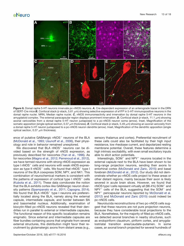
Dorsal raphe 5-HT neurons innervate pc-nNOSneurons
It has been proposed that the brain state-dependentactivity of cortical nNOS� type I cells is powerfully con-trolled by inhibition exerted by neuromodulators releasedin arousal states, such as 5-HT, acetylcholine, noradren-aline, and histamine (Kilduff et al., 2011). Conversely,sleep-promoting peptides and hormones have been sug-gested to promote the recruitment of cortical nNOS� typeI cells (Kilduff et al., 2011). Indeed, the release of 5-HTfrom dorsal raphe neurons is highly dependent on thesleep–wake cycle (Portas et al., 2000). Specifically, thefiring of raphe neurons and, as a consequence, extracel-
lular forebrain 5-HT levels are low during sleep and highduring spontaneous wakefulness (Portas et al., 1998;Sakai, 2011) or SD (Bjorvatn et al., 2002).
Based on this evidence, we hypothesized that dorsalraphe 5-HT neurons target pc-nNOS cells. To this end, wetraced the axons of dorsal raphe 5-HT neurons by inject-ing the viral vector AAV2-EF1a-DIO-EYFP in the dorsalraphe of SERT-Cre mice (Fig. 6A). This resulted in selec-tive expression of enhanced yellow fluorescent protein(eYFP) in dorsal raphe 5-HT neurons (with 100% of eYFP�
cells also expressing 5-HT, n � 3 brains; Fig. 6B). eYFP�
axons innervated the amygdaloid complex, including theBLA (Fig. 6C). Although the LA displayed relatively sparse
Figure 3. Axonal and dendritic arborization of pc-nNOS neurons. A, Biocytin-labeled pc-nNOS neuron coexpressing tdTomato. B,Neurolucida reconstruction from a pc-nNOS cell (MB131202_1) with dendrites (black) running parallel with the intermediate capsuleand axon (green) innervating both the BLA and the caudate–putamen (CPu). C, Neurolucida reconstruction from another pc-nNOS cell(MB151113_2) with dendrites (black) mostly running parallel with the external capsule and axon (green) innervating mostly the BLA,but also the dorsal endopiriform claustrum (DEn), the perirhinal cortex (PRh Ctx), and the amygdalostriatal transition area (AStria)/caudate–putamen (CPu). CeA, Central amygdala; ec, external capsule. Data are presented as the mean � SEM.
New Research 10 of 18
September/October 2016, 3(5) e0177-16.2016 eNeuro.org

eYFP� axons, the pericapsular area, where pc-nNOS arelocated, displayed stronger eYFP� innervation. Examiningsections double labeled for eYFP and nNOS from threetransfected brains, we consistently found eYFP� axonalvaricosities in apposition with pc-nNOS somata (Fig. 6D)or dendrites (Fig. 6E). These observations suggest thatpc-nNOS cells could be modulated by 5-HT released bydorsal raphe neurons.
5-HT inhibits pc-nNOS neuronsTo test the above-mentioned possibility, electrophysi-
ological and pharmacological experiments were per-formed. We recorded pc-nNOS neurons in a cell-attachedconfiguration (n � 18). As reported above, pc-nNOS neu-rons fired spontaneously in control conditions. Bath ap-plication of 50 �M 5-HT produced a significant reductionin firing rate (from 3.6 � 1.6 to 1.6 � 0.4 Hz; p 0.0001,paired t test; Fig. 7A–C), suggesting an inhibitory effect of5-HT. Furthermore, 5-HT enhanced pc-nNOS cell firingirregularity [coefficient of variation (CV) of ISI: 0.5 � 0.06 incontrols; 2.9 � 0.4 in the presence of 5-HT; p 0.0001,paired t test; Fig. 7D] and, in a minority of cells (n � 7),caused a switch from tonic to burst firing (intraburst ISIrange, 25–70 ms; Fig. 7A,E,F). Thus, the action of 5-HT onpc-nNOS is inhibitory, and not excitatory, and contrastswith the previously reported depolarizing effects on otherBLA GABAergic neuron populations (Rainnie, 1999; Boc-chio et al., 2015).
To study the mechanisms through which 5-HT inhibitsthe firing of pc-nNOS neurons, we performed whole-cellpatch-clamp recordings from these neurons in currentclamp. Consistent with the inhibition of firing, the appli-cation of 50 �M 5-HT elicited membrane hyperpolarization(from �59.3 � 0.2 to �64 � 0.7 mV; p � 0.001, one-wayANOVA with Bonferroni post hoc test; n � 10; Fig.8A,B,D), together with a reduction of the Rin (by 11.3 �1.9%; p � 0.0006, one-way ANOVA with Bonferroni post
hoc test; n � 10; Fig. 8). Both effects were blocked byprior incubation with the 5-HT1A antagonist WAY100635(10 �M; p � 0.0235 and p � 0.0064, respectively, pairedt tests; n � 5; Fig. 8). Finally, we confirmed that 5-HT1A-mediated hyperpolarization occurred by a direct effect onpc-nNOS neurons, and was not an indirect network ac-tion, because it persisted in the presence of synapticblockers (10 �M NBQX, 50 �M D-APV, 10 �M SR95531; p� 0.0017, paired t test; n � 5; Fig. 8H). Thus, pc-nNOScells are hyperpolarized by 5-HT via 5-HT1A receptors, inline with the expression of 5-HT1A mRNA in NPY� BLAneurons (Bonn et al., 2013).
Together, these data indicate that pc-nNOS neuronsare distinct from other BLA GABAergic cells in that theyare hyperpolarized and not depolarized by 5-HT. Thishyperpolarization leads to a reduction in firing rate and, ina few cases, to a switch in firing mode. Such 5-HT inhi-bition could mediate, at least in part, the sleep–wake-dependent modulation of pc-nNOS activity, becauseextracellular forebrain 5-HT levels are lower during sleepthan during wakefulness and SD (Portas et al., 1998;Bjorvatn et al., 2002).
DiscussionThe present study provides novel information on
nNOS� type I neurons that surround the BLA. In particu-lar, it describes for the first time the anatomical andphysiological properties of these cells, as well as theirsynaptic connectivity, their activity throughout sleep andwakefulness, and their 5-HT innervation and modulation.We discovered that nNOS� type I neurons are distributedalong the boundaries of the BLA, and express SOM, NPY,and NK1. We observed that pc-nNOS neurons areGABAergic, display high intrinsic excitability, relativelybroad spikes, voltage sag, and rebound depolarizations,and project both inside and outside the BLA. The activityof pc-nNOS (measured by their c-Fos expression) is low
Figure 4. VGAT expression and connectivity of pc-nNOS cells. A, Top, VGAT immunoreactivity of biocytin-filled axonal varicositiesof a pc-nNOS neuron (arrows). Bottom, VGAT immunoreactivity in a biocytin-filled bouton from another pc-nNOS cell (arrow). B, Dualwhole-cell recording (voltage clamp) showing a presynaptic pc-nNOS neuron functionally connected to a postsynaptic principalneuron (PN). Top, Schematic showing the dual whole-cell recording configuration. Middle, Action current evoked in the presynapticpc-nNOS. Bottom, uIPSC recorded in the postsynaptic PN (holding potential, �40 mV; gray, overlap of 10 sweeps repeated every 10s; black, average of the 10 sweeps). Inset, Stereotypical PN firing evoked by 500-ms-long, �100 pA current injection in thepostsynaptic cell held at �65 mV. C, Rate of connectivity between a pc-nNOS cell and BLA cells. One of eleven nearby PN cellsreceived a uIPSC, whereas no uIPSC could be recorded in five nearby pc-nNOS cells or three nearby nNOS-negative interneurons.
New Research 11 of 18
September/October 2016, 3(5) e0177-16.2016 eNeuro.org

during sleep deprivation and high during subsequentsleep. As a putative cellular mechanism of pc-nNOS cellinhibition during sleep deprivation (and more generally
wakefulness), 5-HT, which is known to depolarizeGABAergic cells in the BLA, instead hyperpolarizes pc-nNOS cells. Although previous groups reported the pres-
Figure 5. pc-nNOS neurons are activated during sleep. A, The 24 h profile of EEG SWA (EEG power between 0.5 and 4.0 Hz,displayed as the percentage of mean 24 h baseline; white bar, 12 h light period; dark bar, 12 h dark period) recorded in the frontalcortex and below the distribution of sleep–wake stages (W, wakefulness; N, NREM sleep; R, REM sleep) from a representative mouse.Note, as expected, that sleep predominates, and SWA shows a typical decline during the 12 h light period. B, EEG power spectraldensity during waking, NREM sleep, and REM sleep shown for the frontal EEG (n � 7). Note the state-dependent differences incortical activity. C, Top, Representative profile of SWA during the 4 h SD and subsequent sleep opportunity/RS in one individualmouse. Bottom, The distribution of sleep–wake stages. Mice in the SD group were killed at the end of SD at ZT4 (n � 4), while theremaining mice in the SD�RS group (n � 4) were killed after the sleep opportunity. D, EEG spectral density in NREM sleep (displayedas a ratio of the mean 24 h baseline) during the sleep opportunity after SD (n � 4). Note the typical increase in SWA relative to thecorresponding baseline interval after a period of prolonged waking. Thin lines represent the power density from single mice, whereasthick lines represent the mean power density from all four mice. E, Top panels, Confocal stack (z-stack, 29 �m) showing lack of c-Fosimmunoreactivity in pc-nNOS cells after SD. A median filter was applied (x–y radius, 5 pixels). Arrowhead, A c-Fos� cell immunon-egative for nNOS. Bottom panels, Confocal stack (z-stack, 31 �m) showing c-Fos immunoreactivity in two pc-nNOS cells (arrows)following SD�RS. A median filter was applied (x–y radius, 5 pixels). Insets, Magnification of one the c-Fos� pc-nNOS cells (z-stack,5 �m; no filtering was applied). F, Quantification of c-Fos expression in pc-nNOS neurons. No pc-nNOS neuron expressed c-Fosfollowing SD, whereas 31.4 � 16.4% were c-Fos� after subsequent RS (n � 4 per condition). G, Quantification of c-Fos expressionin paracapsular Nissl-stained cells. Overall, c-Fos� neurons were more abundant following SD (3.7 � 1.1%) than during thesubsequent RS (0.6 � 0.5%, n � 4 per group) ��p 0.01; �p 0.05. Data are presented as the mean � SEM.
New Research 12 of 18
September/October 2016, 3(5) e0177-16.2016 eNeuro.org
ence of putative GABAergic nNOS� neurons of the BLA(McDonald et al., 1993; Usunoff et al., 2006), their physi-ology and role in behavior remained unexplored.
We discovered that BLA nNOS� neurons can be di-vided based on the strength of nNOS expression, aspreviously described for neocortex (Yan et al., 1996). Asfor neocortex (Magno et al., 2012; Perrenoud et al., 2012),we have termed neurons with strong nNOS expression astype I nNOS� cells and neurons with weak nNOS expres-sion as type II nNOS� cells. We found that nNOS� type Ineurons of the BLA coexpress SOM, NPY, and NK1. Thiscombination of neurochemical markers is consistent withthe patterns of expression of cortical type I nNOS� cells(Kubota et al., 2011). These data corroborate the notionthat the BLA exhibits cortex-like GABAergic neuron diver-sity patterns (Spampanato et al., 2011; Capogna, 2014).We found that BLA nNOS� type I neurons are preferen-tially located along the BLA, namely along the externalcapsule, intermediate capsule, and border between BAand basomedial nucleus. Additionally, examination ofbiocytin-filled pc-nNOS neurons revealed that their den-drites run in parallel to external or intermediate capsules.The functional reason of this specific localization remainsenigmatic. Since external and intermediate capsules arefiber bundles containing axons that originate from severalexternal structures, this distribution might favor their re-cruitment by glutamatergic axons from distant areas (e.g.,
sensory thalamus and cortex). Preferential recruitment ofthese cells could also be facilitated by their high inputresistance, low rheobase current, and depolarized restingmembrane potential. Overall, these features determine ahigh intrinsic excitability, with even small excitatory inputsable to elicit action potentials.
Interestingly, SOM� and NPY� neurons located in theexternal capsule next to the BLA have been shown to belong-range projection neurons, sending their axons toentorhinal cortex (McDonald and Zaric, 2015) and basalforebrain (McDonald et al., 2012). Our study did not dem-onstrate whether pc-nNOS cells project to these areas orother distant regions, mainly because their axon is likelysevered in acute brain slices. However, we found thatnNOS type I cells represent virtually all (98.4%) SOM� andNPY� cells of the BLA, suggesting that the SOM� andNPY� pericapsular neurons reported by McDonald et al.(2012) and McDonald and Zaric (2015) could indeed bepc-nNOS cells.
Neurolucida reconstructions of two pc-nNOS cells sug-gest that these neurons are not pure projection neurons,because they have considerable local projections to theBLA. Nonetheless, for the majority of filled pc-nNOS cells,we detected axonal branches in nearby structures, suchas endopiriform claustrum, piriform cortex, and amygda-lostriatal transition area/caudate–putamen. In somecases, an axonal branch projected for several hundreds of
Figure 6. Dorsal raphe 5-HT neurons innervate pc-nNOS neurons. A, Cre-dependent expression of an anterograde tracer in the DRNof SERT-Cre mice B, Confocal stack (z-stack, 5.61 �m) showing selective expression of eYFP in 5-HT-immunopositive neurons in thedorsal raphe nuclei. MRN, Median raphe nuclei. C, nNOS immunoreactivity and innervation by dorsal raphe 5-HT neurons in theamygdaloid complex. The external paracapsular region displays prominent innervation. D, Confocal stack (z-stack, 11.1 �m) showingaxonal varicosities from a dorsal raphe 5-HT neuron juxtaposed to a pc-nNOS neuron soma (arrow). Inset, Magnification of thesomatic apposition (single optical section, 0.37 �m thickness). E, Confocal stack (z-stack, 5.49 �m) showing an axonal varicosity froma dorsal raphe 5-HT neuron juxtaposed to a pc-nNOS neuron dendrite (arrow). Inset, Magnification of the dendritic apposition (singleoptical section, 0.37 �m thickness).
New Research 13 of 18
September/October 2016, 3(5) e0177-16.2016 eNeuro.org

micrometers into caudate–putamen or cortex. However,we never detected a main, thicker axon typical of otherGABAergic long-range projection neurons (e.g., hip-pocamposeptal and septohippocampal cells; Jinno et al.,2007; Unal et al., 2015b). Importantly, we cannot fully ruleout that pc-nNOS cells have a thicker main axon that ismyelinated and therefore could not be visualized usingfluorescence or light microscopy. Nevertheless, our datakeep open the intriguing possibility that pc-nNOS cellscoordinate BLA activity with extra-amygdaloid regions.
Using paired whole-cell recordings, we detected a pre-synaptic pc-nNOS cell functionally connected, likely via aGABAergic connection, to a postsynaptic BLA principalcell. This indicates that BLA principal cells are one of thepostsynaptic targets of pc-nNOS neurons. Crucially, pc-nNOS connection probability to principal cells (1 of 11cellsd) is much lower than the one of other BLA NPY�
interneurons (neurogliaform cells; Ma �nko et al., 2012) or ofBLA PV� interneurons (Woodruff and Sah, 2007a). It is not
clear whether pc-nNOS cells target BLA GABAergic cells,because we did not detect connections from pc-nNOScells and nearby pc-nNOS cells (0 of 5 cells) or nearbynNOS-negative interneurons (0 of 3 cells). In addition toclarifying pc-nNOS postsynaptic targets (both in the BLAand in extra-amygdaloid regions), future studies shouldassess which cellular domains of BLA principal cells aretargeted by pc-nNOS neurons. In the BLA, SOM� neuronstarget distal dendrites of principal neurons, as well asdendrites and cell bodies of interneurons. In line withpotential dendritic targeting, our Neurolucida reconstruc-tions revealed that pc-nNOS cells do not innervate BLA orextra-amygdaloid regions with perisomatic basket-liketerminals.
pc-nNOS cells are likely to modulate other neurons (i.e.,nNOS, NPY, and SOM) not only via GABA release, butalso via other neurochemicals. Nitric oxide signaling hasbeen shown to promote long-term potentiation at inhibi-tory synapses in the LA (Lange et al., 2012), while SOM
Figure 7. 5-HT inhibits pc-nNOS neurons. A, Representative cell-attached recording from a pc-nNOS neuron (voltage-clamp mode)inhibited by bath application of 5-HT (50 �M). In this cell, 5-HT did not trigger burst firing. B, Representative cell-attached recordingfrom a pc-nNOS neuron (voltage-clamp mode) in which bath application of 5-HT elicited a reduction in both firing rate and burst firing.Insets, Magnified examples of tonic firing in control conditions and burst firing upon bath application of 5-HT (50 �M). C, Significantdecrease in firing rate promoted by 5-HT (from 3.6 � 1.6 Hz to 1.6 � 0.4 Hz; p 0.0001, paired t test; n � 18). D, Significant increasein firing irregularity (measured by the CV of the ISI: from 0.5 � 0.06 to 2.9 � 0.4; p 0.0001, paired t test; n � 18) caused by 5-HT.E, 5-HT application enhances the burstiness of pc-nNOS neurons: the peak of the ISI histogram (in Log scale) shifts to the left (n �18). F, 5-HT triggered spike bursts in only 7 of 18 pc-nNOS neurons. The remaining neurons displayed a reduction in firing rate onlyupon 5-HT application. G, In four cells displaying bursts upon 5-HT application, 5-HT was reapplied in the presence of synapticblockers (10 �M NBQX, 50 �M D-APV, and 10 �M SR95531). In these conditions, 5-HT still triggered bursting (the peak of the Log ISIhistogram shifted to the left), suggesting that synaptic inputs are not necessary for bursting activity. ����p 0.0001. Data arepresented as the mean � SEM.
New Research 14 of 18
September/October 2016, 3(5) e0177-16.2016 eNeuro.org

and NPY appear to hyperpolarize LA principal neurons viaG-protein-coupled, inwardly rectifying, potassium chan-nel activation (Meis et al., 2005; Sosulina et al., 2008).
The present study suggests that pc-nNOS cells modu-late amygdaloid and extra-amygdaloid neurons in a vigi-lance state-dependent manner, because our c-Fos datademonstrate that these cells are strongly activated duringsleep (at least when sleep follows sleep deprivation). Incontrast, pc-nNOS� neurons do not express c-Fos afterprolonged wakefulness. To our knowledge, our resultsrepresent the first demonstration of a GABAergic neurontype of the amygdala that dichotomously changes itsactivity as a function of the sleep–wake cycle. Thus,selective sleep activation of nNOS� type I neurons ap-pears to be more widespread and not restricted only tothe cortex (Gerashchenko et al., 2008; Morairty et al.,2013; Dittrich et al., 2015).
In agreement with previous findings (Semba et al.,2001), we show that the overall neuronal activation in theparacapsular area is higher after sleep deprivation thanafter recovery sleep (i.e., a pattern of activation that is theopposite of the one of pc-nNOS neurons). This observa-tion suggests cell type-specific, and not broad, sleepactivation in the BLA. Importantly, it is not clear whether
all nNOS� type I neurons along or inside BLA boundariesare equally inhibited by 5-HT and are activated by recov-ery sleep following sleep deprivation. In this study, welimited our quantification to pc-nNOS neurons adjacent tothe external capsule to match the location of our patch-clamp recordings.
5-HT has been proposed to be one of the neuromodu-lators promoting the inhibition of nNOS type I cells inneocortex (Kilduff et al., 2011; Tricoire and Vitalis, 2012).Our study corroborates this hypothesis, because we de-tected axons from dorsal raphe 5-HT neurons innervatingpc-nNOS cells. In addition, electrophysiological experi-ments demonstrate that 5-HT hyperpolarizes pc-nNOScells via 5-HT1A receptors. As this hyperpolarization wasassociated with a decrease in Rin, it likely arises from theopening of a K� conductance, as described in hippocam-pal neurons (Andrade and Nicoll, 1987). Interestingly, stri-atal nNOS interneurons are also inhibited by 5-HT, but thiseffect is mediated by another class of serotonin receptors(5-HT2C; Cains et al., 2012). Furthermore, in a subset ofcells 5-HT also altered the pc-nNOS the firing mode ofneurons from tonic to bursting. This bursting physiology inresponse to membrane hyperpolarization resembles ef-fects previously reported in striatal low-threshold spike
Figure 8. Direct hyperpolarization of pc-nNOS neurons by 5-HT via 5-HT1A receptors. A, Effect of 5-HT on voltage responses todepolarizing current injection of representative pc-nNOS neuron (20 pA, 300 ms) in control conditions (left) and in the presence of5-HT1A antagonist WAY100635 (10 �M, right). B, Time course of the effect of 5-HT on the Vm of pc-nNOS neurons (n � 10). C, Timecourse of the effect of 5-HT on the Rin of pc-nNOS neurons (n � 10). D, 5-HT significantly hyperpolarizes pc-nNOS cells (from �59.3� 0.2 to �64 � 0.7 mV; p � 0.001, one-way ANOVA with Bonferroni post hoc test; n � 10). E, 5-HT significantly reduced the Rin ofpc-nNOS cells (by 11.3 � 1.9%; p � 0.0006, one-way ANOVA with Bonferroni post hoc test; n � 10). F, G, 5-HT-evokedhyperpolarization and Rin reduction are significantly reduced by 10 �M WAY100635 (p � 0.0235 and p � 0.0064, respectively, pairedt tests; n � 5). H, 5-HT significantly hyperpolarizes pc-nNOS cells, even in the presence of synaptic blockers (10 �M NBQX, 50 �M
D-APV, and 10 �M SR95531; p � 0.0017, paired t test; n � 5), suggesting a direct effect. ���p 0.001; ��p 0.01; �p 0.05. Dataare presented as the mean � SEM.
New Research 15 of 18
September/October 2016, 3(5) e0177-16.2016 eNeuro.org

interneurons (Dehorter et al., 2009; Beatty et al., 2012),cells that are also SOM�, NPY�, and nNOS� (Kawaguchi,1993; Ibáñez-Sandoval et al., 2011). However, this mightoriginate from different mechanisms because pc-nNOSneurons do not display a low-threshold spiking pheno-type. The effect exerted by 5-HT provides a putativecellular mechanism that could explain, at least in part, theactivity of pc-nNOS neurons across sleep and wakeful-ness. Their 5-HT-mediated inhibition, an effect previouslyproposed by Kilduff et al. (2011), could be prominentduring sleep deprivation, when 5-HT release from rapheneurons is high, compared with NREM and REM sleep,when 5-HT release is lower (Portas et al., 1998).
It is unlikely that 5-HT is the only neurotransmitterreleased during wakefulness and arousal that suppressespc-nNOS neuron activity. For example, paracapsularSOM� and NPY� neurons have been shown to coexpressthe muscarinic type 2 acetylcholine receptor (McDonaldand Mascagni, 2011), implying an inhibitory action ofacetylcholine. Since pc-nNOS neurons express NK1 re-ceptors, a putative source of neuromodulatory excitatorydrive on these cells is the NK1 agonist substance P.Importantly, NK1� neurons have been shown to regulateanxiety and reward processing (Gadd et al., 2003; Truittet al., 2009). Future studies should establish whetherpc-nNOS neuron activity is high during sleep due to in-trinsic membrane properties or also because of strongerexcitatory inputs. These inputs could include peptidesand hormones released during sleep, such as adenosine(as proposed by Kilduff et al., 2011), or glutamatergicaxons contained in external/intermediate capsules.
In summary, our work turns the spotlight on a novel BLAGABAergic cell type that is activated by sleep and inhib-ited by wakefulness and 5-HT, and establishes a linkbetween BLA circuits and sleep–wake history. Given thecrucial involvement of the BLA in conditioned fear andanxiety (Tovote et al., 2015), this discovery is particularlycompelling because sleep deprivation has been shown toimpact both emotional phenomena (Graves et al., 2003;Silva et al., 2004). In the future, intersectional geneticapproaches will allow selective tagging of nNOS� type Ineurons (He et al., 2016), for instance by taking advantageof their SOM, NPY, or NK1 expression. In addition tofacilitating their targeting for electrophysiological record-ings, these strategies could also permit specific manipu-lation of BLA nNOS� type I cells during behavior, whichcould probe their precise role in sleep and emotion reg-ulation.
ReferencesAlcami P, Franconville R, Llano I, Marty A (2012) Measuring the firing
rate of high-resistance neurons with cell-attached recording. JNeurosci 32:3118–3130. CrossRef Medline
Andrade R, Nicoll RA (1987) Pharmacologically distinct actions ofserotonin on single pyramidal neurones of the rat hippocampusrecorded in vitro. J Physiol 394:99–124. Medline
Baglioni C, Spiegelhalder K, Lombardo C, Riemann D (2010) Sleepand emotions: a focus on insomnia. Sleep Med Rev 14:227–238.CrossRef Medline
Basheer R, Sherin JE, Saper CB, Morgan JI, McCarley RW, Shiro-mani PJ (1997) Effects of sleep on wake-induced c-fos expression.J Neurosci 17:9746–9750. Medline
Beatty JA, Sullivan MA, Morikawa H, Wilson CJ (2012) Complexautonomous firing patterns of striatal low-threshold spike interneu-rons. J Neurophysiol 108:771–781. CrossRef Medline
Bienvenu TCM, Busti D, Magill PJ, Ferraguti F, Capogna M (2012)Cell-type-specific recruitment of amygdala interneurons to hip-pocampal theta rhythm and noxious stimuli in vivo. Neuron 74:1059–1074. CrossRef Medline
Bjorvatn B, Grønli J, Hamre F, Sørensen E, Fiske E, Bjørkum AA,Portas CM, Ursin R (2002) Effects of sleep deprivation on extra-cellular serotonin in hippocampus and frontal cortex of the rat.Neuroscience 113:323–330. Medline
Bocchio M, Fucsina G, Oikonomidis L, McHugh SB, Bannerman DM,Sharp T, Capogna M (2015) Increased serotonin transporter ex-pression reduces fear and recruitment of parvalbumin interneuronsof the amygdala. Neuropsychopharmacology 40:3015–3026.CrossRef Medline
Bocchio M, McHugh SB, Bannerman DM, Sharp T, Capogna M(2016) Serotonin, amygdala and fear: assembling the puzzle. FrontNeural Circuits 10:10:24. CrossRef Medline
Bonn M, Schmitt A, Lesch KP, Van Bockstaele EJ, Asan E (2013)Serotonergic innervation and serotonin receptor expression ofNPY-producing neurons in the rat lateral and basolateral amygda-loid nuclei. Brain Struct Funct 218:421–435. CrossRef Medline
Bredt DS, Snyder SH (1994) Transient nitric oxide synthase neuronsin embryonic cerebral cortical plate, sensory ganglia, and olfactoryepithelium. Neuron 13:301–313. Medline
Cains S, Blomeley CP, Bracci E (2012) Serotonin inhibits low-threshold spike interneurons in the striatum. J Physiol 590:2241–2252. CrossRef Medline
Capogna M (2014) GABAergic cell type diversity in the basolateralamygdala. Curr Opin Neurobiol 26:110–116. CrossRef Medline
Capogna M, Pearce RA (2011) GABA A,slow: causes and conse-quences. Trends Neurosci 34:101–112. CrossRef Medline
Cirelli C, Tononi G (2000) Gene expression in the brain across thesleep-waking cycle. Brain Res 885:303–321. Medline
Cirelli C, Pompeiano M, Tononi G (1995) Sleep deprivation and c-fosexpression in the rat brain. J Sleep Res 4:92–106. Medline
Cui N, Mckillop LE, Fisher SP, Oliver PL, Vyazoversuskiy VV (2014)Long-term history and immediate preceding state affect EEG slowwave characteristics at NREM sleep onset in C57BL/6 mice. ArchItal Biol 152:156–168. Medline
Dehorter N, Guigoni C, Lopez C, Hirsch J, Eusebio A, Ben-Ari Y,Hammond C (2009) Dopamine-deprived striatal GABAergic in-terneurons burst and generate repetitive gigantic IPSCs in mediumspiny neurons. J Neurosci 29:7776–7787. CrossRef Medline
Dittrich L, Morairty SR, Warrier DR, Kilduff TS (2015) Homeostaticsleep pressure is the primary factor for activation of cortical nNOS/NK1 neurons. Neuropsychopharmacology 40:632–639. CrossRef
Duvarci S, Pare D (2014) Amygdala microcircuits controlling learnedfear. Neuron 82:966–980. CrossRef Medline
Fuentealba P, Begum R, Capogna M, Jinno S, Márton LF, CsicsvariJ, Thomson A, Somogyi P, Klausberger T (2008) Ivy cells: apopulation of nitric-oxide-producing, slow-spiking GABAergicneurons and their Involvement in hippocampal network activity.Neuron 57:917–929. CrossRef Medline
Gadd CA, Murtra P, De Felipe C, Hunt SP (2003) Neurokinin-1receptor-expressing neurons in the amygdala modulate morphinereward and anxiety behaviors in the mouse. J Neurosci 23:8271–8280. Medline
Gao J, Zhang J-X, Xu T-L (2002) Modulation of serotonergic projec-tion from dorsal raphe nucleus to basolateral amygdala on sleep–waking cycle of rats. Brain Res 945:60–70. Medline
Gerashchenko D, Wisor JP, Burns D, Reh RK, Shiromani PJ, SakuraiT, de la Iglesia HO, Kilduff TS (2008) Identification of a populationof sleep-active cerebral cortex neurons. Proc Natl Acad Sci U S A105:10227–10232. CrossRef Medline
Graves LA, Heller EA, Pack AI, Abel T (2003) Sleep deprivationselectively impairs memory consolidation for contextual fear con-ditioning. Learn Mem 10:168–176. CrossRef Medline
New Research 16 of 18
September/October 2016, 3(5) e0177-16.2016 eNeuro.org

Hangya B, Pi H-J, Kvitsiani D, Ranade SP, Kepecs A (2014) Fromcircuit motifs to computations: mapping the behavioral repertoireof cortical interneurons. Curr Opin Neurobiol 26:117–124. Cross-Ref Medline
He M, Tucciarone J, Lee S, Nigro MJ, Kim Y, Levine JM, Kelly SM,Krugikov I, Wu P, Chen Y, Gong L, Hou Y, Osten P, Rudy B, HuangJZ (2016) Strategies and tools for combinatorial targeting ofGABAergic neurons in mouse cerebral cortex. Neuron 91:1228–1243.
Huber R, Deboer T, Tobler I (2000) Topography of EEG dynamicsafter sleep deprivation in mice. J Neurophysiol 84:1888–1893.Medline
Ibáñez-Sandoval O, Tecuapetla F, Unal B, Shah F, Koós T, TepperJM (2011) A novel functionally distinct subtype of striatal neuro-peptide Y interneuron. J Neurosci 31:16757–16769. CrossRefMedline
Janak PH, Tye KM (2015) From circuits to behaviour in the amygdala.Nature 517:284–292. CrossRef Medline
Jasnow AM, Ressler KJ, Hammack SE, Chhatwal JP, Rainnie DG(2009) Distinct subtypes of cholecystokinin (CCK)-containing in-terneurons of the basolateral amygdala identified using a CCKpromoter-specific lentivirus. J Neurophysiol 101:1494–1506.CrossRef Medline
Jinno S, Klausberger T, Marton LF, Dalezios Y, Roberts JDB, Fuen-tealba P, Bushong EA, Henze D, Buzsáki G, Somogyi P (2007)Neuronal diversity in GABAergic long-range projections from thehippocampus. J Neurosci 27:8790–8804. CrossRef Medline
Kawaguchi Y (1993) Physiological, morphological, and histochemicalcharacterization of three classes of interneurons in rat neostriatum.J Neurosci 13:4908–4923. Medline
Kilduff TS, Cauli B, Gerashchenko D (2011) Activation of corticalinterneurons during sleep: an anatomical link to homeostatic sleepregulation? Trends Neurosci 34:10–19. CrossRef Medline
Klausberger T, Somogyi P (2008) Neuronal diversity and temporaldynamics: the unity of hippocampal circuit operations. Science321:53–57. CrossRef Medline
Kubota Y, Shigematsu N, Karube F, Sekigawa A, Kato S, YamaguchiN, Hirai Y, Morishima M, Kawaguchi Y (2011) Selective coexpres-sion of multiple chemical markers defines discrete populations ofneocortical GABAergic neurons. Cereb Cortex 21:1803–1817.CrossRef Medline
Lange MD, Doengi M, Lesting J, Pape HC, Jüngling K (2012) Het-erosynaptic long-term potentiation at interneuron-principal neuronsynapses in the amygdala requires nitric oxide signalling. J Physiol590:131–143. CrossRef Medline
Lee J-E, Jeon C-J (2005) Immunocytochemical localization of nitricoxide synthase-containing neurons in mouse and rabbit visualcortex and co-localization with calcium-binding proteins. Mol Cells19:408–417.
Li G, Stewart R, Canepari M, Capogna M (2014) Firing of hippocam-pal neurogliaform cells induces suppression of synaptic inhibition.J Neurosci 34:1280–1292. CrossRef Medline
Lovett-Barron M, Losonczy A (2014) Behavioral consequences ofGABAergic neuronal diversity. Curr Opin Neurobiol 26:27–33.CrossRef Medline
Magno L, Oliveira MG, Mucha M, Rubin AN, Kessaris N (2012)Multiple embryonic origins of nitric oxide synthase-expressingGABAergic neurons of the neocortex. Front Neural Circuits 6:65.CrossRef
Ma �nko M, Bienvenu TCM, Dalezios Y, Capogna M (2012) Neuroglia-form cells of amygdala: a source of slow phasic inhibition in thebasolateral complex. J Physiol 590:5611–5627. CrossRef Medline
Mascagni F, McDonald AJ (2003) Immunohistochemical character-ization of cholecystokinin containing neurons in the rat basolateralamygdala. Brain Res 976:171–184. Medline
McDonald AJ, Mascagni F (2001) Colocalization of calcium-bindingproteins and GABA in neurons of the rat basolateral amygdala.Neuroscience 105:681–693. Medline
McDonald AJ, Mascagni F (2011) Neuronal localization of M2 mus-carinic receptor immunoreactivity in the rat amygdala. Neurosci-ence 196:49–65. CrossRef Medline
McDonald AJ, Zaric V (2015) GABAergic somatostatin-immunoreactive neurons in the amygdala project to the entorhinalcortex. Neuroscience 290:227–242. CrossRef Medline
McDonald AJ, Payne DR, Mascagni F (1993) Identification of putativenitric oxide producing neurons in the rat amygdala using NADPH-diaphorase histochemistry. Neuroscience 52:97–106. Medline
McDonald AJ, Mascagni F, Zaric V (2012) Subpopulations ofsomatostatin-immunoreactive non-pyramidal neurons in theamygdala and adjacent external capsule project to the basal fore-brain: evidence for the existence of GABAergic projection neuronsin the cortical nuclei and basolateral nuclear com. Front NeuralCircuits 6:46. CrossRef
Meis S, Sosulina L, Schulz S, Höllt V, Pape H-C (2005) Mechanismsof somatostatin-evoked responses in neurons of the rat lateralamygdala. Eur J Neurosci 21:755–762. CrossRef Medline
Morairty SR, Dittrich L, Pasumarthi RK, Valladao D, Heiss JE,Gerashchenko D, Kilduff TS (2013) A role for cortical nNOS/NK1neurons in coupling homeostatic sleep drive to EEG slow waveactivity. Proc Natl Acad Sci U S A 110:20272–20277. CrossRefMedline
Morgan JI, Curran T (1991) Proto-oncogene transcription factors andepilepsy. Trends Pharmacol Sci 12:343–349. Medline
Palchykova S, Winsky-Sommerer R, Meerlo P, Dürr R, Tobler I (2006)Sleep deprivation impairs object recognition in mice. NeurobiolLearn Mem 85:263–271. CrossRef Medline
Paré D, Gaudreau H (1996) Projection cells and interneurons of thelateral and basolateral amygdala: distinct firing patterns and dif-ferential relation to theta and delta rhythms in conscious cats. JNeurosci 16:3334–3350. Medline
Perrenoud Q, Geoffroy H, Gauthier B, Rancillac A, Alfonsi F, KessarisN, Rossier J, Vitalis T, Gallopin T (2012) Characterization of type Iand type II nNOS-expressing interneurons in the barrel cortex ofmouse. Front Neural Circuits 6:36. CrossRef Medline
Portas CM, Bjorvatn B, Fagerland S, Grønli J, Mundal V, Sørensen E,Ursin R (1998) On-line detection of extracellular levels of serotoninin dorsal raphe nucleus and frontal cortex over the sleep/wakecycle in the freely moving rat. Neuroscience 83:807–814. Medline
Portas CM, Bjorvatn B, Ursin R (2000) Serotonin and the sleep/wakecycle: special emphasis on microdialysis studies. Prog Neurobiol60:13–35. Medline
Rainnie DG (1999) Serotonergic modulation of neurotransmission inthe rat basolateral amygdala. J Neurophysiol 82:69–85. Medline
Rainnie DG, Mania I, Mascagni F, McDonald AJ (2006) Physiologicaland morphological characterization of parvalbumin-containing in-terneurons of the rat basolateral amygdala. J Comp Neurol 498:142–161. CrossRef Medline
Sakai K (2011) Sleep-waking discharge profiles of dorsal raphenucleus neurons in mice. Neuroscience 197:200–224. CrossRefMedline
Semba K, Pastorius J, Wilkinson M, Rusak B (2001) Sleepdeprivation-induced c-fos and junB expression in the rat brain:effects of duration and timing. Behav Brain Res 120:75–86. Med-line
Sheng M, Greenberg ME (1990) The regulation and function of c-fosand other immediate early genes in the nervous system. Neuron4:477–485. Medline
Silva RH, Kameda SR, Carvalho RC, Takatsu-Coleman AL, NiigakiST, Abílio VC, Tufik S, Frussa-Filho R (2004) Anxiogenic effect ofsleep deprivation in the elevated plus-maze test in mice. Psycho-pharmacology (Berl) 176:115–122. CrossRef Medline
Smiley JF, McGinnis JP, Javitt DC (2000) Nitric oxide synthaseinterneurons in the monkey cerebral cortex are subsets of thesomatostatin, neuropeptide Y, and calbindin cells. Brain Res 863:205–212. Medline
Sosulina L, Schwesig G, Seifert G, Pape H-C (2008) Neuropeptide Yactivates a G-protein-coupled inwardly rectifying potassium cur-
New Research 17 of 18
September/October 2016, 3(5) e0177-16.2016 eNeuro.org
rent and dampens excitability in the lateral amygdala. Mol CellNeurosci 39:491–498. CrossRef Medline
Spampanato J, Polepalli J, Sah P (2011) Interneurons in the baso-lateral amygdala. Neuropharmacology 60:765–773. CrossRefMedline
Tamamaki N, Tomioka R (2010) Long-range GABAergic connections dis-tributed throughout the neocortex and their possible function. FrontNeurosci 4:202. CrossRef Medline
Taniguchi H, He M, Wu P, Kim S, Paik R, Sugino K, Kvitsiani D,Kvitsani D, Fu Y, Lu J, Lin Y, Miyoshi G, Shima Y, Fishell G, NelsonSB, Huang ZJ (2011) A resource of Cre driver lines for genetictargeting of GABAergic neurons in cerebral cortex. Neuron 71:995–1013. CrossRef Medline
Tomioka R, Okamoto K, Furuta T, Fujiyama F, Iwasato T, Yanagawa Y,Obata K, Kaneko T, Tamamaki N (2005) Demonstration of long-rangeGABAergic connections distributed throughout the mouse neocortex.Eur J Neurosci 21:1587–1600. CrossRef Medline
Tovote P, Fadok JP, Lüthi A (2015) Neuronal circuits for fear andanxiety. Nat Rev Neurosci 16:317–331. CrossRef Medline
Tricoire L, Vitalis T (2012) Neuronal nitric oxide synthase expressingneurons: a journey from birth to neuronal circuits. Front NeuralCircuits 6:82. CrossRef Medline
Truitt WA, Johnson PL, Dietrich AD, Fitz SD, Shekhar A (2009)Anxiety-like behavior is modulated by a discrete subpopulation ofinterneurons in the basolateral amygdala. Neuroscience 160:284–294. CrossRef Medline
Tully K, Li Y, Tsvetkov E, Bolshakov VY (2007) Norepinephrineenables the induction of associative long-term potentiation atthalamo-amygdala synapses. Proc Natl Acad Sci U S A 104:14146–14150. CrossRef Medline
Unal CT, Pare D, Zaborszky L (2015a) Impact of basal forebraincholinergic inputs on basolateral amygdala neurons. J Neurosci35:853–863. CrossRef Medline
Unal G, Joshi A, Viney TJ, Kis V, Somogyi P (2015b) Synaptic targetsof medial septal projections in the hippocampus and extrahip-
pocampal cortices of the mouse. J Neurosci 35:15812–15826.CrossRef Medline
Usunoff KG, Itzev DE, Rolfs A, Schmitt O, Wree A (2006) Nitric oxidesynthase-containing neurons in the amygdaloid nuclear complexof the rat. Anat Embryol (Berl) 211:721–737. CrossRef Medline
Vereczki VK, Veres JM, Müller K, Nagy GA, Rácz B, Barsy B, HájosN (2016) Synaptic organization of perisomatic GABAergic inputsonto the principal cells of the mouse basolateral amygdala. FrontNeuroanat 10:20. CrossRef Medline
Vogel E, et al. (2016) Projection-specific dynamic regulation of inhi-bition in amygdala micro-circuits. Neuron 91:644–651. CrossRefMedline
Vyazovskiy VV, Riedner BA, Cirelli C, Tononi G (2007) Sleep homeo-stasis and cortical synchronization: II. A local field potential studyof sleep slow waves in the rat. Sleep 30:1631–1642. Medline
West M (1999) Stereological methods for estimating the total numberof neurons and synapses: issues of precision and bias. TrendsNeurosci 22:51–61. Medline
Wolff SBE, Gründemann J, Tovote P, Krabbe S, Jacobson G. a,Müller C, Herry C, Ehrlich I, Friedrich RW, Letzkus JJ, Lüthi A(2014) Amygdala interneuron subtypes control fear learningthrough disinhibition. Nature 509:453–458. CrossRef
Woodruff AR, Sah P (2007a) Networks of parvalbumin-positive in-terneurons in the basolateral amygdala. J Neurosci 27:553–563.CrossRef Medline
Woodruff AR, Sah P (2007b) Inhibition and synchronization of basalamygdala principal neuron spiking by parvalbumin-positive in-terneurons. J Neurophysiol 98:2956–2961. CrossRef Medline
Yan XX, Jen LS, Garey LJ (1996) NADPH-diaphorase-positive neu-rons in primate cerebral cortex colocalize with GABA and calcium-binding proteins. Cereb Cortex 6:524–529. Medline
Yoo S-S, Gujar N, Hu P, Jolesz FA, Walker MP (2007) The humanemotional brain without sleep–a prefrontal amygdala disconnect.Curr Biol 17:R877–R878. CrossRef Medline
New Research 18 of 18
September/October 2016, 3(5) e0177-16.2016 eNeuro.org