SL 2009-174 Microbead electrochemiluminescence...
38
1 Microbead electrochemiluminescence immunoassay for detection and identification of Venezuelan equine encephalitis virus Xiaojiang Dai, Rayanne E. Hilsen, Wei-Gang Hu, R. Elaine Fulton Defence Research and Development Canada–Suffield, PO Box 4000, Station Main, Medicine Hat, AB T1A 8K6 Corresponding author: Biotechnology Section, Defence Research and Development Canada–Suffield, PO Box 4000, Station Main, Medicine Hat, Alberta, Canada T1A 8K6; Tel.: +1-403-544-4630; fax: +1-403-544-3388. E-mail address: [email protected] (NON-CONTROLLED GOODS) DMC A REVIEW: GCEC December 2013
-
Upload
trinhnguyet -
Category
Documents
-
view
229 -
download
1
Transcript of SL 2009-174 Microbead electrochemiluminescence...
1
Microbead electrochemiluminescence immunoassay for detection and
identification of Venezuelan equine encephalitis virus
Xiaojiang Dai, Rayanne E. Hilsen, Wei-Gang Hu, R. Elaine Fulton
Defence Research and Development Canada–Suffield, PO Box 4000, Station Main,
Medicine Hat, AB T1A 8K6
Corresponding author: Biotechnology Section, Defence Research and Development
Canada–Suffield, PO Box 4000, Station Main, Medicine Hat, Alberta, Canada T1A 8K6;
Tel.: +1-403-544-4630; fax: +1-403-544-3388.
E-mail address: [email protected]
(NON-CONTROLLED GOODS)DMC AREVIEW: GCEC December 2013
2
ABSTRACT
An electrochemiluminescence (ECL) immunoassay, incorporating chemically
biotinylated and ruthenylated antibodies down-selected from a panel of monoclonal and
polyclonal reagents, was developed to detect and identify Venezuelan equine encephalitis
virus (VEEV). The limit of detection (LOD) of the optimized ECL assay was 103 pfu/ml
VEEV TC-83 virus and 1 ng/ml recombinant (r) VEEV E2 protein. The LOD of the ECL
assay was approximately one log unit lower than that of a sandwich enzyme-linked
immunosorbent assay (ELISA) incorporating the same immunoreagents. Repetition of
ECL assays over time and by different operators demonstrated that the assay was
reproducible (coefficient of variation 4.7–18.5% month-to-month; 3.3–8.8% person-to-
person). The VEEV ECL assay exhibited no cross-reactivity with two closely related
alphaviruses or with 21 heterologous biological agents. A genetically biotinylated
recombinant VEEV antibody, MA116SBP, was evaluated for utility for detection of rE2;
although functional in the ECL assay, the LOD was two log units higher (100 ng/ml vs 1
ng/ml) using MA116SBP than when chemically biotinylated antibody was used. The
ECL assay detected VEEV at the lowest LOD (highest sensitivity) hitherto reported in the
published literature and ECL assay results were generated in 60 min compared to a 6-8
hr period required for ELISA. Results have demonstrated a sensitive, rapid, and fully
automated ECL immunoassay for detection and identification of VEEV.
Keywords: Venezuelan equine encephalitis virus; Envelope E2 protein;
Electrochemiluminescence immunoassay; Enzyme-linked immunosorbent assay
3
1. Introduction
Venezuelan equine encephalitis virus (VEEV), a positive-sense single-stranded RNA
alphavirus, is an important mosquito-borne pathogen in humans and equines (Weaver et
al., 2004). Epizootic VEEV infections cause debilitating disease with a high fatality rate
in equines. In humans, VEEV infection is associated with a potentially life-threatening
severe febrile illness and neurological disease appears in 5% of cases (Griffin, 2001).
The incidence of human infection during equine epizootics can reach 30% and mortality
associated with encephalitis in children is as high as 35%. The outbreaks in Venezuela
and Colombia in 1995 affected around 75,000 people and more than 300 fatal
encephalitis cases occurred (Rivas et al., 1997). VEEV is environmentally stable and
highly infectious by aerosol inhalation, making it a potential biological weapon threat.
As such, it is crucial that rapid and sensitive immunoassays be developed for detection of
VEEV in environmental and clinical samples. A number of immunoassay techniques
utilizing polyclonal or monoclonal antibodies (Wang et al., 2005) or recombinant
antibodies (Duggan et al., 2001; Hu et al., 2002, 2004; Kirsch et al., 2008) has been
described for the detection of VEEV, including enzyme-linked immunosorbent assay
(ELISA), radioimmunosorbent assay (RIA), light addressable potentiometric (LAP)
assay, and dissociation-enhanced lanthanide fluorescent immunoassay (DELFIA)
(Roehrig et al., 1982; Smith et al., 2001; Hu et al., 2002, 2004). Typically, these assays
require multiple operational steps and long incubation periods; acquisition and analysis of
assay results can take many hours.
Electrochemiluminescence (ECL) immunoassay is a magnetic bead-based

4
technology for conducting immunoassays with improved assay performance (Deaver,
1995; Yang et al., 1994). There are three components in an ECL immunoassay: (i) a
biotinylated capture antibody (Cab), pre-bound to streptavidin-coated magnetic beads, (ii)
a detector antibody (Dab), labelled with ruthenium-trisbipyridal, for the emission of light
when electrochemically stimulated, and (iii) an analyte, which reacts with the capture and
detector antibodies resulting in an antigen-antibody sandwich. In ECL reactions, a
precursor molecule (tripropylamine) is activated on an electrode surface resulting in an
electron transfer reaction. This transfer initiates the excitation of ruthenium-trisbipyridal
which ultimately results in the emission of a photon at 620 nm.
Assay techniques utilizing ECL technology have a number of advantages over
conventional assay methods e.g., ELISA and RIA: Limits of detection (LOD) in ECL
assays are lower (higher sensitivity), due to high luminescent signal to noise ratios
(Gatto-Menking et al., 1995; Yu et al., 2000; Garber and O’Brien, 2008; Yoshimura et al.,
2008; Rossi et al., 2008; Kuhle et al., 2010). The dynamic range for analyte detection
extends over a wide range (five orders of magnitude) (Yang et al., 1994; Kijek et al.,
2000; Yu et al., 2000). The time and labour required to complete ECL assays are reduced
compared with conventional immunoassays. The ECL assay is a non-separation
technique, thus does not require plate coating, washing, or aspiration steps. Sample
reading is rapid; the ECL instrumentation used in the present study requires
approximately one minute per sample to read. Labelled ECL reagents are exceptionally
stable and ECL assays are robust and tolerant of analyte detection in the presence of a
variety of sample matrices. These features make the ECL detection system an attractive
alternative to conventional immunoassay techniques. In addition, the instrumentation
5
used in this study is a military hardened version, making it especially useful for military
field use.
This study describes the development of an ECL assay for VEEV. A panel of VEEV
antibodies was screened for optimal performance using VEEV strain TC-83 whole virus
and VEEV recombinant (r) envelope E2 protein as target antigens. ECL assays were
optimized for antibody concentration and assay LOD were determined in comparison to
ELISA incorporating the same antibody reagents. ECL assays were assessed for
reproducibility and precision over time and when conducted by different operators.
Assay specificity for VEEV was evaluated by screening against closely related
alphaviruses and 21 heterologous bacteria, virus, or toxin agents. A genetically
biotinylated recombinant antibody to VEEV was compared to a chemically biotinylated
antibody for utility for detection of VEEV rE2.

6
2. Materials and methods
2.1. Instrumentation
ECL measurements were performed using a M-SERIES® M1MR analyzer (BioVeris
Corp., Gaithersburg, MD).
2.2. Antigens
2.2.1. Homologous antigen
Live VEE TC-83 virus, purified as previously described (Hu et al., 2004), was kindly
provided by Dr. J. Wu, DRDC Suffield. Recombinant E2 protein was expressed in E. coli
and purified at DRDC Suffield (W-G. Hu et al., unpublished data).
2.2.2. Heterologous antigens
Gamma-irradiated Western equine encephalitis virus (WEEV) and Eastern equine
encephalitis virus (EEEV) were purchased from the US Critical Reagents Program (CRP)
(Aberdeen Proving Ground, MD). Yellow fever virus, attenuated vaccine (strain 17D),
was purchased from Aventis Pasteur Inc. (Toronto, ON). Dengue and Klebsiella
pneumoniae were purchased from American Type Culture Collection (Manassas, VA).
Inactivated (60Co irradiated) Brucella melitensis (suis), Bacillus anthracis (vollum),

7
Bacillus globigii (spores), Francisella tularensis, Yersinia pseudotuberculosis, Yersinia
pests (JAVA 9), Escherichia coli, Bacillus cereus, Vaccinia virus (Lister), Bacillus
thuringiensis, Aspergillus niger, Erwinia herbicola, Pseudomonas aeruginosa, Coxiella
burnetii, and MS2 were all acquired from US Army Dugway Proving Ground (DPG)
(Dugway, UT). SEB toxin was purchased from Toxin Technology (Sarasota, FL),
botulinum toxoid A was purchased from WAKO Chemicals Inc. (Richmond, VA), and
ricin A chain was purchased from Sigma-Aldrich Canada Ltd. (Oakville, ON).
2.3. Monoclonal and polyclonal antibodies
Hybridomas 1A4A1 and 1A3A9 were kindly provided by Dr. J.T. Roehrig (Mathews
and Roehrig, 1982; Roehrig et al., 1982; Roehrig and Mathews, 1985). The hybridomas
were grown in BD CellTM MAb Basal Medium (BD-Biosciences, Mississauga, ON)
supplemented with 10% FETALCLONE® fetal calf serum (HyClone Laboratories, Logan
UT), 2mM L-glutamine (Invitrogen, Burlington, ON), and 1% HAT Media Supplement
Hybri-Max® (Sigma-Aldrich Canada). The hybridomas were weaned from FBS medium
and transferred to a CELLineTM 1000 flask (BD-Biosciences) and incubated at 37 oC
under 5% CO2 for two weeks. Supernatant was harvested from the CELLineTM 1000
flask and the monoclonal antibody (mAb) was then purified using a MelonTM Gel
Monoclonal IgG Purification Kit (Pierce, Ottawa, ON) according to the manufacturer’s
instructions.
Rabbit and goat anti-VEEV polyclonal antibodies (pAb) were previously developed
and purified for IgG under a DRDC Suffield contract by SciLab Consulting Inc.
8
(Redcliff, AB). Both the rabbit and the goat pAb had been purified on a Bio-Gel® Protein
G column (Bio-Rad Laboratories, Mississauga, ON) by High-Performance Liquid
Chromatography (Spectral Physics, San Jose, CA).
2.4. Genetically biotinylated recombinant antibody
A genetically biotinylated recombinant VEEV single-chain variable fragment
antibody, MA116SBP, was previously generated at DRDC Suffield (Alvi et al., 1999,
2002, 2003; Hu et al., 2002).
2.5. ECL assay reagents
The following ECL assay reagents and buffers were purchased from BioVeris Corp.:
Biotin-LC-Sulfo-NHS ester, Ruthenium (II) tris-bipyridine-NHS ester, Streptavidin-
coated Dynabeads® M-280, M-SERIES® Positive Calibrator, M-SERIES® Negative
Calibrator, StabilCoat® Immunoassay Stabilizer, BV-CLEANTM Plus solution, BV-
GLOTM Plus solution, BV-DILUENTTM solution, BV-STORETM solution, and BV-
SANITIZETM solution.
2.6. Matrix powders and soils
Flour (white, enriched, all purpose) (Safeway brand), cornstarch (Safeway brand),
baking powder (Safeway brand), baking soda (Safeway brand), laundry detergent (Tide
9
Original, Procter and Gamble), coffee creamer (Coffee-Mate®, Carnation, Nestlé), skim
milk, and powdered sugar were all purchased from Safeway Canada Ltd. (Medicine Hat,
AB). Talcum powder, powdered cleanser, and spackling powder were purchased through
Foreign Military Sales from the US Critical Reagents Program. The four soil samples
including sand, sand loam, loamy sand, and clay loam were from the DRDC Suffield
Experimental Proving Ground and had been previously characterized by the Alberta
Environmental Centre (Vegreville, AB).
2.7. Preparation of labeled antibodies
Prior to labeling, all antibodies were desalted using NAP-5 columns (Amersham
Biosciences, Baie d’Urfé, QC) according to the manufacturer’s instructions. In separate
labeling reactions, the four antibodies (1A4A1 mAb, 1A3A9 mAb, rabbit pAb, and goat
pAb) were each biotinylated with biotin-LC-Sulfo-NHS ester at a molar ratio of 10:1
(Biotin:Ab) and ruthenylated with ruthenium (II) tris-bipyridine-NHS ester at 7.5:1
(Ru:Ab) by labeling procedures recommended by BioVeris. In brief, the antibodies and
respective labeling solutions were incubated separately and simultaneously using an end-
over-end shaker with gentle rotation for 60 min at RT. The labeling reactions were
quenched by the addition of 20 l of 2 M glycine and then incubated at RT for an
additional 10 min. Uncoupled biotin and ruthenium esters were removed by dialysis in
four changes of sterile phosphate buffered saline (PBS) (pH 7.4) at 4 oC over two days.
The protein concentrations of labeled antibodies were determined using a Micro BCATM
assay (Pierce) or a ND-1000 Spectrophotometer (LabX, Midland, ON) with bovine serum
10
albumin (BSA) (Pierce) as the standard. Aliquots of each labeled antibody were stored at
4 oC until used.
2.8 Preparation of capture antibodies
For the preparation of Cab (antibody-functionalized beads), biotinylated antibodies
were pre-bound to streptavidin-coated Dynabeads® (100 g antibody per ml of
DynabeadsR). Biotinylated antibodies and beads were incubated together with an end-
over-end rotation for 60 min at RT. Unbound antibody was removed by washing the
beads three times with sterile PBS-0.1% Tween 20 (PBST) using a MPCTM-S magnetic
particle concentrator (Dynal Biotech ASA, Oslo), followed by three washes with sterile
PBS.
2.9. ECL immunoassay
Cab (biotinylated antibody pre-bound to streptavidin-coated Dynabeads®) was
diluted in StabilCoat® Immunoassay Stabilizer and the detector antibody (Dab)
(ruthenylated antibody) (Ab-Ru) was diluted in PBS-2% BSA-0.1% Tween 20. Antigen
was diluted in PBS-0.3% Tween 20. ECL reaction solutions were prepared in 0.75 ml
round-bottom tubes (Matrix Technologies) (VWR International, Mississauga, ON) by
adding 25 l of Cab, 25 l of Dab, and 50 l of antigen solution (or 50 l of PBS-0.3%
Tween 20 for the antigen negative control). The tubes were placed in a specialized 96-
well assay plate (BioVeris Corp.), which was then loaded into the M1MR analyzer, where
it was incubated with shaking for 15 min at RT and then analyzed.
11
2.10. ELISA
Initial ELISA experiments were performed in sandwich format to optimize the
concentrations of the Cab, Dab, and indicator antibody (Iab) (data not shown). For the
sandwich ELISA, the format utilized the rabbit-Cab/1A4A1-Dab antibody pair identical
to that used in the ECL assay. Nunc MaxisorpTM flat-bottomed 96-well plates (Life
Technologies, Burlington, ON) were coated overnight at 4oC with 3 g/ml of Cab (rabbit
pAb-biotin) in carbonate bicarbonate buffer, pH 9.6. The plates were washed five times
with PBST in an ELx50 plate washer (Biotek Instruments, Winooski, VT) and then
blocked with 2% BSA for 1 h at 37 oC. After five washes with PBST, plates were
incubated with TC-83 virus at 101106 pfu/ml or VEEV rE2 protein at 0.0110,000 ng/ml
for 1 h at 37 oC. Following five washes with PBST, the plates were incubated with 2
g/ml of Dab (1A4A1-Ru) for 1 h at 37 oC. The plates were again washed five times
with PBST and then incubated with a 1:2000 dilution of horseradish peroxidase (HRP)-
conjugated anti-mouse IgG (Caltag Laboratories, Burlingame, CA). Finally, the plates
were washed five times with PBST and developed for 30 min at RT with 3,3’,5,5’-
tetramethylbenzidine (TMB) microwell perioxidase substrate system (KPL, Gaithersburg,
USA). The reactions were read at an absorbance of 635 nm by a microplate autoreader
(Molecular Devices, Sunnyvale, CA, USA).
2.11. ECL and ELISA data analysis
LOD was determined by calculating a cutoff value that was equal to 20% above the
12
mean ECL or ELISA reading for the negative control. Therefore, a signal was considered
positive if the ECL or ELISA ratio of the sample signal reading to background reading
(S/B) was 1.2 or greater.

13
3. Results
3.1. Screening and optimization of antibody pairs for VEEV ECL assay
Four Abs (1A4A1 mAb, 1A3A9 mAb, rabbit pAb, and goat pAb) were each labeled
with biotin-LC-sulfo-NHS ester and ruthenium (II) tris-bipyridine-NHS ester. A total of
16 combinations of antibody pairs was tested for utility in the ECL assays at a Cab
dilution of 1:25, Dab concentration of 4 g/ml, TC-83 virus concentration of 105 pfu/ml,
and VEEV rE2 concentration of 100 ng/ml. Amongst the 16 antibody pairs, three pairs
(1A4A1-Cab/1A4A1-Dab, 1A4A1-Cab/1A3A9-Dab, and 1A3A9-Cab/1A4A1-Dab)
exhibited high reactivity with TC-83 virus, five pairs exhibited moderate reactivity, and
eight other pairs produced only background ECL signals (data not shown). Using VEEV
rE2 as the antigen, four antibody pairs (rabbit-Cab/1A4A1-Dab, goat-Cab/1A4A1-Dab,
1A4A1-Cab/1A4A1-DAb, and 1A4A1-Cab/rabbit-Dab) exhibited high reactivity, five
pairs exhibited moderate reactivity, and the remaining seven pairs produced only
background ECL signals (data not shown). In total, approximately 50% of the antibody
pairs tested had little or no reactivity in the ECL assay with either TC-83 virus or rE2.
Based on these results, three antibody pairs (1A3A9-Cab/1A4A1-Dab with high
reactivity to TC-83 virus, rabbit-Cab/1A4A1-Dab with high reactivity to recombinant E2,
and 1A4A1-Cab/1A4A1-Dab with high reactivity to both TC-83 virus and rE2) were
chosen for further optimization in the ECL assay.
Experiments were performed to optimize the concentrations of Cab and Dab for each
of the three selected antibody pairs. Concentrations of Cab and Dab that yielded the

14
highest S/B ratio and the lowest LOD were determined to be optimal. If two or more
concentrations produced comparable results, the lowest concentration of antibody was
selected for use. Cabs were first optimized by testing dilutions of 1:16, 1:25, and 1:50
with 4 g/ml of Dab against three different concentrations of antigen (VEEV TC-83 virus
at 103, 104 and 105 pfu/ml, and VEEV rE2 protein at 1, 10, and 100 ng/ml). Then, Dabs
were optimized by testing concentrations of 4, 10, and 20 g/ml with the optimal dilution
of Cab against the same three concentrations of antigen. As a result of these experiments,
the three antibody pairs were optimized for both VEEV TV83 virus and VEEV E2
protein as follows: antibody pair 1A4A1-Cab/1A4A1-Dab: Cab 1:35 and Dab 10 g/ml;
1A3A9-Cab/1A4A1-Dab: Cab 1:25 and Dab 10 g/ml; and rabbit-Cab/1A4A1-Dab: Cab
1:16 and Dab 4 g/ml (data not shown). These concentrations of Cab and Dab were used
with the three noted antibody pairs in all subsequent experiments.
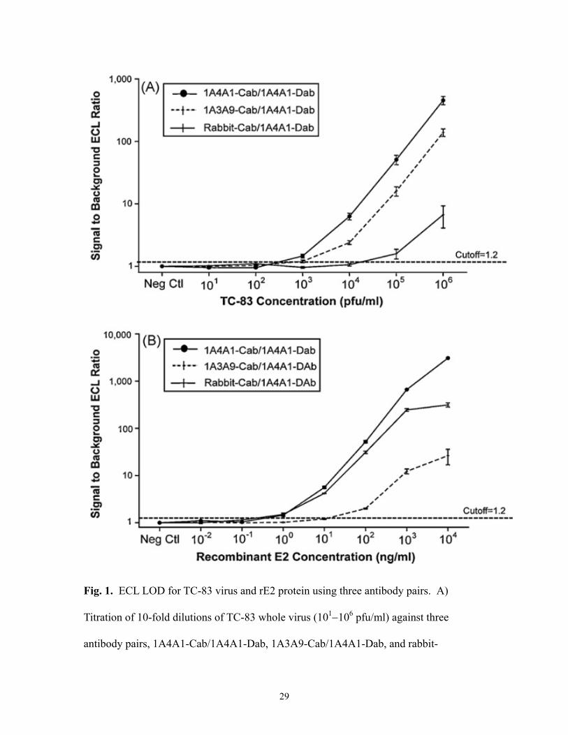
3.2. ECL LOD for TC-83 virus and VEEV recombinant E2
Experiments were performed to determine the assay LOD and dynamic range for
each optimized antibody pair. Results showed that antibody pair 1A4A1-Cab/1A4A1-
Dab produced the lowest assay LOD for both TC-83 virus and rE2 protein; with this
antibody pair, the LOD was 102–103 pfu/ml for TC-83 virus and 1 ng/ml for rE2 (Fig. 1A
and B and Table 1). The LOD for antibody pair 1A3A9-Cab/1A4A1-Dab was
approximately103 pfu/ml for TC 83 and 10 ng/ml for rE2 protein, while antibody pair
rabbit-Cab/1A4A1-Dab produced a LOD of 104–105 pfu/ml for TC83 and 1 ng/ml for rE2
protein (Fig. 1A and B and Table 1). Antibody pair 1A4A1-Cab/1A4A1-Dab exhibited

15
an assay dynamic range of 45 orders of magnitude, from approximately 102–106 pfu/ml
for TC-83 whole virus and 0.1–10,000 ng/ml for rE2 protein (Fig. 1A and B).
3.3. Comparison of ECL and ELISA
To compare assay LOD of ECL and ELISA, a sandwich ELISA, which used a rabbit-
Cab/1A4A1-Dab antibody pair format identical to that used in the ECL assay, was
performed for detection of TC-83 whole virus and VEEV rE2. The ELISA LOD obtained
with this antibody pair was 106 pfu/ml TC-83 whole virus and 10 ng/ml rE2 (Table 2).
These values were one to two log units higher than that observed in the ECL assay when
the same antibody pair was used (i.e., ECL LOD 104–105 pfu/ml for TC83 virus and 1
ng/ml for rE2 protein). Thus, the LOD of the VEEV ECL assay was approximately 10
times lower (higher sensitivity) than that of a comparable sandwich ELISA for detection
of VEEV whole virus and VEEV rE2 protein.
3.4. ECL LOD for VEEV recombinant E2 using recombinant antibody MA116SBP
To eliminate the need for chemical biotinylation of antibody, a genetically
biotinylated recombinant antibody, MA116SBP, was tested in the ECL assay for utility
for detection of rE2 protein. MA116SBP was pre-bound to streptavidin-coated
Dynabeads® following the same procedures as described above for mAbs and pAbs.
MA116SBP Cab at a dilution of 1:25 was paired with each of four Dabs, 1A4A1-Dab,
1A3A9-Dab, rabbit-Dab, and goat-Dab, each at a concentration of 4 g/ml, and evaluated
16
in the ECL assay with VEEV rE2 at a concentration of 100 ng/ml. Among the four
antibody pairs, MA116SBP-Cab/1A4A1-Dab pair produced the highest S/B values for
rE2 antigen (data not shown), thus this antibody pair was chosen for optimization of Cab
and Dab. Cab was optimized by testing dilutions of 1:8, 1:16, 1:25, 1:50 with 4 g/ml
Dab and three concentrations of VEEV rE2 (1, 10, and 100 ng/ml), followed by
optimization of Dab by testing concentrations of 4, 10, and 20 g/ml against the optimal
dilution of Cab (1:8) and VEEV rE2 at the same antigen concentrations. The assay LOD
obtained using genetically biotinylated MA116SBP as Cab was compared with that
obtained using chemically biotinylated Cab. Under optimal conditions, with the Cab
dilution of 1:8 and the Dab concentration of 10 g/ml, the LOD for rE2 obtained using
the pair MA116SBP-Cab/1A4A1-Dab was 10–100 ng/ml (Fig. 2). This value was two to
three logs higher than that observed when antibody pairs 1A4A1-Cab/1A4A1-Dab and
rabbit-Cab/1A4A1-Dab were used to detect rE2 (Fig. 1B and Table 1).
Antibody pair MA116SBP-Cab/1A4A1-DAb exhibited a dynamic range of
approximately three orders of magnitude, from approximately 100–100,000 ng/ml for
detection of rE2 protein (Fig. 2). By comparison, antibody pairs 1A4A1-Cab/1A4A1-
Dab and rabbit-Cab/1A4A1-DAb each exhibited a dynamic range of approximately four
orders of magnitude (1–10,000 ng/ml) for detection of rE2 (Fig. 1B).
The findings suggest that, although the genetically biotinylated recombinant
antibody MA116SBP was functional as Cab in ECL, the assay LOD obtained by its use
for detection of VEEV rE2 protein was higher (lower sensitivity) than that obtained using
chemically biotinylated Cab.
17
3.5. Assay reproducibility and precision
The assay reproducibility and precision were evaluated by titrating samples of VEEV
whole virus from month-to-month and from person-to-person (Fig. 3 and Tables 3 and 4).
For month-to-month reproducibility, LOD assays were performed on three separate
occasions over a period of five months (May 2007 to September 2007) with the same
antibody pair (1A4A1-Cab/1A4A1-Dab), stored at 4 oC over the five-month period, and
the same 10-fold dilutions of VEEV TC83, freshly prepared for each experiment (Fig. 3).
For person-to-person reproducibility, two different persons conducted standard LOD
assays using the same antibody pair (1A4A1-Cab/1A4A1-Dab) and same 10-fold
dilutions of VEEV TC83, prepared freshly by each person on the day of the experiment.
The coefficient of variation for the month-to-month assay precision determinations
ranged from 4.7%–18.5% (Table 3) and for the person-to-person assay precision
determinations from 3.3%–8.8% (Table 4).
3.6. Assay specificity
The specificity of the ECL assay was evaluated by testing two closely related
members of the alphavirus genus, WEEV and EEEV, and 21 unrelated bacterial, viral,
and toxin agents. Using the antibody pair 1A4A1-Cab/1A4A1-Dab, no cross-reactivity
was detected with either WEEV or EEEV whole viruses (Fig. 4A and B). While VEEV
produced a positive S/B ECL signal at a concentration of 104 pfu/ml, WEEV and EEEV
exhibited only a background level signal even at a concentration of 107 pfu/ml (Fig. 4A
18
and B). Similarly, there was no cross-reactivity observed with 21 unrelated agents,
including unrelated viruses: dengue, MS2, vaccinia Lister, and yellow fever (Fig. 5).
These results demonstrated that, with respect to the related and unrelated microorganisms
tested, this ECL assay was specific for VEEV.
3.7. Interference assay with powder and soil matrices
To evaluate matrix effects in the ECL assay, 15 environmental matrices, including 11
different powders and four types of soils, were examined in either non-spiked or 104
pfu/ml VEEV-spiked ECL assays. All powder and soil suspensions were used in the
assay at a final concentration of 1 mg of powder or soil per ml of PBS-0.3% Tween 20.
The positive control was prepared in PBS-0.3% Tween 20 containing 104 pfu/ml TC-83
whole virus and the negative control was PBS-0.3% Tween 20 only. A signal was
considered positive with interference effect if the S/B ratio was 20% above or below the
mean negative control (for non-spiked) or mean positive control (for VEEV-spiked). As
shown in Fig. 6, talcum powder, powdered cleanser, sand, sand loam, loamy sand, and
clay loam tested positive by increasing the background in the non-spiked ECL assay. In
the VEEV-spiked assay, sand and loamy sand exhibited significant interference by
quenching the ECL signal by 31%, relative to the positive control (Fig. 6). There was no
significant interference detected with 13 other powders and soils in the VEEV-spiked
assay. These results indicated that, at the level of significance examined, two powders
and four soils interfered with the ECL assays by increasing background or quenching the
ECL signals.

19
4. Discussion
A fully automated ECL immunoassay has been developed to detect VEEV TC-83
whole virus and VEEV rE2 protein.. The sensitivity of this ECL assay was the highest
(lowest LOD) hitherto reported in the published literature. Use of the 1A4A1-
Cab/1A4A1-Dab antibody combination yielded a LOD for TC-83 whole virus of 102–103
pfu/ml and for VEEV rE2 protein of 1 ng/ml. In a comparison of the ECL assay and
ELISA in which the same antibody pairs and assay format were used, rabbit-
Cab/1A4A1-Dab yielded a LOD of 104–105 pfu/ml whole virus by ECL assay and 1
ng/ml rE2 protein; in comparison, the ELISA LOD was 106 pfu/ml for TC-83 whole virus
and 10 ng/ml for rE2 protein. In a previously published study, the LOD for the Trinidad
donkey strain of VEEV was reported to be 1.25107 pfu/ml by ELISA and 3.13106
pfu/ml by DELFIA (Smith et al., 2001). In the current study, ECL assay results were
generated rapidly (30–60 min, including 15 min incubation) compared with the time
required to perform a comparable ELISA (6–8 hrs). Results also indicated that the biotin-
and ruthenium-labeled VEEV mAb and pAb Cab and Dab could be stored at 4 oC for at
least 10 months without deterioration of ECL signal. This finding is consistent with a
previous report stating that biotin- and ruthenium-labeled antibodies were stable at 4 oC
for up to one year (Kijek et al., 2000).
There are two envelope glycoproteins on VEEV, E1 and E2. Eight epitopes on the
VEEV E2 glycoprotein (E2a–h) have been identified (Mathews and Roehrig, 1982;
Roehrig et al., 1982; Roehrig and Mathews, 1985). MAbs 1A4A1 and 1A3A9 have been
20
previously well characterized and found to be specific for E2c and E2g epitopes,
respectively (Roehrig et al., 1982; Roehrig and Mathews, 1985). Studies on the spatial
arrangement of these epitopes have indicated that the E2c and E2g epitopes are closely
linked on VEEV TC-83 and that an anti-E2c antibody (e.g., 1A4A1) and an anti-E2g
antibody (e.g., 1A3A9) will compete with each other due to steric hindrance (Roehrig and
Mathews, 1985). In the present study, antibody pairs 1A3A9-Cab/1A4A1-Dab and
1A4A1-Cab/1A4A1-Dab exhibited high reactivity with TC-83 whole virus, suggesting
that 1A4A1 and 1A3A9 did not compete with each other and that the competition of
1A4A1 with itself did not affect binding. This observation also suggests that the amount
of Cab and Dab relative to the amount of available antigenic epitope was not limiting.
This is not unexpected as there are multiple copies of E2 on the surface of the VEEV
whole virus. By comparison, antibody pair 1A3A9-Cab/1A4A1-Dab pair exhibited poor
reactivity when rE2 was used as antigen. This observation suggests that one or more of
the antibody pair may have been directed against a conformational epitope on the whole
virus or that rE2 may have lacked important glycosylation and/or may have been
improperly folded for optimal activity with 1A3A9-Cab. The antibody pair rabbit-
Cab/1A4A1-Dab exhibited good reactivity with rE2 but, when compared to antibody pair
1A4A1-Cab/1A4A1-Dab, relatively poor reactivity with TC-83 whole virus. Overall, our
data showed that, in the context of developing an ECL assay, some antibody pairs had
good reactivity with both whole virus and recombinant antigen, while other antibody
pairs had good reactivity with only whole virus or recombinant antigen, but not both.
This finding suggests that, while recombinant component antigen can be a useful
substitute for whole pathogen antigen in development of immunoassays with some

21
antibody combinations, this may not always be the case, thus illustrating the importance
of utilizing the whole pathogen as antigen when developing assays for clinical or
environmental use.
During the labeling of antibody with biotin-LC-Sulfo-NHS ester or ruthenium (II)
tris-bipyridine-NHS ester, the N-hydroxysulfo-succinimide ester of biotin or ruthenium
combines with the –amide group of lysine to form a stable amide bond (Miralles et al.,
1991; Deaver, 1995). These reactions have been shown to lead sometimes to loss of
antigen-binding activity of the antibody. In the present study, ELISA results showed no
observable loss of binding activity of biotinylated or ruthenylated antibodies, in
comparison with unlabeled antibodies, with either TC83 whole virus or rE2 antigen (data
not shown). This observation agrees with previously published findings (Kijek et al.,
2000). However, eight of the 16 antibody pairs tested (50%) had little or no reactivity
with either TC-83 virus or rE2 in the ECL assay. This could have been a reflection of the
sandwich format in which two antibodies may have competed for the same epitope on the
antigen or may have been sterically hindered by each other. For example, antibody pairs
1A3A9-Cab/1A3A9-Dab, rabbit-Cab/rabbit-Dab, and goat-Cab/goat-Dab produced poor
ECL signals. However, antibody pair 1A4A1-Cab/1A4A1-Dab pair was an exception,
exhibiting excellent ECL signals for both TC-83 whole virus and recombinant E2 protein;
the reason for this apparent inconsistency remains unclear.
The ECL microbead immunoassay described in this paper requires the biotinylation
of antibody. Chemical biotinylation of antibody followed by removal of unbound biotin
is time-consuming, typically requiring 3–4 days to complete. Furthermore, the degree of
conjugation with biotin often varies, batch-to-batch (Miralles et al., 1991). To eliminate

22
these problems and the need for chemical biotinylation, a genetically biotinylated
recombinant antibody, MA116SBP, was tested for utility in the ECL assay. MA116SBP
was found to be functional as Cab in the ECL assay, but its use resulted in a higher assay
LOD (lower sensitivity) than when a chemically biotinylated Cab was used (LOD = 100
ng/ml vs 1 ng/ml). This could have been due to (i) MA116SBP being improperly folded
and hence lacking spatial conformation following expression in E. coli, (ii) the
recombinant protein containing only 11 residues of the streptavidin-binding peptide
rather than the full biotin molecule, or (iii) the antibody being a single-chain variable
fragment rather than the full length antibody. Function of the genetically biotinylated
recombinant antibody might be improved by constructing new expression vectors
containing full length biotin and antibody or by expression in a eukaryotic expression
system to gain proper post translational modification and spatial conformation. Further
studies would be required to address these issues.
23
5. Conclusions
An ECL immunoassay reactive with VEEV TC83 whole virus and the E2
glycoprotein on the VEEV virion surface has been developed for detection and
identification of VEEV. Antibody pairs, in all possible combinations, were down-
selected from a panel of four monoclonal and polyclonal antibody reagents for utility in
the assay. The LOD of the optimized ECL assay was 103 pfu/ml for TC-83 whole virus
and 1 ng/ml for VEEV rE2 component. The LOD of the VEEV ECL assay for both TC83
whole virus and rE2 was approximately one log unit lower (higher sensitivity) than that
of a sandwich ELISA incorporating the same immunoreagents. The ECL assay was
reproducible over time and when conducted by different persons. The VEEV ECL assay
exhibited no cross-reactivity with two closely related alphaviruses or with 21 unrelated
heterologous agents. In experiments designed to evaluate the effect of sample matrices
on assay performance, several powder and soil types were shown to interfere with the
assay by increasing background or quenching of ECL signal. A genetically biotinylated
VEEV recombinant antibody was evaluated in the assay for utility for detection of rE2,
but was found to lack reactivity when compared to incorporation in the assay of a
chemically biotinylated antibody reagent. This study has demonstrated a rapid, sensitive,
and specific assay for detection and identification of VEEV in environmental or clinical
samples.
24
Acknowledgments
The authors wish to thank Dr. Josh Wu for providing the TC-83 virus for this study.
The technical assistance of Mr. Jeffrey Ranches is also gratefully acknowledged. This
study was supported by a grant from the Chemical, Biological, Radiological-Nuclear
(CBRN) Research and Technology Initiative (CRTI), #03-0021TD (Assay Development
and Production Team for the Development, Validation, Production, and Distribution of
Assays for the Identification of Bioterrorist Agents).
25
References
Alvi, A.Z., Fulton, R.E., Chau, D., Suresh, M.R., Nagata, L.P., 2002. Development of a
second generation monoclonal single chain variable fragment antibody against
Venezuelan equine encephalitis virus: expression and functional analysis. Hybrid.
Hybridomics 21, 169–178.
Alvi, A.Z., Hu, W-G.., Fulton, R.E., Coles, J.E., Long, M.C., Nagata, L.P., 2003.
Functional enhancement of a partially active single-chain variable fragment
antibody to Venezuelan equine encephalitis virus. Viral Immunol. 16, 213–222.
Alvi, A.Z., Stadnyk, L.L., Nagata, L.P., Fulton, R.E., Bader, D.E., Roehrig, J.T., Suresh,
M.R., 1999. Development of a functional monoclonal single-chain variable
fragment antibody against Venezuelan equine encephalitis virus. Hybridoma 18,
413–421.
Deaver, D.R., 1995. A new non-isotopic detection system for immunoassays. Nature 377,
758–760.
Duggan, J.M., Coates, D.M., Ulaeto, D.O., 2001. Isolation of single-chain antibody
fragments against Venezuelan equine encephalomyelitis virus from two different
immune sources. Viral Immunol. 14, 263–273.
Garber, E.A., O'Brien, T.W., 2008. Detection of ricin in food using
electrochemiluminescence-based technology. J. AOAC Int. 91, 376–382.
Gatto-Menking, D.L., Yu, H., Bruno, J.G., 1995. Sensitive detection of biotoxoids and
bacterial spores using an immunomagnetic electrochemiluminescence sensor.
Biosensors & Bioelectronics 10, 501–507.

26
Griffin, D. E., 2001. Alphaviruses, in: Knipe, D.M., Howley, P. M. (Eds.), Fields
Virology. Lippincott-Raven, Philadelphia, pp. 937–941.
Hu, W-G., Alvi, A.Z., Fulton, R.E., Suresh, M.R., Nagata, L.P., 2002. Genetic engineering
of streptavidin-binding peptide tagged single-chain variable fragment antibody to
Venezuelan equine encephalitis virus. Hybrid. Hybridomics 21, 415–420.
Hu, W-G.., Thompson, H.G.., Alvi, A.Z., Nagata, L.P., Suresh, M.R., Fulton, R.E., 2004.
Development of immunofiltration assay by light addressable potentiometric
sensor with genetically biotinylated recombinant antibody for rapid identification
of Venezuelan equine encephalitis virus. J. Immunol. Methods 289, 27–35.
Kijek, T.M., Rossi, C.A., Moss, D., Parker, R.W., Henchal, E.A., 2000. Rapid and
sensitive immunomagnetic-electrochemiluminescent detection of staphyloccocal
enterotoxin B. J. Immunol. Methods 236, 9–17.
Kirsch, M.I., Hülseweh, B., Nacke, C., Rülker, T., Schirrmann, T., Marschall, H.-J., Hust,
M., Dübel, S., 2008. Development of human antibody fragments using antibody
phage display for the detection and diagnosis of Venezuelan equine encephalitis
virus (VEEV). BMC Biotechnol. 8, 66–80.
Kuhle, J., Regeniter, A., Leppert, D., Mehling, M., Kappos, L., Lindberg, R.L., Petzold,
A., 2010. A highly sensitive eletrochemiluminescence immunoassay for the
neurofilament heavy chain protein. J. Neuroimmunol. 220, 114–119.
Mathews, J.H., Roehrig, J.T., 1982. Determination of the protective epitopes on the
glycoproteins of Venezuelan equine encephalomyelitis virus by passive transfer of
monoclonal antibodies. J. Immunol. 129, 2763–2767.
Miralles, F., Takeda, Y., Escribano, M.J., 1991. Comparison of carbohydrate and peptide
27
biotinylation on the immunological activity of IgG1 murine monoclonal
antibodies. J. Immunol. Methods 140, 191–196.
Rivas, F., Diaz, L. A., Cardenas, V. M., Daza, E., Bruzon, L., Alcala, A., De la Hoz, O.,
Caceres, F. M., Aristizabal, G., Martinez, J. W., Revelo, D., De la Hoz, F., Boshell,
J., Camacho, T., Calderon, L., Olano, V. A., Villarreal, L. I., Roselli, D., Alvarez,
G., Ludwig, G., Tsai, T., 1997. Epidemic Venezuelan equine encephalitis in La
Guajira, Colombia. J. Infect. Dis. 175, 828–832.
Roehrig, J.T., Day, J.W., Kinney, R.M., 1982. Antigenic analysis of the surface
glycoproteins of a Venezuelan equine encephalomyelitis virus (TC-83) using
monoclonal antibodies. Virology 118, 269–278.
Roehrig, J.T., Mathews, J.H., 1985. The neutralization site on the E2 glycoprotein of
Venezuelan equine encephalomyelitis (TC-83) virus is composed of multiple
conformationally stable epitopes. Virology 142, 347–256.
Rossi, C.A., Ulrich, M., Norris, S., Reed, D.S., Pitt, L.M., Leffel, E.K., 2008.
Identification of a surrogate marker for infection in the African green monkey
model of inhalation anthrax. Infect. Immun. 76, 5790–5801.
Smith, D.R., Rossi, C.A., Kijek, T.M., Henchal, E.A., Ludwig, G.V., 2001. Comparison of
dissociation-enhanced lanthanide fluorescent immunoassays to enzyme-linked
immunosorbent assays for detection of staphylococcal enterotoxin B, Yersinia
pestis-specific F1 antigen, and Venezuelan equine encephalitis virus. Clin. Diagn.
Lab. Immunol. 8, 1070–1075.
Wang, E., Paessler, S., Aguilar, P.V., Smith, D.R., Coffey, L.L., Kang, W., Pfeffer, M.,
Olson, J., Blair, P.J., Guevara, C., Estrada-Franco, J., Weaver, S.C., 2005. A
28
novel, rapid assay for detection and differentiation of serotype-specific antibodies
to Venezuelan equine encephalitis complex alphaviruses. Am. J. Trop. Med. Hyg.
72, 805–810.
Weaver, S.C., Ferro, C., Barrera, R., Boshell, J., Navarro, J.C., 2004. Venezuelan equine
encephalitis. Annu. Rev. Entomol. 49, 141–174.
Yang, H., Leland, J.K., Yost, D., Massey, R.J., 1994. Electrochemiluminescence: a new
diagnostic and research tool. ECL detection technology promises scientists new
"yardsticks" for quantification. Biotechnology 12, 193–194.
Yoshimura, N.J., Miyazaki, N., Ito, K., Takeda, K., Hiramatsu, S., Morita, S., Miyauchi,
A., Murakami, T., Inomata, K., Noguchi, S., Satoh, T., Amino, N., 2008.
Evaluation of a new rapid and fully automated electrochemiluminescence
immnuoassay for thyrotropin receptor autoantibodies. Thyroid 18, 1157–1164.
Yu, H., Raymonda, J.W., McMahon, T.M., Campagnari, A.A., 2000. Detection of
biological threat agents by immunomagnetic microsphere-based solid phase
fluorogenic- and electro-chemiluminescence. Biosens. Bioelectron. 14, 829–840.
29
Fig. 1. ECL LOD for TC-83 virus and rE2 protein using three antibody pairs. A)
Titration of 10-fold dilutions of TC-83 whole virus (101106 pfu/ml) against three
antibody pairs, 1A4A1-Cab/1A4A1-Dab, 1A3A9-Cab/1A4A1-Dab, and rabbit-

30
Cab/1A4A1-Dab. B) Titration of 10-fold dilutions of rE2 (0.0110,000 ng/ml) against
three antibody pairs. The concentrations of Cab and Dab for each antibody pair were as
follows: 1A4A1-Cab/1A4A1-Dab pair at a Cab dilution of 1:35 and Dab concentration of
10 g/ml, 1A3A9-Cab/1A4A1-Dab pair at Cab dilution of 1:25 and Dab concentration of
10 g/ml, and rabbit-Cab/1A4A1-Dab pair at Cab dilution of 1:16 and Dab concentration
of 4 g/ml. The data was from three separate experiments, each with three replicates of
each concentration of antigen (n9). Error bars represent one standard deviation of the
mean. Neg Ctl: PBS-0.3% Tween 20 (n18). Signal to Background ECL Ratio: average
ECL reading divided by average ECL reading of negative control.
Fig. 2. ECL LOD for VEEV rE2 protein using recombinant Cab MA116SBP. Titration
of 10-fold dilutions of rE2 (0.1100,000 ng/ml) using the antibody pair MA116SBP-
31
Cab/1A4A1-Dab. MA116SBP-Cab dilution was at 1:8 and 1A4A1-Dab at 10 g/ml.
The data was from three separate experiments, each with three replicates of each
concentration of antigen (n9). Error bars represent one standard deviation of the mean.
Neg Ctl: PBS-0.3% Tween 20 (n18). Signal to Background ECL Ratio: average ECL
reading divided by average ECL reading of negative control.
Fig. 3. Reproducibility of VEEV ECL assay from month-to-month and from person-to-
person. The assays were performed with the antibody pair 1A4A1-Cab/1A4A1-Dab at a
Cab dilution of 1:35 and a Dab concentration of 10 g/ml. Assays 1, 2 and 3 represent
three separate titrations of 10-fold dilutions of TC-83 whole virus (101106 pfu/ml),
performed by one person, over a period of five months, each with three replicates of each
concentration of antigen (n=3 for each assay). Assay 4 represents a single titration of
virus using the same antibody pair, performed in triplicate by a second person, in
September 2007 (n=3). Error bars represent one standard deviation of the mean. Neg Ctl:

32
PBS-0.3% Tween 20 (n=3 for each assay). Signal to Background ECL Ratio: average
ECL reading divided by average ECL reading of negative control.
Fig. 4. Cross-reactivity assays with alphaviruses WEEV and EEEV. The assays were
performed with the antibody pair 1A4A1-Cab/1A4A1-Dab at a Cab dilution of 1:35 and a
Dab concentration of 10 g/ml. The data was from two separate assays, each with three
replicates of each concentration of agent (n6). Error bars represent one standard
deviation of the mean. Neg Ctl: PBS-0.3% Tween 20 (n12). Signal to Background
Ratio: average ECL reading divided by average ECL reading of negative control.
33
Fig. 5. Cross-reactivity assays with 21 unrelated agents. The assay was performed with
the antibody pair 1A4A1-Cab/1A4A1-Dab at a Cab dilution of 1:35 and a Dab
concentration of 10 g/ml. A concentration of 1 g/ml was used for most of the 21
agents with the exception of: Vaccinia, Lister: 106 pfu/ml; Aspergillus niger: 104
spores/ml; Staphylococcal enterotoxin B (SEB): 2 ng/ml; Ricin: 3 ng/ml; Yellow fever:
109 pfu/ml; Dengue: 104 pfu/ml. The data was from two separate assays, each with three
replicates of each concentration of agent (n6). Error bars represent one standard
deviation of the mean. Negative control: PBS-0.3% Tween 20 (n=12). Signal to
Background ECL Ratio: average ECL reading divided by average ECL reading of
negative control.

34
Fig. 6. Spiked and non-spiked interference assay. The assay was performed with the
antibody pair 1A4A1-Cab/1A4A1-Dab at a Cab dilution of 1:35 and a Dab concentration
of 10 g/ml. The data was from two separate assays, each with two replicates of each
concentration (n4). Positive control: 104 pfu/ml TC-83 whole virus in PBS-0.3% Tween
20 (n8). Error bars represent one standard deviation of the mean. Neg Ctl: PBS-0.3%
Tween 20 (n8). Signal to Background Ratio: average ECL reading divided by average
ECL reading of negative control.

35
Table 1. LOD for VEEV TC83 and rE2 protein by ECL using three different antibody pairsa 1A4A1-Cab/1A4A1-Dab 1A3A9-Cab/1A4A1-Dab Rbt-Cab/1A4A1-Dab ECL ECL ECL virus (pfu/ml)
rE2 (ng/ml)
virus (pfu/ml)
rE2 (ng/ml)
virus (pfu/ml)
rE2 (ng/ml)
102–103 1 103 10 104–105 1 aData was from three separate experiments; each concentration of antigen was tested in triplicate (n=9).
36
Table 2. Comparison of LOD for VEEV TC83 and E2 protein by ECL and sandwich ELISAa ECL ELISA Virus (pfu/ml) rE2 (ng/ml) Virus (pfu/ml) rE2 (ng/ml) 104–105 1 106 10 aData was from three separate experiments; each concentration of antigen was tested in triplicate by ECL
(n=9) and in duplicate by ELISA (n=6).
37
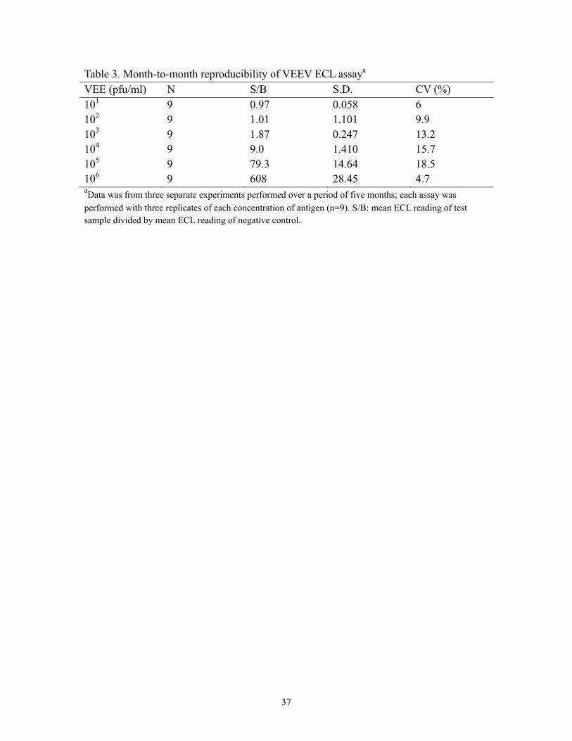
Table 3. Month-to-month reproducibility of VEEV ECL assaya VEE (pfu/ml) N S/B S.D. CV (%) 101 9 0.97 0.058 6 102 9 1.01 1.101 9.9 103 9 1.87 0.247 13.2 104 9 9.0 1.410 15.7 105 9 79.3 14.64 18.5 106 9 608 28.45 4.7 aData was from three separate experiments performed over a period of five months; each assay was
performed with three replicates of each concentration of antigen (n=9). S/B: mean ECL reading of test sample divided by mean ECL reading of negative control.

38
Table 4. Person-to-person reproducibility of VEEV ECL assaya VEE (pfu/ml) N S/B S.D. CV (%) 101 6 0.97 0 0 102 6 0.99 0.035 3.6 103 6 1.27 0.057 4.5 104 6 4.8 0.424 8.8 105 6 36.9 1.202 3.3 106 6 327 28.45 3.7 aData was from two separate experiments performed by two different people; each assay was performed
with three replicates of each concentration of antigen. S/B: mean ECL reading of test sample divided by mean ECL reading of negative control.