Skull
23
SKULL By: Ramez Maqsood Student GDC Srinagar
-
Upload
ramez-maqsood -
Category
Health & Medicine
-
view
33 -
download
5
Transcript of Skull

SKULLBy: Ramez Maqsood
Student GDC Srinagar
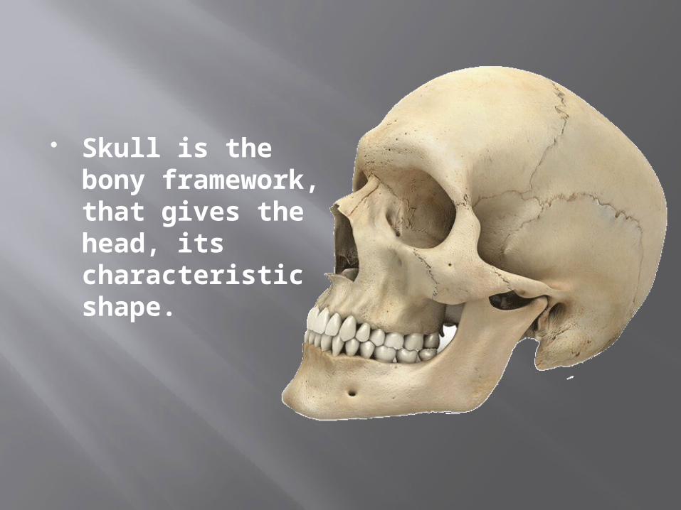
Skull is the bony framework, that gives the head, its characteristic shape.
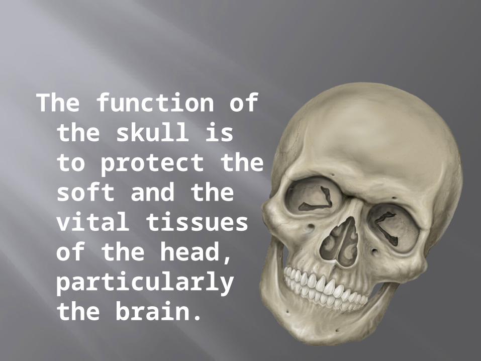
The function of the skull is to protect the soft and the vital tissues of the head, particularly the brain.

The Skull consists of the Cranium (the bony box housing the brain) and the face.

The skull is composed of 22 bones, 8 in the cranium and 14 in the face.
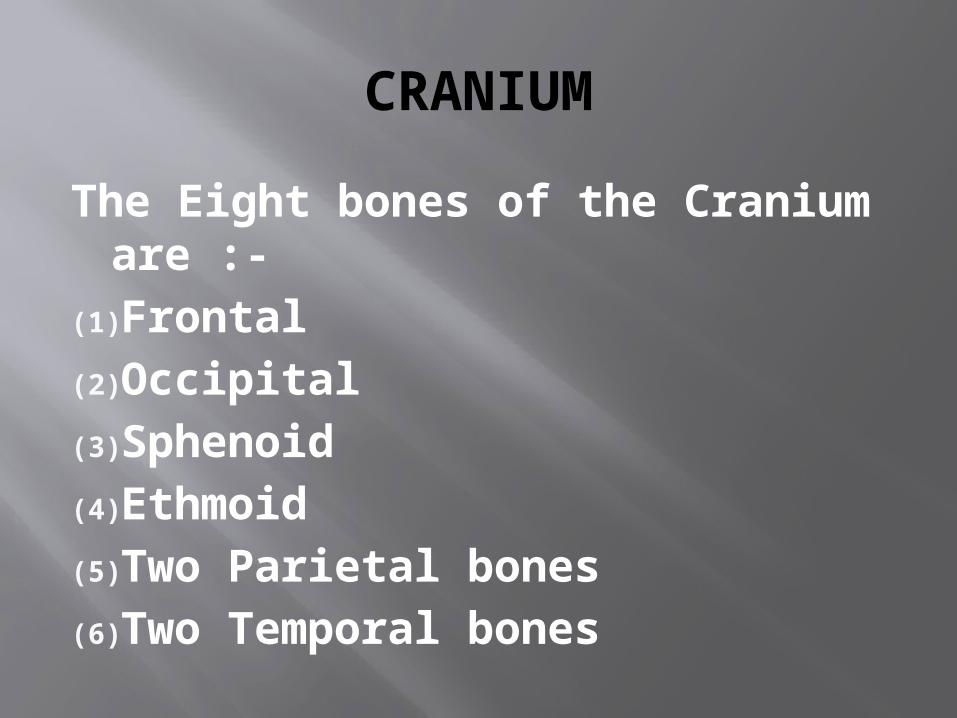
CRANIUM
The Eight bones of the Cranium are :-
(1)Frontal(2)Occipital(3)Sphenoid(4)Ethmoid(5)Two Parietal bones(6)Two Temporal bones


(i) FRONTAL : The frontal bone forms the forehead , the anterior (front) part of the cranial vault and the roof of the orbits (eye sockets).Inside the bone, just behind the eyebrows are two air spaces called the frontal sinuses.

(ii) OCCIPITAL : The occipital bone forms the posterior (back) part of the floor and vault of the cranium. It is the bone which supports the head upon the spinal column.
The spinal cord leaves the cranium through an opening in the occipital bone called the foramen magnum

(iii) SPHENOID : The sphenoid bone is the central part of the base of the cranium. It forms part of the orbits, transmits the optic nerve and supports the posterior part of the maxilla.The sphenoid air sinuses lie in this bone. The pituitary gland lies in a bony socket called the sella turcica, located on the superior aspect of the sphenoid bone.Dental Tip: When a patient is seated in the dental chair, the headrest should support the occipital bone and thereby support the entire head.

(iv) ETMOID : The Ethmoid bone lies between the eyes and extends from the frontal bone to the sphenoid bone. It forms the anterior part of the skull, the medial wall of each orbit, part of the nasal septum and the roof of the nose.It transmits the olfactory nerve (nerve of smell)

(v) PARIETAL : The Parietal bones forms a large part of the cranial vault and extend from the frontal bone to the occipital bone. The two bones join at the midline on the top of the cranium and form the saggital suture.
From this suture, these bones extend down and out to about the level of the top of the external ear where they meet the temporal bones.

(vi) TEMPORAL : Temporal bones complete the sides and part of the base of the cranium. These bones contain organs of hearing and equilibrium.
The external acoustic meatus in the side of each bone forms a passage from the external ear to the middle ear which lies within each bone.

FACE
There are 14 bones in the face.(i) Mandible (1)(ii) Maxillae (2)(iii) Zygomatic Bones (2)(iv) Lacrimal Bones (2)(v) Nasal Bones (2)(vi) Inferior conchae (2)(vii) Palatine Bones (2)(viii) Vomer (1)


(i) ZYGOMATIC : The right and left zygomatic bones form the lower and outer edges of each orbit and that part of each zygomatic arch nearest the eye.
The Zygomatic bone and Zygomatic process of the temporal bone form the Zygomatic arch. The anterior edge of the zygomatic bone joins the maxilla.That part of the maxilla which joins the zygomatic bone is called the zygomatic process.

(ii) LACRIMAL : The paired (right and left) Lacrimal bones form small parts of the medial walls of the orbits.
The lacrimal bones transmits the naso-lacrimal duct from the eye to the nose or nasal fossa.

(iii) NASAL : The nasal bones (right and left) are long, thin pieces of bone that form the upper part of the bridge of the nose.
The anterior lower part of the nasal septum is composed of cartilage

(iv) INFERIOR CONCHAE: The inferior nasal conchae (right and left) are scroll like bones lying horizontally along the lateral walls of the nasal cavity.
The bony elements of the middle and superior conchae are extensions of the lateral parts of the ethmoid bones.
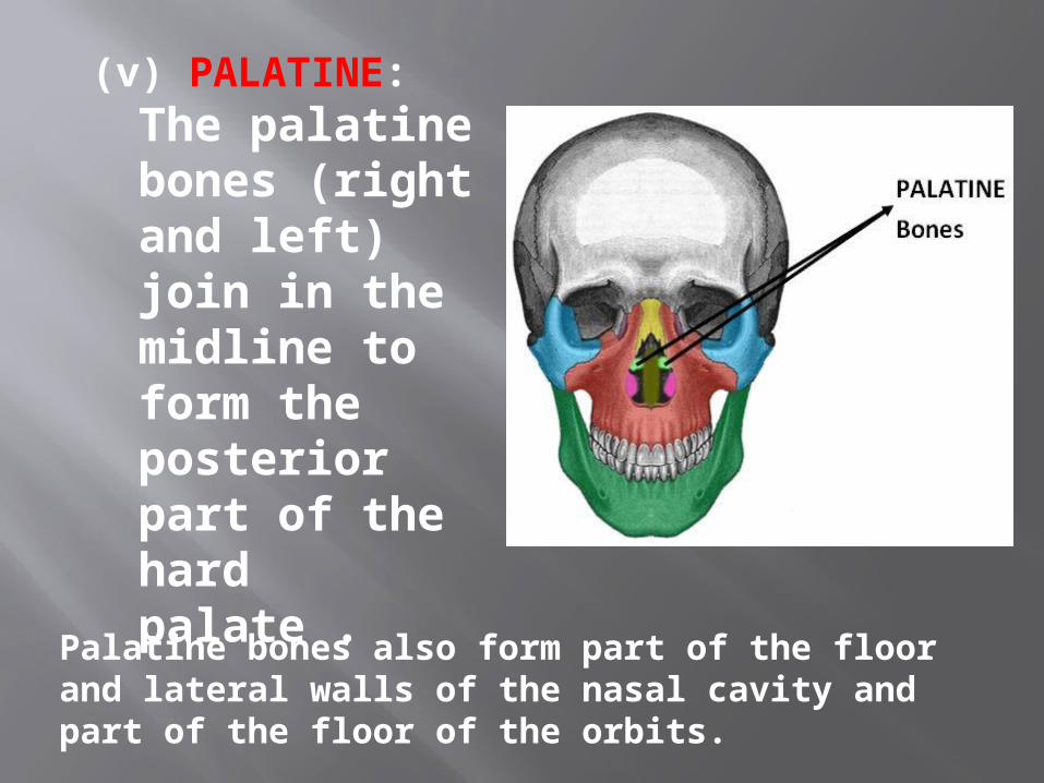
(v) PALATINE: The palatine bones (right and left) join in the midline to form the posterior part of the hard palate .
Palatine bones also form part of the floor and lateral walls of the nasal cavity and part of the floor of the orbits.

(vi) VOMER: The vomer forms the inferior part of the nasal septum, the vertical partition separating the right and left nasal cavities.

(vii) MAXILLA: The right and left maxillary bones forms the upper jaw and palate of the mouth.
The two halves are fused at the intermaxillary suture to form the upper jaw.

(viii) MANDIBLE: The horse shoe-shaped bone forming the lower jaw, articulating with the skull at the temporomandi - -bular joint.
Mandible is the largest, strongest and lowest bone in the face.