Skin Tumors Revealing Hematological MalignancySM Journal of Hematology and Oncology SM Gr u How to...
2
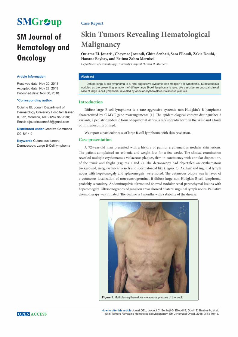
SM Journal of Hematology and Oncology Gr up SM How to cite this article Jouari OEL, Jroundi C, Senhaji G, Elloudi S, Douhi Z, Baybay H, et al. Skin Tumors Revealing Hematological Malignancy. SM J Hematol Oncol. 2018; 3(1): 1011s. OPEN ACCESS Case Report Skin Tumors Revealing Hematological Malignancy Ouiame EL Jouari*, Cheymae Jroundi, Ghita Senhaji, Sara Elloudi, Zakia Douhi, Hanane Baybay, and Fatima Zahra Mernissi Department of Dermatology University Hospital Hassan II, Morocco Article Information Received date: Nov 20, 2018 Accepted date: Nov 28, 2018 Published date: Nov 30, 2018 *Corresponding author Ouiame EL Jouari, Department of Dermatology University Hospital Hassan II, Fez, Morocco, Tel: 212677879830; Email: [email protected] Distributed under Creative Commons CC-BY 4.0 Keywords Cutaneous tumors; Dermoscopy; Large B-Cell lymphoma Abstract Diffuse large B-cell lymphoma is a rare aggressive systemic non-Hodgkin’s B lymphoma. Subcutaneous nodules as the presenting symptom of diffuse large B-cell lymphoma is rare. We describe an unusual clinical case of large B-cell lymphoma, revealed by annular erythematous violaceous plaques. Introduction Diffuse large B-cell lymphoma is a rare aggressive systemic non-Hodgkin’s B lymphoma characterized by C-MYC gene rearrangements [1]. e epidemiological context distinguishes 3 variants, a pediatric endemic form of equatorial Africa, a rare sporadic form in the West and a form of immunocompromised. We report a particular case of large B-cell lymphoma with skin revelation. Case presentation A 72-year-old man presented with a history of painful erythematous nodular skin lesions. e patient complained an asthenia and weight loss for a few weeks. e clinical examination revealed multiple erythematous violaceous plaques, firm in consistency with annular disposition, of the trunk and thighs (Figures 1 and 2). e dermoscopy had objectified an erythematous background, irregular linear vessels and spermatozoid like (Figure 3). Axillary and inguinal lymph nodes with hepatomegaly and splenomegaly, were noted. e cutaneous biopsy was in favor of a cutaneous localization of non-centrogerminat if diffuse large non-Hodgkin B-cell lymphoma, probably secondary. Abdominopelvic ultrasound showed nodular renal parenchymal lesions with hepatomegaly. Ultrasonography of ganglion areas showed bilateral inguinal lymph nodes. Palliative chemotherapy was initiated. e decline is 4 months with a stability of the disease. Figure 1: Multiples erythematous violaceous plaques of the truck.
Transcript of Skin Tumors Revealing Hematological MalignancySM Journal of Hematology and Oncology SM Gr u How to...
SM Journal of Hematology and Oncology
Gr upSM
How to cite this article Jouari OEL, Jroundi C, Senhaji G, Elloudi S, Douhi Z, Baybay H, et al. Skin Tumors Revealing Hematological Malignancy. SM J Hematol Oncol. 2018; 3(1): 1011s.OPEN ACCESS
Case Report
Skin Tumors Revealing Hematological MalignancyOuiame EL Jouari*, Cheymae Jroundi, Ghita Senhaji, Sara Elloudi, Zakia Douhi, Hanane Baybay, and Fatima Zahra MernissiDepartment of Dermatology University Hospital Hassan II, Morocco
Article Information
Received date: Nov 20, 2018 Accepted date: Nov 28, 2018 Published date: Nov 30, 2018
*Corresponding author
Ouiame EL Jouari, Department of Dermatology University Hospital Hassan II, Fez, Morocco, Tel: 212677879830; Email: [email protected]
Distributed under Creative Commons CC-BY 4.0
Keywords Cutaneous tumors; Dermoscopy; Large B-Cell lymphoma
Abstract
Diffuse large B-cell lymphoma is a rare aggressive systemic non-Hodgkin’s B lymphoma. Subcutaneous nodules as the presenting symptom of diffuse large B-cell lymphoma is rare. We describe an unusual clinical case of large B-cell lymphoma, revealed by annular erythematous violaceous plaques.
Introduction
Diffuse large B-cell lymphoma is a rare aggressive systemic non-Hodgkin’s B lymphoma characterized by C-MYC gene rearrangements [1]. The epidemiological context distinguishes 3 variants, a pediatric endemic form of equatorial Africa, a rare sporadic form in the West and a form of immunocompromised.
We report a particular case of large B-cell lymphoma with skin revelation.
Case presentation
A 72-year-old man presented with a history of painful erythematous nodular skin lesions. The patient complained an asthenia and weight loss for a few weeks. The clinical examination revealed multiple erythematous violaceous plaques, firm in consistency with annular disposition, of the trunk and thighs (Figures 1 and 2). The dermoscopy had objectified an erythematous background, irregular linear vessels and spermatozoid like (Figure 3). Axillary and inguinal lymph nodes with hepatomegaly and splenomegaly, were noted. The cutaneous biopsy was in favor of a cutaneous localization of non-centrogerminat if diffuse large non-Hodgkin B-cell lymphoma, probably secondary. Abdominopelvic ultrasound showed nodular renal parenchymal lesions with hepatomegaly. Ultrasonography of ganglion areas showed bilateral inguinal lymph nodes. Palliative chemotherapy was initiated. The decline is 4 months with a stability of the disease.
Figure 1: Multiples erythematous violaceous plaques of the truck.

Citation: Jouari OEL, Jroundi C, Senhaji G, Elloudi S, Douhi Z, Baybay H, et al. Skin Tumors Revealing Hematological Malignancy. SM J Hematol Oncol. 2018; 3(1): 1011s.
Page 2/2
Gr upSM Copyright Jouari OEL
Figure 2: Annular erythematous plaques on the members.
Figure 3: Dermoscopy showing an erythematous background, irregular linear vessels and spermatozoid like.
DiscussionNon-Hodgkin’s lymphomas (NHLs) are a group of malignant
disorders that can arise from any part of the lymphatic system. However, extra-nodal lymphomas are uncommon accounting for
around 6-18% of NHLs. The reported incidence of B-cell malignancies is extremely rare at <1% of all cutaneous lymphomas [1]. Cutaneous involvement is indeed very rare. A subcutaneous nodule as the presenting symptom of diffuse large B-cell lymphoma is rare. Skin or soft tissue nodules are more commonly seen in subcutaneous panniculitis-like T-cell lymphoma [2]. The cutaneous involvement is usually secondary to the spread of the disease but can sometimes reveal it as in our observation. The specific dermatological lesions which can reveal hematological diseases [3], are mainly represented by leukaemides (leukaemia cutis), which are red brown to purple dermal papules, plaques or nodules. Granulocytic sarcoma as an extra-medullary tumor masses, ulcerated plaques and gingival hypertrophy [4,5]. In our case, clinically the lesions evoked cutaneous lymphoma, metastasis or angiosarcoma. Histology allowed the diagnosis of large cell L-cell lymphoma in a sporadic form. Histologically, there are 2 variants: classical B lymphoma and atypical B lymphoma. In our case, the histology was that of a classical B lymphoma but with an unusual clinical presentation. We describe an unusual clinical case of large B-cell lymphoma, revealed by annular erythematous violaceous plaques. Unfortunately, these secondary localizations are generally considered as poor prognostic factors.
ConclusionAny patient complained asthenia and weight loss with cutaneous
tumors, is required to screen for hematological malignancy and allowed to realize the necessary assessment.
References
1. Attili VS, Batra U, Bapsy PP, Lokanatha D, Clementeena R, Varma PP, et al. Skin nodules as a presenting feature of diffuse large B-cell gastric lymphoma. Indian J Dermatol. 2008; 53: 39-40.
2. Bik Ling Man, Kit Kwan Ma and Yat Pang Fu. Subcutaneous nodules as the presenting symptom of diffuse large B cell lymphoma. BMJ Case Rep. 2014; 2014: bcr2014206300.
3. Blessis D, Frances C, Grillot J. Manifestations dermatologiques des maldies du système hématologique et oncologie dermatologique. Dermatologie et medicine, Springer Verlag, Framce. 2009.
4. Evans KG. Acute Onset of Leg Nodules in a Sporotrichoid Pattern. Arch Dermatol. 2012; 148: 1199-1200.
5. H Bay Bay. Les manifestations cutanées au cours des hémopathies malignes, EM consulte. 2011.