Skin melanocytes: biology and development - Tor...
12
Postępy Dermatologii i Alergologii XXX; 2013/1 30 Address for correspondence: Mirosława Cichorek PhD, Department of Embryology, Medical University of Gdansk, 1 Dębinki Str., 80-211 Gdansk, Poland, phone: +48 58 349 14 95, fax: +48 58 349 14 95, e-mail: [email protected] Received: 17.07.2012, accepted: 24.10.2012. Skin melanocytes: biology and development Mirosława Cichorek, Małgorzata Wachulska, Aneta Stasiewicz, Agata Tymińska Department of Embryology, Medical University of Gdansk, Poland Head: Mirosława Cichorek PhD Postep Derm Alergol 2013; XXX, 1: 30-41 DOI: 10.5114/pdia.2013.33376 Review paper Abstract In the human skin, melanocytes are present in the epidermis and hair follicles. The basic features of these cells are the ability to melanin production and the origin from neural crest cells. This last element is important because there are other cells able to produce melanin but of different embryonic origin (pigmented epithelium of retina, some neurons, adipocytes). The life cycle of melanocyte consists of several steps including differentiation of melanocyte lineage/s from neural crest, migration and proliferation of melanoblasts, differentiation of melanoblasts into melanocytes, proliferation and maturation of melanocytes at the target places (activity of melanogenic enzymes, melanosome formation and transport to keratinocytes) and eventual cell death (hair melanocytes). Melanocytes of the epider- mis and hair are cells sharing some common features but in general they form biologically different populations living in unique niches of the skin. Key words: melanocytes, neural crest cells, stem cells. Introduction Melanocytes form a heterogeneous group of cells in the human body. Although all of them have ability to produce melanin and originate from embryonic cells named neur- al crest cells (NCC), their particular functions in all target places are much wider than the melanin synthesis only [1]. In the human body melanocytes’ presence does not con- firm only epidermis, hair and iris where they give a color of these structures. Melanocytes have been also found in the inner ear, nervous system, heart and probably it is not the end of a list where these cells exist [2, 3]. It is neces- sary to stress that not only melanocytes have ability to pro- duce melanin but also other cells e.g. cells of pigmented epithelium of retina, epithelia of iris and ciliary body of the eye, some neurons, adipocytes [4, 5]. The life cycle of melanocytes consists of several steps including lineage specification from embryonic neural crest cells (melanoblasts), migration and proliferation of me- lanoblasts, differentiation of melanoblasts into mela- nocytes, maturation of melanocytes (melanin production in special organelles – melanosomes, dendritic morpho- logy), transport of mature melanosomes to keratinocytes and eventual cell death. Several populations of neural crest cells (cranial, dorsal trunk, ventral trunk) give melanocytes of the skin. The embryonic origin of epidermal and hair melanocytes is the same but development is different [6, 7]. The problem of melanocyte stem cells’ localization in the adult skin is still a matter of debate. The first such place was a hair bulge, but if only...? [8]. Experimental data clearly show that the trunk NCC migrating through a ven- tral pathway could remain in a myelin sheath of the cuta- neous nerves and in particular situations give melanoblasts [9, 10]. The embryonic development of melanocytes give an opportunity to better understand the skin diseases e.g. melanoma and its heterogeneity, vitiligo. Thus, in this review the epidermal and hair melanocytes’ biology and devel- opment are characterized. Melanocyte in the skin as the epidermal melanin unit element Melanocytes molecularly are recognizable by identifi- cation of melanocyte-specific proteins as tyrosinase (TYR), tyrosinase-related protein 1 and 2 (TYRP1, TYRP2/DCT), melanosomal matrix proteins (Pmel17, MART-1), microph- thalmia transcription factor (MITF) [1]. The microscopic analysis indicates that mature melanocytes are oval or fusiform, dendritic cells, smaller than keratinocytes. In the
Transcript of Skin melanocytes: biology and development - Tor...

Postępy Dermatologii i Alergologii XXX; 2013/130
AAddddrreessss ffoorr ccoorrrreessppoonnddeennccee:: Mirosława Cichorek PhD, Department of Embryology, Medical University of Gdansk, 1 Dębinki Str., 80-211 Gdansk, Poland, phone: +48 58 349 14 95, fax: +48 58 349 14 95, e-mail: [email protected]:: 17.07.2012, aacccceepptteedd:: 24.10.2012.
Skin melanocytes: biology and development
Mirosława Cichorek, Małgorzata Wachulska, Aneta Stasiewicz, Agata Tymińska
Department of Embryology, Medical University of Gdansk, PolandHead: Mirosława Cichorek PhD
Postep Derm Alergol 2013; XXX, 1: 30-41
DOI: 10.5114/pdia.2013.33376
Review paper
AbstractIn the human skin, melanocytes are present in the epidermis and hair follicles. The basic features of these cells arethe ability to melanin production and the origin from neural crest cells. This last element is important because thereare other cells able to produce melanin but of different embryonic origin (pigmented epithelium of retina, some neurons, adipocytes). The life cycle of melanocyte consists of several steps including differentiation of melanocytelineage/s from neural crest, migration and proliferation of melanoblasts, differentiation of melanoblasts into melanocytes,proliferation and maturation of melanocytes at the target places (activity of melanogenic enzymes, melanosomeformation and transport to keratinocytes) and eventual cell death (hair melanocytes). Melanocytes of the epider-mis and hair are cells sharing some common features but in general they form biologically different populations living in unique niches of the skin.
KKeeyy wwoorrddss:: melanocytes, neural crest cells, stem cells.
Introduction
Melanocytes form a heterogeneous group of cells in thehuman body. Although all of them have ability to producemelanin and originate from embryonic cells named neur-al crest cells (NCC), their particular functions in all targetplaces are much wider than the melanin synthesis only [1].In the human body melanocytes’ presence does not con-firm only epidermis, hair and iris where they give a colorof these structures. Melanocytes have been also found inthe inner ear, nervous system, heart and probably it is notthe end of a list where these cells exist [2, 3]. It is neces-sary to stress that not only melanocytes have ability to pro-duce melanin but also other cells e.g. cells of pigmentedepithelium of retina, epithelia of iris and ciliary body of theeye, some neurons, adipocytes [4, 5].
The life cycle of melanocytes consists of several stepsincluding lineage specification from embryonic neural crestcells (melanoblasts), migration and proliferation of me -lanoblasts, differentiation of melanoblasts into mela -nocytes, maturation of melanocytes (melanin productionin special organelles – melanosomes, dendritic morpho -logy), transport of mature melanosomes to keratinocytesand eventual cell death. Several populations of neural crestcells (cranial, dorsal trunk, ventral trunk) give melanocytes
of the skin. The embryonic origin of epidermal and hairmelanocytes is the same but development is different [6, 7]. The problem of melanocyte stem cells’ localizationin the adult skin is still a matter of debate. The first suchplace was a hair bulge, but if only...? [8]. Experimental dataclearly show that the trunk NCC migrating through a ven-tral pathway could remain in a myelin sheath of the cuta-neous nerves and in particular situations give melanoblasts[9, 10]. The embryonic development of melanocytes givean opportunity to better understand the skin diseases e.g.melanoma and its heterogeneity, vitiligo. Thus, in this reviewthe epidermal and hair melanocytes’ biology and devel-opment are characterized.
Melanocyte in the skin as the epidermal melaninunit element
Melanocytes molecularly are recognizable by identifi-cation of melanocyte-specific proteins as tyrosinase (TYR),tyrosinase-related protein 1 and 2 (TYRP1, TYRP2/DCT),melanosomal matrix proteins (Pmel17, MART-1), microph-thalmia transcription factor (MITF) [1]. The microscopicanalysis indicates that mature melanocytes are oval orfusiform, dendritic cells, smaller than keratinocytes. In the
Postępy Dermatologii i Alergologii XXX; 2013/1 31
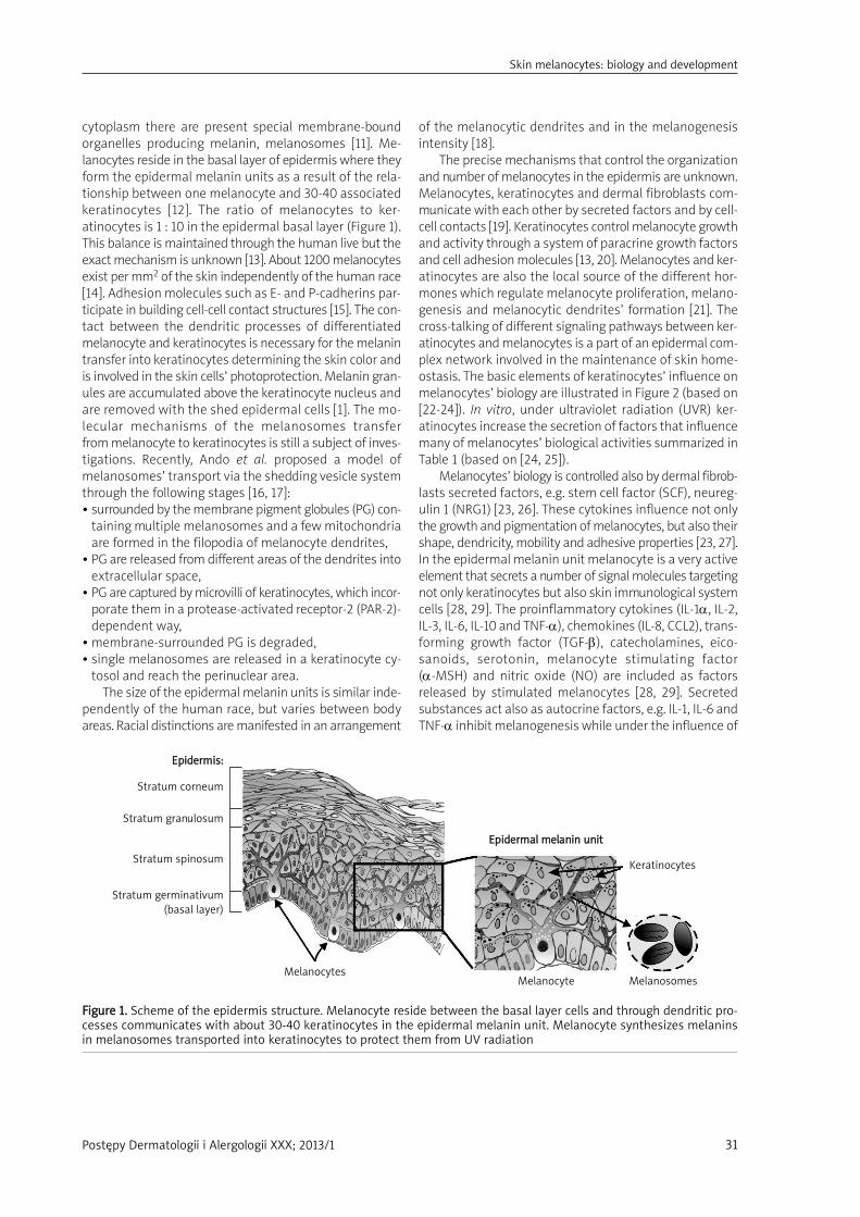
cytoplasm there are present special membrane-boundorganelles producing melanin, melanosomes [11]. Me -lanocytes reside in the basal layer of epidermis where theyform the epidermal melanin units as a result of the rela-tionship between one melanocyte and 30-40 associatedkeratinocytes [12]. The ratio of melanocytes to ker-atinocytes is 1 : 10 in the epidermal basal layer (Figure 1).This balance is maintained through the human live but theexact mechanism is unknown [13]. About 1200 melanocytesexist per mm2 of the skin independently of the human race[14]. Adhesion molecules such as E- and P-cadherins par-ticipate in building cell-cell contact structures [15]. The con-tact between the dendritic processes of differentiatedmelanocyte and keratinocytes is necessary for the melanintransfer into keratinocytes determining the skin color andis involved in the skin cells’ photoprotection. Melanin gran-ules are accumulated above the keratinocyte nucleus andare removed with the shed epidermal cells [1]. The mo-lecular mechanisms of the melanosomes transfer from melanocyte to keratinocytes is still a subject of inves-tigations. Recently, Ando et al. proposed a model ofmelanosomes’ transport via the shedding vesicle systemthrough the following stages [16, 17]:• surrounded by the membrane pigment globules (PG) con-
taining multiple melanosomes and a few mitochondriaare formed in the filopodia of melanocyte dendrites,
• PG are released from different areas of the dendrites intoextracellular space,
• PG are captured by microvilli of keratinocytes, which incor-porate them in a protease-activated receptor-2 (PAR-2)-dependent way,
• membrane-surrounded PG is degraded,• single melanosomes are released in a keratinocyte cy -
tosol and reach the perinuclear area.The size of the epidermal melanin units is similar inde-
pendently of the human race, but varies between bodyareas. Racial distinctions are manifested in an arrangement
of the melanocytic dendrites and in the melanogenesisintensity [18].
The precise mechanisms that control the organizationand number of melanocytes in the epidermis are unknown.Melanocytes, keratinocytes and dermal fibroblasts com-municate with each other by secreted factors and by cell-cell contacts [19]. Keratinocytes control melanocyte growthand activity through a system of paracrine growth factorsand cell adhesion molecules [13, 20]. Melanocytes and ker-atinocytes are also the local source of the different hor-mones which regulate melanocyte proliferation, melano-genesis and melanocytic dendrites’ formation [21]. Thecross-talking of different signaling pathways between ker-atinocytes and melanocytes is a part of an epidermal com-plex network involved in the maintenance of skin home-ostasis. The basic elements of keratinocytes’ influence onmelanocytes’ biology are illustrated in Figure 2 (based on[22-24]). In vitro, under ultraviolet radiation (UVR) ker-atinocytes increase the secretion of factors that influencemany of melanocytes’ biological activities summarized inTable 1 (based on [24, 25]).
Melanocytes’ biology is controlled also by dermal fibrob-lasts secreted factors, e.g. stem cell factor (SCF), neureg-ulin 1 (NRG1) [23, 26]. These cytokines influence not onlythe growth and pigmentation of melanocytes, but also theirshape, dendricity, mobility and adhesive properties [23, 27].In the epidermal melanin unit melanocyte is a very activeelement that secrets a number of signal molecules targetingnot only keratinocytes but also skin immunological systemcells [28, 29]. The proinflammatory cytokines (IL-1α, IL-2,IL-3, IL-6, IL-10 and TNF-α), chemokines (IL-8, CCL2), trans-forming growth factor (TGF-β), catecholamines, eico -sanoids, serotonin, melanocyte stimulating factor (α-MSH) and nitric oxide (NO) are included as factorsreleased by stimulated melanocytes [28, 29]. Secreted substances act also as autocrine factors, e.g. IL-1, IL-6 and TNF-α inhibit melanogenesis while under the influence of
FFiigguurree 11.. Scheme of the epidermis structure. Melanocyte reside between the basal layer cells and through dendritic pro-cesses communicates with about 30-40 keratinocytes in the epidermal melanin unit. Melanocyte synthesizes melaninsin melanosomes transported into keratinocytes to protect them from UV radiation
EEppiiddeerrmmiiss::
Stratum corneum
Stratum granulosum
Stratum spinosum
Stratum germinativum (basal layer)
Melanocytes
EEppiiddeerrmmaall mmeellaanniinn uunniitt
Melanocyte
Keratinocytes
Melanosomes
Skin melanocytes: biology and development

Postępy Dermatologii i Alergologii XXX; 2013/132
Plasma membranewith receptors
Cytoplasm
Nuclear membrane
Nucleus
NGF
TYRTYRP1TYRP2
MMeellaannooccyyttee
KKeerraattiinnooccyyttee
Proliferation, differentiation, melanogenesis, dendritogenesis
ET-1 GM-CSF bFGF SCFα-MSHPGE2PGF2α
?
CREB
ATP
Factors secretedby keratinocyte
Signal transductionpathways
in melanocyte
NGFR EP1/EP3/FP ETBR GM-CSFR FGFR1/2 c-Kit
PLC PKA PKC MAPK
MITF-MMITF-M
promoter
Plasma membrane
FFiigguurree 22.. Graphical presentation of the basic elements in keratinocytes-melanocytes cooperation. The melanocyte proli-feration, differentiation, melanogenesis are under control of factors secreted by surrounding keratinocytesSCF – stem cell factor, bFGF – basic fibroblast growth factor, GM-CSF – granulocyte-macrophage colony-stimulating factor, ET-1 – endothe-lin 1, α-MSH – melanocyte-stimulating hormone, PGE2 – prostaglandin E2, PGF2α – prostaglandin F2α, NGF – nerve growth factor, c-Kit – tyrosine kinase receptor, FGFR1/2 – fibroblast growth factor receptor, GM-CSFR – granulocyte-macrophage colony-stimulating fac-tor receptor, ETBR – endothelin B receptor, MC1R – melanocortin 1 receptor, EP1/EP3/FP – prostanoid receptors, NGFR – nerve growth fac-tor receptor, MAPK – mitogen-activated protein (MAP) kinases, PKC – protein kinase C, PKA – protein kinase A, PLC – phospholipase C, TYR – tyrosinase, TYRP1 – tyrosinase-related protein 1, TYRP2 – tyrosinase-related protein 2, MITF-M – melanocyte-specific MITF (micro-phthalmia-associated transcription factor) isoform, CRE – cAMP response elements, CREB – cAMP response element-binding
MC1R
cAMP
CRE
TTaabbllee 11.. The paracrine factors secreted by keratinocytes afterUV radiation that influence on melanocyte biology
TThhee ffaaccttoorrss ddeerriivveedd TThhee eeffffeeccttss oonn mmeellaannooccyytteeffrroomm kkeerraattiinnooccyytteess
bFGF ↑ Proliferation
ET-1 ↓ Proliferation, ↑ dendricity, ↓ melanogenesis
IL-1α/1β ↑ Proliferation, ↑ melanogenesis, ↑ survival
ACTH ↑ Proliferation, ↑ dendricity, ↑ melanogenesis, ↑ survival
α-MSH ↑ Dendricity, ↑ melanogenesis, ↑ melanosomal transfer
PGE2/PGF2α ↑ Proliferation, ↑ melanogenesis
GM-CSF ↑ Melanogenesis
NO ↓ Melanogenesis
TNF-α ↑ Dendricity, ↑ survival
NGF ↓ Melanogenesis
BMP-4 ↑ Proliferation, ↑ dendricity, ↑ melanogenesis
eicosanoids and α-MSH the level of melanin synthesis iselevated [30]. Thus, melanocytes and cooperating ker-atinocytes form well-organized units in the epidermis. Thestable element in each unit is the melanocyte that liveslong, keratinocytes die and are shedding. It is an open ques-tion how long melanocyte lives in the human skin.
Hair follicle melanocytes
Melanocytes are located in the proximal bulb of eachhair follicle and also near hair, e.g. in the sebaceous gland[31]. The bodies of bulbar melanocytes are located at theapex on the dermal papilla. Melanocyte dendrites enterbetween the cortical and medullar keratinocytes [32]. Theratio of melanocytes to keratinocytes is 1 : 5, it is moredense than in the epidermis (Figure 3) [33]. Follicular pig-mentation is a result of structural and functional inter-actions between follicular melanocytes, matrix ker-atinocytes and dermal papilla fibroblasts. This tripartitesystem is described as the hair melanin unit or follicularmelanin unit. The process of hair pigmentation includesthe melanogenic activity of follicular melanocytes, the trans-
Mirosława Cichorek, Małgorzata Wachulska, Aneta Stasiewicz, Agata Tymińska

Postępy Dermatologii i Alergologii XXX; 2013/1 33
fer of melanin granules into keratinocytes and the formationof pigment ed hair shafts [32-34]. It is considered thata transport of melanin granules to keratinocytes in the grow-ing hair shaft is similar to the epidermal phagocytosis ofmelanosomes mediated by receptor PAR2 on keratinocytes.But differences concern degradation of melanosomes andtheir quality. Hair melanocytes, in contrast to epidermalones, include mainly bigger mature Stage IV melanosomes(melanogenesis and stages of melanosomes are describedfurther in this work) and more expanded Golgi apparatusand rough endoplasmatic reticulum (RER). These pigmentcells are larger and more dendritic than epidermal ones [31,33]. Moreover, in epidermis almost whole transportedmelanin is degraded in the differentiating keratinocytes,but in hair cortical keratinocytes pigment granules aredigested only slightly [33]. Diversity of hair color arises most-ly from the quantity and ratio of the brown-black eume-lanin and the yellow-red pheomelanin [35].
Melanin synthesis in the hair occurs under control ofproducts secreted by neighboring cells as keratinocytes,fibroblasts and endothelial cells, which act through
paracrine or autocrine mechanisms and may be modifiedby hormonal signals. In pigmentation determining hair color, the following elements are involved: melanocortinreceptor 1 (MCR1) and its α-MSH, adrenocorticotropic hor-mone (ACTH), receptor c-Kit and its ligand SCF, endothe-lins, different neurotransmitters, cytokines, growth factorand other regulators similar as for epidermal melanogenesiscontrol [31, 33]. The biochemical pathway of pigment for-mation and melanosomes biogenesis run likewise in theepidermis, but it is stressed that hair follicle melanocytesare more sensitive to aging influences than epidermalmelanocytes, what results in hair greying [33]. It seems thatfibroblast of dermal papilla derived factors: insulin growthfactor (IGF-1), keratinocyte growth factor (KGF), noggin, SCFhave special significance for control the hair matrix ker-atinocyte and melanocyte proliferation and activity dur-ing the hair growth [32, 36]. Epidermal melanocytes arelong-living cells, while hair melanocytes die at the end ofthe hair cycle which lasts 3-8 years [31]. The melanogen-esis process takes place only during the anagen stage (grow-ing phase) of the hair growth cycle; pigment formation is
FFiigguurree 33.. Melanocyte localization in the hair between cells covering the hair papilla in the hair bulb. Stem cells for mela-nocytes are located in the region named the hair bulge
Epidermis
Sebaceous gland
Arrector pili muscle
Bulge (melanocytes stem cells)
MMeellaannooccyytteess
Hair bulb
Dermis
Skin melanocytes: biology and development

Postępy Dermatologii i Alergologii XXX; 2013/134
turned off in the catagen stage (regressing phase) andabsent in the telogen stage (resting phase) [31]. Additionally,it is marked that during catagen completely differentiat-ed bulbar melanocytes die through apoptosis, but newmelanocytes develop from melanoblasts residing in the hairbulge [8, 32]. Summarizing, survival, proliferation and dif-ferentiation of melanocytes are regulated by microenvi-ronment of the hair follicles.
Melanocyte biology
MMeellaannooggeenneessiiss
Melanogenesis is a biochemical pathway responsiblefor melanin synthesis [37]. It takes place in melanocytes,in separate cytoplasmic organelles called melanosomes [11].Two major types of melanin are produced – pheomelaninand eumelanin. They differ in color and the way of synthesis.Melanin has numerous properties which are beneficial tothe body: UV light absorption and scattering, free radicalscavenging, coupled oxidation-reduction reactions and ionstorage [23, 38, 39]. The availability of substrates and thefunction of melanogenesis enzymes decide about the types
of melanins produced (Figure 4). Tyrosinase (TYR) carriesout tyrosine hydroxylation to L-3,4-dihydro xyphenylalanine(DOPA) which is rapidly oxidized to DOPAquinone [40]. Inthe presence of cysteine DOPAquinone react with it,yielding 3- or 5-cysteinylDOPAs, which then oxidize and poly-merize, giving rise to yellow-red soluble melanin – pheome-lanin [37, 41]. In the absence of thiols (cysteine, glutathioneor thioredoxin) brown-black eumelanin is produced.DOPAquinone spontaneously undergoes cyclization toDOPAchrome [42]. The DOPAchrome spontaneously losescarboxylic acid and generates 5,6-dihydroxyindole (DHI), which rapidly oxidizes and polymerizes to form darkbrown-black, insoluble DHI-melanin. However, if DOPA -chrome tautomerase (TYRP2/DCT) is present, DOPAchromewill form DHI-2-carboxylic acid (DHICA) [43]. Tyrosinase andTYRP1 catalyze further conversions obtaining finallya lighter brown color DHICA-melanin [30, 37]. Human skincontains a mixture of all melanin types, and the ratio ofthose in part determines visible pigmentation [19]. Diver-sity of skin pigmentation among different ethnic groupsis preserved and depends on eumelanin content. The ratioof eumelanin to total melanin decide about skin color [30].Pheomelanin does not correlate with skin pigmentation,a similar amount of this pigment is observed in the darkand light skin. While in hair, the ratio of eumelanin topheomelanin decides about the color [35]. Eumelanin com-paring to pheomelanin has better photoprotecting prop-erties – higher resistance to degradation and ability to reac-tive oxygen species (ROS) neutralization [44]. Eumelaninsare considered to be more effective in terms of photo-protection than the reddish pheomelanin. As a conse-quence, the risk of skin cancer in lighter skin is 30-40-foldhigher than in the darker one [41]. Products of genes reg-ulating melanogenesis act at subcellular, cellular, tissue andenvironmental levels [21]. During melanogenesis, as inter-mediate products, cytotoxic molecules are synthesized(quinones, hydrogen peroxide). Thus, melanocyte protectsitself by separating areas of melanogenesis in melanosomesand increases the level of antiapoptotic protein Bcl-2 [1, 21].
Melanosomes probably originate from endoplasmicreticulum of melanocytes, but it still remains a matter ofdebate [25]. Their development requires tyrosinase (TYR)and tyrosinase-related proteins (TYRP1, TYRP2). Of thesethree enzymes, tyrosinase is crucial to melanogenesis andis synthesized on the ribosomes of the RER and transportedto the Golgi complex where it undergoes glycosylation,which is a process essential for its normal structure andfunction [45, 46].
There are four stages in melanosome development(Table 2). Premelanosomes (Stage I) are a round, small vesi-cles with an amorphous matrix. Melanosomes at Stage IIhave an organized, structured fibrillar matrix (mainlyfrom gp100 family) and tyrosinase is present but pigmentsynthesis has not been noted. The beginning of melaninproduction takes place at Stage III, where pigment is deposit-ed on protein fibrils. At the last Stage IV pigment fills the
FFiigguurree 44.. Simplified scheme of the melanin synthesis inmelanocytes during melanogenesis. Tyrosine under influ-ence of the basic enzymes such as tyrosinase (TYR), tyro-sine-related protein 1 (TYRP1) and 2 (TYRP2) changes intoa polymer of melanin, a mixture of pigments named eume-lanin (black-brown) and pheomelanin (yellow-red)
TYR
TYR
+ CysteineLack of cysteine
Oxidation polymerization
DHICA DHI
DHICA-melanin DHI-melanin
CysteinylDOPA
PPhheeoommeellaanniinnyellow to red
TRP2
TRP1
EEuummeellaanniinnssbrown to black
Tyrosine
L-DOPA
DOPAchrome
DOPAquinone
Mirosława Cichorek, Małgorzata Wachulska, Aneta Stasiewicz, Agata Tymińska

Postępy Dermatologii i Alergologii XXX; 2013/1 35
whole melanosome [41, 47]. Fully melanized melanosomeslose tyrosinase activity and are transported to surround-ing keratinocytes by elements of the cytoskeletal system(Figure 1) [48].
MMeellaannooccyytteess’’ aabbiilliittyy ooff pprroolliiffeerraattiioonn aanndd aaggee--rreellaatteeddcchhaannggeess
The precise mechanisms that control the organizationand number of melanocytes in the epidermis are unknownalthough keratinocytes may interact with melanocytes viagrowth factors, cell surface molecules, or other factors relat-ed to proliferation and differentiation of the epidermis.Melanocyte is a highly differentiated cell that produces a pig-ment melanin inside melanosomes. This cell is dark anddendritic in shape. Melanin production is the basic func-tion of melanocyte. With the process of differentiation thiscell loses the proliferative potential. Epidermal melanocytesare thought as a very stable population which proliferateextremely rarely under normal circumstances.
All we know about melanocytes’ proliferation controlcome from in vitro studies. Human melanocyte prolifera-tion requires the cross-talking of several signaling pathways including the MAPK-kinase signaling, α-MSH/cAMP/PKA, Endothelin/PKC (PKA protein kinase A,PKC protein kinase C) [23, 49]. As the potent mitogens aregrowth factors and hormones as stem cell factor (SCF),hepatocyte growth factor (HGF), endothelins, α-MSH,ACTH. While, transforming growth factor-β (TGF-β), inter-feron-β (INF-β), IL-1, IL-6, TNF-α, cause the opposite effect
and arrest melanocytes’ growth [21, 29]. The MITF as themain melanocyte transcription factor influences prolifer-ation, dendrite formation, melanin synthesis and inducesthe expression of antiapoptotic bcl-2 gene [50].
Epidermal melanocytes are long-living cells while hair melanocytes live as the hair cycle lasts (median: 3-5 years) [31]. Density of melanocytes in the skindepends on the environment (mainly UVR) and factorssecreted by keratinocytes and fibroblasts. After 30years of age 10-20% of epidermal melanocytes are lostevery decade [51]. In the older people, apart froma decreased number of melanocytes morphology ischanged (melano cytes are larger, more dendritic) andtyrosinase activity is reduced [19, 31, 52]. The relationshipsbetween ageing and the proliferative activity of me lano -cytes have been observed. In vitro, adult melanocytes pro-liferate less times than fetal and neonatal melanocytes[53]. Also, melanocytes from patients with a prematureageing disorder have reduced proliferative potential [50].Terminally differentiated melanocyte proliferative poten-tial is inhibited by changes in the cell cycle control ele-ments, e.g. accumulation of cyclin-dependent kinaseinhibitors (p27Kip1, p16INK4a and p21Cip1), hypophospho-rylation of pRB (retinoblastoma protein), decrease lev-el of cyclin D1 [50, 53]. Table 3 lists basic cell cycle reg-ulators involved in the regulation of melanocyticsenescence (based on [53]).
Furthermore, the reason for a decreased number ofmelanocytes is programmed cell death of terminally dif-
TTaabbllee 22.. Characteristics of the developmental stages of melanosomes during melanin synthesis. The melanogenesis takesplace in special organelles named melanosomes. As first develops a vesicle (Stage I) which builds inside a fibrillar matrixformed by glycoproteins (Pmel17, MART-1) and gets tyrosinase and other enzymes of melanogenesis (Stage II). The mela-nosome produces melanin, which polymerizes and settles on the internal fibrils (Stage III). In the last stage (Stage IV)melanosome is filled up with melanin
MMeellaannoossoomm ffeeaattuurreess SSttaaggee II SSttaaggee IIII SSttaaggee IIIIII SSttaaggee IIVV
Shape Spherical Elongated Elliptical, ellipsoidal Elliptical, ellipsoidal
Internal structure – Matrix fibrils are visible Matrix fibrils are visible Matrix fibrils are covered by polymerized melanin
TYR – + + +
TYRP1 – + + +
TYRP2 – + + +
Melanin synthesis – – Begins, settle on internal fibrils Filled by melanin
Color Brown Dark brown to black
Skin melanocytes: biology and development

Postępy Dermatologii i Alergologii XXX; 2013/136
ferentiated cells. Accumulation of reactive oxygen sub-strates (ROS) as a result of reduced content/activity ofcatalase (key antioxidant enzyme) and downregulationof BCL-2 seem to be the main inducer of melanocyte apop-tosis [50, 52].
In the melanocyte proliferation, the mitogen-activat-ed protein kinase (MAPK) pathway is involved, which is stim-
ulated by many growth factors. In the terminally differ-entiated melanocytes this main proliferative pathway isnot active [50]. Discoveries in the field of molecular reg-ulations of melanocyte proliferation and death help usunderstand disorders such as melanoma or vitiligo [54, 55].
Summarizing, the proliferation and differentiation ofmelanocytes during development are regulated by differentgenetic and epigenetic factors derived from keratinocytes,fibroblasts, melanocytes, the pituitary gland, other organsand environmental factors (such as UV radiation) [24].
Embryonic origin of skin melanocytes
MMoorree tthhaann oonnee ppooppuullaattiioonn ooff nneeuurraall ccrreesstt cceellllss iiss tthhee ssoouurrccee ooff sskkiinn mmeellaannooccyytteess
Neural Crest Cells is a group of cells originating fromthe embryonic germ layer named ectoderm. Under induc-tive influence of the notochord, the middle area of theembryonic disc differentiates into neuroectoderm that isvisible as a neural plate at 4-week-old human embryo (Figure 5 A). This plate folds and changes into the neuraltube, future central nervous system elements – brain, spinalcord. During this process named neurulation, a group ofcells from edges of the neural plate (crests), separates,changes the phenotype from epithelial to mesenchyme andmigrate out from neuroepithelium (Figures 5 A and 5 B).These neuroectodermal cells migrating to many places ofthe forming embryo’s body are neural crest cells – NCC (Figure 5 C). Neural crest cells are initially multipotent cellsbut gradually become lineage-restricted in developmen-
TTaabbllee 33.. The basic activity of the cell cycle regulators andother factors associated with melanocytes senescence
TThhee cceellll ccyyccllee rreegguullaattoorr TThhee lleevveell ooff eexxpprreessssiioonn//aaccttiivviittyy dduurriinngg sseenneesscceennccee
Cyclin E ↓ Protein level
p16INK4a ↑ Expression
Protein RB1 Dephosphorylation
CDK2 and CDK4 ↓ Activities
Transcription factor E2F4 ↑ Association with RB1
p300/CBP histone ↓ Activityacetyltransferases
Extracellular matrix proteins ↑ Protein level
MAPK-signaling pathway Inactivation
p53 Melanocytes senescence is independent of p53, probably except for situation with deficient p16/RB1 pathway
Neural crests
Neural plate
Neural plate folding
Notochord
NCC
Neural tube
Neu
roec
tode
rm
Somites
FFiigguurree 55.. Development of the neural crest cells (NCC) during early embryogenesis at a 4-week-old embryo from neuroec-toderm. The neuroectodermal cells proliferate, form the neural plate that folds, fuses and changes into neural tube (AA, BB). During this neurulation process, cells from edges (crests) of the neural plate separate from the neural tube as inde-pendent population of embryonic cells named neural crest cells, that is located above the neural tube (future brain andspinal cord) and beneath surface ectoderm (future epidermis) (CC)
AA BB CC
CC
Mirosława Cichorek, Małgorzata Wachulska, Aneta Stasiewicz, Agata Tymińska

Postępy Dermatologii i Alergologii XXX; 2013/1 37
tal potential. This potential is determined by anatomicallocalization along the cranial-caudal axis, e.g. cranialNCC can differentiate into neurons, glial cells but also chon-drocytes, osteocytes, muscle cells, whereas trunk NCC formneurons and glial cells in the peripheral nervous system,endocrinal cells (Figure 6). These cells proliferate and startto express distinct molecular markers [7, 56].
Neural crest cells are traditionally grouped into fourregionally distributed populations: cranial, vagal, trunk andsacral. Melanocytes mainly origin from cranial and trunk-located NCC.
Melanocytes residing in skin of the head origin from the cranial NCC while in the remaining parts of thehuman body mainly from the trunk NCC. Except me -lanocytes, cranial NCC together with mesodermal cells formthe ectomesenchyme of the head, that gives skeleton, mus-cles and dermis of the head (Figure 6 B) [57].
According to embryonic migratory pathways, thetrunk NCC is divided into two populations, dorsally (be -tween surface ectoderm and somites) and ventrally (be -tween neural tube and somites) migrating cells (Figure 6 A). Traditionally, the dorso-laterally migrating cells
NNCCCC
TTrruunnkk NNCCCC
CCrraanniiaall NNCCCC
Surface ectoderm
Somite
Neural tube
DDoorrssaall
Melanocyte
Melanocyte Neurons, glia of ganglia
Melanocyte Neurons, glia of ganglia Dermis of head Smooth muscles Chondrocyte osteocyte
Adrenal medulla Schwann cells
VVeennttrraall
FFiigguurree 66.. Derivatives of the trunk (AA) and cranial (BB) neural crest cells (NCC) and the basic pathways of the trunk NCCmigration during early embryonic time. Dorsally migrating trunk NCC move between the surface ectoderm and somites,finally develop into melanocytes of the epidermis and hair (A). Ventrally migrating trunk NCC move between the neuraltube and somites, give elements of the peripheral nervous system (ganglionic cells, Schwann cells), medulla of the adre-nal glands and according to latest investigations, melanocytes of the skin (A)
AA
BB
Skin melanocytes: biology and development

Postępy Dermatologii i Alergologii XXX; 2013/138
are thought to be the main source for melanocytes whilethe ventrally migrating cells give rise to the peripheral ner-vous system and adrenal medulla (Figure 6 B). But, thereis strong evidence that a fraction of melanocytes arise fromcells migrating first ventrally and then along the nerves [9, 58]. Cells present in a nerve sheath (Schwann cells) havethe potential to produce melanocytes also after birth. In vitro, Schwann cells cultured in melanocytes mediumde-differentiate into glial-melanocytic progenitor able togive melanocytes [59].Thus, cells migrating ventrallyeither differentiate into neurons or are maintained as mul-tipotent cells that differentiate into cells forming myelinsheath or melanocytes (Figure 6) [7]. These cells invade theepidermis during the process of embryonic cutaneous inner-vation [9]. The recent findings that congenital (prenatal)nevi begin as intradermal nevi seems to support the hypoth-esis that precursors for melanocytes could origin from der-mis cells [60]. It is suggested that prenatal nevi may devel-op from the precursors for Schwann cells, which arrive nearepidermis along cutaneous nerve, may respond to factorssecreted by epidermal cells and differentiate intomelanocytes. As cutaneous nerves grow from deep dermisnear the epidermis they branch and form the candelabrapattern (a neurocutaneous unit). Along these branches pre-cursors for melanocytes migrate to the epidermis and asa result the congenital nevi may develop [60]. During humandevelopment, melanoblast migration and cutaneousnerve growth take place at the same time between 6 and8 weeks [9, 60]. Communication between the nervous sys-tem and epidermal melanocytes has been proven [61]. Theobservation that epidermal melanocytes molecularly dif-fer from dermal melanocytes seems to support thehypothesis about double origin of skin melanocytes [62].Thus, melanocytes in the skin either derive directly fromNCC populating the skin via a dorsolateral migratory path-way or arise from ventrally migrating precursors formingthe myelin around the cutaneous nerves [63].
As melanoblasts travel through the dermis, they mul-tiply. While traveling to their final destinations, melanoblastssequentially express additional melanogenic genes, manyof them regulated by transcription factor MITF. The mostimportant in the maturation of melanocyte is the ap pear-ance of tyrosinase, enzyme of melanin synthesis.Melanocytes finally reside in the skin and hair follicles, theoral mucosa, the choroid of the eye, the iris, and some inter-nal sites, such as meninges and the inner ear (the stria vas-cularis). The fate of NCC depends on environmental fac-tors they meet on the migratory pathways [7]. After cellspecification, melanoblasts proliferate and spread totheir final destinations in the epidermis and hair follicleswhere they differentiate. It takes place at 6-8 weeks andby 12-13 weeks the majority are localized in the epidermis[60, 64]. Whether all of them reach the epidermis is an unre-solved developmental problem. Dermal melanocytes areseen during human fetal development but they are not evident after birth. There are suggestions that some
melanoblasts could stay in the dermis [64, 65]. It remainsunknown how the stream of melanoblasts to the epider-mis is controlled. The time when melanoblast presence inthe dermis is detectable is also time for cutaneous nervedevelopment [60]. The time between 9 and 12 weeks is alsothe beginning of hair buds’ development and melanoblastmigration to them. On the 18th week of intrauterine lifea hair which comes through the skin surface havemelanocytes present in the hair bulb [64, 66] but the activ-ity of tyrosinase is very low [67]. The fetal first hair presenton the whole embryo skin is named lanugo. At the end ofpregnancy (30-33 weeks) lanugo degenerates and final hairdevelops, the first hair cycle is done [68].
Spaces between cells are filled with a rich extracellu-lar matrix formed by many fibrillar proteins such as col-lagens, fibronectin. Cell adhere to one another using adhe-sion molecules such as cadherins or adhere to matrix byintegrins. These adhesion mechanisms are of special impor-tance during melanoblast migration. Melanoblasts migrateover very long distances throughout the embryo, proliferateand promote their own survival. Thus, melanoblast devel-opment is a highly dynamic process, which requires rapidactivation of different signaling pathways.
BBaassiicc mmoolleeccuullaarr eelleemmeennttss ooff eemmbbrryyoonniicc//ffeettaall mmeellaannooccyytteess ddeevveellooppmmeenntt
The results of experiments have not found out ananswer to a question – at what stage NCC get features ofmelanoblasts, precursors to melanocytes [7, 69]. Me -lanoblasts have features of melanocytes but do not pro-duce melanin, there is no agreement about a set of mol-ecular markers for this early stage of melanocyte inhuman development. The most commonly listed molec-ular markers for the precursors for melanocytes are:a tyrosine kinase receptor KIT (c-kit); transcription factorssuch as MITF, SOX10, Pax 3 and melanogenic enzyme tyrosi-nase-related protein (TYRP-2) [7, 24, 56, 70]. The exact mech-anisms responsible for melanoblast migration are not wellunderstood, although adhesion molecules such as cad-herins, integrins, and extracellular matrix elements areinvolved in it. The ephrin receptor (EphR) and the endothe-lin receptor (EDNRB2) allow melanoblasts to migratealong extracellular matrix containing ephrin and endothe-lin-3 [69, 71].
The most important growth factors regulatingmelanocyte development from the NCC are endothelins,ligand for c-kit (SCF stem cell factor), Wnt proteins andneuregulin-1 [72, 73]. This regulations are described in detailin other works [24, 63, 72, 73].
The c-Kit receptor binds the stem cell factor (SCF) secret-ed by the dermal cells and as a result of this pathway acti-vation melanoblasts avoid apoptosis and proliferate [24,73]. The Wnt/Frizzled protein/β-catenin-signaling pathway,the Notch pathway and the MAPK-signaling pathways areessential for melanoblast/melanocyte development. Stud-ies in several model organisms suggest that compo-
Mirosława Cichorek, Małgorzata Wachulska, Aneta Stasiewicz, Agata Tymińska

Postępy Dermatologii i Alergologii XXX; 2013/1 39
nents of the Wnt/β-catenin signaling pathway are requiredfor induction of melanocyte fate [74]. The Notch signalingpathway is of special importance during the embryonic peri-od. The Notch receptor requires the contact with anoth-er cell because the ligand for this receptor is a plasma mem-brane protein (e.g. Delta). MAPK-signaling pathwayRas/Raf/Mek/Erk) has been of special interest accordingto BRAF role in melanoma development [75].
Many of the factors listed above induce expression ofthe MITF protein functioning as a master of melanocyte pro-liferation, differentiation and survival. The MITF activatesgenes responsible for the migration to the skin, prevent-ing apoptosis in migrating cells, melanin production [70, 76].
Stem cells for human melanocytes in adults
Melanocytes proliferate rarely but some situations pointout that in the skin there are precursors for melanocytese.g. repigmentation of the skin of vitiligo patients after pho-totherapy [77].
What do we know about the presence of melanoblastsin the adult skin?
The first documented reservoir for melanocyte stemcells was the bulge area of the hair follicles either in themouse or human [78, 79]. The bulge area is a part of out-er root sheath that provides the insertion point for the arrec-tor pili muscle and points to the bottom of the permanentportion of hair follicles (Figure 3). Each time a hair is lostthe hair follicle regenerates and melanocyte stem cells areactivated [80]. The bulge region contains pluripotential, mor-phologically undifferentiated cells which develop into keratinocyte progenitors and melanoblasts not only for hair but also epidermis [8]. After stimulation these cellsmigrate in the basal layer of the epidermis and differen-tiate into mature melanocytes [64, 78].
However, there is growing evidence that the dermisis also a reservoir of melanocytes [64, 77, 81]. Majority ofthe progress on melanocyte development has beenmade in murine models, little progress has been made onthe identification of melanocyte-producing stem cells inhuman skin. Where are melanoblasts located in the dermis? The answer to this question is not satisfactoryenough. Recent developmental studies using modelorganisms and lineage tracing have been able to tracemelanocytes arising from migration of a multipotent pre-cursor cell along nerve projections. With great probabil-ity there could be cells with stem cell’s properties locat-ed in the cutaneous nerves, but if only [9, 58]? These cellsare retained in a stem cell-like state until the signal sentby the end of the cutaneous nerve promotes these cellsto differentiate into melanocytes [82]. But there are alsoobservations that some melanoblasts from the dorsal wayof NCC migration stay in the dermis after the end of epi-dermis inoculation, up to the second trimester during fetaltime [64, 71, 83]. Whether they survive in the human adultdermis is still an open problem.
There are difficulties in the identification of mela -noblasts in the adult skin because of lack of a set of me -lanoblast markers. It seems that the epidermis and dermismelanocytes are biologically different populations [62].
Epidermis and dermis as places for the melanoblasts’reservoir give an opportunity for a new look at the me -lanoma heterogeneity. Melanoma is a tumor which devel-ops from melanocytes but there is no agreement if themature melanocytes or cells from earlier developmentalstages may follow the tumoricidal transformation [65, 84-86]. Recently, Whiteman et al. made an attempt to sepa-rate melanoma(s) into melanoma arising from epithelialmelanocytes and not associated with epithelia [86].
References
1. Plonka PM, Passeron T, Brenner DJ, et al. What are melanocytesreally doing all day long…? Exp Dermatol 2009; 18: 799-819.
2. Tachibana M. Sound needs sound melanocytes to be heard.Pigment Cell Res 1999; 12: 344-54.
3. Brito FC, Kos L. Timeline and distribution of melanocyte pre-cursors in the mouse heart. Pigment Cell Melanoma Res 2008;21: 464-70.
4. Hu DN, Simon J, Sarna T. Role of ocular melanin in ophthalmicphysiology and pathology. Photochem Photobiol 2008; 84: 639-44.
5. Randhawa M, Huff T, Valencia JC, et al. Evidence for the ectopicsynthesis of melanin in human adipose tissue. FASEB J2009; 23: 835-43.
6. O'Rahilly R, Müller F. The development of the neural crest inthe human. J Anat 2007; 211: 335-51.
7. Ernfors P. Cellular origin and developmental mechanisms dur-ing the formation of skin melanocytes. Exp Cell Res 2010; 316:1397-407.
8. Nishimura EK. Melanocyte stem cells: a melanocyte reservoirin hair follicles for hair and skin pigmentation. Pigment CellMelanoma Res 2011; 24: 401-4.
9. Adameyko I, Lallemend F, Aquino JB, et al. Schwann cell precursors from nerve innervation are a cellular origin ofmelanocytes in skin. Cell 2009; 139: 366-79.
10. Cramer SF. Stem cells for epidermal melanocytes – a challengefor students of dermatology. Am J Dermatopathol 2009; 31:331-431.
11. Seiji H, Fitzpatrick TB. The reciprocal relationship betweenmelanization and tyrosinase activity in melanosomes (melaningranules). J Biochem 1961; 49: 700-6.
12. Fitzpatrick TB, Breathnach AS. The epidermal melanin unit sys-tem. Dermatol Wochenschr 1963; 147: 481-9.
13. Haass NK, Herlyn M. Normal human melanocyte homeosta-sis as a paradigm for understanding melanoma. J Invest Der-matol Symp Proc 2005; 10: 153-63.
14. Miot LD, Miot HA, Silva MG, et al. Physiopathology of melas-ma. An Bras Dermatol 2009; 84: 623-35.
15. Haass NK, Smalley KS, Li L, et al. Adhesion, migration and com-munications in melanocytes and melanoma. Pigment Cell Res2005; 18: 150-9.
16. Ando H, Niki Y, Yoshida M, et al. Involvement of pigment glob-ules containing multiple melanososmes in the transfer ofmelanosomes from melanocytes to keratinocytes. Cell Logist2011; 1: 12-20.
17. Ando H, Niki Y, Masaaki I, et al. Melanosomes are transferredfrom melanocytes to keratinocytes through the processes of
Skin melanocytes: biology and development

Postępy Dermatologii i Alergologii XXX; 2013/140
packing, release, uptake, and dispersion. J Invest Dermatol 2012;132: 1222-9.
18. Miyamura Y, Coelho S, Wolber R. Regulation of human skin pig-mentation and responses to ultraviolet radiation. Pigment CellRes 2006; 20: 2-13.
19. Yamaguchi Y, Brenner M, Hearing VJ. The regulations of skinpigmentation. J Biol Chem 2007; 13: 1-11.
20. Lee AY. Role of keratinocytes in the development of vitiligo.Ann Dermatol 2012; 24: 115-25.
21. Sulaimon SS, Kitchell BE. The biology of melanocytes. Vet Der-matol 2003; 14: 57-65.
22. Hirobe T. Role of keratinocyte-derived factors involved in reg-ulating the proliferation and differentiation of mammalian epi-dermal melanocytes. Pigment Cell Res 2004; 18: 2-12.
23. Costin GE, Hearing VJ. Human skin pigmentation: melanocytesmodulate skin colour in response to stress. FASEB J 2007; 21:976-94.
24. Hirobe T. How are proliferation and differentiation ofmelanocytes regulated? Pigment Cell Melanoma Res 2011; 24:462-78.
25. Park HY, Kasmadaki M, Gilchrest YBA. Cellular mechanismsregulating melanogenesis. Cell Mol Life Sci 2009; 66: 1493-506.
26. Choi W, Kolbe L, Hearing VJ. Characterization of the bioactivemotif of neuregulin-1, a fibroblast-derived paracrine factor that regulates the constitutive color and the function ofmelanocytes in human skin. Pigment Cell Melanoma Res 2012;25: 1-5.
27. Wang Z, Coleman DJ, Bajaj G, et al. RXRalpha ablation in epi-dermal keratinocytes enhances UVR-induced DNA damage,apoptosis, and proliferation of keratinocytes and melanocytes.J Invest Dermatol 2011; 131: 177-87.
28. Lu Y, Zhu WY, Tan C, et al. Melanocytes are potential immuno-competent cells: evidence from recognition of immunologi-cal characteristics of cultured human melanocytes. PigmentCell Res 2002; 15: 454-60.
29. Tam I, Stępień K. Melanocytes – immunocompetent pigmentcells. Postep Derm Alergol 2007; 4: 188-93.
30. Slominski A, Tobin DJ, Shibahara S, et al. Melanin pigmenta-tion in mammalian skin and its hormonal regulation. PhysiolRev 2004; 84: 1155-228.
31. Tobin DJ. The cell biology of human hair follicle pigmentation.Pigment Cell Melanoma Res 2011; 24: 75-88.
32. Commo S, Bernard BA. Melanocyte subpopulation turnoverduring the human hair cycle: an immunohistochemical study.Pigment Cell Res 2000; 13: 253-9.
33. Slominski A, Wortsman J, Plonka PM, et al. Hair follicle pig-mentation. J Invest Dermatol 2005; 124: 13-21.
34. Commo S, Gaillard O, Thibaut S, et al. Absence of TRP-2 inmelanogenic melanocytes of human hair. Pigment Cell Res2004; 17: 488-97.
35. Ito S, Wakamatsu K. Diversity of human hair pigmentation asstudied by chemical analysis of eumelanin and pheomelanin.J Eur Acad Dermatol Venereol 2011; 25: 1369-80.
36. Randall VA. Androgens and hair growth. Dermatol Ther2008; 21: 314-28.
37. Simon DJ, Peles D, Wakamatsu K, et al. Current challenges inunderstanding melanogenesis: bridging chemistry, biologicalcontrol, morphology and function. Pigment Cell Melanoma Res2009; 22: 563-79.
38. Riley PA. Melanin. Int J Biochem Cell Biol 1997; 29: 1235-9.39. Bush WD, Simon JD. Quantification of Ca(2+) binding to
melanin supports the hypothesis that melanosomes servea functional role in regulating calcium homeostasis. PigmentCell Res 2007; 20: 134-9.
40. Fitzpatrick TB, Miyamoto M, Ishikawa K. The evolution of con-cepts of melanin biology. Arch Dermatol 1967; 96: 305-23.
41. Hearing VJ. Determination of melanin synthetic pathway. J Invest Dermatol 2011; 131: 8-11.
42. Sugumaran M. Molecular mechanism for mammalian melano-genesis. Comparison with insect cuticular sclerotization.FEBS Lett 1991; 295: 233-9.
43. Del Marmol V, Beerman F. Tyrosinase and related proteins inmammalian pigmentation. FEBS Lett 1996; 381: 165-8.
44. Abdel-Malek ZA, Kadekaro AL, Swope VB. Stepping upmelanocytes to the challenge of UV exposure. Pigment CellMelanoma Res 2010; 23: 171-86.
45. Hearing VJ. Biogenesis of pigment granules: a sensitive way to regulate melanocyte function. J Dermatol Sci 2005; 37:3-14.
46. Watabe H, Valencia JC, Le Pape E, et al. Involvement of dyneinand spectrin with early melanosome transport and melano -somal protein trafficking. J Invest Dermatol 2008; 128: 162-73.
47. Schiaffino MV. Signaling pathways in melanosome biogen-esis and pathology. Int J Biochem Cell Biol 2010; 42: 1094-104.
48. Schallreuter K, Slominski A, Pawelek JM. What controlsmelanogenesis? Exp Dermatol 1998; 7: 143-50.
49. Mizoguchi M. Melanocyte development: with a messenger ofencouragement to young women scientists. Pigment Cell Res2004; 17: 533-544.
50. Rizos H, Becker TM, Holland EA. Cell cycle regulation in themelanocyte. In: Textbook of melanoma. Thomson JF, MortonDL, Kroon BBR (eds.). Martin Dunitz Taylor & Francis GroupUK, London 2004; 100: 13-25.
51. Whiteman DC, Parsons PG, Green AC. Determinants ofmelanocyte density in adult human skin. Arch Dermatol Res1999; 291: 511-6.
52. Kauser S, Westgate GE, Green MR, et al. Human hair follicleand epidermal melanocytes exhibit striking differences in theiraging profile which involves catalase. J Invest Dermatol 2011;131: 979-82.
53. Bennet DC, Medrano EE. Molecular regulation of melanocytesenescence. Pigment Cell Res 2002; 15: 242-50.
54. Imokawa G. Autocrine and paracrine regulation of melanocytesin human skin and in pigmentary disorders. Pigment Cell Res2004; 17: 96-110.
55. Misterska M, Szulczynska-Gabor J, Zaba R. Aetiopathogene-sis, clinical picture and treatment of vitiligo. Postep Derm Aler-gol 2009; 26: 212-23.
56. Betters E, Liu Y, Kjaeldgaard A, et al. Analysis of early humanneural crest development. Dev Biol 2010; 344: 578-92.
57. Le Douarin NM, Creuzet S, Couly G, et al. Neural crest cell plas-ticity and its limits. Development 2004; 131: 4637-50.
58. Cramer SF. The histogenesis of acquired melanocytic nevi-basedon a new concept of melanocytic differentiation. Am J Der-matopathol 1984; 6: 289-98.
59. Dupin E, Real C, Glavieux-Pardanaud C, et al. Reversal of devel-opmental restrictions in neural crest cells lineages: transitionfrom Schwann cells to glial-melanocytic precursors in vitro.Proc Natl Acad Sci USA 2003; 100: 5229-33.
60. Cramer SF, Fesyuk A. On the development of neurocutaneousunits-implications for the histogenesis of congenital, acquired,and dysplastic nevi. Am J Dermatopathol 2012; 34: 60-81.
61. Hara M, Toyoda M, Yaar M, et al. Innervation of melanocytesin human skin. J Exp Med 1996; 184: 1385-95.
62. Aoki H, Yamada Y, Hara A, et al. Two distinct types of mousemelanocyte: differential signaling requirement for the main-tenance of non-cutaneous and dermal versus epidermalmelanocytes. Development 2009; 136: 2511-21.
Mirosława Cichorek, Małgorzata Wachulska, Aneta Stasiewicz, Agata Tymińska

Postępy Dermatologii i Alergologii XXX; 2013/1 41
63. Sommer L. Generation of melanocytes from neural crest cells.Pigment Cell Melanoma Res 2011; 24: 411-21.
64. Gleason BC, Crum CP, Murphy GF. Expression patterns of MITFduring human cutaneous embryogenesis: evidence forbulge epithelial expression and persistence of dermalmelanoblasts. J Cutan Pathol 2008; 35: 615-22.
65. Zabierowski SE, Fukunaga-Kalabis M, Li L, et al. Dermis-derivedstem cells: a source of epidermal melanocytes and melanoma?Pigment Cell Melanoma Res 2011; 24: 422-9.
66. Muller M, Jasmin JR, Monteil RA, Loubiere R. Embryology ofthe hair follicle. Early Hum Dev 1991; 26: 159-66.
67. Gerschoni-Baruch R, Benderly A, Brandes JM, et al. DOPA reac-tion test in hair bulbs of fetuses and its application to the pre-natal diagnosis albinism. J Am Dermatol 1991; 24: 220-2.
68. Gareri J, Koren G. Prenatal hair development : implicationsfor drug exposure determination. Forensic Science Interna-tional 2010; 196: 27-31.
69. Harris ML, Erickson CA. Lineage specification in neuralcrest cell path finding. Dev Dyn 2007; 236: 1-19.
70. Vance KW, Goding CR. The transcription network regulatingmelanocyte development and melanoma. Pigment Cell Res2004; 17: 318-25.
71. Dupin E, Sommer L. Neural crest progenitors and stemcells: from early development to adulthood. Dev Biol 2012;366: 83-95.
72. Cooper CD, Raible DW. Mechanisms for reaching the differ-entiated state: insights from neural crest-derived melanocytes.Semin Cell Dev Biol 2009; 20: 105-10.
73. Hari L, Miescher I, Shakhova O, et al. Temporal control of neur-al crest lineage generation by Wnt/beta-catenin signaling.Development 2012; 139: 2107-17.
74. Raible DW, Ragland JW. Reiterated Wnt and BMP signals in neural crest development. Semin Cell Dev Biol 2005; 16: 673-82.
75. Smalley KS. A pivotal role for ERK in the oncogenic behaviorof malignant melanoma? Int J Cancer 2003; 104: 527-32.
76. McGill GG, Horstmann M, Widlund HR, et al. Bcl2 regulationby the melanocyte master regulator Mitf modulates lineagesurvival and melanoma cell viability. Cell 2002; 109: 707-18.
77. Davids LM, du Toit E, Kidson SH, et al. A rare repigmentationpattern in a vitiligo patient: a clue to an epidermal stem-cellreservoir of melanocytes? Clin Exp Dermatol 2009; 34: 246-8.
78. Nishimura E, Jordan SA, Oshima H, et al. Dominant role of theniche in melanocyte stem-cell fate determination. Nature2002; 416: 854-60.
79. Nishimura EK, Granter SR, Fisher DE. Mechanisms of hair gray-ing: incomplete melanocyte stem cell maintenance in theniche. Science 2005; 307: 720-4.
80. Goding CR. Melanocytes, the new black. Int J Biochem CellBiol 2007; 39: 275-9.
81. Toma JG, McKenzie IA, Bagli D, et al. Isolation and charac-terization of multipotent skin-derived precursors fromhuman skin. Stem Cells 2005; 23: 727-37.
82. Adameyko I, Lallemend F, Furlan A, et al. Sox2 and Mitf cross-regulatory interactions consolidate progenitor and melanocytelineages in the cranial neural crest. Development 2012; 139:397-410.
83. Suder E, Bruzewicz S. Melanocytes of fetal dermis – studieswith anti-HMB-45 antibody. Med Sci Monit 2004; 10: 229-32.
84. Schatton T, Frank MH. Cancer stem cells and human malig-nant melanoma. Pigment Cell Melanoma Res 2007; 21: 39-55.
85. Hoek KS, Goding CR. Cancer stem cells versus phenotype-switching in melanoma. Pigment Cell Melanoma Res 2010;23: 746-59.
86. Whiteman DC, Pavan WJ, Bastian BC. The melanomas: a synthesis of epidemiological, clinical, histopathological, genetic, and biological aspects, supporting distinct sub-types, casual pathways, and cells of origin. Pigment CellMelanoma Res 2011; 24: 879-97.
Skin melanocytes: biology and development


![arXiv:1806.04765v1 [cs.CV] 12 Jun 2018 · 2018. 6. 14. · of skin cancer originating in melanocytes which are the cells responsible for the pigmentation of skin, hair and eyes. Melanoma](https://static.fdocuments.in/doc/165x107/5fc71822141b566fcd3bf37b/arxiv180604765v1-cscv-12-jun-2018-2018-6-14-of-skin-cancer-originating.jpg)