Skin and Wound Assessment -...
42
Content Creators: Members of the South West Regional Wound Care Program’s Clinical Practice and Knowledge Translation Learning Collaborative Wound Assessment and Measurement
Transcript of Skin and Wound Assessment -...

Content Creators:Members of the South West Regional Wound Care Program’s Clinical Practice and Knowledge Translation Learning Collaborative
Wound Assessment and Measurement

2
So
uth
West R
egio
na
l W
ound
Care
Pro
gra
m
1. Explain the importance and frequency of accurate wound assessments
2. Describe common data collection and documentation forms, and why and how to use them
3. Understand how to assess common wound characteristics
Learning Objectives

3
So
uth
West R
egio
na
l W
ound
Care
Pro
gra
m
WOUND ASSESSMENT AND DATA COLLECTION AND DOCUMENTATION

4
So
uth
West R
egio
na
l W
ound
Care
Pro
gra
m
• Systematic process of assigning numbers or grades to the wound/wound characteristics during an examination. This process includes:
• Tests:
• Wound/wound characteristics are measured/assessed
• Examination:
• Process of determining the values of the tests
• Evaluation:
• Process of making clinical judgments based on the data obtained from the exam
What is Wound Assessment?

5
So
uth
West R
egio
na
l W
ound
Care
Pro
gra
m
• Purpose:
• Examine the severity of the wound
• Determine the status of wound healing
• Establish a baseline for the wound
• Prepare a plan of care
• Report observed changes in the wound over time
Wound Assessment:

6
So
uth
West R
egio
na
l W
ound
Care
Pro
gra
m
Recording method should allow for tracking of each assessment item over time, in objective terms and show the changes in the wound status, including:
• Periwound skin attributes
• Wound tissue attributes
• Wound exudate characteristics
Examples of valid, reliable wound healing tools:
• Pressure Ulcer Scale for Healing (PUSH)
• Bates-Jensen Wound Assessment Tool (BWAT)
• Photographic Wound Assessment Tool (PWAT)
Wound Assessment

7
So
uth
West R
egio
na
l W
ound
Care
Pro
gra
m
• Developed by the National Pressure Ulcer Advisory Panel (NPUAP) 1996 to address practice of back staging pressure ulcers
• Tool assesses three components:
• Surface area measurement (scored from 0-10)
• Exudate amount [scored from 0 (none) to 3 (heavy)]
• Tissue type [scored from 0 (closed) to 4 (necrotic tissue)]
Pressure Ulcer Scale for Healing (PUSH Tool)

8
So
uth
West R
egio
na
l W
ound
Care
Pro
gra
m
• To ensure consistency, definitions for each scored item are found at the bottom of the tool
• Studies have found the tool to have content validity, correlation validity, prospective validity, and is sensitive to change1-4
• Tool has been validated to assess healing of venous and diabetic foot ulcers in addition to pressure ulcers5
PUSH Tool Continued
9
So
uth
West R
egio
na
l W
ound
Care
Pro
gra
m
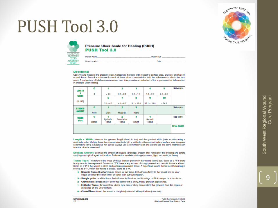
PUSH Tool 3.0

10
So
uth
West R
egio
na
l W
ound
Care
Pro
gra
m
• Evaluates 13 wound characteristics with a numerical rating scale and rates from best (1) to worst (5)
• Total score ranges from 13 (skin closed) to 65 (profound tissue degeneration) – watch total score to see if wound healing or not
• Valid and reliable tool which has evolved to include measuring and predicting wound healing6
Bates-Jensen Wound Assessment Tool (BWAT)

11
So
uth
West R
egio
na
l W
ound
Care
Pro
gra
m
BWAT Tool

12
So
uth
West R
egio
na
l W
ound
Care
Pro
gra
m
• Consists of 6 items: edges, necrotic tissue type, necrotic tissue amount, skin color surrounding wound, granulation tissue, and epithelialization
• Can be used with a wound photograph and therefore very versatile
• Valid, reliable, and responsive7-9
Photographic Wound Assessment Tool (PWAT)

13
So
uth
West R
egio
na
l W
ound
Care
Pro
gra
m
PWAT

14
So
uth
West R
egio
na
l W
ound
Care
Pro
gra
m
These forms and their instructions can be found online at:
swrwoundcareprogram.ca
• Depending on the patient’s medical diagnosis and/or medical impairments, multiple assessment forms may be required, i.e.:
• Initial Wound Assessment Screen
• Interdisciplinary Lower Leg Assessment Form
• Interdisciplinary Diabetic/Neuropathic Foot Assessment Form
• Interdisciplinary Pressure Ulcer Contributing Factors Assessment Tool
Data Collection Forms

15
So
uth
West R
egio
na
l W
ound
Care
Pro
gra
m
ASSESSMENT OF WOUND CHARACTERISTICS

16
So
uth
West R
egio
na
l W
ound
Care
Pro
gra
m
• Wound characteristics6:
• Location
• Age of wound
• Size of wound
• Stage or depth of tissue involvement
• Undermining or tunneling
• Necrotic tissue
• Granulation tissue
• Epithelium
• Exudate
Wound Characteristics

17
So
uth
West R
egio
na
l W
ound
Care
Pro
gra
m
• Where the wound occurs on the person’s anatomy• Use anatomic terms
• If there are multiple wounds in a similar location:
• Identify with letters, i.e. wound A, B, C
• Use references such as lateral, medial, proximal, distal, etc.
• Measure as a single wound
Location

18
So
uth
West R
egio
na
l W
ound
Care
Pro
gra
m
• Location may help determine etiology, i.e.:
• Venous ulcers:
• Above ankle
• Medial lower leg
• Arterial ulcers:
• Lower leg dorsum
• Foot, lateral border of foot, toe joints, over boney prominences
• Malleolus
• Neuropathic ulcers:
• Plantar surface of foot and heel
• Metatarsal heads
• Lateral border of foot and mid-foot deformities
Wound Location

19
So
uth
West R
egio
na
l W
ound
Care
Pro
gra
m
• Describe in terms of days, weeks, months, years
• Wound duration will help guide treatment
• Age of the wound is the highest predictor of healing potential
Wound Duration

20
So
uth
West R
egio
na
l W
ound
Care
Pro
gra
m
• Accurate, complete, uniform, and consistent wound size measurements are required to establish diagnosis, plan of care, and to evaluate
• Three components:
• Area
• Depth
• Volume
• No current gold standard for wound measurement
Size of Wound

21
So
uth
West R
egio
na
l W
ound
Care
Pro
gra
m
• Take measurements the same way each time from noted reference point on the body
• Use same units of measure and terminology for each measurement
• Have same person take measurements
• Use an assistant to record measurements
• Use a prepared form
Tips to Measure Accurately

22
So
uth
West R
egio
na
l W
ound
Care
Pro
gra
m
• A variety of methods are available to measure wound area:
• Ruler method
• Acetate tracing
• Digital tracings
• Wound photography
• Regardless of the method used, it should be consistently applied, and the results should be documented to assess progress of healing
• Wound surface area (l x w) is a geometric formula for a rectangle – can inflate the area of the wound up to 44%
Wound Area10

23
So
uth
West R
egio
na
l W
ound
Care
Pro
gra
m
• Simple, inconsistent
• Not reliable for irregular or large wounds
• Accuracy is increased by taking an average of three measurements
• Surface Area Measurement:
• The longest length
• The greatest width perpendicular to it
• Multiply length x width for surface area
Ruler Measurement10

24
So
uth
West R
egio
na
l W
ound
Care
Pro
gra
m
• Tracing of a wound shape on acetate paper – repeated tracings show changes in size/shape over time
• When tracing is made on metric graph paper, it’s called planimetry. Size is determined by counting graph squares
• Tracing can become a wound map, showing different areas of non-viable tissue and areas of undermining
Acetate Tracing10

25
So
uth
West R
egio
na
l W
ound
Care
Pro
gra
m
• Benefits:
• Permanent record of wound
• Serial photos can show progression towards healing, can be used as teaching tool
• Reliable, accurate, improves measurement consistency
• No contamination
• No damage to wound bed
• Less painful
• Disadvantages/challenges:
• Need consent
• Lighting may affect color of wound characteristics
• Difficulty measuring wounds on a curved surface
• Cost of camera
Wound Photography6

26
So
uth
West R
egio
na
l W
ound
Care
Pro
gra
m
• Use a good lighting source
• Screen private areas from the camera
• Position ruler to show relative size
• Use a string of known length and position camera from wound the same distance every time
• Use ID signs with patient ID, wound location, and date
• Use a ring flash attachment to reduce shadows
• Use an assistant to position the person and id sign
Tips for Good Photos6

27
So
uth
West R
egio
na
l W
ound
Care
Pro
gra
m
• Wound care providers must regularly re-evaluate the rate of wound surface area closure to help determine whether or not the wound is closing at an expected rate
• The precision of wound measurement and the method of calculating the rate of change can influence clinical decisions
Using Wound Measurements to Track Healing6

28
So
uth
West R
egio
na
l W
ound
Care
Pro
gra
m
Etiology % Reduction in Surface Area as a Predictor of Wound Healing
Venous Leg Ulcer11 > 28.79% at 4 weeks will close by 24 weeks
Diabetic Foot Ulcer12,13 > 50% a 4 weeks will close by 12 weeks
Pressure Ulcer14,15 > 39% after 2 weeks will close more quickly
Open Surgical Wound (average size of 10cm2)16
50% at 13 days will close by 21 days
• In general, a 20-30% reduction in surface area over a three to four week period is a reliable predictive indicator of chronic wound healing
• Specific wound closure rates based on wound etiology:
Calculating % Reduction in Wound Size Over Time6

29
So
uth
West R
egio
na
l W
ound
Care
Pro
gra
m
• Distanced from the visible skin surface to the wound bed
• Associated with extent of tissue damage
• Crude method of tracking growth of granulation tissue
• Methods:• Find deepest site and measure
• Clock method
Wound Depth6

30
So
uth
West R
egio
na
l W
ound
Care
Pro
gra
m
Indistinct, diffuse
• Normal tissues have blended into the wound bed
Attached
• Even or flush with the wound base, no sides or walls present, flat
Unattached
• Sides or walls are present; floor or base of wound is deeper than edge
Rolled under, thickened
• Soft to firm and flexible to touch
• Hyperkeratosis
• Callus like tissue formation around wound and at the edges
Wound Edges6

31
So
uth
West R
egio
na
l W
ound
Care
Pro
gra
m
Tunnel
• A separation of the fascial planes leading to sinus tracts
• Involves a small % of the wound margins
• Narrow and long, and seems to have a destination
• Measure and record depth of tunnel
Undermining
• Involves a greater % of the wound margins, with more shallow length than tunneling
• Usually involves subcutaneous tissues
• An erosion under the edge of the wound
Tunneling and Undermining6
32
So
uth
West R
egio
na
l W
ound
Care
Pro
gra
m
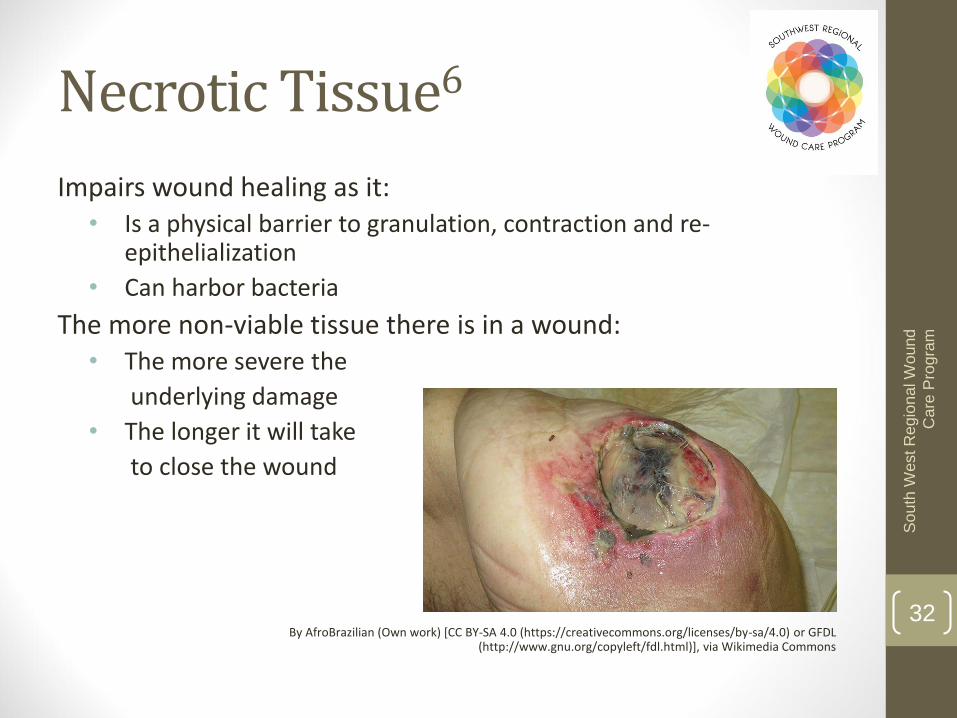
Impairs wound healing as it:• Is a physical barrier to granulation, contraction and re-
epithelialization
• Can harbor bacteria
The more non-viable tissue there is in a wound:• The more severe the
underlying damage
• The longer it will take
to close the wound
By AfroBrazilian (Own work) [CC BY-SA 4.0 (https://creativecommons.org/licenses/by-sa/4.0) or GFDL (http://www.gnu.org/copyleft/fdl.html)], via Wikimedia Commons
Necrotic Tissue6

33
So
uth
West R
egio
na
l W
ound
Care
Pro
gra
mRed• Wound bed is clean and tissue is red/pink• Goal: maintain moist wound healing environment
Yellow
• Wound bed has slough/fibrin present and tissue may be combo of red/pink + ivory/canary yellow/green (depending if infection is present)
• Not all yellow is bad – granulation grows through yellow fibrin and healthy tendon may appear as white/yellow
• Goal: maintain moist environment whilst managing excess exudate and remove slough
Black• Non-viable tissue present. Color may be dark brown/black/grey +/-
red/pink +/- canary yellow/green• Goal: remove non-viable tissue, except stable eschar on a heel
• The type of non-viable tissue present can help identify the phase of wound healing that the wound is in, and as such, can help to direct treatment options.
Red/Yellow/Black System17

34
So
uth
West R
egio
na
l W
ound
Care
Pro
gra
m
• The growth of small blood vessels and connective tissue into the wound cavity
• Healthy when bright, beefy red, shiny, and granular with a velvety appearance
• A paler appearance with spontaneous bleeding may indicate ischemia, infection, or a co-morbidity such as anemia
Granulation Tissue6

35
So
uth
West R
egio
na
l W
ound
Care
Pro
gra
m
• Process of epidermal resurfacing
• Appears as red or pink skin
• May migrate from islands on the wound surface, the wound edges, or both
Epithelial Tissue

36
So
uth
West R
egio
na
l W
ound
Care
Pro
gra
m
• Use clinical judgment to estimate the percentage of the wound covered and the tissue type in quarters
• E.g. • < 25%
• 25 50%
• > 50 < 75%
• 75 100%
By Acdx, R. S. Shaw (Own work) [Public domain], via Wikimedia Commons
Measuring Necrotic, Granulation and Epithelial Tissue Amount

37
So
uth
West R
egio
na
l W
ound
Care
Pro
gra
m
• The term given to the fluid that leaks from a wound
• Exudate characteristics are influenced by:
• Wound etiology
• Wound healing physiology
• Wound environment
• Compounding pathological factors
• Asses color, consistency, amount and odor by looking at:
• The wound itself, post wound cleansing and debridement
• The dressing
Exudate6

38
So
uth
West R
egio
na
l W
ound
Care
Pro
gra
m
ExudateDescriptor
Color and Consistency
Serous Clear/light yellow, thing/watery
Sero-sang Pink light red, thin/watery
Sang Bright red, thin/watery
Purulent Darker yellow/tan or blue/green, thin thick, watery opaque
Other Some dressings and topicals can alter the appearance of exudate, i.e. silver, cadexomer iodine, etc.
Exudate Color and Consistency6

39
So
uth
West R
egio
na
l W
ound
Care
Pro
gra
m
Descriptor Definition
None No visible exudate on the dressing or on the wound.
ScantNo measurable exudate on the dressing; however the wound tissues are moist.
Small • < 25% of the dressing has drainage on it• Wound tissues are visibly moist• Moisture is evenly distributed in the wound
Moderate• Drainage involves > 25% to < 75% of the dressing• Wound tissues are saturated• Moisture is/isn’t evenly distributed in the wound
Large
• Drainage involves > 75% of the dressing• Wound tissues are saturated• Drainage is freely expressed from the tissue• Moisture is/isn’t evenly distributed in the wound
Exudate Amount

40
So
uth
West R
egio
na
l W
ound
Care
Pro
gra
m
• Odor
• No standard terminology
• All occluded wounds have an odor
• Necrotic tissue in a wound contaminated with anaerobes may produce a foul odor
• Pseudomonas has a sickening sweet odor along with blue/green exudate
• Odor is significant when it is new or when it has changed
Exudate Odor6

41
So
uth
West R
egio
na
l W
ound
Care
Pro
gra
m
1. Thomas DR, Rodeheaver GT, Bartolucci AA, et al. Pressure Ulcer Scale for Healing: Derivation and validation of the PUSH tool. Adv Wound Care. 1997;10(5):96-101.
2. Gardner SE, Frantz RA, Bergouist S, Shin CD. A prospective study of the pressure ulcer scale for healing (PUSH). J of Gerontology. 2005;60A(1):93-97.
3. Stotts NA, Thomas DR, Frantz RA, et al. An instrument to measure healing in pressure ulcers: Development and validation of the pressure ulcer scale for healing (PUSH). J Gerontol Series A. 2001;56(12):M795-799.
4. Lee S, Kwon PME, Dorner B, et al. Pressure ulcer healing with a concentrated, fortified collagen protein hydrolysate supplement: A randomized controlled trial. Advances in Skin and Wound Care. 2006;19(2):92-96.
5. Hon J, Lagden K, McLaren AM, et al. A prospective multicenter study to validate use of the PUSH in patients with diabetic, venous, and pressure ulcers. Ostomy Wound Management. 2010;56(2):26-36
6. Sussman C, Bates-Jensen B. Wound care: A collaborative practice manual for health professionals. USA:Lippincott Williams & Wilkins;2007.
7. Houghton PE, Kincaid CB, Campbell KE, et al. Photographic assessment of the appearance of chronic pressure and leg ulcers. Ostomy Wound Management. 2000;46(4):20-30.
8. Houghton PE, Kincaid CB, Lovell M, et al. Effect of Electrical Stimulation on Chronic Leg Ulcer Size and Appearance. Physical Therapy. 2003;83(1):17-28.
9. Thawer HA, Houghton PE, Woodbury MG, et al. A Comparison of Computer-assisted and Manual Wound Size Measurement. Ostomy Wound Management . 2002;48(10
10. Keast DH, Bowering CK, Evans AW, et al. MEASURE: A proposed assessment framework for developing best practice recommendations for wound assessment. Wound Rep Reg. 2004;12:S1-S17.
11. Kantor J, Margolis DJ. A multicenter study of percentage change in venous leg ulcer area as a prognostic index of healing at 24 weeks. Br J Dermatol. 2000;142:960-964.
12. Snyder RJ, Cardinal M, Dauphinee DM, et al. A post-hoc analysis of reduction in diabetic foot ulcer size at 4 weeks as a predictor of healing by 12 weeks. Ostomy Wound Management. 2010;56(3):44-50.
13. Bolton L. Chronic wounds and delayed healing risk. Wounds. 2010;22(6):8-12. 14. Van Rijswijk L. Full-thickness pressure ulcers: Patient and wound healing characteristics. Decubitus. 1993;6:16-21. 15. Gunes UY. A prospective study evaluating the pressure ulcer scale for healing to assess stage II, stage III, and stage
IV pressure ulcers16. Ramirez AT, Soroff HS, Schwartz MS, et al. Experimental wound healing in man. Surg Gynecol Obstet. Feb.
1969;128(2):283-293. 17. Krasner D. Wound care: how to use the red-yellow-black system. Am J Nurs. 1995:95(5):44–47.
References

42
So
uth
West R
egio
na
l W
ound
Care
Pro
gra
m
• VanRijswijk L, Polansky M. Predictors of time to healing deep pressure ulcers. Ostomy Wound Management. October 1994;40(8):40-42, 44, 46-48.
• Bergstrom N, Allman RM, Alvarez OM, et al. Clinical practice guideline: Treatment of pressure ulcers. Rockville MD: US Department of Health and Human Services Public Health Service Agency for Health Care Policy and Research; 1994. 15.
• www.npuap.org• Bates-Jensen BM, Vredevoe D, Brecht ML. Validity and reliability of the pressure sore status tool. Decubitus. 1992;5(6):20-28.• Bolton L, McNees P, Van Rijswijk L, et al. Wound healing outcomes using standardized assessment and care in clinical practices. J
Wound Ostomy Continence Nursing. 2004;31(2):65-71.• Ferrell BA, Artinian BM, Sessing D. The sessing scale for assessment of pressure ulcer healing. J Am Geriatr Soc. 1995;43(1):37–40.• Ferrell BA, Keeler E, Siu AL, Ahn S-H, Osterweil D. Cost-effectiveness of low-air-loss beds for treatment of pressure ulcers. J Gerontol A
Biol Sci Med Sci. 1995;50A(3):M141–M146.• Ferrell BA. The Sessing Scale for measurement of pressure ulcer healing. Adv Wound Care. 1997;10(5):78-80.• ):46-53.• Woodbury MG, Houghton PE, Keast DH, Campbell KE. Development, Validity, Reliability and Responsiveness of a New Leg Ulcer
Measurement Tool. Advances in Skin & Wound Care 2004;17:187-196.• Woodbury MG, Houghton PE, Campbell KE, Keast DH. Leg ulcer measurement tool (LUMT): more about its ability to detect change.
Ostomy Wound Management . 2004;50(10):78.• Nachbar F SW, Merkle T, Cognetta AB, et al. The ABCD rule of dermatoscopy, High prospective value in the diagnosis of doubtful
melanocytic skin lesions. J Am Acad Dermatol. Apr 1994;30(4):551-559.• Throne N. The problem of black skin. Nursing Times; 1969”999-1001.• Weiss EL. Connective tissue in wound healing. McCulloch J KL, Feedar J, ed. Wound Healing Alternatives in Management. Second ed.
Philadelphia: FA Davis; 1995:26-28.• Makelbust J, Sieggreen M. Etiology and pathophysiology of pressure ulcers. In: Makelbust J SM, ed. Makelbust J, Sieggreen M. First
ed. West Dundee, IL:S.N. Publications; 1991:19-27.• Dorland. Dorland’s Illustrated Medical Dictionary. W.B. Saunders (Harcourt Health Services) [electronic]. Available at:
http://www.mercksource.com/pp/us/cns/cns_hl_dorlands. Accessed September 19, 2005.• Flanagan M. Improving accuracy of wound measurement in clinical practice. Ostomy/Wound Management. 2003;49(10):28-40.
References Continued