(SKILLS/HANDS-ON) Airway Margaret Baldwin, PharmD, BCPS ... · (SKILLS/HANDS-ON) Airway Margaret...
93
(SKILLS/HANDS-ON) Airway Margaret Baldwin, PharmD, BCPS Pharmacist, Intermountain Medical Center, Intermountain Healthcare Joseph R. Bledsoe, MD Medical Director, Intermountain Medical Center, Intermountain Healthcare Bradley J. Morris, RN, CFRN, PA-C Physician Assistant, Trauma Service, Intermountain Medical Center, Intermountain Healthcare Objectives: • Recognize a difficult or challenging airway • Differentiate and familiarize yourself with available algorithms, instruments, and techniques to secure an airway • Practice with direct laryngoscopy, video laryngoscopy, and a 4 step technique to a surgical airway • Discuss alternative medications used in rapid sequence intubation
Transcript of (SKILLS/HANDS-ON) Airway Margaret Baldwin, PharmD, BCPS ... · (SKILLS/HANDS-ON) Airway Margaret...

(SKILLS/HANDS-ON) Airway
Margaret Baldwin, PharmD, BCPS
Pharmacist, Intermountain Medical Center, Intermountain Healthcare
Joseph R. Bledsoe, MD
Medical Director, Intermountain Medical Center, Intermountain Healthcare
Bradley J. Morris, RN, CFRN, PA-C
Physician Assistant, Trauma Service, Intermountain Medical Center, Intermountain Healthcare
Objectives: • Recognize a difficult or challenging airway • Differentiate and familiarize yourself with available algorithms,
instruments, and techniques to secure an airway • Practice with direct laryngoscopy, video laryngoscopy, and a 4 step
technique to a surgical airway • Discuss alternative medications used in rapid sequence intubation

Airway EducationETCCC14
Brad Morris, PA‐C, RN, CFRNJoey Bledsoe, MD

Airway Priorities
1.Oxygenate2.Ventilate3.Protect Airway

Airway Management
• Spontaneous ventilation• Assisted mask/bag ventilation• Controlled mask/bag ventilation• Intubation + controlled ventilation• Surgical airway + controlled
ventilation
Use the least aggressive means necessary for airway management

Indications for Intubation• Insufficient Oxygenation• Insufficient Ventilation• Loss of airway protection• Impending airway problems (CNS,
Trauma)

Preparation
• Oxygen• Ambu bag with mask• Suction• Laryngoscope (working)• different size ETT • Suction• Plan B (Adjuncts)

Prevention of Failure• Assess situation• Decision for specific airway management• Communicate• Plan B• Reassess (change plan, if needed)

Prevention of Failure
Do not mess with a perfectly fine airway.

Difficult Airway

Preparation
• Oxygen• Ambu bag with mask• Suction• Laryngoscope (working)• different size ETT • Suction• Plan B

Tools


Glidescope

Glidescope

Endotracheal Intubation Depends Upon Manipulation of:
• Cervical spine• Atlanto‐occipital Joint• Mandible• Oral soft tissues• Neck hyoid bone
• Additionally:– Dentition– Pathology ‐ Acquired and
Congenital

The Normal Airway
• History of one or more easy intubations w/o sequelae
• Normal appearing face with regular features• Normal clear voice• Absence of scars, burns, swelling, infections, tumour, or hematoma
• No history of radiation of the head or neck• Ability to lie supine asymptomatically; no history of snoring or sleep apnea

The Normal Airway• Patent nares• Ability to open mouth widely
with TMJ rotation and subluxation (3 – 4 cm or two finger breaths)
• Mallampati Class I– Patient sitting straight up,
opening mouth as wide as possible, with protruding tongue; the uvula, posterior pharyngeal wall, entire tonsillar pillars, and fauces can be seen
• At least 6 cm (3 finger breaths) from tip of mandible to thyroid notch with neck extension
• At least 9 cm from symphysis of mandible to mandible angle

• Slender supple neck w/o masses; full range of neck motion
• Larynx moveable with swallowing and manually moveable laterally (about 1.5 cm each side)
• Slender to moderate body build• Ability to extend atlanto‐occipital joint (normal extension is 35°)
The Normal Airway

Risk Factors For Difficult Intubation• El‐Canouri et al. ‐ prospective study of 10, 507 patients demonstrating difficult intubation with objective airway risk criteria– Mouth opening < 4 cm– Thyromental distance < 6 cm– Mallampati grade 3 or greater– Neck movement < 80%– Inability to advance mandible (prognathism)– Body weight > 110 kg – Positive history of difficult intubation

Signs Indicative of a Difficult Intubation
• Trauma, deformity: burns, radiation therapy, infection, swelling, hematoma of face, mouth, larynx, neck
• Stridor or air hunger• Intolerance in the supine position• Hoarseness or abnormal voice• Mandibular abnormality
– Decreased mobility or inability to open the mouth at least 3 finger breaths– Micrognathia, receding chin
• Treacher Collins, Peirre Robin, other syndromes• Less than 6 cm (3 finger breaths) from tip of the mandible to thyroid notch with
neck in full extension– < 9 cm from the angle of the jaw to symphysis– Increased anterior or posterior mandibular length

• Laryngeal Abnormalities– Fixation of larynx to other structures of neck, hyoid, or floor of mouth.
• Macroglossia• Deep, narrow, high arched oropharynx• Protruding teeth• Mallampati Class 3 and 4
Signs Indicative of a Difficult Intubation

• Neck Abnormalities– Short and thick– Decreased range of motion (arthritis, spondylitis, disk disease)– Fracture (subluxation)– Trauma
• Thoracoabdominal abnormalities– Kyphoscoliosis– Prominent chest or large breasts– Morbid obesity– Term or near term pregnancy
• Age 50 – 59• Male gender
Signs Indicative of a Difficult Intubation

• Previous Intubations• Dental problems (bridges, caps, dentures, loose teeth)• Respiratory Disease (sleep apnea, smoking, sputum, wheeze)• Arthritis (TMJ disease, ankylosing spondylitis, rheumatoid
arthritis)• Clotting abnormalities (before nasal intubation)• Congenital abnormalities• Type I DM• NPO status
Difficult Intubation ‐ History

Difficult Intubation ‐ Physical Exam
• General:– LOC, facies and body habitus, presence or absence of cyanosis, posture,
pregnancy• Facies:
– Abnormal facial features• Pierre Robin• Treacher Collins• Klippel – Feil• Apert’s syndrome• Fetal Alcohol syndrome• Acromegaly
• Nose:– For nasal intubation– Patency

Pierre Robin

Treacher Collins

• TMJ Joint – articulation and movement between the mandible and cranium
• Diseases:– Rheumatoid arthritis– Ankylosing spondylitis– Psoriatic arthritis– Degenerative join disease
• Movements: rotational and advancement of condylar head
• Normal opening of mouth 5 – 6 cm
Difficult Intubation ‐ Physical Exam

Difficult Intubation ‐ Physical Exam
• Oral Cavity – Foreign bodies
• Teeth:– Long protruding teeth can restrict access– Dental damage 25% of all anesthesia litigations– Loose teeth can aspirate– Edentulous state
• Rarely associated with difficulty visualizing airway
• Tongue:– Size and mobility

Structured Approach to Airway Management
• MOUTHSComponent Description Assessment Activities
Mandible Length and subluxation Measure hyomental distance and anterior displacement of mandible
Opening Base, symmetry, range Assess and measure mouth opening in centimetres
Uvula Visibility Assess pharyngeal structures and classify
Teeth Dentition Assess for presence of loose teeth and dental appliances
Head Flexion, extension, rotation of head/neck and cervical spine
Assess all ranges and movement
Silhouette Upper body abnormalities, both anterior and posterior
Identify potential impact on control of airway of large breasts, buffalo hump, kyphosis, etc.

Bag/Valve/Mask Ventilation• Always need to anticipate difficult mask ventilation• Langeron et al. 1502 patients reported a 5% incidence of difficult mask
ventilation• 5 independent risk factors of difficult mask ventilation:
– Beard– BMI > 26– Edentulous– Age > 55 years of age– History of snoring (obstruction)
• Two of these predictors of DMV– Sensitivity and specificity > 70%
• DMV Difficult Intubation in 30% of cases

Pre ‐ oxygenation
• Traditional:– 3 minutes of tidal volume breathing at 5 ml/kg 100% O2
• Rapid– 8 deep breaths within 60 seconds at 10 L/min
• Always ensure pulse oximetry on patient

Positioning• Optimal Position – “sniffing position”
– Flexion of the neck and extension of the antlanto‐occipital joint

Mandible and Floor of Mouth• Optimal position:
– flexing neck and extending the atlantooccipital joint

Positioning

Visualization
• Insert blade into mouth• Sweep to right side and
displace tongue to the left• Advance the blade until it lies
in the valeculla and then pull it forward and upward using firm steady pressure without rotating the wrist
• Avoid leaning on upper teeth• May need to place pressure on
cricoid to bring cords into view

Laryngoscopy Grade • Grade I ‐ 99%• Grade II ‐ 1%• Grade III ‐ 1/2000• Grade IV ‐ 1/ 10,000

Insertion
• Insert cuff to ~ 3 cm beyond cords• Tendency to advance cuff too far
– Right mainstem intubation
• Cuff Inflation– Inflate to 20 cm H2O– Listen for leak at patients mouth– Over inflation can lead to ischemia of trachea

Confirmation ETT Position• Continuous CO2 monitoring or capnometry
– Gold standard
• Must have at least 3 continuous readings without declining CO2

Other Methods to Determine Placement of ETT tube
• Auscultation• Visualization of tube through cords• Fiberoptic bronchoscopy• Pulse oximetry not improving or worsening• Movement of the chest wall• Condensation in ET tube• Negative Pressure Test• CXR

Airway Maneuvers
• BURP – Improves visualization of airway1. Posterior pressure on the larynx against cervical
vertebrae (Backward)2. Superior pressure on the larynx as far as possible
(Upward)3. Lateral pressure on the larynx to the right (Right)4. With pressure (Pressure)

Causes of Failed Intubation
• Poor positioning of the head• Tongue in the way• Pivoting laryngoscope against upper teeth• Rushing• Being overly cautious• Inadequate sedation• Inappropriate equipment• Unskilled laryngoscopist

Some Predictors of a Difficult Airway
• C‐spine immobilized trauma patient
• Protruding tongue• Short, thick neck• Prominent upper incisors
(“buckteeth”)• Receding mandible• High, arched palate• Beard or facial hair
• Dentures• Limited jaw opening• Limited cervical mobility• Upper airway conditions• Face, neck, or oral trauma• Laryngeal trauma• Airway edema or
obstruction• Morbidly obese

Additional Predictors:Medical History
• Joint disease • Acromegaly• Thyroid or major neck
surgeries• Tumors, known abnormal
structures• Genetic anomalies• Epiglottitis
• Previous problems in surgery
• Diabetes• Pregnancy• Obesity• Pain issues

Assess the Risk
• Identifying a potentially difficult airway is essential to preparing and developing a strategy for successful ETI and also preparing an alternate plan in the event of a failed ETI.

Difficult to Bag (MOANS)
• Mask Seal• Obesity or Obstruction• Age > 55• No Teeth• Stiff

Mask Seal
• Small Hands• Wrong Mask Size• Oddly Shaped Face• Bushy Beard• Blood/Vomit• Facial Trauma
MOANS

Obesity or Obstruction
• Obesity– Heavy chest– Abdominal contents inhibit movement of the diaphragm
– Increased supraglottic airway resistance– Billowing cheeks– Difficult mask seal– Quicker desaturation
MOANS

Obesity or Obstruction
• 3rd Trimester Pregnancy– Increased body mass– Quick desaturation– Increased Mallampati Score– Gravid uterus inhibits movement of the diaphragm
MOANS

Obesity or Obstruction
• Obstructions– Foreign Body– Angioedema– Abscesses– Epiglottitis– Cancer– Traumatic Disruption/Hematoma/Burns
MOANS

Age > 55
• Associated with BVM difficulty, possibly due to loss of tone in the upper airway
MOANS

No Teeth
• Face tends to “cave in”• Consider leaving dentures in for BVM and remove for intubation
MOANS

Stiff
• Refers to Poor Compliance• Reactive Airway Disease• COPD• Pulmonary Edema/Advance Pneumonia• History of Snoring/Sleep Apnea
– Also predicts a higher Mallampati score
MOANS

Difficult Laryngoscopy & Intubation
• LEMONS– Look Externally– Evaluate 3‐3‐2 –Mallampati Score–Obstruction–Neck Mobility– Scene and Situation

LOOK Externally
• Beards or facial hair• Short, fat neck• Morbidly obese patients• Facial or neck trauma• Broken teeth (can lacerate balloons)• Dentures (should be removed)• Large teeth• Protruding tongue• A narrow or abnormally shaped face
LEMONS

EVALUATE 3‐3‐2
• Bottom of Jaw/Chin to Neck > 3 fingers
• Jaw/Palate > 3 fingers wide• Mouth opens > 2 fingers wide
Any single indicator has poor specificity
LEMONS

EVALUATE 3‐3‐2
• Mouth Opens at least 3 finger widths.
• Three finger widths thyromental distance.
• Two finger widths mandibulohyoid distance.
LEMONS

EVALUATE 3‐3‐2
• Will patients mouth open wide enough to accommodate 3 fingers?
• Will 3 fingers fit between the mentum and hyoid bone?
• Will 2 fingers fit between the hyoid and thyroid notch?– If not, expect a difficult intubation
LEMONS

Mouth opens at least 3 fingers width?LEMONS
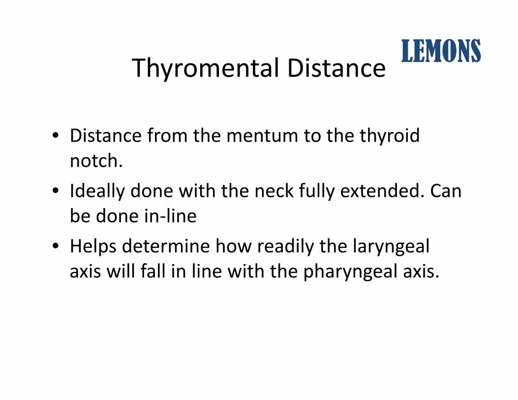
Thyromental Distance
• Distance from the mentum to the thyroid notch.
• Ideally done with the neck fully extended. Can be done in‐line
• Helps determine how readily the laryngeal axis will fall in line with the pharyngeal axis.
LEMONS

Thyromental Distance
• If the thyromental distance is short,
<3 finger widths, the laryngeal axis
makes a more acute angle with the
pharyngeal axis and it will be difficult
to achieve alignment.
• Less space to displace the tongue.
LEMONS

Thyromental Distance‐3 fingers?LEMONS

Mandibulohyoid Distance‐ 2 fingers?
• Measured from the mentum to the top of the hyoid bone.
• The epiglottis arises from the thyroid and remains dorsal to the hyoid bone.
• Therefore, the position of the hyoid bone marks the entrance to the larynx.
LEMONS

Mandibulohyoid DistanceLEMONS

Mandibulohyoid Distance
• When the position of the hyoid bone is caudal or relatively caudal, a large portion of the tongue is situated in the hypopharynx instead of the mouth.
• During laryngoscopy, this large hypopharyngeal tongue mass further compromises the compliance needed for its displacement
LEMONS

Mandibulohyoid Distance
• Patients who have a longer mandibulohyoid distance, greater then 2 finger widths, tend to be more difficult to intubate.
• A more caudal hyoid bone thus indicates a relatively caudal larynx.
LEMONS

Upper & Lower Face
• Measure the size of the upper face as compared to the lower face.
• Should be roughly the same.
• If the lower face is longer than the upper face then you should anticipate some degree of difficulty lining up the structures.
LEMONS

Upper and lower face equal?LEMONS

Upper and lower face equal?LEMONS

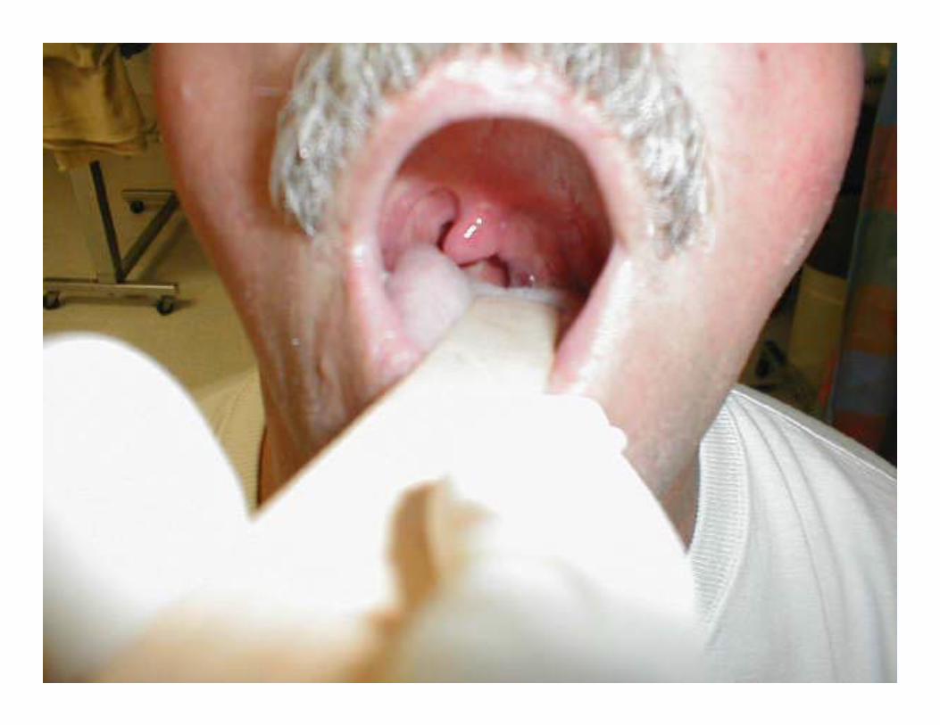
Mallampati Score LEMONS

Mallampati Score
• Have patient sit up, and stick out tongue without phonating
• May be unable to properly assess this in an emergent field situation
• Modified version is to use a laryngoscope blade like a tongue blade to visualize the oropharynx – (not as sensitive or specific)
LEMONS

Mallampati Classification
• Relates to tongue size to pharyngeal size.• Performed with patient in a sitting position, head neutral, mouth open wide and tongue protruding to the maximum.
• The Subsequent Classification is assigned based upon the pharyngeal structures visible.
LEMONS

Mallampati Classification
• Class I: Visualization of the soft palate, fauces, uvula, and anterior & posterior pillars
LEMONS

Mallampati Classification
• Class II: Visualization of the Soft palate, fauces and uvula.
LEMONS

Mallampati Classification
• Grade III: Visualization of the soft palate and the base of the uvula.
LEMONS

Mallampati Classification
• Grade IV: The soft palate is not visible at all.
LEMONS

LEMONS

Mallampati ClassificationLEMONS

• Laryngoscopy or intubation may be more difficult in the presence of an obstruction– Anatomy– Trauma– Foreign body obstruction– Edema (burns)
LEMONSObstruction

ObstructionsLaryngoscopic View Grades
Grade 1: Full aperture visibleGrade 2: Lower part of cords visibleGrade 3: Only epiglottis visibleGrade 4: Epiglottis not visible
LEMONS

ObstructionsLaryngoscopic View Grades
Graded in order from the best view to worst.• Grade 1: Visualization of the entire laryngeal
apeture
LEMONS

ObstructionsLaryngoscopic View Grades
• Grade 2: Visualization of just the posterior portion of the laryngeal aperture.
• Grade 3: Visualization of only the epiglottis
• Grade 4: Visualization of the soft palate only.
LEMONS

ObstructionsLaryngoscopic View Grades
• A severe grade III or IV view with failed endotracheal intubation occurs in 0.05‐0.35% of patients
LEMONS


Cormack & Lehane Grading
Grade I = success & ease of intubation
<1%<5%
10-30%
% listed = incidence
LEMONS

NeckMobility
• Ideally the neck should be able to extend back approximately 35°
• Problems:– Cervical Spine Immobilization– Ankylosing Spondylitis– Rheumatoid Arthritis– Halo fixation
LEMONS

“BURP” – a.k.a.“External Laryngeal Manipulation”
• Backward, Upward, Rightward Pressure: manipulation of the trachea
• 90% of the time the best view will be obtained by pressing over the thyroid cartilage
Differs from the Sellick Maneuver