SKELETON FROM PREI KHMENG, ANGKOR. Venkatesh SK I I&II I III
5
MALUNITED SUPRACONDYLAR FRACTURE OF FEMUR IN A 2000 YEAR-OLD HUMAN SKELETON FROM PREI KHMENG, ANGKOR. Venkatesh SK I , Chhem R I&II , Wong KM I , Wang SC I , Siew PY I , Pottier C III Introduction The purpose of this article is to describe the radiological investigation of a malunited supracondylar fracture of the femur in a 2000-year-old skeleton from Prei Khmeng site, Angkor, Siemreap, Cambodia. 1 Materials and Methods Skeleton The skeletal remains were unearthed in January 2001 at the excavation site of Prei Khmeng temple in Angkor region, province of Siemreap, Cambodia. The burial site had 7 human skeletons. One adult skeleton (referred from hereon as Bony I) was lying stretched on its back in an East-West direction, with head turned towards the East (Figure1). A Carbon-14 isotope dating of charcoal found in the stratum confirmed that the skeleton is approximately 2000 year-old. Despite the fragmentation of the skull and pubis, the morphometric analysis of long bones clearly indicates that the skeleton was that of an adult female. The entire skeleton almost completely recovered by its soil matrix was cut into 11 blocks and transported from Cambodia to the National University Hospital, National University of Singapore, Singapore for a scientific investigation. 33 I Osteoarchaeology Research Group, Department of Diagnostic Imaging, National University of Singapore, Singapore. II Paleoradiology Research Unit, Department of Radiology, University of Western Ontario, Canada. III École Française d' Extrême-Orient (EFEO), Siemreap, Cambodia. 1 The authors like to thank the APSARA Authority for having lent skeletal remains to our Department for a scientific investigation. Figure 1 (a, b): Bony I at the site of excavation. Note the smooth bowing of the lower end of the right femur. Close up view of the malunited fracture of the right femur (b)
Transcript of SKELETON FROM PREI KHMENG, ANGKOR. Venkatesh SK I I&II I III

MALUNITED SUPRACONDYLAR FRACTURE OF FEMUR IN A 2000 YEAR-OLD HUMANSKELETON FROM PREI KHMENG, ANGKOR.
Venkatesh SK I, Chhem R I&II, Wong KM I, Wang SC I, Siew PY I, Pottier C III
Introduction
The purpose of this article is to describe the radiological investigation of a malunited supracondylar fractureof the femur in a 2000-year-old skeleton from Prei Khmeng site, Angkor, Siemreap, Cambodia.1
Materials and Methods
SkeletonThe skeletal remains were unearthed in
January 2001 at the excavation site of PreiKhmeng temple in Angkor region, province ofSiemreap, Cambodia. The burial site had 7human skeletons. One adult skeleton (referredfrom hereon as Bony I) was lying stretched onits back in an East-West direction, with headturned towards the East (Figure1). A Carbon-14isotope dating of charcoal found in the stratumconfirmed that the skeleton is approximately2000 year-old. Despite the fragmentation of theskull and pubis, the morphometric analysis oflong bones clearly indicates that the skeleton wasthat of an adult female. The entire skeletonalmost completely recovered by its soil matrixwas cut into 11 blocks and transported fromCambodia to the National University Hospital,National University of Singapore, Singapore fora scientific investigation.
33
I Osteoarchaeology Research Group, Department of Diagnostic Imaging, National University of Singapore, Singapore.II Paleoradiology Research Unit, Department of Radiology, University of Western Ontario, Canada.III École Française d' Extrême-Orient (EFEO), Siemreap, Cambodia.1 The authors like to thank the APSARA Authority for having lent skeletal remains to our Department for a scientific investigation.
Figure 1 (a, b): Bony I at the site of excavation. Note thesmooth bowing of the lower end of the right femur. Close upview of the malunited fracture of the right femur (b)
Malunited fracture jsrc.qxp 6/6/2005 6:25 PM Page 33
ImagingEach block of Bony I were cleaned separately. The soil was carefully removed to free the bones
from the thick blocks of clay. The bones were not fully cleaned, but were cleaned enough to adequatelyx-ray the material, as presence of soil more than two or three centimeters severely hinders good x-ray imaging.Intensive cleaning results in further damage of the fragile material and dis-articulation of many of thejoints, which was avoided after the first cleaning. After the first cleaning the blocks were subjected to x-raysand CT-scanning. Following the CT scan, each block was carefully cleaned. After complete cleaning, theCT scan was performed again. Bones and bone fragments were kept in an exploded view as much aspossible, labeled separately, bagged separately, and the information entered into a database. Informationon bone fragments that conjoin with other fragments was also entered. An ongoing journal was maintainedand data entered about each step of the process for future reference.
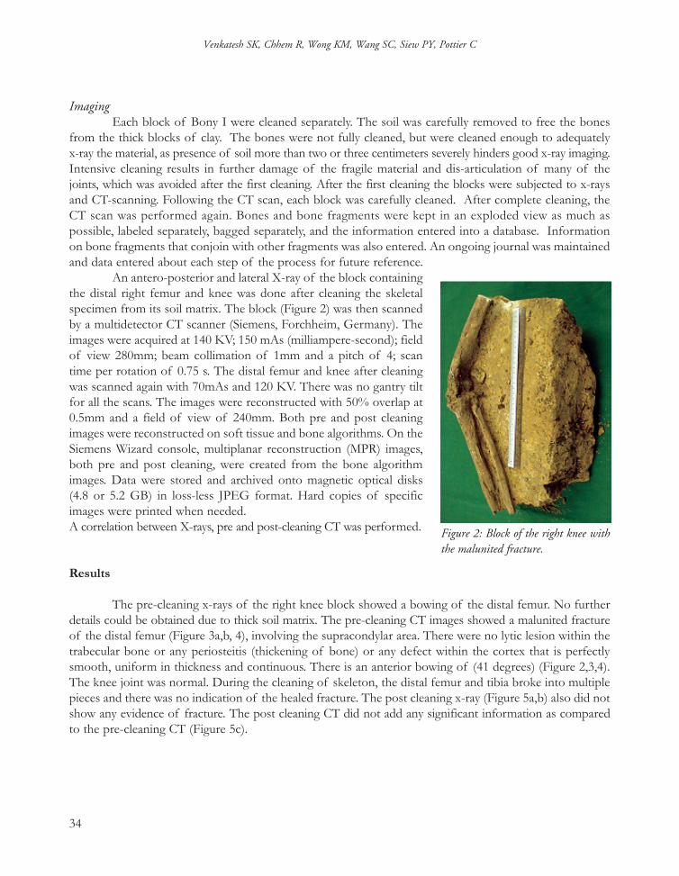
An antero-posterior and lateral X-ray of the block containingthe distal right femur and knee was done after cleaning the skeletalspecimen from its soil matrix. The block (Figure 2) was then scannedby a multidetector CT scanner (Siemens, Forchheim, Germany). Theimages were acquired at 140 KV; 150 mAs (milliampere-second); fieldof view 280mm; beam collimation of 1mm and a pitch of 4; scantime per rotation of 0.75 s. The distal femur and knee after cleaningwas scanned again with 70mAs and 120 KV. There was no gantry tiltfor all the scans. The images were reconstructed with 50% overlap at0.5mm and a field of view of 240mm. Both pre and post cleaningimages were reconstructed on soft tissue and bone algorithms. On theSiemens Wizard console, multiplanar reconstruction (MPR) images,both pre and post cleaning, were created from the bone algorithmimages. Data were stored and archived onto magnetic optical disks(4.8 or 5.2 GB) in loss-less JPEG format. Hard copies of specificimages were printed when needed.A correlation between X-rays, pre and post-cleaning CT was performed.
Results
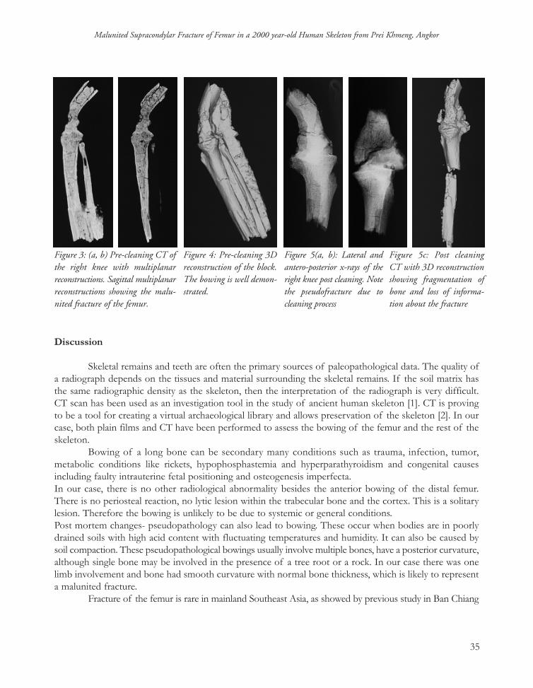
The pre-cleaning x-rays of the right knee block showed a bowing of the distal femur. No furtherdetails could be obtained due to thick soil matrix. The pre-cleaning CT images showed a malunited fractureof the distal femur (Figure 3a,b, 4), involving the supracondylar area. There were no lytic lesion within thetrabecular bone or any periosteitis (thickening of bone) or any defect within the cortex that is perfectlysmooth, uniform in thickness and continuous. There is an anterior bowing of (41 degrees) (Figure 2,3,4).The knee joint was normal. During the cleaning of skeleton, the distal femur and tibia broke into multiplepieces and there was no indication of the healed fracture. The post cleaning x-ray (Figure 5a,b) also did notshow any evidence of fracture. The post cleaning CT did not add any significant information as comparedto the pre-cleaning CT (Figure 5c).
34
Venkatesh SK, Chhem R, Wong KM, Wang SC, Siew PY, Pottier C
Figure 2: Block of the right knee withthe malunited fracture.
Malunited fracture jsrc.qxp 6/6/2005 6:25 PM Page 34
Discussion
Skeletal remains and teeth are often the primary sources of paleopathological data. The quality ofa radiograph depends on the tissues and material surrounding the skeletal remains. If the soil matrix hasthe same radiographic density as the skeleton, then the interpretation of the radiograph is very difficult.CT scan has been used as an investigation tool in the study of ancient human skeleton [1]. CT is provingto be a tool for creating a virtual archaeological library and allows preservation of the skeleton [2]. In ourcase, both plain films and CT have been performed to assess the bowing of the femur and the rest of theskeleton.
Bowing of a long bone can be secondary many conditions such as trauma, infection, tumor,metabolic conditions like rickets, hypophosphastemia and hyperparathyroidism and congenital causesincluding faulty intrauterine fetal positioning and osteogenesis imperfecta.In our case, there is no other radiological abnormality besides the anterior bowing of the distal femur.There is no periosteal reaction, no lytic lesion within the trabecular bone and the cortex. This is a solitarylesion. Therefore the bowing is unlikely to be due to systemic or general conditions.Post mortem changes- pseudopathology can also lead to bowing. These occur when bodies are in poorlydrained soils with high acid content with fluctuating temperatures and humidity. It can also be caused bysoil compaction. These pseudopathological bowings usually involve multiple bones, have a posterior curvature,although single bone may be involved in the presence of a tree root or a rock. In our case there was onelimb involvement and bone had smooth curvature with normal bone thickness, which is likely to representa malunited fracture.
Fracture of the femur is rare in mainland Southeast Asia, as showed by previous study in Ban Chiang
35
Malunited Supracondylar Fracture of Femur in a 2000 year-old Human Skeleton from Prei Khmeng, Angkor
Figure 3: (a, b) Pre-cleaning CT ofthe right knee with multiplanarreconstructions. Sagittal multiplanarreconstructions showing the malu-nited fracture of the femur.
Figure 4: Pre-cleaning 3Dreconstruction of the block.The bowing is well demon-strated.
Figure 5(a, b): Lateral andantero-posterior x-rays of theright knee post cleaning. Notethe pseudofracture due tocleaning process
Figure 5c: Post cleaningCT with 3D reconstructionshowing fragmentation ofbone and loss of informa-tion about the fracture
Malunited fracture jsrc.qxp 6/6/2005 6:25 PM Page 35

burials [3]. In a total of 136 burials, there was only one fracture femur. There were seven healed fracturesat other sites, one bone contusion, and one avulsion fracture. This last case has been found to be difficultto distinguish from an osteochondroma
The most important historical inference from this case is that people living in Angkor region 2000years ago were able to treat major fracture such as a supracondylar fracture of the femur. The bonesetterswere able to immobilize the fracture by using splint, and allow the fracture to heal. However, as expectedtraction was not known during this period, and thus explain why this fracture has healed in a wrong position.Also, the cause and mechanism of the fracture may be inferred from this case. A major trauma is necessary tocause injury to a very strong bone like the femur. This may have been caused by a fall from height or froma stilt house, or by a fight against wild animal. There was no definite evidence to determine the exact causeand mechanism of the fracture.
In conclusion, a case of supracondylar malunited fracture in a 2000 year-old human skeleton hasbeen investigated by X-rays examination and CT. CT scan was useful to make the final diagnosis of amalunion in a healed fracture and to exclude an underlying lesion, such as an infection or a tumor that mayhave led to this fracture. The use of CT with 3D reconstruction has been highlighted with particularreference to preservation of pathological data and measurement information, which may be lost during thecleaning process for anthropological analysis.
36
Venkatesh SK, Chhem R, Wong KM, Wang SC, Siew PY, Pottier C
Malunited fracture jsrc.qxp 6/6/2005 6:25 PM Page 36

References
Harwood-Nash DCF.,1979, Computed Tomography of ancient Egyptian mummies. J Comput Assist Tomogr ; 3: 768-773.
Marx, M.,1986 D'Auria SH. Three-dimensional CT reconstructions of ancient human Egyptian mummy.AJR Am J Roentgenol; 150:147-149.
Ban Chiang, 2001, a prehistoric village site in northeast Thailand. I, the human skeletal remains / Michael Pietrusewsky and Michele Toomay Douglas. Philadelphia: University of Pennsylvania Museum of Archaeology and Anthropology.
37
Malunited Supracondylar Fracture of Femur in a 2000 year-old Human Skeleton from Prei Khmeng, Angkor
Malunited fracture jsrc.qxp 6/6/2005 6:25 PM Page 37