
Growth & Development of Skeletal Muscle. Skeletal, Striated, Voluntary Muscle.
Upload
truongtuyenCategory
view
219download
3
Skeletal muscle fatigue – regulation of excitation–contraction coupling to avoid metabolic catastrophe
Brian R. MacIntosh1,*, Robert J. Holash1 and Jean-Marc Renaud2
1Human Performance Laboratory, Faculty of Kinesiology, University of Calgary, Calgary, AB T2N 1N4, Canada2Department of Cellular and Molecular Medicine, Neuromuscular Research Centre, Faculty of Medicine, University of Ottawa, Ottawa, ON K1H 8M5,Canada
*Author for correspondence ([email protected])
Journal of Cell Science 125, 2105–2114� 2012. Published by The Company of Biologists Ltddoi: 10.1242/jcs.093674
SummaryATP provides the energy in our muscles to generate force, through its use by myosin ATPases, and helps to terminate contraction bypumping Ca2+ back into the sarcoplasmic reticulum, achieved by Ca2+ ATPase. The capacity to use ATP through these mechanisms issufficiently high enough so that muscles could quickly deplete ATP. However, this potentially catastrophic depletion is avoided. It has been
proposed that ATP is preserved not only by the control of metabolic pathways providing ATP but also by the regulation of the processes thatuse ATP. Considering that contraction (i.e. myosin ATPase activity) is triggered by release of Ca2+, the use of ATP can be attenuated bydecreasing Ca2+ release within each cell. A lower level of Ca2+ release can be accomplished by control of membrane potential and by direct
regulation of the ryanodine receptor (RyR, the Ca2+ release channel in the terminal cisternae). These highly redundant control mechanismsprovide an effective means by which ATP can be preserved at the cellular level, avoiding metabolic catastrophe. This Commentary willreview some of the known mechanisms by which this regulation of Ca2+ release and contractile response is achieved, demonstrating thatskeletal muscle fatigue is a consequence of attenuation of contractile activation; a process that allows avoidance of metabolic catastrophe.
Key words: Endurance, Membrane excitability, Skeletal muscle contraction
IntroductionTypically, when exercise scientists think of control of skeletal
muscle contraction, they consider motor unit recruitment and ratecoding, the primary means by which the brain regulates the
magnitude of muscle contraction. However, the magnitude ofskeletal muscle contraction can also be regulated at the cellular
level. This level of control is necessary to avoid cellular
metabolic catastrophe, i.e. the situation in which ATP depletionreaches a level that is damaging to the fiber.
ATP is the common final source for energy in the cell, and itsconcentration [along with the concentrations of ADP and
inorganic phosphate (Pi)] affects the magnitude of energy thatis made available from its hydrolysis. At rest, skeletal muscle has
a low metabolic rate, not unlike many other passive tissues, and
the intracellular concentration of ATP is in the 5–7 mM range(Hochachka and Matheson, 1992). However, during muscle
activity, there are three major ATPases that require ATP for their
function (Box 1); during exercise, skeletal muscle can increase itsrate of ATP use by more than 100 times (Hochachka and
McClelland, 1997), a rate that is much faster than can bereplenished by aerobic metabolism. This latter point is clear from
the fact that the muscle power output can greatly exceed the level
that can be supported by aerobic metabolism (MacIntosh et al.,2000). To accommodate this additional energy requirement, ATP
is provided by non-aerobic means (through phosphocreatine
hydrolysis and glycolysis resulting in lactate formation), but thisalternative source of ATP has limited capacity (Monod and
Scherrer, 1965). For this reason, a high rate of ATP use cannot besustained in muscle and ATP use must thus be regulated
(Hochachka, 1994; Hochachka and Matheson, 1992; Myburgh,
2004). We know that muscle fibers can preserve ATP because theATP level usually decreases by only 20–25% during exercise(Argov et al., 2000; Dawson et al., 1978) and rarely falls below
50% of its initial value (Karatzaferi et al., 2001; Spriet et al.,1987).
We have proposed that the cellular regulation of ATP use is the
principal mechanism for the decreased contractile response that isassociated with acute repetitive muscle use, typically calledmuscle fatigue (MacIntosh and Shahi, 2011). We also have
recognized that regulating Ca2+ release from the sarcoplasmaticreticulum (SR), and thus the intracellular Ca2+ concentration
([Ca2+]i), dictates the rate of ATP use in the muscle cell becauseit affects Ca2+ ATPase and myosin ATPase activity, whichtogether account for 90% of ATP use (Box 1). It should be kept in
mind that a multitude of highly redundant factors contribute tothis regulation of ATP usage. We will not present all of thesehere. Instead, this Commentary concentrates on the following
three major areas: (1) regulation of the contractile proteins actinand myosin by Ca2+ and myosin light chain phosphorylation, (2)
regulation of membrane excitability through K+ and Cl2
channels, and (3) the regulation of Ca2+ availability and the SRCa2+ release channel. A summary of the process of excitation–
contraction coupling is presented in Fig. 1 and described in detailin Box 1. This figure gives the context for the remainingdiscussion in this Commentary.
Regulation of the interaction between actinand myosinMuscle contraction is the consequence of the interaction betweenactin and myosin. When the myosin head binds to actin, forming
Commentary 2105
Journ
alof
Cell
Scie
nce
a cross-bridge, it undergoes a sequence of reactions referred to as
the cross-bridge cycle (Box 2). The force of isometric contraction
is primarily a function of the number of cross-bridges that are
engaged simultaneously in the strong-binding state. This number
is governed, at a given sarcomere length, by the proportional
occupation of troponin with Ca2+ and the rate constants for the
transition to the strong-binding state, as well as the dissociation
of the cross-bridge. Troponin is a regulatory protein associated
with the thin filament. As explained in Box 1, binding of Ca2+ to
troponin allows cross-bridge cycling to occur.
When force is plotted as a function of [Ca2+], a sigmoid curve
is obtained. At very low [Ca2+], no force is generated (Fig. 2).
Resting [Ca2+] is in this range. Next, there is a range for which
increases in [Ca2+] are associated with increases in force, as
additional binding of Ca2+ to troponin reveals an increased
number of actin myosin interaction sites. This range of [Ca2+] is
referred as submaximal [Ca2+]. Finally, there is a range, for
which increases in [Ca2+] produce no further increase in force
because, in that range, all Ca2+-binding sites of troponin are
occupied; i.e. the contractile apparatus is maximally activated.
The [Ca2+], at which force is half-maximum is known as Ca50
and reflects the Ca2+ sensitivity of the contractile components. A
decrease in Ca50 is observed when the force–[Ca2+] curve is
shifted towards lower [Ca2+] and represents an increased Ca2+
sensitivity.
There are two primary mechanisms that affect Ca2+ sensitivity,
altered binding of Ca2+ to troponin and altered cross-bridge
kinetics. Cross-bridge kinetics is the more dominant of these
mechanisms and its kinetics is affected by several factors,
including: temperature, pH, sarcomere length, myosin regulatory
light chain (RLC) phosphorylation, and concentration of Pi. How
these factors affect cross-bridge kinetics and Ca2+ sensitivity has
been discussed in detail elsewhere (MacIntosh, 2003). Here, we
will discuss the mechanism for increased Ca2+ sensitivity that is
affected by myosin RLC phosphorylation.
Myosin RLC phosphorylation increasesCa2+ sensitivityThere are two myosin light chains (small peptides) associated
with each myosin head. One of these light chains, the RLC, can
be phosphorylated by myosin light chain kinase (MLCK). In
resting muscle, the RLCs of myosin are mostly unphosphorylated
(MacIntosh et al., 1993). When [Ca2+]i increases during
contraction, Ca2+ binds to another small peptide, calmodulin,
and the Ca2+–calmodulin complex subsequently binds to MLCK,
thereby activating it. Therefore, the repetitive activation of
muscle leads to a progressive phosphorylation of RLCs
(MacIntosh et al., 1993). MLCK is eventually deactivated when
[Ca2+]i decreases to resting levels and RLC is dephosphorylated
over the course of a few minutes by the slow activity of myosin
light chain phosphatase (Hartshorne et al., 1998).
When muscles are elicited to contract with a single stimulus,
force rapidly rises then falls in a contraction called a twitch. Peak
twitch force represents about 5–20% of the maximum force a
muscle can generate when stimulated at high frequency
(.150 Hz, yielding a completely fused tetanic contraction).
When a twitch is elicited immediately after a 1-second long
tetanus, the peak force is much higher than that measured before
the tetanic contraction; this phenomenon is known as post-tetanic
twitch potentiation. When muscles are stimulated at frequencies
below 10 Hz, each subsequent contraction develops more force
for the first several seconds, a phenomenon known as staircase.
During staircase and post-tetanic potentiation, the twitch peak
rate of force development increases proportionally to the increase
in active force, whereas the timecourse of the twitch is not altered
(Hainaut and Desimedt, 1968; MacIntosh and Gardiner, 1987).
Both staircase and post-tetanic twitch potentiation are thought to
be due to RLC phosphorylation (MacIntosh et al., 1993;
MacIntosh and Rassier, 2002; Moore and Stull, 1984). Skinned
fiber experiments have confirmed that RLC phosphorylation, as
achieved with addition of MLCK and calmodulin, results in
increased Ca2+ sensitivity (Persechini et al., 1985), which now
has been related to changes in cross-bridge kinetics, as explained
below.
Sweeney and Stull (Sweeney and Stull, 1990) addressed the
question of whether potentiation relies on an increased number of
engaged cross-bridges or an increased force per cross-bridge.
They used the quick longitudinal stretch of the muscle to measure
stiffness, which is assumed to be proportional to the number of
cross-bridges engaged in the strong-binding state. They reported
that longitudinal stiffness increases in proportion to the increased
active force at submaximal [Ca2+]i when myosin light chains are
phosphorylated by MLCK. This was interpreted as indicating that
potentiation is a function of increased number of cross-bridges
engaged, not an increase in force per cross-bridge. They also
reported that myosin ATPase activity increases in proportion to
the increased active force, indicating that the cycle time per
cross-bridge is not altered. This allowed them to conclude that
the increase in the number of cross-bridges is a function of an
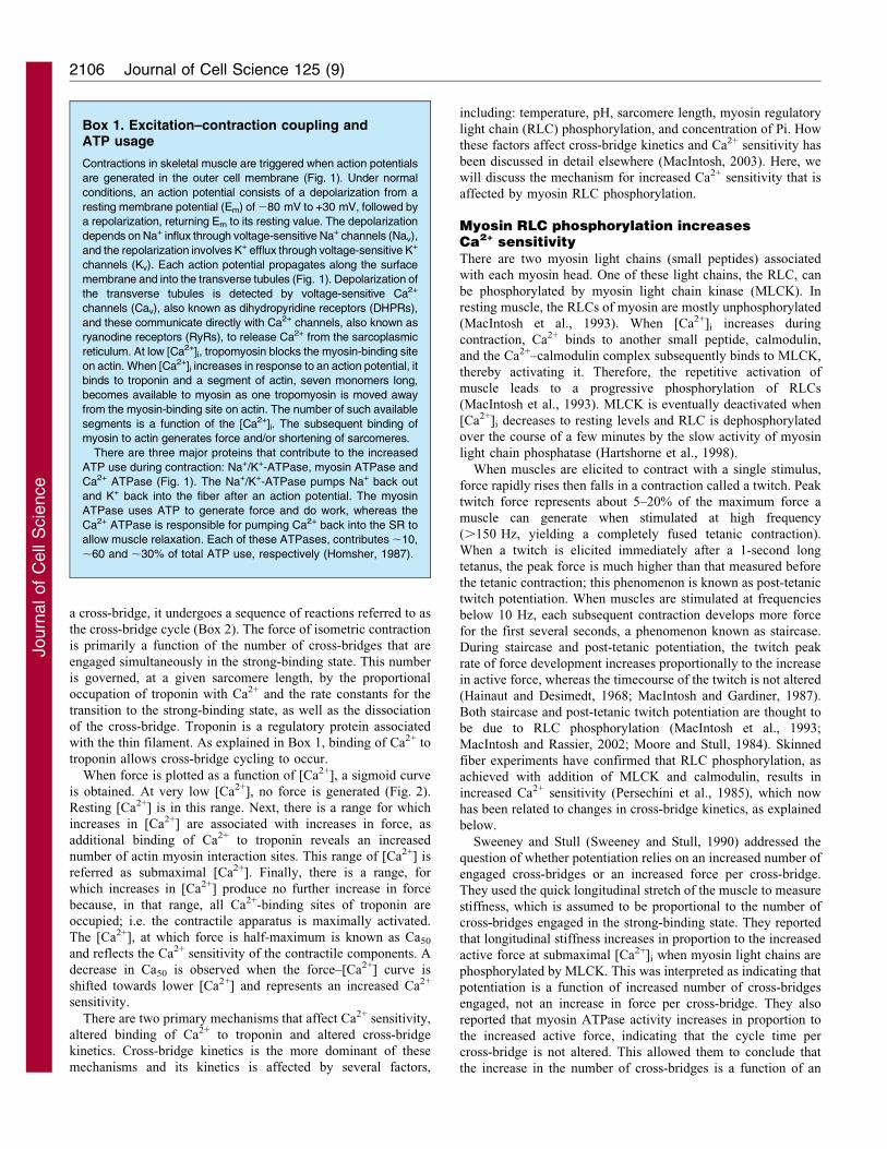
Box 1. Excitation–contraction coupling andATP usage
Contractions in skeletal muscle are triggered when action potentials
are generated in the outer cell membrane (Fig. 1). Under normal
conditions, an action potential consists of a depolarization from a
resting membrane potential (Em) of 280 mV to +30 mV, followed by
a repolarization, returning Em to its resting value. The depolarization
depends on Na+ influx through voltage-sensitive Na+ channels (Nav),
and the repolarization involves K+ efflux through voltage-sensitive K+
channels (Kv). Each action potential propagates along the surface
membrane and into the transverse tubules (Fig. 1). Depolarization of
the transverse tubules is detected by voltage-sensitive Ca2+
channels (Cav), also known as dihydropyridine receptors (DHPRs),
and these communicate directly with Ca2+ channels, also known as
ryanodine receptors (RyRs), to release Ca2+ from the sarcoplasmic
reticulum. At low [Ca2+]i, tropomyosin blocks the myosin-binding site
on actin. When [Ca2+]i increases in response to an action potential, it
binds to troponin and a segment of actin, seven monomers long,
becomes available to myosin as one tropomyosin is moved away
from the myosin-binding site on actin. The number of such available
segments is a function of the [Ca2+]i. The subsequent binding of
myosin to actin generates force and/or shortening of sarcomeres.
There are three major proteins that contribute to the increased
ATP use during contraction: Na+/K+-ATPase, myosin ATPase and
Ca2+ ATPase (Fig. 1). The Na+/K+-ATPase pumps Na+ back out
and K+ back into the fiber after an action potential. The myosin
ATPase uses ATP to generate force and do work, whereas the
Ca2+ ATPase is responsible for pumping Ca2+ back into the SR to
allow muscle relaxation. Each of these ATPases, contributes ,10,
,60 and ,30% of total ATP use, respectively (Homsher, 1987).
Journal of Cell Science 125 (9)2106
Journ
alof
Cell
Scie
nce

increased rate of engagement, not a decrease in the rate of
dissociation of cross-bridges (Sweeney and Stull, 1990).
Levine and colleagues have shown that RLC phosphorylation is
associated with a change in the way in which the myosin heads are
ordered along the thick filament backbone (Levine et al., 1996). In
the unphosphorylated state, the myosin heads are packed tightly
along the thick filament. However, phosphorylation of the RLC
results in disorder of the myosin heads. This disorder was
interpreted as an increased mobility, such that the myosin head
can swing away from the thick filament, bringing the myosin head
into close proximity to actin and, hence, increasing the probability
of its interaction with actin.
So, the regulation of [Ca2+]i during contraction and
phosphorylation of myosin light chain can have significant
impact on ATP usage. Although RLC phosphorylation is
typically associated with potentiation, this phosphorylation
persists when fatigue begins (MacIntosh et al., 1993). The
same Ca2+–calmodulin complex that activates MLCK also
inhibits Ca2+ release (Boschek et al., 2007; Xiong et al., 2006).
The muscle is capable of decreasing the release of Ca2+, while
+ +
+ +
+ +
+ +
+ +
+ +
+ +
+ +
+
+ + + + + + + + + + + +
Myosin and actin filaments
+
+ +
+ + +
+ ++ + + +
+ + + + + +
+ +
+ + +
+ + + + + + + + + + + + + + +
++
Sarcolemma
Sarcoplasm
Sarcolemma
3Na+
2K+
K+
K+
Na+
Na+
Na+
K+
ClC–
Ca2+
Ca2+
+ + + +
2Ca2+ 2Ca2+
K+ATP
3Na+
2K+
+ + + + + + + + +
Calmodulin(apoCaM andCaCaM)
DHPR II–III loop
Cytosolic side
Luminal side
S100A1
Calsequestrin
RyR
(DHPR)
(RyR)
Na+ channel
K+ channel
CIC channel
Ca2+-ATPase
Na+/K+-ATPase
K+ATP channel
Sarcoplasmic reticulum
MyosinATPase
2Ca2+
Mg�ATP ADP +Pi +Mg2+
Mg�ATP ADP +Pi +Mg2+
Mg�ATP
ADP +Pi +Mg2+
Key
T-tubule30 A
Myosin
Fig. 1. Excitation–contraction coupling and ATP usage in the cell. Here, the processes that use ATP are illustrated together with the proteins involved in
excitation–contraction coupling. ATP is used by the Na+/K+-ATPase at the surface and transverse tubule membranes, by myosin ATPase throughout the cell and
by Ca2+ ATPase in the membranes of the sarcoplasmic reticulum. The sequence of events in excitation–contraction coupling involve the following: action
potential is generated at the neuromuscular junction and propagates along the surface membrane and into the transverse tubules (gray arrows). The voltage sensor
DHPR detects the associated depolarization and opens the RyR to release Ca2+ into the sarcoplasm. Ca2+ diffuses throughout the cell and binds to several ligands,
including troponin and calmodulin. Binding of Ca2+ to troponin moves the tropomyosin away from the myosin-binding site on actin, permitting cross-bridge
cycling (Box 2). Removal of Ca2+ from the cytoplasm by Ca2+ ATPase results in the recovery of tropomyosin to its blocked position and relaxation occurs. The
detailed image of the RyR is reproduced from Song et al. (Song et al., 2011) with permission.
Regulation of excitation–contraction coupling in fatigue 2107
Journ
alof
Cell
Scie
nce

preserving its contractile function through increasing Ca2+
sensitivity. Such a mechanism helps in preserving ATP and
prolonging the duration of any muscle activity. The importance
of this process is further illustrated by the fact that muscle
activation by motor neurons is at frequencies (Hennig and Lømo,
1985) that do not give rise to maximal increases in [Ca2+]i and
full force development (Westerblad and Allen, 1991); i.e. most of
the time [Ca2+]i is submaximal.
Analysis of the force–Ca2+ relationship also suggests that the
attenuation of Ca2+ release and thus lower [Ca2+]i can also be an
effective way of decreasing the activity of myosin ATPases as
well as Ca2+ ATPases. Myosin ATPase activity decreases
because less Ca2+ will bind to troponin, and therefore there
will be a reduced level of myosin–actin interaction. A reduction
in Ca2+ ATPase activity occurs because there would be less Ca2+
to pump back into the SR. Decreased Ca2+ release can therefore
be an effective way to preserve ATP during a metabolic stress.
There is considerable evidence that a reduction in Ca2+ release
does occur during repeated muscle activation, either through
reduced Ca2+ availability (Allen et al., 2011; MacIntosh and
Kupsh, 1987) or by regulating the process of its release (Li et al.,
2002). In the next sections, we will look at how changes in
membrane excitability affect Ca2+ release. This is followed by a
discussion of the regulation of the ryanodine receptor (RyR),
which is the Ca2+ release channel of the SR.
Control of membrane excitationAs mentioned above, the release of Ca2+ is triggered by an action
potential propagating through transverse tubules (Fig. 1). The
magnitude of Ca2+ release can be modulated by the amplitude
and shape of the action potential. Shape and amplitude are
affected by both ion concentration gradients and by modulation
0
0.2
0.4
0.6
0.8
1
8 7 6 5 4
Rel
ativ
e fo
rce
pCa2+
Fig. 2. Ca2+–force relationship. The relationship between [Ca2+] (expressed
as negative log) and active force is presented here for three conditions, control
(black), increased Ca2+ sensitivity (shifted to the left, red) and decreased Ca2+
sensitivity (shifted to the right, blue). Force is primarily a function of [Ca2+],
but clearly the force at a given [Ca2+] can be modified. Ca2+ sensitivity is
altered by several factors including sarcomere length, regulatory light chain
phosphorylation, pH, Pi concentration and temperature.
Box 2. Cross-bridge cycle
In the resting state, most of the myosin heads exist in a primed state [step (ii) in the figure]; that is, with the ATP partially hydrolyzed and the myosin
head cocked, ready for the power stroke. Although it is thought that a weak binding of the myosin head to actin can occur, even when the
tropomyosin is in the ‘blocked’ position [step (i) in the figure], the binding of Ca2+ to troponin moves tropomyosin to the ‘closed’ position, thereby
increasing the probability of this weak binding and permitting the rapid transition from weak to strong binding. The existence of one myosin head in
the strong binding state causes the tropomyosin to move further from the blocked position to a position referred to as ‘open’. This is a form of
cooperativity. The rate constant for the transition to strong binding is low without Ca2+ bound to troponin, but increases with binding of Ca2+ to
troponin. On going from a weak-binding state to a strong-binding state [step (iii) in the figure], inorganic phosphate (Pi) is released from the myosin
head and a swinging lever action generates force and/or permits relative motion of the thick and thin filaments. This strong-binding state will persist
until ATP replaces the ADP that remains after the Pi is released. ATP binding permits dissociation of the myosin head from actin [step (iv) in the
figure]. It is generally accepted that each cycle as described here results in the hydrolysis of one ATP molecule (Barclay, 2003; Gordon et al., 2000).
(ii) Closed (iii) Open
Pi
PartialATP hydrolysis
Ca2+
Ca2+(ii) Blocked
(i) Closed (i) Blocked (iv) Open
Ca2+
Tropomyosin
Myosin
Actin
ADP ADP*Pi ATPKey
Myosin ATPase
Journal of Cell Science 125 (9)2108
Journ
alof
Cell
Scie
nce

of ion channel activity. During metabolic stress, it is desirable to
decrease Ca2+ release. Here, we will present the mechanisms by
which the cell modifies the amplitude and shape of the action
potential to decrease Ca2+ release. Through the discussion below,
we will see that some of the changes are meant to preserve
contractile function, but when ATP levels are threatened, the
changes result in decreased excitability, decreased Ca2+ release
and preservation of ATP.
To facilitate an understanding of the impact of ion
concentration gradients and permeabilities on the action
potential, it is worthwhile to begin with the resting condition.
The resting state of the cell and the changes in ion permeability
that occur during the action potential are presented in Box 3.
The role of K+ in membrane excitability
At rest, the normal extracellular [K+] ([K+]e) varies between 3.5
and 4.0 mM. However, this increases to 10–14 mM during
muscle activity (Juel et al., 2000; Mohr et al., 2004; Nielsen et al.,
2004; Street et al., 2005). In vitro at 37 C, maximal tetanic force
is not affected until the [K+]e reaches 10 mM; at or above that
concentration, tetanic force decreases (Cairns et al., 2011).
Twitch and submaximal tetanic forces, are potentiated when
[K+]e is increased up to 12 mM (Holmberg and Waldeck, 1980),
whereas above that concentration twitch force is depressed
(Yensen et al., 2002). The mechanism of the K+-induced
potentiation is unknown, but is no doubt beneficial for
maximizing muscle performance at the onset of exercise. The
K+-induced force depression, on the other hand, is due to a K+-
induced depolarization of the cell membrane resulting in
decreased action potential overshoot as Na+ channels are
inactivated (Yensen et al., 2002) (Fig. 3; Box 3). There is
indirect evidence that the amount of Ca2+ that is released by SR is
reduced once the action potential overshoot decreases below 5
mV; twitch force is unaffected when overshoot varies between 5
and 30 mV (Cairns et al., 2003; Yensen et al., 2002). It is also
important to note that the force–[K+]e relationship is dynamic;
i.e. the concentration at which K+ potentiates or depresses force is
modulated by several factors. Two of these factors are the
activities of the voltage-dependent chloride channel protein (ClC-
1, also known as CLC1 and CLCN1) and the ATP-sensitive K+
(KATP) channel, which are discussed in the following two
sections. Ion channel activity can influence action potential
amplitude because the amplitude of the action potential is
dependent not only on the resting membrane potential but, more
importantly, also on the exchange of ions during the action
potential through channels in addition to voltage-gated Na+ and
K+ channels (Nav and Kv, respectively), such as the Cl2 and the
KATP channels (Box 3).
Regulation and impact of the ClC-1
ClC-1 is the main channel in skeletal muscle by which Cl2
crosses the cell membrane; this channel therefore controls the
muscle membrane Cl2 permeability. In the resting state, Cl2
permeability largely exceeds that of K+ (Palade and Barchi, 1977;
Pedersen et al., 2009a; Pedersen et al., 2005; Pedersen et al.,
2009b) and plays a major role in the maintenance of resting
membrane potential (Em). For example, when [K+]e increases, the
resulting membrane depolarization is slower and smaller when
Cl2 is present in the muscle bathing solution than in its complete
absence (Dulhunty, 1978). This observation indicates that inward
movement of Cl2 counteracts the depolarizing effects of the
accumulation of K+ outside the cell.
ClC-1 can, under some conditions, directly influence the
amplitude of the action potential (Box 3). Under normal
conditions (i.e. 4 mM K+), removal of extracellular Cl2 does
not affect the kinetics of an action potential (Cairns et al., 2004),
because Na+ and K+ fluxes largely exceed that of Cl2. However,
the same is not true at high [K+]e. At 11 mM K+, ,50% of soleus
fibers are completely unexcitable and the remaining fibers
generate action potentials with peaks that only reach 210 mV
(Pedersen et al., 2005). Reducing Cl2 permeability by 50%
allows for a recovery of membrane excitability; the number of
fibers generating action potentials increases to 95% and action
potential peak reaches +10 mV. This recovery occurs because at
11 mM K+, a large number of Na+ channels are inactivated
resulting in a considerably reduced Na+ influx during the action
potential compared with that at 4 mM K+. This inactivation of
Na+ channels allows the Cl2 influx to strongly counteract the
smaller Na+ depolarizing effect, decreasing the action potential
amplitude. Lowering Cl2 permeability by closing Cl2 channels
lowers Cl2 influx, thus diminishing its ability to counteract the
Na+ depolarization and allowing for greater action potential
amplitude. Associated with the increase in membrane excitability
is an increase in force production (Pedersen et al., 2005).
Therefore, Cl2 has two opposing effects on membrane
excitability and force when the [K+]e is elevated; a complete
removal of Cl2 from the extracellular fluid exaggerates the K+-
induced membrane depolarization, loss of membrane excitability
and, subsequently, force, whereas the small decreases in Cl2
permeability that are achieved when up to 50% of the Cl2
Box 3. Action potential and its modulation by Cl2
and K+ permeability
Transmembrane movement of ions is affected by three factors, ion
permeability, concentration gradient and membrane potential (Em).
At rest, Em is about 280 mV and the cell membrane is impermeable
to Na+, somewhat permeable to K+ and most permeable to Cl2. Na+
and Cl2 are at a high concentration outside the membrane and a
low concentration inside. [K+] is high inside and low outside. The
action potential results from increased permeability to Na+ and K+,
while Cl2 permeability does not change. The depolarization phase
involves the activation of Na+ channels (Nav) and the resulting influx
brings Em to +30 mV. In normal conditions, this occurs despite a Cl2
influx due to the predominance of Nav activity. Inactivation of Nav
channels shortly after they open, and increasing activation of K+
channels (Kv) resulting in K+ efflux, and the continuing Cl2 influx
allow the repolarization of the membrane back to 280 mV.
Repolarization of the membrane reprimes Nav channels, so they
can be activated for another action potential. However, small
membrane depolarizations (i.e. less than 15 mV) at rest (e.g. an
increased [K+]e during muscle activity) inactivates a portion of the
Nav channels, decreasing their availability. As a consequence of
lower Nav channel availability there is less predominance of the Na+
inward movement during the depolarization phase. Now, any
changes in Cl2 and K+ channel activity will substantially impact
the rate of depolarization and the amplitude of action potentials. For
example, closure of Cl2 channels will decrease the Cl2 influx during
depolarization allowing for faster and greater depolarization. By
contrast, activation of Cl2 and K+ channels (to increase Cl2 influx
and K+ efflux) will slow down the depolarization and decrease the
action potential amplitude.
Regulation of excitation–contraction coupling in fatigue 2109
Journ
alof
Cell
Scie
nce
channels are closed improves membrane excitability and forcegeneration.
The modulation of the effect of K+ by regulation of ClC-1discussed above is based on studies that use resting unfatigued
muscle fibers. If the observed modulation has any physiologicalsignificance, one would expect that there would be someregulation of the Cl2 permeability by controlling the ClC-1
activity. In fact, during repeated contractions in rat extensordigitorum longus (EDL) fibers, Cl2 permeability initiallydecreases by 50% within 1 minute (Pedersen et al., 2009a). As
the stimulation is prolonged, Cl2 permeability remains constantfor 3 minutes and then suddenly increases by threefold above thepre-stimulation level, corresponding with opening of a very largenumber of ClC-1 channels. Associated with the increase in Cl2
permeability is a decrease in membrane excitability (Pedersenet al., 2009a). Thus, ClC-1 is controlled during muscle activity.
Here, we consider an exercise example to illustrate the impact
of the regulation of Cl2 channels. Interstitial [K+] reaches 10–11 mM within 5 minutes during 30-watt single leg extensionexercise (Nielsen et al., 2004). Such [K+] largely depresses
tetanic force in vitro (Cairns et al., 2011; Pedersen et al., 2003)and yet the exercise continues without difficulty for 30 minutes.Perhaps, the decreased Cl2 permeability at the onset of muscle
activity is essential in preventing any K+-induced forcedepression, while at the same time favoring a K+-induced forcepotentiation (Fig. 3). In other words, the combined increase in
[K+]e and decrease in Cl2 permeability might help to maximize
muscle performance at the onset of muscle activity. Conversely,any increase in Cl2 permeability might be linked to a fatigueprocess triggered by a metabolic stress to increase muscle
sensitivity to the K+-induced force depression (Pedersen et al.,2009b). In the latter situation, membrane excitability decreases,thereby lowering Ca2+ release and force generation (i.e. fatigue)in order to lower the activity of the Ca2+ and myosin ATPases.
The ultimate benefit is a reduced ATP demand, avoidingmetabolic catastrophe.
Regulation and impact of the KATP channel
The KATP channel is an ATP-sensitive K+ channel. The channelwas named after it was discovered that ATP binding closes thechannel (Noma, 1983). It is now established that KATP channels
are primarily activated by the changes in metabolite levels thatoccur during metabolic stress, including decreases in intracellularATP and pH, and increases in intracellular ADP and extracellular
adenosine (Barrett-Jolley et al., 1996; Davies, 1990; Noma, 1983;Vivaudou et al., 1991). The KATP channel is thus considered to bean energy sensor that is activated during metabolic stress. Beingan ion channel, it also links the excitability of the sarcolemma to
the metabolic state of the fiber. There is now clear evidence thatactivation of KATP channels during repetitive stimulationcoincides with the activation of Cl2 channels as discussed
above (Pedersen et al., 2009a), and that the channel is crucial inpreventing fiber damage and severe muscle dysfunction duringexercise and fatigue by decreasing muscle cell excitability
(Cifelli et al., 2008; Cifelli et al., 2007; Kane et al., 2004;Stoller et al., 2009; Thabet et al., 2005).
One mechanism by which the KATP channel prevents fiberdamage was revealed by studies in which channel openers were
used, which allows for a large K+ efflux during the actionpotential (Box 3) and results in a reduction in the action potentialamplitude (Gong et al., 2003; Matar et al., 2000). The resulting
decreases in membrane excitability and action potentialamplitude lead to a decrease in Ca2+ release and force (Burtonand Smith, 1997; Duty and Allen, 1995; Gong et al., 2003; Matar
et al., 2001; Weselcouch et al., 1993; Wickenden et al., 1996).The physiological benefit of a reduced Ca2+ release would bereduced activity of Ca2+ ATPases and myosin ATPases, therebypreserving ATP (Fig. 3). Indeed, decreases in ATP levels during
metabolic inhibition are faster in the presence KATP channelblockers (Gramolini and Renaud, 1997; Weselcouch et al., 1993).However, modulation of KATP channel activity during muscle
fatigue has no effect on ATP levels in mouse EDL comparedwith that in control conditions, possibly as other regulatorymechanisms prevail. Nevertheless, in soleus muscle, blocking the
KATP channel accentuates ATP loss compared with that in controlconditions, whereas channel activation prevents a reduction ofATP (Matar et al., 2000). These results therefore support the
hypothesis that KATP channels affect energy metabolism, butmore studies are necessary to fully understand how exactly theseeffects prevent fiber damage and muscle dysfunction.
A second mechanism by which KATP channels prevent fiber
damage and dysfunction is through the maintenance of restingEm. This finding came from studies in which the KATP channelactivity is abolished by either exposing normal muscles to
channel blockers or by using muscles from Kcnj11-null mice[mice in which the Kcnj11 (Kir6.2) gene that encodes the channelpore protein has been deleted]. Normally, resting Em depolarizes
Action potential
Contraction Myosin ATPase
Ca2+ release
ClC1 channel ( Cl– permeability)
ATP
RelaxationCa2+ ATPase
[K+]e
ClC1 and KATP channel ( Cl– and K+ permeability)
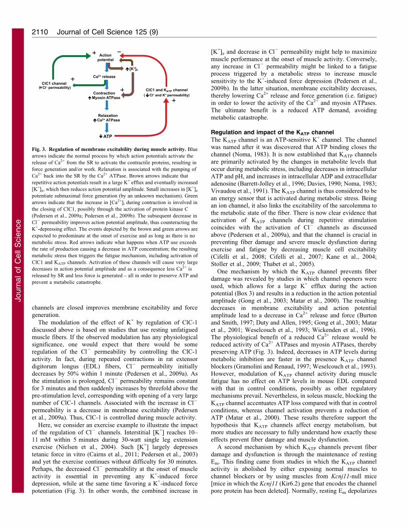
Fig. 3. Regulation of membrane excitability during muscle activity. Blue
arrows indicate the normal process by which action potentials activate the
release of Ca2+ from the SR to activate the contractile proteins, resulting in
force generation and/or work. Relaxation is associated with the pumping of
Ca2+ back into the SR by the Ca2+ ATPase. Brown arrows indicate that
repetitive action potentials result in a large K+ efflux and eventually increased
[K+]e, which then reduces action potential amplitude. Small increases in [K+]e
potentiate submaximal force generation (by an unknown mechanism). Green
arrows indicate that the increase in [Ca2+]i during contraction is involved in
the closing of ClC1, possibly through the activation of protein kinase C
(Pedersen et al., 2009a; Pedersen et al., 2009b). The subsequent decrease in
Cl2 permeability improves action potential amplitude, thus counteracting the
K+-depressing effect. The events depicted by the brown and green arrows are
expected to predominate at the onset of exercise and as long as there is no
metabolic stress. Red arrows indicate what happens when ATP use exceeds
the rate of production causing a decrease in ATP concentration; the resulting
metabolic stress then triggers the fatigue mechanism, including activation of
ClC1 and KATP channels. Activation of these channels will cause very large
decreases in action potential amplitude and as a consequence less Ca2+ is
released by SR and less force is generated – all in order to preserve ATP and
prevent a metabolic catastrophe.
Journal of Cell Science 125 (9)2110
Journ
alof
Cell
Scie
nce

at between 10 and 15 mV during repeated muscle contractions(Cifelli et al., 2008; Comtois et al., 1995; Light et al., 1994),
whereas it does not change during metabolic inhibition(Gramolini and Renaud, 1997). In the absence of KATP channelactivity, the depolarization can be as high as 50 mV under both
conditions. This depolarization has been shown to be sufficientlylarge enough to activate the Ca2+ channels in transverse tubules,leading to an uncontrolled Ca2+ influx, large increases in restingintracellular Ca2+ and in force (Cifelli et al., 2008; Cifelli et al.,
2007; Gong et al., 2000; Light et al., 1994; Matar et al., 2000). Asa consequence of the increases in resting [Ca2+], there is greateruse of ATP by the Ca2+ and myosin ATPases, which worsen the
possibility of a metabolic catastrophe. Furthermore, chronicelevation of [Ca2+]i is a well known factor that causes fiberdamage (Jackson et al., 1984; Jones et al., 1984).
Thus, the regulation of membrane excitability is complex anddepends not only on changes in extra- and intra-cellular ionconcentrations, such as that of K+, but also on changes in ion
channel activity. At the onset of exercise, [K+]e increases rapidlyand is likely to potentiate force generation similar to that upon anincreased RLC phosphorylation. At the same time, the activity of
ClC-1 decreases, possibly to prevent any K+-induced forcedepression. When metabolic stress occurs, the activity of ClC-1increases and this results in a depression of membraneexcitability, which is further enhanced by a concomitant
activation of KATP channels. As membrane excitability isreduced by these two channels, less Ca2+ is released and lessforce is generated. The outcome of these mechanisms is the
preservation of the limited supply of ATP through reducing itsuse by the Ca2+ and myosin ATPases.
Control of Ca2+ releaseAction potentials on the membrane are translated into a release of
Ca2+ in a process referred to as excitation–contraction coupling(Box 1). The dihydropyridine receptors (DHPRs) are specializedchannels that detect the action potential in the transverse tubulemembrane (Fig. 1) and, through a direct connection (in skeletal
muscle), open and close the Ca2+ release channels in the SR, theRyRs. The release of Ca2+ into the myofibril initiates musclecontraction and, as a result, increases ATP demand through the
activity of the myosin and Ca2+ ATPases. Although an actionpotential is the main controlling factor detected by the DHPR, andtherefore the main determinant that results in opening and closing
of the RyR, this ion channel is further regulated by a large numberof ionic and protein ligands that influence the opening probability(Po). We will examine further a small well-known collection of
these ligands and how they prevent ATP depletion. It is also knownthat reactive oxygen species can modulate Ca2+ release through theRyR (Oba et al., 2002), but this will not be discussed further here.
Regulation of RyR by ATP, Ca2+ and Mg2+
The three most studied regulatory ligands of the RyR are ATP,
Ca2+ and Mg 2+ (Lamb and Stephenson, 1992; Laver et al., 1997;Meissner, 1984; Meissner et al., 1986). There are two distinctsites on RyR for these ligands that either enhance or reduce its
Po. These sites are referred to as the high-affinity A-site,regulating activation, and the low affinity I-site, leading toinhibition (Lamb, 2000). Both Ca2+ and ATP bind to the A-site to
increase the Po of RyR (Meissner, 1986), whereas both Ca2+ andMg2+ bind to the lower affinity I-site to reduce its Po (Laver et al.,1997; Meissner et al., 1986). In the absence of Mg2+, Ca2+ opens
RyR by binding to the A-site if it is present at a concentration of1 mM and closes the receptor through binding at the I-site at high
concentrations of 1 mM (Meissner et al., 1986). Under normalresting conditions with [Mg2+] being present at 1 mM, the I-siteis occupied with Mg2+ and the subsequent inhibition of the
channel overrides any activating effect of Ca2+ and ATP. Mg2+
also competes with Ca2+ for binding to the A-site, but this doesnot result in an activation of the channel (Laver et al., 1997). Thestrong activating effect of ATP is most clearly observed when
Mg2+ is removed from the solution (Lamb and Stephenson,1991). ATP, in the absence of external Ca2+, by itself is capableof fully activating RyR and initiating Ca2+ release (Meissner
et al., 1986). This result further emphasizes how important theinhibitory effect of Mg2+ at rest is.
Considering the effects of Mg2+ described above, it would
appear that the RyR would never be activated! However, with eachaction potential, some RyRs are activated and Ca2+ is released intothe cell. The fact that Mg2+ reduces the Po of RyR becomes more
important when the concentration of ATP decreases, because asshown in Fig. 1, a net ATP hydrolysis results in an increase in[Mg2+]. This occurs because Mg2+ has a lower affinity for ADP
than for ATP. This increase in [Mg2+] and decrease in theconcentration of ATP combine to cause a decrease in Po,attenuating Ca2+ release in subsequent activations.
Several studies have examined the effect of higher [Mg2+] onexcitation–contraction coupling (Blazev and Lamb, 1999; Lamband Stephenson, 1991; Laver et al., 1997; Owen et al., 1997). The
general consensus of these studies is that [Mg2+] at rest is a stronginhibitor of RyR, which keeps the channel closed and unresponsiveto the strong activating effect of ATP and Ca2+ until an actionpotential activates the cell. Ca2+-induced activation of RyR, a key
mechanism in cardiac muscle, is apparently not relevant in skeletalmuscle (Endo, 2009). At 3 mM [Mg2+], a concentration that can bereached during fatiguing contractions (Westerblad and Allen,
1992), the magnitude of Ca2+ release achieved by transverse tubuledepolarization is reduced (Blazev and Lamb, 1999; Laver et al.,1997; Owen et al., 1996). Thus, inhibition by Mg2+ is important to
prevent spontaneous Ca2+ release at rest and it provides anothermechanism, by which less Ca2+ is released during metabolic stress.
The reduced activation of RyR, which results from low
concentrations of ATP, has also been explored. When theconcentration of ATP is held artificially low (,0.5 mM ATP),Ca2+ release is significantly reduced when RyR is directly
activated by DHPR (Blazev and Lamb, 1999; Owen et al., 1996).Although this ATP decrease might appear extreme, it has beenreasoned that a localized decrease in the concentration of ATP to
below 1 mM could occur in the vicinity of RyR (Owen et al.,1996). Therefore, both the concomitant decrease in ATPconcentration and increase in [Mg2+] upon an impendingmetabolic catastrophe can contribute to a decrease in Ca2+
release by lowering RyR Po to eventually preserve ATP. Thesensitivity of the RyR to changes in [Mg2+] and ATPconcentration, can also make it an energy sensor, in a similar
manner to the KATP channel.
Regulation of RyR by calmodulin and the S100 Ca2+-binding protein A1 (S100A1)
There are several additional ligands that interact with RyR. Here
we will discuss two that have known regulatory impact on Po ofthe RyR, calmodulin and S100 Ca2+-binding protein A1(S100A1).
Regulation of excitation–contraction coupling in fatigue 2111
Journ
alof
Cell
Scie
nce

The small ubiquitous peptide calmodulin (CaM) was one of thefirst ligand regulators of RyR to be discovered (Chen and
MacLennan, 1994; Hamilton et al., 2000; Yang et al., 1994). Atrest, when [Ca2+] is low, calmodulin exists in the apocalmodulin(apoCaM) form, and upon increase in [Ca2+]i binds to Ca2+,becoming Ca2+-bound (CaCaM), and resulting in a change of its
shape and its effect as a ligand (Boschek et al., 2007; Boscheket al., 2008; Xiong et al., 2006). At low [Ca2+]i (up to 0.1 mM),the Ca2+-free apoCaM binds to RyR and increases its Po.
Conversely, as [Ca2+]i increases, the amount of the CaCaMcomplex also increases, which binds to RyR but decreases its Po
(Boschek et al., 2007; Xiong et al., 2006).
RyR has many distinct sites for ligand binding (Box 1) (Songet al., 2011), although it will probably be many years before wefully understand the range of ligands for RyR and their functions.Recent work has provided some insight. One of the important
ligands for RyR is S100A1. Prosser and colleagues used aknockout mouse (S100A1) to demonstrate that S100A1 enhancesCa2+ release (Prosser et al., 2008). Twitch and tetanic contractile
amplitudes are attenuated, and the corresponding Ca2+ transientsare smaller in the S100A1 mice compared with muscles of wild-type control mice. Yamaguchi et al. contributed to our
understanding of ligand control of RyR by using a mouse withan altered RyR CaCaM-binding site (RyRD/D) (Yamaguchi et al.,2011). Similar to the S100A1 mice, the RyRD/D mice produce
lower twitch forces, suggesting that they lack the S100A1-binding site. However, Ca2+ release during a maximal tetaniccontraction is enhanced and there is a greater peak [Ca2+]i
compared with that in muscles of wild-type mice (Yamaguchi
et al., 2011). On the basis of these results, the authors suggest thatthere is one site on the RyR that binds both CaCaM and S100A1,and that with S100A1 bound, this site is important for promoting
Ca2+ release at low [Ca2+], such as occurs during a twitch. Thesame site is also important for inhibiting Ca2+ release duringrepeated or sustained activation by binding CaCaM at higher
[Ca2+]i. In this way, Ca2+ is able to slow energy expenditure laterin contraction. It is interesting to note that CaCaM activatesMLCK and inhibits Ca2+ release. These two processes will haveopposing effects on muscle, enhancing contraction by promoting
RLC phosphorylation and attenuating contraction by decreasingCa2+ release.
ConclusionsThis Commentary has aimed to illustrate, with a few examples,how skeletal muscle contraction is regulated at the cellular level.
Although most researchers concentrate on the depression ofcontraction during fatigue, we point out here that there areregulatory processes that can enhance muscle performance. Thefirst example is that of myosin RLC phosphorylation, which
allows for increased force production at a given [Ca2+]i. Thiseffect permits a decrease in Ca2+ release without any loss of forceor power, but with the advantage of a reduced Ca2+ ATPase
activity, as less Ca2+ needs to be pumped back into the SR.Overall, this effect might be important in reducing ATP usageand prolonging the muscle activity. The second example is that of
K+-induced force potentiation that occurs when [K+]e increases.The underlying mechanism of this potentiation is still unknown.Finally, a third mechanism that helps maintain a high muscle
performance at the onset of exercise is the decrease in Cl2
channel activity; this effect is important to counteract the likelydepression of force that would otherwise be the result of
increasing [K+]e. We also discuss some mechanisms thatprevent a metabolic catastrophe when energy demand exceedsthe capability to replenish ATP. This regulation is highlyredundant and appears to concentrate on diminishing Ca2+
release, because this simultaneously reduces the activity ofboth Ca2+ ATPase and myosin ATPase and serves to preserveATP. This regulation occurs at the level of membrane
excitability, where the activation of both Cl2 and KATP
channels reduces excitability, thereby reducing the Ca2+ releaseby RyR. There is also regulation at the level of the RyR, which
responds to a decrease in [ATP] and increase in [Mg2+], whichoccur during repetitive or sustained contractions. In this case,both of these changes reduce the Po of RyR. RyR Po is also
regulated by Ca2+, either directly or through CaM as sustainedelevation of [Ca2+]i results in an inhibition of RyR by the CaCaMcomplex. Collectively, these regulatory processes allow exerciseto continue, as long as additional motor units are available for
recruitment, while preventing depletion of ATP in individualcells.
AcknowledgementsThe authors would like to thank Barbara Holash for creating Fig. 1.Research in the laboratories of the authors is supported by theNatural Sciences and Engineering Research Council of Canada.
ReferencesAllen, D. G., Clugston, E., Petersen, Y., Roder, I. V., Chapman, B. and Rudolf, R.
(2011). Interactions between intracellular calcium and phosphate in intact mousemuscle during fatigue. J. Appl. Physiol. 111, 358-366.
Argov, Z., Lofberg, M. and Arnold, D. L. (2000). Insights into muscle diseases gainedby phosphorus magnetic resonance spectroscopy. Muscle Nerve 23, 1316-1334.
Barclay, C. J. (2003). Models in which many cross-bridges attach simultaneously canexplain the filament movement per ATP split during muscle contraction. Int. J. Biol.
Macromol. 32, 139-147.
Barrett-Jolley, R., Comtois, A., Davies, N. W., Stanfield, P. R. and Standen, N. B.(1996). Effect of adenosine and intracellular GTP on KATP channels of mammalianskeletal muscle. J. Membr. Biol. 152, 111-116.
Blazev, R. and Lamb, G. D. (1999). Low [ATP] and elevated [Mg2+] reducedepolarization-induced Ca2+ release in rat skinned skeletal muscle fibres. J. Physiol.
520, 203-215.
Boschek, C. B., Jones, T. E., Squier, T. C. and Bigelow, D. J. (2007). Calciumoccupancy of N-terminal sites within calmodulin induces inhibition of the ryanodinereceptor calcium release channel. Biochemistry 46, 10621-10628.
Boschek, C. B., Sun, H., Bigelow, D. J. and Squier, T. C. (2008). Differentconformational switches underlie the calmodulin-dependent modulation of calciumpumps and channels. Biochemistry 47, 1640-1651.
Burton, F. L. and Smith, G. L. (1997). The effect of cromakalim on intracellular[Ca2+] in isolated rat skeletal muscle during fatigue and metabolic blockade. Exp.
Physiol. 82, 469-483.
Cairns, S. P., Buller, S. J., Loiselle, D. S. and Renaud, J. M. (2003). Changes of actionpotentials and force at lowered [Na+]o in mouse skeletal muscle: implications forfatigue. Am. J. Physiol. Cell Physiol. 285, C1131-C1141.
Cairns, S. P., Ruzhynsky, V. and Renaud, J. M. (2004). Protective role of extracellularchloride in fatigue of isolated mammalian skeletal muscle. Am. J. Physiol. Cell
Physiol. 287, C762-C770.
Cairns, S. P., Leader, J. P. and Loiselle, D. S. (2011). Exacerbated potassium-inducedparalysis of mouse soleus muscle at 37 C vis-a-vis 25 C: implications for fatigue. K+-induced paralysis at 37 C. Pflugers Arch. 461, 469-479.
Chen, S. R. and MacLennan, D. H. (1994). Identification of calmodulin-, Ca(2+)-, andruthenium red-binding domains in the Ca2+ release channel (ryanodine receptor) ofrabbit skeletal muscle sarcoplasmic reticulum. J. Biol. Chem. 269, 22698-22704.
Cifelli, C., Bourassa, F., Gariepy, L., Banas, K., Benkhalti, M. and Renaud, J. M.
(2007). KATP channel deficiency in mouse flexor digitorum brevis causes fibredamage and impairs Ca2+ release and force development during fatigue in vitro.J. Physiol. 582, 843-857.
Cifelli, C., Boudreault, L., Gong, B., Bercier, J. P. and Renaud, J. M. (2008).Contractile dysfunctions in ATP-dependent K+ channel-deficient mouse muscleduring fatigue involve excessive depolarization and Ca2+ influx through L-type Ca2+channels. Exp. Physiol. 93, 1126-1138.
Comtois, A., Light, P. and Renaud, J. M. (1995). Effect of tolbutamide on the rate offatigue and recovery in frog sartorius muscle. J. Pharmacol. Exp. Ther. 274, 1061-1066.
Davies, N. W. (1990). Modulation of ATP-sensitive K+ channels in skeletal muscle byintracellular protons. Nature 343, 375-377.
Journal of Cell Science 125 (9)2112
Journ
alof
Cell
Scie
nce
Dawson, M. J., Gadian, D. G. and Wilkie, D. R. (1978). Muscular fatigue investigatedby phosphorus nuclear magnetic resonance. Nature 274, 861-866.
Dulhunty, A. F. (1978). The dependence of membrane potential on extracellularchloride concentration in mammalian skeletal muscle fibres. J. Physiol. 276, 67-82.
Duty, S. and Allen, D. G. (1995). The effects of glibenclamide on tetanic force andintracellular calcium in normal and fatigued mouse skeletal muscle. Exp. Physiol. 80,529-541.
Endo, M. (2009). Calcium-induced calcium release in skeletal muscle. Physiol. Rev. 89,1153-1176.
Gong, B., Miki, T., Seino, S. and Renaud, J. M. (2000). A K(ATP) channel deficiencyaffects resting tension, not contractile force, during fatigue in skeletal muscle. Am. J.
Physiol. Cell Physiol. 279, C1351-C1358.
Gong, B., Legault, D., Miki, T., Seino, S. and Renaud, J. M. (2003). KATP channelsdepress force by reducing action potential amplitude in mouse EDL and soleusmuscle. Am. J. Physiol. Cell Physiol. 285, C1464-C1474.
Gordon, A. M., Homsher, E. and Regnier, M. (2000). Regulation of contraction instriated muscle. Physiol. Rev. 80, 853-924.
Gramolini, A. and Renaud, J. M. (1997). Blocking ATP-sensitive K+ channel duringmetabolic inhibition impairs muscle contractility. Am. J. Physiol. 272, C1936-C1946.
Hainaut, K. and Desimedt, J. E. (1968). Staircase potentiation in normal humanmuscle: instantaneous relation between force and speed of the twitch. Arch. Int.
Physiol. Biochim. 76, 554-556.
Hamilton, S. L., Serysheva, I. and Strasburg, G. M. (2000). Calmodulin andexcitation-contraction coupling. News Physiol. Sci. 15, 281-284.
Hartshorne, D. J., Ito, M. and Erdodi, F. (1998). Myosin light chain phosphatase:subunit composition, interactions and regulation. J. Muscle Res. Cell Motil. 19, 325-341.
Hennig, R. and Lømo, T. (1985). Firing patterns of motor units in normal rats. Nature
314, 164-166.
Hochachka, P. W. (1994). Muscles As Molecular And Metabolic Machines. London,UK: Informa Healthcare.
Hochachka, P. W. and Matheson, G. O. (1992). Regulating ATP turnover rates overbroad dynamic work ranges in skeletal muscles. J. Appl. Physiol. 73, 1697-1703.
Hochachka, P. W. and McClelland, G. B. (1997). Cellular metabolic homeostasisduring large-scale change in ATP turnover rates in muscles. J. Exp. Biol. 200, 381-386.
Holmberg, E. and Waldeck, B. (1980). On the possible role of potassium ions in theaction of terbutaline on skeletal muscle contractions. Acta Pharmacol. Toxicol.
(Copenh.) 46, 141-149.
Homsher, E. (1987). Muscle enthalpy production and its relationship to actomyosinATPase. Annu. Rev. Physiol. 49, 673-690.
Jackson, M. J., Jones, D. A. and Edwards, R. H. (1984). Experimental skeletal muscledamage: the nature of the calcium-activated degenerative processes. Eur. J. Clin.
Invest. 14, 369-374.
Jones, D. A., Jackson, M. J., McPhail, G. and Edwards, R. H. (1984). Experimentalmouse muscle damage: the importance of external calcium. Clin. Sci. 66, 317-322.
Juel, C., Pilegaard, H., Nielsen, J. J. and Bangsbo, J. (2000). Interstitial K(+) inhuman skeletal muscle during and after dynamic graded exercise determined bymicrodialysis. Am. J. Physiol. Regul. Integr. Comp. Physiol. 278, R400-R406.
Kane, G. C., Behfar, A., Yamada, S., Perez-Terzic, C., O’Cochlain, F., Reyes, S.,
Dzeja, P. P., Miki, T., Seino, S. and Terzic, A. (2004). ATP-sensitive K+ channelknockout compromises the metabolic benefit of exercise training, resulting in cardiacdeficits. Diabetes 53 Suppl 3:, S169-S175.
Karatzaferi, C., de Haan, A., Ferguson, R. A., van Mechelen, W. and Sargeant, A. J.
(2001). Phosphocreatine and ATP content in human single muscle fibres before andafter maximum dynamic exercise. Pflugers Arch. 442, 467-474.
Lamb, G. D. (2000). Excitation-contraction coupling in skeletal muscle: comparisonswith cardiac muscle. Clin. Exp. Pharmacol. Physiol. 27, 216-224.
Lamb, G. D. and Stephenson, D. G. (1991). Effect of Mg2+ on the control of Ca2+release in skeletal muscle fibres of the toad. J. Physiol. 434, 507-528.
Lamb, G. D. and Stephenson, D. G. (1992). Importance of Mg2+ in excitation-contraction coupling in skeletal muscle. News Physiol. Sci. 7, 270-274.
Laver, D. R., Baynes, T. M. and Dulhunty, A. F. (1997). Magnesium inhibition ofryanodine-receptor calcium channels: evidence for two independent mechanisms.J. Membr. Biol. 156, 213-229.
Levine, R. J. C., Kensler, R. W., Yang, Z. H., Stull, J. T. and Sweeney, H. L. (1996).Myosin light chain phosphorylation affects the structure of rabbit skeletal musclethick filaments. Biophys. J. 71, 898-907.
Li, J. L., Wang, X. N., Fraser, S. F., Carey, M. F., Wrigley, T. V. and McKenna,
M. J. (2002). Effects of fatigue and training on sarcoplasmic reticulum Ca(2+)regulation in human skeletal muscle. J. Appl. Physiol. 92, 912-922.
Light, P. E., Comtois, A. S. and Renaud, J. M. (1994). The effect of glibenclamide onfrog skeletal muscle: evidence for K+ATP channel activation during fatigue.J. Physiol. 475, 495-507.
MacIntosh, B. R. (2003). Role of calcium sensitivity modulation in skeletal muscleperformance. News Physiol. Sci. 18, 222-225.
MacIntosh, B. R. and Gardiner, P. F. (1987). Posttetanic potentiation and skeletalmuscle fatigue: interactions with caffeine. Can. J. Physiol. Pharmacol. 65, 260-268.
MacIntosh, B. R. and Kupsh, C. C. (1987). Staircase, fatigue, and caffeine in skeletalmuscle in situ. Muscle Nerve 10, 717-722.
MacIntosh, B. R. and Rassier, D. E. (2002). What is fatigue? Can. J. Appl. Physiol. 27,42-55.
MacIntosh, B. R. and Shahi, M. R. S. (2011). A peripheral governor regulates musclecontraction. Appl. Physiol. Nutr. Metab. 36, 1-11.
MacIntosh, B. R., Grange, R. W., Cory, C. R. and Houston, M. E. (1993). Myosinlight chain phosphorylation during staircase in fatigued skeletal muscle. Pflugers
Arch. 425, 9-15.
MacIntosh, B. R., Neptune, R. R. and Van den Bogert, A. J. (2000). Intensity ofcycling and cycle ergometry: power output and energy cost. In Biomechanics And
Biology Of Movement (ed. B. M. Nigg, B. R. MacIntosh and J. Mester), pp. 129-148.Champaign, IL: Human Kinetics.
Matar, W., Nosek, T. M., Wong, D. and Renaud, J. M. (2000). Pinacidil suppressescontractility and preserves energy but glibenclamide has no effect during musclefatigue. Am. J. Physiol. Cell Physiol. 278, C404-C416.
Matar, W., Lunde, J. A., Jasmin, B. J. and Renaud, J. M. (2001). Denervationenhances the physiological effects of the K(ATP) channel during fatigue in EDL andsoleus muscle. Am. J. Physiol. Regul. Integr. Comp. Physiol. 281, R56-R65.
Meissner, G. (1984). Adenine nucleotide stimulation of Ca2+-induced Ca2+ release insarcoplasmic reticulum. J. Biol. Chem. 259, 2365-2374.
Meissner, G. (1986). Ryanodine activation and inhibition of the Ca2+ release channel ofsarcoplasmic reticulum. J. Biol. Chem. 261, 6300-6306.
Meissner, G., Darling, E. and Eveleth, J. (1986). Kinetics of rapid calcium release bysarcoplasmic reticulum. Effects of calcium, magnesium, and adenine nucleotides.Biochemistry 25, 236-244.
Mohr, M., Nordsborg, N., Nielsen, J. J., Pedersen, L. D., Fischer, C., Krustrup, P.
and Bangsbo, J. (2004). Potassium kinetics in human muscle interstitium duringrepeated intense exercise in relation to fatigue. Pflugers Arch. 448, 452-456.
Monod, H. and Scherrer, J. (1965). The work capacity of a synergistic muscular group.Ergonomics 8, 329-338.
Moore, R. L. and Stull, J. T. (1984). Myosin light chain phosphorylation in fast andslow skeletal muscles in situ. Am. J. Physiol. 247, C462-C471.
Myburgh, K. H. (2004). Can any metabolites partially alleviate fatigue manifestationsat the cross-bridge? Med. Sci. Sports Exerc. 36, 20-27.
Nielsen, J. J., Mohr, M., Klarskov, C., Kristensen, M., Krustrup, P., Juel, C. and
Bangsbo, J. (2004). Effects of high-intensity intermittent training on potassiumkinetics and performance in human skeletal muscle. J. Physiol. 554, 857-870.
Noma, A. (1983). ATP-regulated K+ channels in cardiac muscle. Nature 305, 147-148.
Oba, T., Murayama, T. and Ogawa, Y. (2002). Redox states of type 1 ryanodinereceptor alter Ca(2+) release channel response to modulators. Am. J. Physiol. Cell
Physiol. 282, C684-C692.
Owen, V. J., Lamb, G. D. and Stephenson, D. G. (1996). Effect of low [ATP] ondepolarization-induced Ca2+ release in skeletal muscle fibres of the toad. J. Physiol.
493, 309-315.
Owen, V. J., Lamb, G. D., Stephenson, D. G. and Fryer, M. W. (1997). Relationshipbetween depolarization-induced force responses and Ca2+ content in skeletal musclefibres of rat and toad. J. Physiol. 498, 571-586.
Palade, P. T. and Barchi, R. L. (1977). Characteristics of the chloride conductance inmuscle fibers of the rat diaphragm. J. Gen. Physiol. 69, 325-342.
Pedersen, T. H., Clausen, T. and Nielsen, O. B. (2003). Loss of force induced by highextracellular [K+] in rat muscle: effect of temperature, lactic acid and beta2-agonist.J. Physiol. 551, 277-286.
Pedersen, T. H., de Paoli, F. and Nielsen, O. B. (2005). Increased excitability ofacidified skeletal muscle: role of chloride conductance. J. Gen. Physiol. 125, 237-246.
Pedersen, T. H., de Paoli, F. V., Flatman, J. A. and Nielsen, O. B. (2009a). Regulationof ClC-1 and KATP channels in action potential-firing fast-twitch muscle fibers.J. Gen. Physiol. 134, 309-322.
Pedersen, T. H., Macdonald, W. A., de Paoli, F. V., Gurung, I. S. and Nielsen, O. B.(2009b). Comparison of regulated passive membrane conductance in action potential-firing fast- and slow-twitch muscle. J. Gen. Physiol. 134, 323-337.
Persechini, A., Stull, J. T. and Cooke, R. (1985). The effect of myosin phosphorylationon the contractile properties of skinned rabbit skeletal muscle fibers. J. Biol. Chem.
260, 7951-7954.
Prosser, B. L., Wright, N. T., Hernandez-Ochoa, E. O., Varney, K. M., Liu, Y.,Olojo, R. O., Zimmer, D. B., Weber, D. J. and Schneider, M. F. (2008). S100A1binds to the calmodulin-binding site of ryanodine receptor and modulates skeletalmuscle excitation-contraction coupling. J. Biol. Chem. 283, 5046-5057.
Song, D. W., Lee, J.-G., Youn, H.-S., Eom, S. H. and Kim, H. (2011). Ryanodinereceptor assembly: a novel systems biology approach to 3D mapping. Prog. Biophys.
Mol. Biol. 105, 145-161.
Spriet, L. L., Soderlund, K., Bergstrom, M. and Hultman, E. (1987). Anaerobicenergy release in skeletal muscle during electrical stimulation in men. J. Appl.
Physiol. 62, 611-615.
Stoller, D., Pytel, P., Katz, S., Earley, J. U., Collins, K., Metcalfe, J., Lang, R. M.and McNally, E. M. (2009). Impaired exercise tolerance and skeletal musclemyopathy in sulfonylurea receptor-2 mutant mice. Am. J. Physiol. Regul. Integr.
Comp. Physiol. 297, R1144-R1153.
Street, D., Nielsen, J.-J., Bangsbo, J. and Juel, C. (2005). Metabolic alkalosis reducesexercise-induced acidosis and potassium accumulation in human skeletal muscleinterstitium. J. Physiol. 566, 481-489.
Sweeney, H. L. and Stull, J. T. (1990). Alteration of cross-bridge kinetics by myosinlight chain phosphorylation in rabbit skeletal muscle: implications for regulation ofactin-myosin interaction. Proc. Natl. Acad. Sci. USA 87, 414-418.
Thabet, M., Miki, T., Seino, S. and Renaud, J.-M. (2005). Treadmill running causessignificant fiber damage in skeletal muscle of KATP channel-deficient mice. Physiol.
Genomics 22, 204-212.
Regulation of excitation–contraction coupling in fatigue 2113
Journ
alof
Cell
Scie
nce

Vivaudou, M. B., Arnoult, C. and Villaz, M. (1991). Skeletal muscle ATP-sensitiveK+ channels recorded from sarcolemmal blebs of split fibers: ATP inhibition isreduced by magnesium and ADP. J. Membr. Biol. 122, 165-175.
Weselcouch, E. O., Sargent, C., Wilde, M. W. and Smith, M. A. (1993). ATP-sensitive potassium channels and skeletal muscle function in vitro. J. Pharmacol. Exp.
Ther. 267, 410-416.Westerblad, H. and Allen, D. G. (1991). Changes of myoplasmic calcium concentration
during fatigue in single mouse muscle fibers. J. Gen. Physiol. 98, 615-635.Westerblad, H. and Allen, D. G. (1992). Myoplasmic free Mg2+ concentration during
repetitive stimulation of single fibres from mouse skeletal muscle. J. Physiol. 453,413-434.
Wickenden, A. D., Prior, H., Kelly, E., Russell, K., Poucher, S. M. and Kumar, P.
(1996). The effects of pharmacological modulation of KATP on the guinea-pigisolated diaphragm. Eur. J. Pharmacol. 302, 79-88.
Xiong, L., Zhang, J. Z., He, R. and Hamilton, S. L. (2006). A Ca2+-binding domain inRyR1 that interacts with the calmodulin binding site and modulates channel activity.Biophys. J. 90, 173-182.
Yamaguchi, N., Prosser, B. L., Ghassemi, F., Xu, L., Pasek, D. A., Eu, J. P.,
Hernandez-Ochoa, E. O., Cannon, B. R., Wilder, P. T., Lovering, R. M. et al.
(2011). Modulation of sarcoplasmic reticulum Ca2+ release in skeletal muscleexpressing ryanodine receptor impaired in regulation by calmodulin and S100A1. Am.
J. Physiol. Cell Physiol. 300, C998-C1012.Yang, H. C., Reedy, M. M., Burke, C. L. and Strasburg, G. M. (1994). Calmodulin
interaction with the skeletal muscle sarcoplasmic reticulum calcium channel protein.Biochemistry 33, 518-525.
Yensen, C., Matar, W. and Renaud, J.-M. (2002). K+-induced twitch potentiation isnot due to longer action potential. Am. J. Physiol. Cell Physiol. 283, C169-C177.
Journal of Cell Science 125 (9)2114
Journ
alof
Cell
Scie
nce


