Skeletal Joints I. Skeletal Joints- also called articulations (place where two or more bones meet)...
12
Skeletal Joints Skeletal Joints I. Skeletal Joints- also called I. Skeletal Joints- also called articulations articulations (place where two or (place where two or more bones meet) more bones meet) A. Functions of Joints: A. Functions of Joints: 1. 1. Give skeleton mobility Give skeleton mobility (allow you to (allow you to move!) move!) 2. Hold skeleton together 2. Hold skeleton together (protective (protective function) function)
-
Upload
horatio-taylor -
Category
Documents
-
view
221 -
download
0
Transcript of Skeletal Joints I. Skeletal Joints- also called articulations (place where two or more bones meet)...
Skeletal JointsSkeletal Joints
I. Skeletal Joints- also called I. Skeletal Joints- also called articulationsarticulations (place where two or more bones meet)(place where two or more bones meet)
A. Functions of Joints:A. Functions of Joints:
1. 1. Give skeleton mobilityGive skeleton mobility (allow you (allow you to to move!)move!)
2. Hold skeleton together (protective 2. Hold skeleton together (protective function)function)
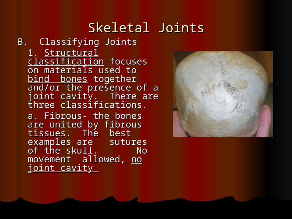
Skeletal JointsSkeletal JointsB. Classifying JointsB. Classifying Joints
1. 1. Structural classificationStructural classification focuses on materials used focuses on materials used to to bind bonesbind bones together together and/or the presence of a and/or the presence of a joint cavity. There are joint cavity. There are three classifications.three classifications.
a. Fibrous- the bones a. Fibrous- the bones are united by fibrous are united by fibrous tissues. The best tissues. The best examples are examples are
sutures of the skull. sutures of the skull. No No movement movement allowed, allowed, no no joint cavity joint cavity
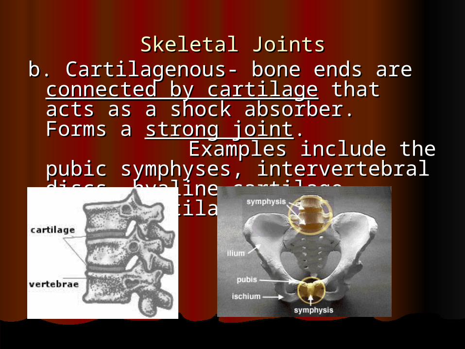
Skeletal JointsSkeletal Jointsb. Cartilagenous- bone ends are b. Cartilagenous- bone ends are
connected by cartilageconnected by cartilage that acts as a that acts as a shock absorber. Forms a shock absorber. Forms a strong strong jointjoint. . Examples Examples include the pubic symphyses, include the pubic symphyses, intervertebral discs, hyaline intervertebral discs, hyaline cartilage, costal cartilage.cartilage, costal cartilage.
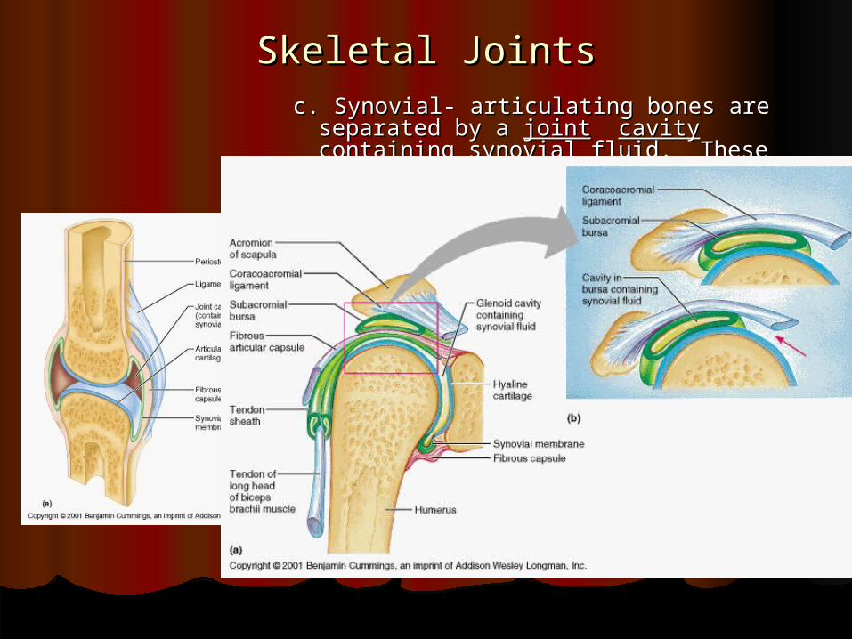
Skeletal JointsSkeletal Jointsc. Synovial- articulating bones are c. Synovial- articulating bones are
separated by a separated by a jointjoint cavity containing cavity containing synovial fluidsynovial fluid. These joints are . These joints are moveable, and have four moveable, and have four distinguishing features.distinguishing features.1) 1) Articular cartilageArticular cartilage- covers the - covers the ends of the ends of the bones forming the bones forming the jointjoint2) 2) Fibrous Articular CapsuleFibrous Articular Capsule- the - the sleeve or capsule of fibrous sleeve or capsule of fibrous connective tissue connective tissue 3) 3) Joint CavityJoint Cavity- Cavity enclosed - Cavity enclosed by by the capsule that contains the the capsule that contains the synovial synovial fluidfluid4) 4) Reinforcing LigamentsReinforcing Ligaments- the - the capsule is supported with capsule is supported with ligaments (connect bone to ligaments (connect bone to bone)bone)
Skeletal JointsSkeletal Joints
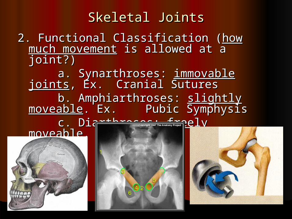
2. Functional Classification (2. Functional Classification (how much how much movementmovement is allowed at a joint?) is allowed at a joint?)
a. Synarthroses: a. Synarthroses: immovable jointsimmovable joints, , Ex. Ex. Cranial SuturesCranial Sutures
b. Amphiarthroses: b. Amphiarthroses: slightly moveableslightly moveable. . Ex. Ex. Pubic SymphysisPubic Symphysis
c. Diarthroses: c. Diarthroses: freely moveablefreely moveable. Ex. . Ex. Ball and Ball and SocketSocket
Question of the weekQuestion of the week What makes joints “crack” or have a popping sound? What makes joints “crack” or have a popping sound? All of the joints in our bodies are surrounded by synovial fluid, a thick, All of the joints in our bodies are surrounded by synovial fluid, a thick,
clear liquid. When you stretch or bend your finger to pop the knuckle, clear liquid. When you stretch or bend your finger to pop the knuckle, you're causing the bones of the joint to pull apart. As they do, the you're causing the bones of the joint to pull apart. As they do, the connective tissue capsule that surrounds the joint is stretched. By connective tissue capsule that surrounds the joint is stretched. By stretching this capsule, you increase its volume. And as we know from stretching this capsule, you increase its volume. And as we know from chemistry class, with an increase in volume comes a decrease in chemistry class, with an increase in volume comes a decrease in pressure. So as the pressure of the synovial fluid drops, gases pressure. So as the pressure of the synovial fluid drops, gases dissolved in the fluid become less soluble, forming bubbles through a dissolved in the fluid become less soluble, forming bubbles through a process called process called cavitationcavitation. When the joint is stretched far enough, the . When the joint is stretched far enough, the pressure in the capsule drops so low that these bubbles burst, pressure in the capsule drops so low that these bubbles burst, producing the pop that we associate with knuckle cracking. It takes producing the pop that we associate with knuckle cracking. It takes about 25 to 30 minutes for the gas to redissolve into the joint fluid. about 25 to 30 minutes for the gas to redissolve into the joint fluid. During this period of time, your knuckles won't crack. Once the gas is During this period of time, your knuckles won't crack. Once the gas is redissolved, cavitation is once again possible, and you can start redissolved, cavitation is once again possible, and you can start popping your knuckles again.popping your knuckles again.
Discoveryhealth.comDiscoveryhealth.com
Skeletal JointsSkeletal Joints
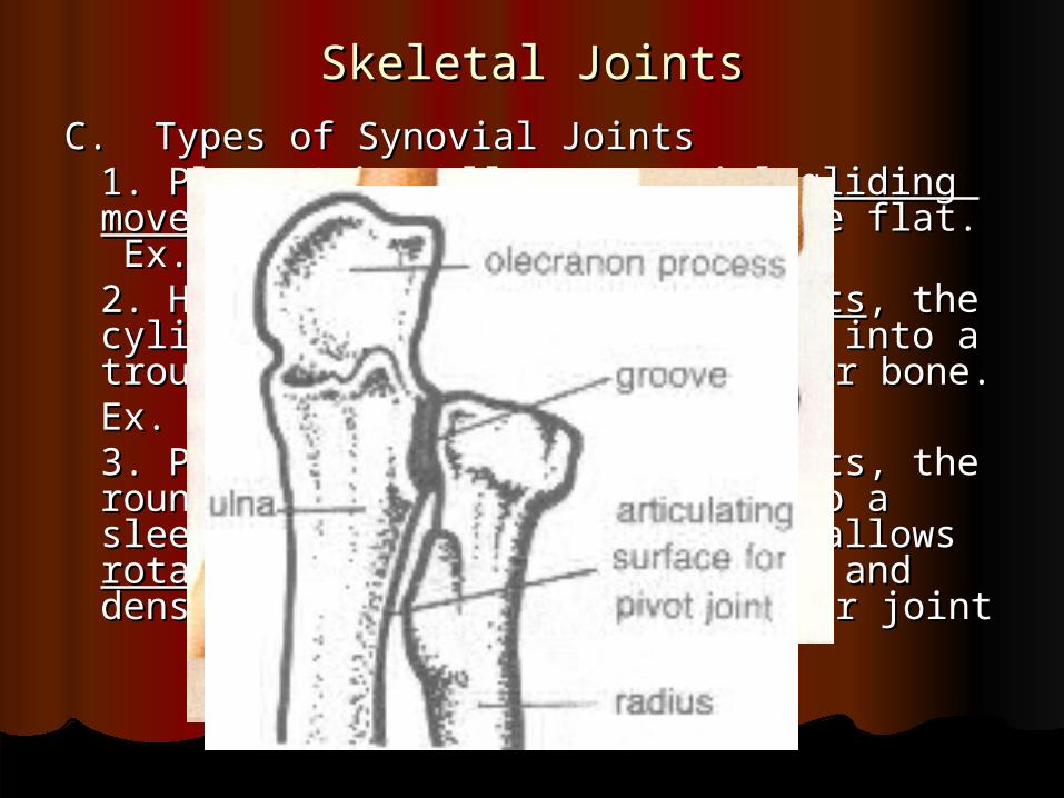
C. Types of Synovial JointsC. Types of Synovial Joints1. Plane Joint-allows 1. Plane Joint-allows nonaxial gliding nonaxial gliding movementsmovements, articular surfaces are flat. , articular surfaces are flat.
Ex. Ex. intercarpal jointsintercarpal joints2. Hinge Joint- 2. Hinge Joint- uniaxial movementsuniaxial movements, the , the cylindrical end of one bone fits into a cylindrical end of one bone fits into a
trough-trough- shaped surface of another bone. shaped surface of another bone. Ex. Ex. Elbow jointElbow joint, phalanges, knee, phalanges, knee3. Pivot Joint- 3. Pivot Joint- uniaxialuniaxial movements, the movements, the rounded end of one bone fits into a rounded end of one bone fits into a sleeve or ring of another, this allows sleeve or ring of another, this allows rotationrotation of one bone. Ex. Atlas and of one bone. Ex. Atlas and
dens of dens of axis, proximal radioulnar jointaxis, proximal radioulnar joint
Skeletal JointsSkeletal Joints
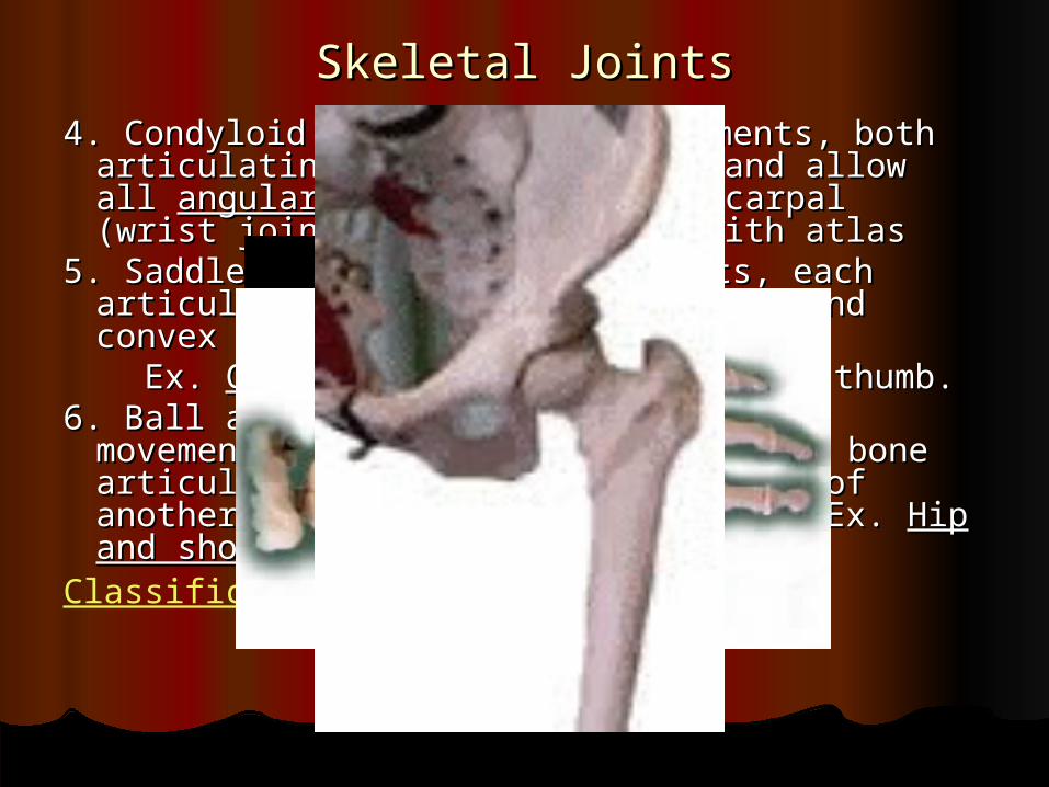
4. Condyloid Joint- 4. Condyloid Joint- biaxial biaxial movements, both movements, both articulating surfaces are oval and allow all articulating surfaces are oval and allow all angular motionsangular motions. Ex. Radio carpal (wrist joint), . Ex. Radio carpal (wrist joint), occipital bone with atlasoccipital bone with atlas
5. Saddle Joint- biaxial movements, each 5. Saddle Joint- biaxial movements, each articular surface has both concave and articular surface has both concave and convex areas that fit together. convex areas that fit together.
Ex. Ex. Carpo-metacarpalCarpo-metacarpal joint of the thumb. joint of the thumb. 6. Ball and Socket Joint- 6. Ball and Socket Joint- multiaxialmultiaxial movements, movements,
spherical head spherical head of one bone articulates with of one bone articulates with the cuplike socket of another, most freely the cuplike socket of another, most freely moving joint. Ex. moving joint. Ex. Hip and shoulderHip and shoulder joints joints
Classification of Joints
Skeletal JointsSkeletal Joints
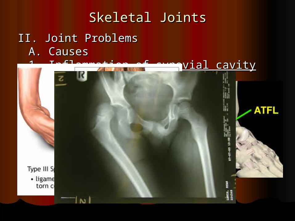
II. Joint ProblemsII. Joint ProblemsA. CausesA. Causes
1. 1. Inflammation of synovial cavityInflammation of synovial cavity2. 2. DegenerationDegeneration of the joint or of the joint or
articular articular cartilagecartilageB. Most common trauma-induced joint B. Most common trauma-induced joint injuriesinjuries
1. Sprain: the 1. Sprain: the ligaments reinforcing a ligaments reinforcing a joint joint are stretched orare stretched or torn.torn.
2. 2. DislocationDislocation: bones are forced out : bones are forced out of of normal positions at the joint.normal positions at the joint.
Skeletal JointsSkeletal Joints
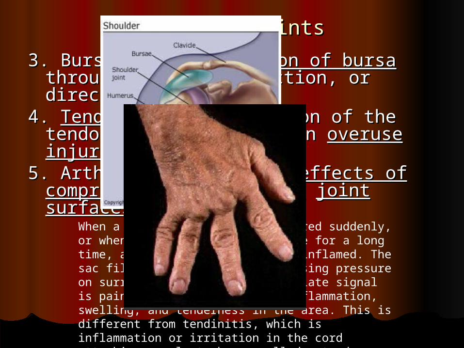
3. Bursitis: 3. Bursitis: inflammation of bursainflammation of bursa through through excessive friction, or direct injuryexcessive friction, or direct injury
4. 4. TendonitisTendonitis: inflammation of the tendon : inflammation of the tendon sheath, usually an sheath, usually an overuse injuryoveruse injury
5. Arthritis: cumulative 5. Arthritis: cumulative effects of effects of compression abrasions atcompression abrasions at joint surfacesjoint surfaces
When a joint is overused or injured suddenly, or when it remains under pressure for a long time, a nearby bursa can become inflamed. The sac fills with excess fluid, causing pressure on surrounding tissue. The immediate signal is pain, often accompanied by inflammation, swelling, and tenderness in the area. This is different from tendinitis, which is inflammation or irritation in the cord attaching muscle to bone, called a tendon.
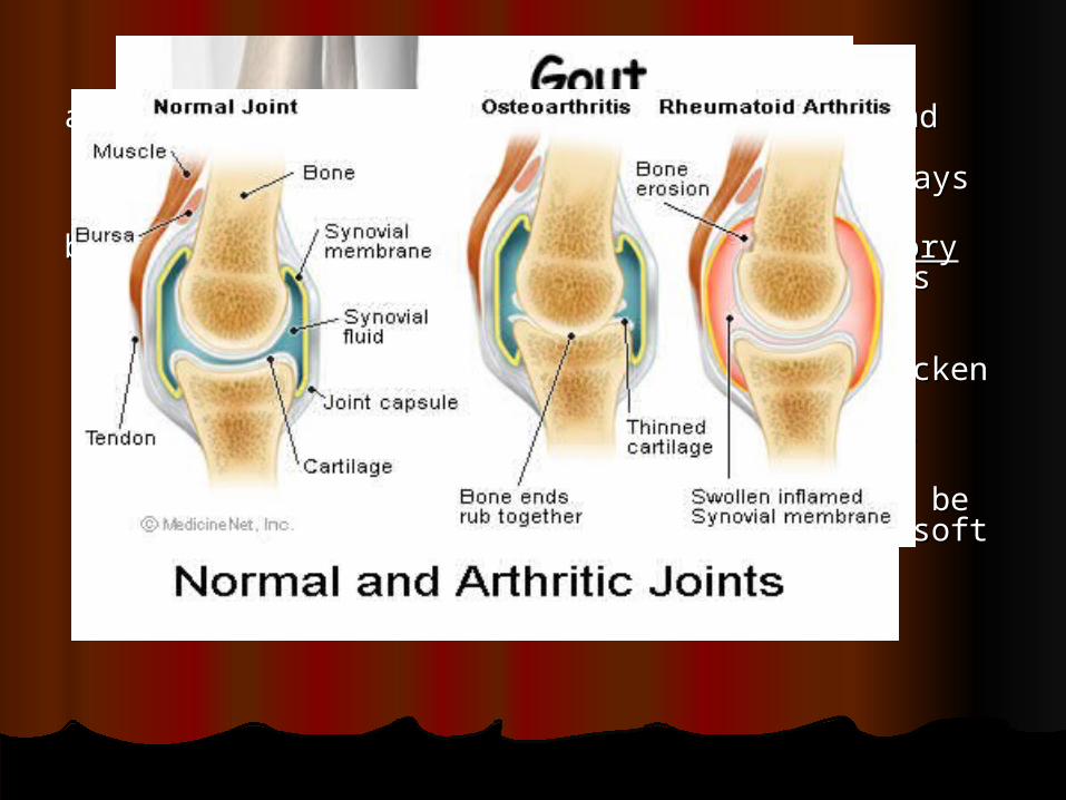
Skeletal JointsSkeletal Jointsa. Osteoarthritis: most common form, “wear and tear a. Osteoarthritis: most common form, “wear and tear
arthritis” that commonly affects older people. The arthritis” that commonly affects older people. The articular cartilage softens,articular cartilage softens, frays and begins to frays and begins to break down.break down.
b. Rheumatoid Arthritis: A b. Rheumatoid Arthritis: A Chronic inflammatory Chronic inflammatory disorderdisorder. An autoimmune disorder (the body’s . An autoimmune disorder (the body’s immune system attempts to destroy its own immune system attempts to destroy its own tissues). The disorder begins with an inflammation tissues). The disorder begins with an inflammation of synovial membranes that thicken and of synovial membranes that thicken and accumulate fluid. The scar tissue can eventually accumulate fluid. The scar tissue can eventually ossify and bones look ossify and bones look deformed.deformed.c. Gouty Arthritis or Gout: a disease which c. Gouty Arthritis or Gout: a disease which uric aciduric acid accumulates in the blood and may be deposited as accumulates in the blood and may be deposited as needle-shaped crystals in the soft tissues or needle-shaped crystals in the soft tissues or joints.Gout tends to run in families.joints.Gout tends to run in families.
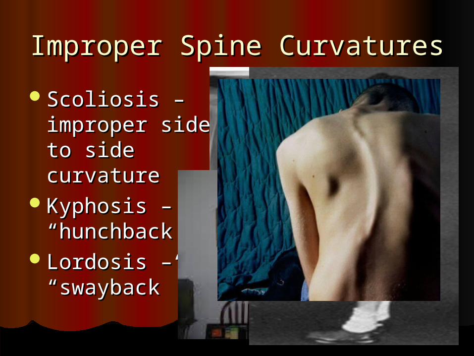
Improper Spine CurvaturesImproper Spine Curvatures
Scoliosis – Scoliosis – improper side to improper side to side curvatureside curvature
Kyphosis – Kyphosis – “hunchback”“hunchback”
Lordosis – Lordosis – “swayback”“swayback”