Size Exclusion Chromatography - Wolfson Centre Home...
139
Size Exclusion Chromatography Principles and Methods GE Healthcare Life Sciences
Transcript of Size Exclusion Chromatography - Wolfson Centre Home...

Size Exclusion Chromatography – Principles and M
ethods
18-1022-18 AL 11/2014
GE and GE monogram are trademarks of General Electric Company.
ÄKTA, Biacore, BioProcess, HiLoad, HiPrep, HiScale, HiTrap, MabSelect, MidiTrap, MiniTrap, MultiTrap, Sephacryl, Sephadex, Sepharose, SpinTrap, Superdex, Superose, Tricorn, and UNICORN are trademarks of GE Healthcare Company or one of its subsidiaries.
BCA is a trademark of Thermo Fisher Scientific LLC. Coomassie is a trademark of Imperial Chemical Industries Limited. Microsoft and Excel are registered trademarks of the Microsoft Corporation. Triton is a trademark of Union Carbide Chemicals and Plastic Company Inc. Tween is a trademark of Uniqema Americas LLC.
All other third-party trademarks are the property of their respective owners.
IMAC Sepharose products, Ni Sepharose products and Fe Sepharose products: These products are sold under a license from Sigma-Aldrich under patent number EP 1276716 (Metal chelating compositions) and equivalent patents and patent applications in other countries.
© 2000–2014 General Electric Company—All rights reserved. First published Dec. 2000.
All goods and services are sold subject to the terms and conditions of sale of the company within GE Healthcare which supplies them. A copy of these terms and conditions is available on request. Contact your local GE Healthcare representative for the most current information.
GE Healthcare UK Limited Amersham Place Little Chalfont Buckinghamshire, HP7 9NA UK
GE Healthcare Europe, GmbH Munzinger Strasse 5 D-79111 Freiburg Germany
GE Healthcare Bio-Sciences Corp. 800 Centennial Avenue, P.O. Box 1327 Piscataway, NJ 08855-1327 USA
GE Healthcare Bio-Sciences KK Sanken Bldg., 3-25-1, Hyakunincho Shinjuku-ku, Tokyo 169-0073 Japan
For local office contact information,please visit www.gelifesciences.com/contact
www.gelifesciences.com/sizeexclusion
GE Healthcare Bio-Sciences ABBjörkgatan 30751 84 UppsalaSweden
Size Exclusion ChromatographyPrinciples and Methods
GE HealthcareLife Sciences
imagination at workimagination at work
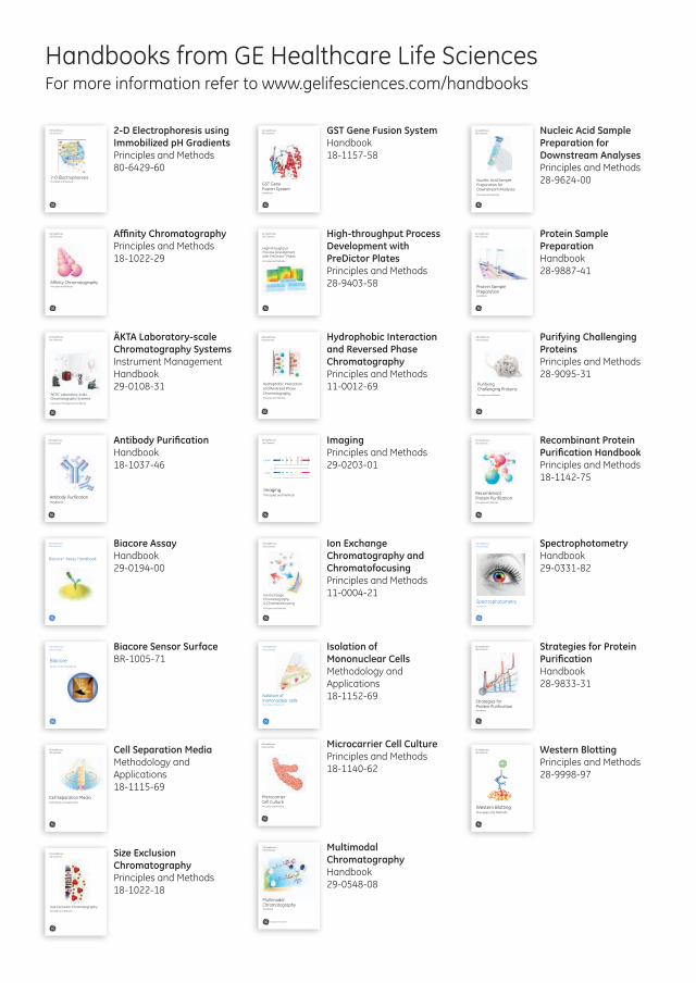
Handbooks from GE Healthcare Life SciencesFor more information refer to www.gelifesciences.com/handbooks
2-D ElectrophoresisPrinciples and Methods
GE HealthcareLife Sciences 2-D Electrophoresis using
Immobilized pH Gradients Principles and Methods 80-6429-60
Affinity ChromatographyPrinciples and Methods
GE HealthcareLife Sciences Affinity Chromatography
Principles and Methods 18-1022-29
GE HealthcareLife Sciences
ÄKTA™ Laboratory-scale Chromatography SystemsInstrument Management Handbook
ÄKTA Laboratory-scale Chromatography Systems Instrument Management Handbook 29-0108-31
GE HealthcareLife Sciences
Antibody PurificationHandbook
Antibody Purification Handbook 18-1037-46
GE HealthcareLife Sciences
Biacore™ Assay Handbook
Biacore Assay Handbook 29-0194-00
GE HealthcareLife Sciences
BiacoreSensor Surface Handbook
Biacore Sensor Surface BR-1005-71
GE HealthcareLife Sciences
Cell Separation MediaMethodology and applications
Cell Separation Media Methodology and Applications 18-1115-69
Size Exclusion ChromatographyPrinciples and Methods
GE HealthcareLife Sciences Size Exclusion
Chromatography Principles and Methods 18-1022-18
GE Healthcare Life Sciences
GST Gene Fusion System Handbook
GST Gene Fusion System Handbook 18-1157-58
GE HealthcareLife Sciences
High-throughputProcess Developmentwith PreDictor™ PlatesPrinciples and Methods
High-throughput Process Development with PreDictor Plates Principles and Methods 28-9403-58
Hydrophobic Interactionand Reversed PhaseChromatographyPrinciples and Methods
GE HealthcareLife Sciences Hydrophobic Interaction
and Reversed Phase Chromatography Principles and Methods 11-0012-69
GE HealthcareLife Sciences
ImagingPrinciples and Methods
Laser
CCD IRUV
IRUV
trans
epi
630 710520
W
460365
312
473 532 635 650 685 785
epi
Imaging Principles and Methods 29-0203-01
GE HealthcareLife Sciences
Ion Exchange Chromatography & ChromatofocusingPrinciples and Methods
Ion Exchange Chromatography and Chromatofocusing Principles and Methods 11-0004-21
GE HealthcareLife Sciences
Isolation of mononuclear cellsMethodology and applications
Isolation of Mononuclear Cells Methodology and Applications 18-1152-69
GE HealthcareLife Sciences
Microcarrier Cell CulturePrinciples and Methods
Microcarrier Cell Culture Principles and Methods 18-1140-62
imagination at work
GE Healthcare Life Sciences
Multimodal Chromatography Handbook
Multimodal Chromatography Handbook 29-0548-08
GE Healthcare Life Sciences
Nucleic Acid Sample Preparation for Downstream AnalysesPrinciples and Methods
Nucleic Acid Sample Preparation for Downstream Analyses Principles and Methods 28-9624-00
GE HealthcareLife Sciences
Protein Sample PreparationHandbook
Protein Sample Preparation Handbook 28-9887-41
GE HealthcareLife Sciences
Purifying Challenging Proteins Principles and Methods
Purifying Challenging Proteins Principles and Methods 28-9095-31
GE HealthcareLife Sciences
Recombinant Protein Purification Principles and Methods
Recombinant Protein Purification Handbook Principles and Methods 18-1142-75
GE HealthcareLife Sciences
SpectrophotometryHandbook
Spectrophotometry Handbook 29-0331-82
GE HealthcareLife Sciences
Strategies for Protein Purif icationHandbook
Strategies for Protein Purification Handbook 28-9833-31
GE HealthcareLife Sciences
Western BlottingPrinciples and Methods
Western Blotting Principles and Methods 28-9998-97

Size exclusion chromatography Principles and Methods
2 18-1022-18 AL
Contents
Introduction .............................................................................................................................................. 7Symbols ............................................................................................................................................................................. 8
Common acronyms and abbreviations .............................................................................................................. 9
Chromatography terminology ............................................................................................................................. 10
Chapter 1 Size exclusion chromatography (SEC) in practice ............................................................................15
Introduction .................................................................................................................................................................. 15
Purification by SEC .................................................................................................................................................... 15Group separation ............................................................................................................................................. 16High-resolution fractionation ..................................................................................................................... 17Rapid purity check and screening ............................................................................................................ 18Resolution in SEC .............................................................................................................................................. 18Sample volume and column dimensions .............................................................................................. 18
Media selection ........................................................................................................................................................... 20
Sample and buffer preparation .......................................................................................................................... 24Sample buffer composition ......................................................................................................................... 24Sample concentration and viscosity ....................................................................................................... 24Sample volume .................................................................................................................................................. 26Buffer composition ......................................................................................................................................... 26Denaturing (chaotropic) agents and detergents ............................................................................... 26
Column and media preparation ......................................................................................................................... 27Setting column pressure limits................................................................................................................... 28
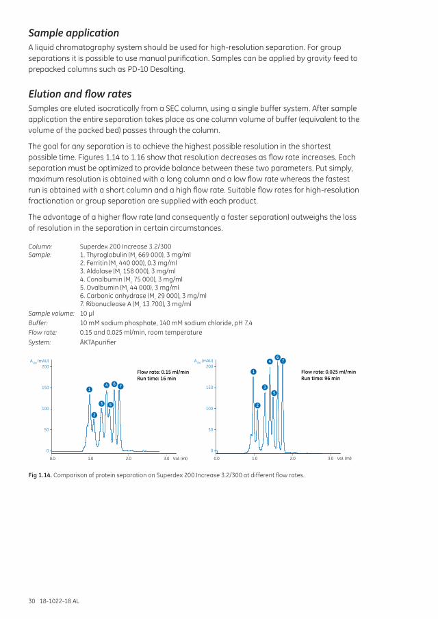
Sample application ................................................................................................................................................... 30
Elution and flow rates .............................................................................................................................................. 30Controlling flow rates ..................................................................................................................................... 32
Method development for high-resolution fractionation .......................................................................... 32
Maintenance of SEC columns .............................................................................................................................. 33
Equipment selection ................................................................................................................................................. 33
Scaling up ...................................................................................................................................................................... 33
BioProcess media for large-scale production .............................................................................................. 34
Troubleshooting .......................................................................................................................................................... 35
Chapter 2 Superdex: for high resolution, short run times, and high recovery ..................................................39
Superdex Increase .......................................................................................................................................... 40Superdex ............................................................................................................................................................. 42Superdex prep grade ...................................................................................................................................... 43
Separation options .................................................................................................................................................... 45
Separation examples .............................................................................................................................................. 46
Performing a separation ........................................................................................................................................ 48First-time use or after long-term storage ............................................................................................. 48Cleaning ................................................................................................................................................................ 49Removing severe contamination .............................................................................................................. 49
Media characteristics ............................................................................................................................................... 50Chemical stability ............................................................................................................................................. 50Storage .................................................................................................................................................................. 50
18-1022-18 AL 3
Chapter 3 Superose: broad fractionation range for laboratory scale ............................................................51
Superose Increase ........................................................................................................................................... 51Superose ............................................................................................................................................................... 53Superose prep grade ...................................................................................................................................... 53
Separation options .................................................................................................................................................... 54
Separation examples ............................................................................................................................................... 55
Performing a separation ........................................................................................................................................ 56First-time use or after long-term storage ............................................................................................. 56Cleaning ................................................................................................................................................................ 57
Media characteristics ............................................................................................................................................... 57Chemical stability ............................................................................................................................................. 57Storage .................................................................................................................................................................. 57
Chapter 4 Sephacryl: fast, high-recovery separations at laboratory and industrial scale ........................59
Separation options .................................................................................................................................................... 62
Separation examples ............................................................................................................................................... 63
Performing a separation ........................................................................................................................................ 64First-time use or after long-term storage ............................................................................................. 64Cleaning ................................................................................................................................................................ 64To remove severe contamination ............................................................................................................. 65
Media characteristics ............................................................................................................................................... 65Chemical stability ............................................................................................................................................. 65Storage .................................................................................................................................................................. 66
Chapter 5 Sephadex: desalting, buffer exchange, and sample clean up .......................................................67
Separation options .................................................................................................................................................... 68
Separation examples ............................................................................................................................................... 72
Performing a separation ........................................................................................................................................ 73
General considerations ........................................................................................................................................... 73Small-scale desalting of samples ............................................................................................................. 73Desalting larger sample volumes using HiTrap and HiPrep columns ..................................... 73Buffer preparation ........................................................................................................................................... 74Sample preparation ........................................................................................................................................ 74Buffer exchange ............................................................................................................................................... 74
HiTrap Desalting columns...................................................................................................................................... 74Manual purification with a syringe .......................................................................................................... 75
Simple desalting with ÄKTAprime plus ............................................................................................................. 76
Desalting on a gravity-feed PD-10 column ................................................................................................... 77Buffer preparation ........................................................................................................................................... 77Optimization of desalting ............................................................................................................................. 77
Scale-up and processing larger sample volumes ...................................................................................... 78Increasing sample loading capacity from 1.5 ml up to 7.5 ml .................................................... 79Increasing sample loading capacity from 15 ml up to 60 ml ...................................................... 79For sample volumes greater than 60 ml ............................................................................................... 80
Media characteristics ............................................................................................................................................... 80Column packing ................................................................................................................................................ 81Cleaning ................................................................................................................................................................ 81Chemical stability ............................................................................................................................................. 81Storage .................................................................................................................................................................. 81
4 18-1022-18 AL
Chapter 6 Sephadex LH-20: size exclusion chromatography in organic solvents ........................................83
Media characteristics ............................................................................................................................................... 83
Separation examples ............................................................................................................................................... 84
Packing a column ...................................................................................................................................................... 85
Performing a separation ........................................................................................................................................ 86
Cleaning ......................................................................................................................................................................... 86
Chemical stability ....................................................................................................................................................... 87
Storage ........................................................................................................................................................................... 87
Transferring Sephadex LH-20 from aqueous solution to organic solvents ................................... 87
Chapter 7 Size exclusion chromatography in theory.........................................................................................89
Defining the process................................................................................................................................................. 89
Selectivity curves and media selection ........................................................................................................... 91
Resolution ...................................................................................................................................................................... 92
Chapter 8 Size exclusion chromatography in a purification strategy ............................................................95
The purification strategy according to CIPP .................................................................................................. 95Purification of humanized IgG4 monoclonal antibody .................................................................... 97
Appendix 1 Column packing and preparation .......................................................................................................99
Columns for packing SEC media......................................................................................................................... 99
Checking column efficiency ................................................................................................................................100
Column packing for high-resolution fractionation using Superdex prep grade and Sephacryl High Resolution .........................................................................................................................101
Column packing for group separations using Sephadex .....................................................................103
Controlling flow rates .............................................................................................................................................105
Appendix 2 Sephadex and Darcy’s law .................................................................................................................106
Appendix 3 Sample preparation ............................................................................................................................107
Sample clarification ................................................................................................................................................107Centrifugation ..................................................................................................................................................107Filtration ..............................................................................................................................................................107Desalting ............................................................................................................................................................108
Denaturation ..............................................................................................................................................................108
Precipitation and resolubilization .....................................................................................................................108Ammonium sulfate precipitation ...........................................................................................................109Removal of lipoproteins ...............................................................................................................................111
Appendix 4 Selection of purification equipment .................................................................................................112
System recommendations for high-resolution SEC columns ....................................................112
18-1022-18 AL 5
Appendix 5 Converting from linear flow (cm/h) to volumetric flow rates (ml/min) and vice versa ................114
From linear flow (cm/h) to volumetric flow rate (ml/min) ...........................................................114From volumetric flow rate (ml/min) to linear flow (cm/h) ............................................................114From ml/min to using a syringe ..............................................................................................................115
Appendix 6 Conversion data ...................................................................................................................................116
Proteins .........................................................................................................................................................................116
Nucleic Acids ..............................................................................................................................................................116
Column pressures ....................................................................................................................................................116
Appendix 7 Amino acids table ...............................................................................................................................118
Appendix 8 Analysis and characterization ..........................................................................................................120
Protein detection and quantitation .................................................................................................................120
Purity check and protein characterization ..................................................................................................120Purity ....................................................................................................................................................................120Characterization .............................................................................................................................................121
Appendix 9 Storage of biological samples ...........................................................................................................122
General recommendations .................................................................................................................................122
Common storage conditions for purified proteins ...................................................................................122
Appendix 10 Molecular weight estimation and molecular weight distribution analysis ..............................123
Performing a molecular weight determination .........................................................................................125
Related literature ................................................................................................................................128
Ordering information ..........................................................................................................................129High-resolution SEC ......................................................................................................................................129Desalting and group separations ...........................................................................................................130Empty columns ................................................................................................................................................131Accessories and spare parts .....................................................................................................................132

6 18-1022-18 AL
18-1022-18 AL 7
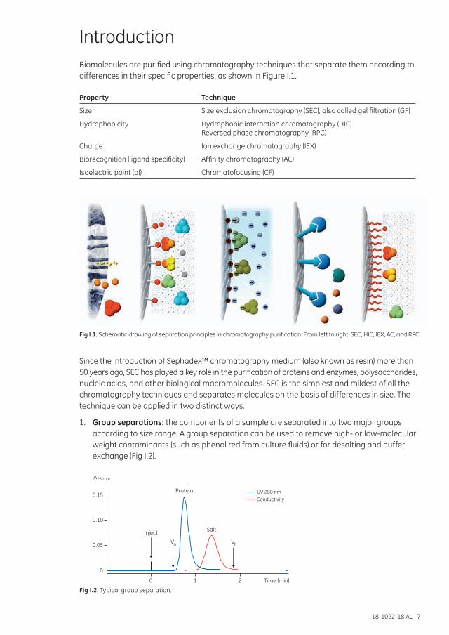
IntroductionBiomolecules are purified using chromatography techniques that separate them according to differences in their specific properties, as shown in Figure I.1.
Property Technique
Size Size exclusion chromatography (SEC), also called gel filtration (GF)
Hydrophobicity Hydrophobic interaction chromatography (HIC) Reversed phase chromatography (RPC)
Charge Ion exchange chromatography (IEX)
Biorecognition (ligand specificity) Affinity chromatography (AC)
Isoelectric point (pI) Chromatofocusing (CF)
Fig I.1. Schematic drawing of separation principles in chromatography purification. From left to right: SEC, HIC, IEX, AC, and RPC.
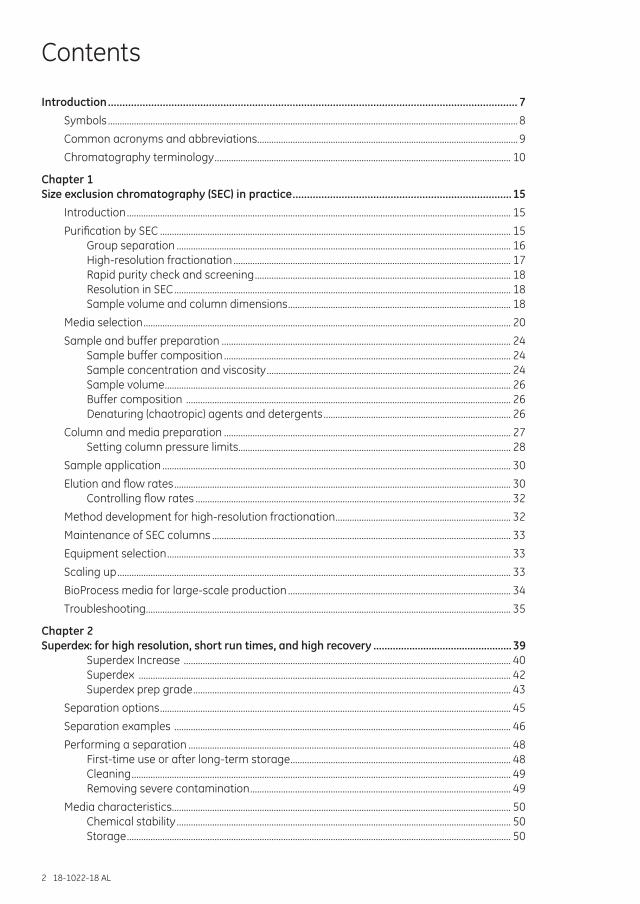
Since the introduction of Sephadex™ chromatography medium (also known as resin) more than 50 years ago, SEC has played a key role in the purification of proteins and enzymes, polysaccharides, nucleic acids, and other biological macromolecules. SEC is the simplest and mildest of all the chromatography techniques and separates molecules on the basis of differences in size. The technique can be applied in two distinct ways:
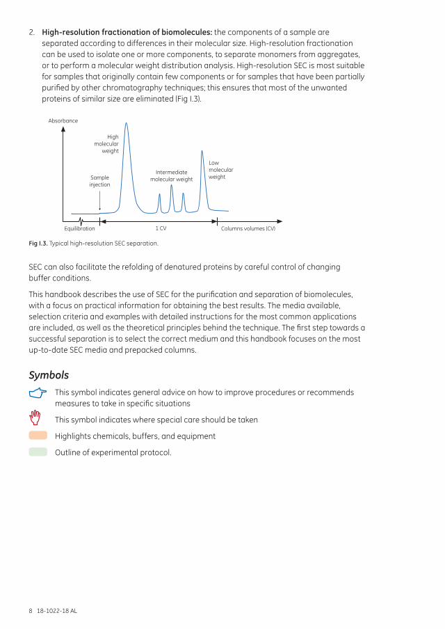
1. Group separations: the components of a sample are separated into two major groups according to size range. A group separation can be used to remove high- or low-molecular weight contaminants (such as phenol red from culture fluids) or for desalting and buffer exchange (Fig I.2).
0
0.05
0.10
0.15
0 1 2 Time (min)
UV 280 nmConductivity
Protein
SaltInject
Vo
A280 nm
Vt
Fig I.2. Typical group separation.
8 18-1022-18 AL
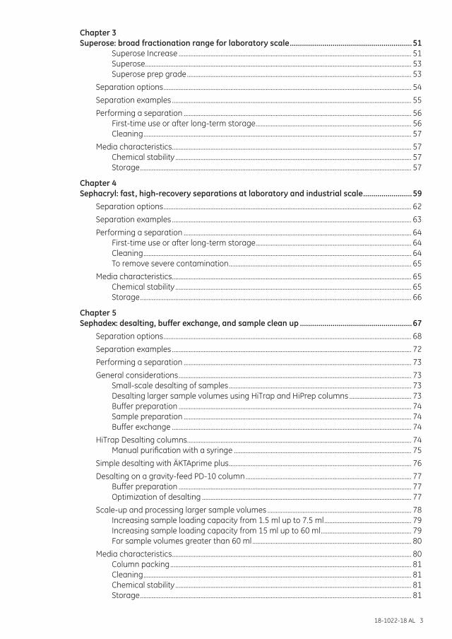
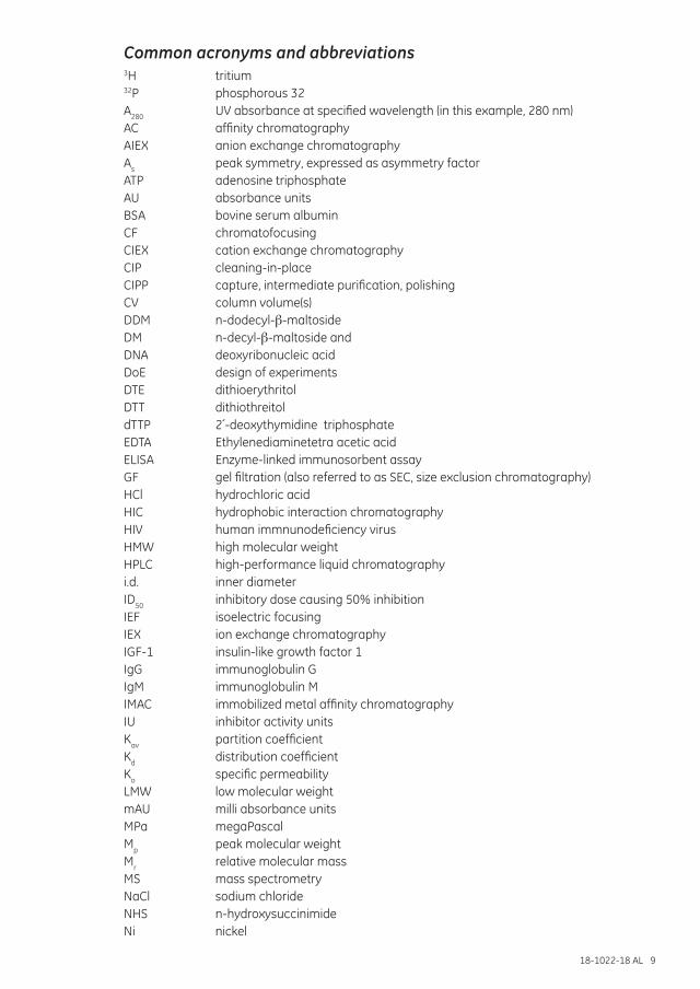
2. High-resolution fractionation of biomolecules: the components of a sample are separated according to differences in their molecular size. High-resolution fractionation can be used to isolate one or more components, to separate monomers from aggregates, or to perform a molecular weight distribution analysis. High-resolution SEC is most suitable for samples that originally contain few components or for samples that have been partially purified by other chromatography techniques; this ensures that most of the unwanted proteins of similar size are eliminated (Fig I.3).
Absorbance
High molecular
weight
Low molecular weight
Equilibration Columns volumes (CV)
Sample injection
Intermediate molecular weight
1 CV
Fig I.3. Typical high-resolution SEC separation.
SEC can also facilitate the refolding of denatured proteins by careful control of changing buffer conditions.
This handbook describes the use of SEC for the purification and separation of biomolecules, with a focus on practical information for obtaining the best results. The media available, selection criteria and examples with detailed instructions for the most common applications are included, as well as the theoretical principles behind the technique. The first step towards a successful separation is to select the correct medium and this handbook focuses on the most up-to-date SEC media and prepacked columns.
Symbols This symbol indicates general advice on how to improve procedures or recommends
measures to take in specific situations
This symbol indicates where special care should be taken
Highlights chemicals, buffers, and equipment
Outline of experimental protocol.
18-1022-18 AL 9
Common acronyms and abbreviations3H tritium32P phosphorous 32 A280 UV absorbance at specified wavelength (in this example, 280 nm)AC affinity chromatographyAIEX anion exchange chromatographyAs peak symmetry, expressed as asymmetry factorATP adenosine triphosphate AU absorbance unitsBSA bovine serum albuminCF chromatofocusingCIEX cation exchange chromatography CIP cleaning-in-placeCIPP capture, intermediate purification, polishingCV column volume(s)DDM n-dodecyl-ơ-maltoside DM n-decyl-ơ-maltoside and DNA deoxyribonucleic acidDoE design of experimentsDTE dithioerythritolDTT dithiothreitol dTTP 2´-deoxythymidine triphosphateEDTA Ethylenediaminetetra acetic acidELISA Enzyme-linked immunosorbent assay GF gel filtration (also referred to as SEC, size exclusion chromatography)HCl hydrochloric acidHIC hydrophobic interaction chromatographyHIV human immnunodeficiency virus HMW high molecular weightHPLC high-performance liquid chromatographyi.d. inner diameterID50 inhibitory dose causing 50% inhibitionIEF isoelectric focusingIEX ion exchange chromatographyIGF-1 insulin-like growth factor 1IgG immunoglobulin GIgM immunoglobulin MIMAC immobilized metal affinity chromatographyIU inhibitor activity unitsKav partition coefficientKd distribution coefficientKo specific permeabilityLMW low molecular weight mAU milli absorbance unitsMPa megaPascalMp peak molecular weightMr relative molecular massMS mass spectrometryNaCl sodium chlorideNHS n-hydroxysuccinimideNi nickel
10 18-1022-18 AL
N/m, Nm-1 column efficiency expressed as number of theoretical plates per meterNM n-nonyl-E-maltoside PAGE polyacrylamide gel electrophoresisPBS phosphate-buffered salinepI isolectric pointpsi pounds per square inchPVDF polyvinylidene fluorideRI refractive indexRNA ribonucleic acid RPC reversed phase chromatographyRs resolution, the degree of separation between peaksSDS sodium dodecyl sulfateSEC size exclusion chromatography (also referred to as GF, gel filtration)ssDNA, ssRNA single-stranded DNA, RNASR solvent resistantTCM traditional Chinese medicineUDM n-undecyl-E-maltoside UV ultravioletVe peak elution (retention) volumeV0 void volumeVi volume of buffer inside the matrixVs volume of stationary phase Vsep separation volume Vt total volume of the packed bedv/v volume/volume W1/2 peak width at half peak heightw/v weight/volume
Chromatography terminology
Adapter Often used for the movable end pieces of columns; contains filter, flow distributor, and possibility to connect tubing.
Adsorption Binding. The process of interaction between the solute (for example, a protein) and the stationary phase.
Affinity chromatography A group of methods based on various types of specific affinities between target molecule(s), for example, a protein and a specific ligand coupled to a chromatography medium.
Asymmetry (asymmetry factor) Factor describing the shape of a chromatographic peak.
Backpressure The pressure drop across a column and/or a chromatography system.
Band broadening The widening of a zone of solute (for example, a protein) when passing through a column or a chromatography system. Gives rise to dilution of the solute and reduces resolution. Also often called peak broadening or zone broadening.
Binding Adsorption. The process of interaction between a solute (for example, a protein) and the stationary phase.
Binding buffer Buffer/solution/eluent used for equilibration of the column before sample loading.
18-1022-18 AL 11
Binding capacity The maximum amount of material that can be bound per ml of chromatography medium. See also Dynamic binding capacity.
Capacity factor The degree of retention of a solute (for example, a protein) relative to an unretained peak.
Chromatofocusing Method that separates proteins on the basis of pI.
Chromatogram A graphical presentation of detector response(s) indicating the concentration of the solutes coming out of the column during the purification (volume or time).
Chromatography From Greek chroma, color, and graphein, to write.
Chromatography medium/media
The stationary phase, also called resin. The chromatography medium is composed of a porous matrix that is usually functionalized by coupling of ligands to it . The matrix is in the form of particles (beads) or, rarely, a single polymer block (monolith).
CIP (cleaning-in-place) Common term for cleaning chromatography columns and/or systems with the purpose of removing unwanted/nonspecifically bound material.
Column Usually column hardware packed with chromatography medium.
Column equilibration Passage of buffer/solution through the chromatography column to establish conditions suitable for binding of selected sample components. For example, to establish correct pH and ionic strength, and ensure that proper counter ions or counter ligands are present.
Column hardware The column tube and adapters. All pieces of the column except the chromatography medium/the packed bed.
Column hardware pressure The pressure inside the column. Column hardware pressure that is too high can break the column.
Column packing Controlled filling of the column hardware with chromatography medium to obtain a packed bed.
Column volume The geometrical volume of the column interior/the chromatography bed.
Counter ion Ion of opposite charge that interacts with an ion exchange chromatography medium after the column equilibration. The counter ion is displaced by a protein that binds to the ion exchanger. If a high concentration of the counter ion is applied, it will compete with the bound protein and elute it from the chromatography column.
Counter ligand Substances that interact with ligands of a chromatography medium and can be displaced by a solute (for example, protein) binding to the ligand.
Dead volume The volume outside the packed chromatography bed. Can be column dead volume or chromatography system dead volume. The dead volume contributes to band broadening.
Degassing Removal of dissolved air from buffers/solutions.
Desorption Elution. Release or removal of bound substances from the chromatography medium.
12 18-1022-18 AL
Design of experiments (DoE) DoE allows use of a minimum number of experiments, in which several experimental parameters can be varied simultaneously. Based on the obtained data, a mathematical model of the studied process (e.g., a protein purification protocol or a chromatography step) is created. The model can be used to understand the influence of the experimental parameters on the outcome and to find an optimum for the process.
Dynamic binding capacity The binding capacity determined by applying the target using flow through a column, as opposed to equilibrium binding capacity determined by batch experiment.
Efficiency Measured as number of theoretical plates. High efficiency means that sharp peaks will be obtained.
Effluent The mobile phase leaving the column (= eluate).
Eluate The mobile phase leaving the column (= effluent).
Eluent The buffer/solution used during chromatography (= mobile phase).
Elution buffer Buffer/solution used for elution (desorption) of bound solutes (for example, proteins) from a column.
Elution volume The volume of buffer/solution (eluent) required to elute the solute for example, a protein (= retention volume).
Elution time The time required for elution of a solute (protein) (= retention time).
Flow rate Volumetric flow (ml/min) or linear flow rate (cm/h). Measurement of flow through a column and/or chromatography system.
Flowthrough Material passing the column during sample loading (without being bound).
Frit Type of deep filter often used at top and bottom of columns.
Gel filtration (GF) Size-exclusion chromatography. Separates solutes (for example, proteins) according to size.
Gradient elution Continuous increased or decreased concentration of a substance (in the eluent) that causes elution of bound solutes (for example, proteins).
Hydrophobic interaction chromatography (HIC)
Method based on the hydrophobic interaction between solutes (for example, proteins) and the chromatography medium in the presence of high salt concentration.
Hydroxyapatite chromatography
Mixed-mode ion exchange chromatography method.
Immobilized metal ion affinity chromatography (IMAC)
Method based on the affinity of proteins with His, Cys, or Trp amino residues on their surface and metal ions on the chromatography medium.
Ion exchange chromatography (IEX)
Method based on electrostatic interactions between solutes (for example, proteins) and chromatography medium.
Isocratic elution Elution of the solutes without changing the composition of the buffer/solution (eluent).
Ligand The specific molecular group that is coupled to the matrix to give some decided function to the chromatography medium.
Ligand density Related to ligand concentration. The distribution of ligands on the surfaces (also surfaces inside pores) of the chromatography matrix.
Linear velocity The flow rate normalized by the column cross section (cm/h).
18-1022-18 AL 13
Mass transfer Movement of a solute (for example, a protein) in and out of the stationary phase. Important factor for column efficiency.
Matrix The matrix is the nonfunctional base for the chromatography medium. The matrix has a porous structure that provides a large surface that can be modified with ligands that introduce possibilities for protein binding.
Mobile phase The fluid (buffer/solution) carrying the solutes during chromatography (= eluent).
Peak broadening Same as band broadening.
Peak capacity The number of peaks that can be separated using a chromatography column.
Peak tailing Broadening at the end of a peak due to additional delay of a fraction of the solute. Results in increased asymmetry factor.
Pore Cavity in a chromatography matrix.
Pore volume The total volume of the pores in a chromatography medium.
Pressure over the packed bed The pressure drop across the packed bed upon passage of solution through the column. Caused by flow resistance in the packed bed.
Recovery The relative amount of target protein that is retrieved after purification compared with amount loaded on the column.
Resin The term is sometimes used instead of the more generic term, chromatography medium.
Resolution Measurement of the ability of a packed column to separate two solutes (peaks).
Retention volume Same as elution volume.
Retention time Same as elution time.
Reversed phase chromatography (RPC)
Method based on hydrophobic interactions between solutes (sample components) and ligands coupled to the chromatography medium. Organic modifiers (for example, acetonitrile) in the eluent are used for elution.
Sample The material loaded on the chromatography column/medium, or to be analyzed.
Sample application Applying/loading sample on the column.
Sample loading Loading/applying sample on the column.
Sample volume Usually the volume of the sample loaded on the chromatography column/medium.
Selectivity Measure of the relative retention of two solutes in a column. Related to the distance between two peaks.
Solute The dissolved substance (for example, a protein) in, for example, the mobile phase.
Stationary phase Often called resin, chromatography beads, chromatography material, chromatography medium or media.
Step gradient elution Stepwise increase in concentration of the substance that affects elution of bound solutes.
Void volume The elution volume of solutes that do not enter the pores or interact with the chromatography medium, thus passing between the beads in the packed bed.
Wash Wash step. Removal of unbound or weakly bound material from a column after the sample loading.
14 18-1022-18 AL
Wash buffer Buffer/solution used for washing the column after sample loading.
Wash volume Volume of buffer/solution used for the wash step.
Yield Amount of target protein (or other solute) obtained after a purification step, or after the entire purification (multiple steps).
Zone broadening Same as peak broadening.
18-1022-18 AL 15
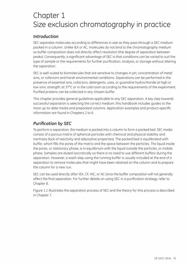
Chapter 1 Size exclusion chromatography in practiceIntroductionSEC separates molecules according to differences in size as they pass through a SEC medium packed in a column. Unlike IEX or AC, molecules do not bind to the chromatography medium so buffer composition does not directly affect resolution (the degree of separation between peaks). Consequently, a significant advantage of SEC is that conditions can be varied to suit the type of sample or the requirements for further purification, analysis, or storage without altering the separation.
SEC is well-suited to biomolecules that are sensitive to changes in pH, concentration of metal ions, or cofactors and harsh environmental conditions. Separations can be performed in the presence of essential ions, cofactors, detergents, urea, or guanidine hydrochloride at high or low ionic strength; at 37°C or in the cold room according to the requirements of the experiment. Purified proteins can be collected in any chosen buffer.
This chapter provides general guidelines applicable to any SEC separation. A key step towards successful separation is selecting the correct medium; this handbook includes guides to the most up-to-date media and prepacked columns. Application examples and product-specific information are found in Chapters 2 to 6.
Purification by SECTo perform a separation, the medium is packed into a column to form a packed bed. SEC media consist of a porous matrix of spherical particles with chemical and physical stability and inertness (lack of reactivity and adsorptive properties). The packed bed is equilibrated with buffer, which fills the pores of the matrix and the space between the particles. The liquid inside the pores, or stationary phase, is in equilibrium with the liquid outside the particles, or mobile phase. Samples are eluted isocratically so there is no need to use different buffers during the separation. However, a wash step using the running buffer is usually included at the end of a separation to remove molecules that might have been retained on the column and to prepare the column for a new run.
SEC can be used directly after IEX, CF, HIC, or AC since the buffer composition will not generally affect the final separation. For further details on using SEC in a purification strategy, refer to Chapter 8.
Figure 1.1 illustrates the separation process of SEC and the theory for this process is described in Chapter 7.
16 18-1022-18 AL
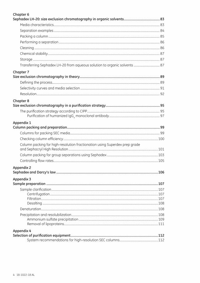
Fig 1.1. Process of SEC. (A) Schematic picture of a bead with an electron microscopic enlargement. (B) Schematic drawing of sample molecules diffusing into bead pores. (C) Graphical description of separation: (i) sample is applied on the column; (ii) the smallest molecule (yellow) is more delayed than the largest molecule (red); (iii) the largest molecule is eluted first from the column. Band broadening causes significant dilution of the protein zones during chromatography. (D) Schematic chromatogram.
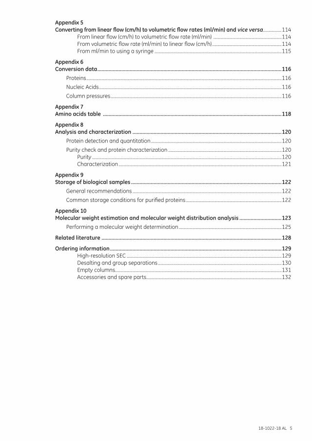
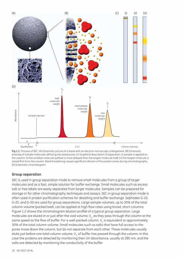
Group separationSEC is used in group separation mode to remove small molecules from a group of larger molecules and as a fast, simple solution for buffer exchange. Small molecules such as excess salt or free labels are easily separated from larger molecules. Samples can be prepared for storage or for other chromatography techniques and assays. SEC in group separation mode is often used in protein purification schemes for desalting and buffer exchange. Sephadex G-10, G-25, and G-50 are used for group separations. Large sample volumes, up to 30% of the total column volume (packed bed), can be applied at high flow rates using broad, short columns. Figure 1.2 shows the chromatogram (elution profile) of a typical group separation. Large molecules are eluted in or just after the void volume, Vo, as they pass through the column at the same speed as the flow of buffer. For a well-packed column, Vo is equivalent to approximately 30% of the total column volume. Small molecules such as salts that have full access to the pores move down the column, but do not separate from each other. These molecules usually elute just before one total column volume, Vt, of buffer has passed through the column. In this case the proteins are detected by monitoring their UV absorbance, usually at 280 nm, and the salts are detected by monitoring the conductivity of the buffer.
(A)
(D)
(B) (C) (i) (ii) (iii)
1 CVEquilibration
Sample injection
High molecular
weightIntermediate
molecularweight
Lowmolecular
weight
Column volumes0
Vt
Vo
VR
Abso
rban
ce
18-1022-18 AL 17
Column: HiTrap™ Desalting 5 ml
Sample: (Histidine)6 protein eluted from HiTrap Chelating HP with 20 mM sodium phosphate, 500 mM sodium chloride 500 mM imidazole, pH 7.4
Buffer: 20 mM sodium phosphate, 150 mM sodium chloride, pH 7.0
0
0.05
0.10
0.15
UV 280 nm
Conductivity
(Histidine) -protein
Salt
6
Inject
2 Time (min)10
VtVo
A 280 (mAU)
Fig 1.2. Typical chromatogram of a group separation. UV (protein) and conductivity (salt) detection enable pooling of the desalted fractions and facilitate optimization of the separation Vo = void volume; Vt = total column volume.
Refer to Chapter 5 for detailed information on how Sephadex is used in group separation of high- and low-molecular weight substances in applications like desalting, buffer exchange, and sample clean up.
Refer to Chapter 7 for detailed information on the theory of SEC.
High-resolution fractionationSEC is used in fractionation mode to separate multiple components in a sample on the basis of differences in their size. The goal can be to isolate one or more of the components, or to analyze the molecular-weight distribution in the sample. Optimal results for high-resolution fractionation will be achieved with samples that originally contain few components or with samples that have been partially purified by other chromatography techniques to eliminate most of the unwanted proteins of similar size.
High-resolution fractionation by SEC is well-suited for the final polishing step in a purification scheme. Monomers are easily separated from aggregates. Samples can be transferred to a suitable buffer for assay or storage.
Prepacked SEC columns are highly recommended for optimal performance during high-resolution fractionation and are available for three different purposes:
• Preparative purification for sample volumes in the milliliter range.• Small-scale preparative purification for sample volumes up to 500 µl.• Analytical runs for sample volumes up to 500 µl.
HiLoad™ columns prepacked with Superdex™ prep grade media, or HiPrep™ columns prepacked with Sephacryl™ media, are used for preparative purification, which is characterized by collection of the sample. The sample volume is in the milliliter range and the collected fractions are usually in milligram amounts.
18 18-1022-18 AL
Small-scale preparative purification and analytical runs are performed using Superdex or Superose™ media prepacked in Tricorn™ or Precision Columns (PC columns). Compared with preparative purification, the sample size and collected amounts are smaller (microliter and microgram to milligram scale) in small-scale preparative purification. Depending on sample volume, either Tricorn 10/300 GL columns should be used (25 µl to 500 µl) or 3.2/300 columns (4 µl to 50 µl). While samples are collected in preparative purification, no collection of sample in analytical purification is necessary—in this case it is the data from the run that is in focus.
Rapid purity check and screeningShort columns with small bed volumes such as Superdex 75 5/150 GL, Superdex 200 Increase 5/150 GL, and Superose 6 Increase 5/150 GL are suitable for rapid purity check or size analysis of proteins and other biomolecules. Short cycle times, together with small sample volume and low buffer consumption make this column a good choice in screening experiments to check protein homogeneity. However, when using the same media, shorter columns give lower resolution than longer columns.
Resolution in SECThe success of SEC depends primarily on choosing conditions that give sufficient selectivity and counteract peak broadening effects during the separation. After selection of SEC medium, sample volume and column dimensions are the two most critical parameters that will affect the resolution of the separation. Chromatography system-related factors can also affect resolution.
The final resolution is influenced by many factors (Table 1.1). The molecular-weight range over which an SEC medium can separate molecules is referred to as the selectivity of the medium (see fractionation range guide for SEC media in Media selection in this chapter). Resolution is a function of the selectivity of the medium and the efficiency of that medium to produce narrow peaks (minimal peak broadening), as illustrated in Chapter 7.
Table 1.1. Factors that influence resolution
Medium-related factors Particle sizeParticle uniformityMatch between pore size and analyte size
Column-related factors Bed heightColumn packing quality
Chromatography system-related factors Tubing dimensions (diameter and length) Volumes in system components
Experimental-related factors Flow rateSample volumeViscosity
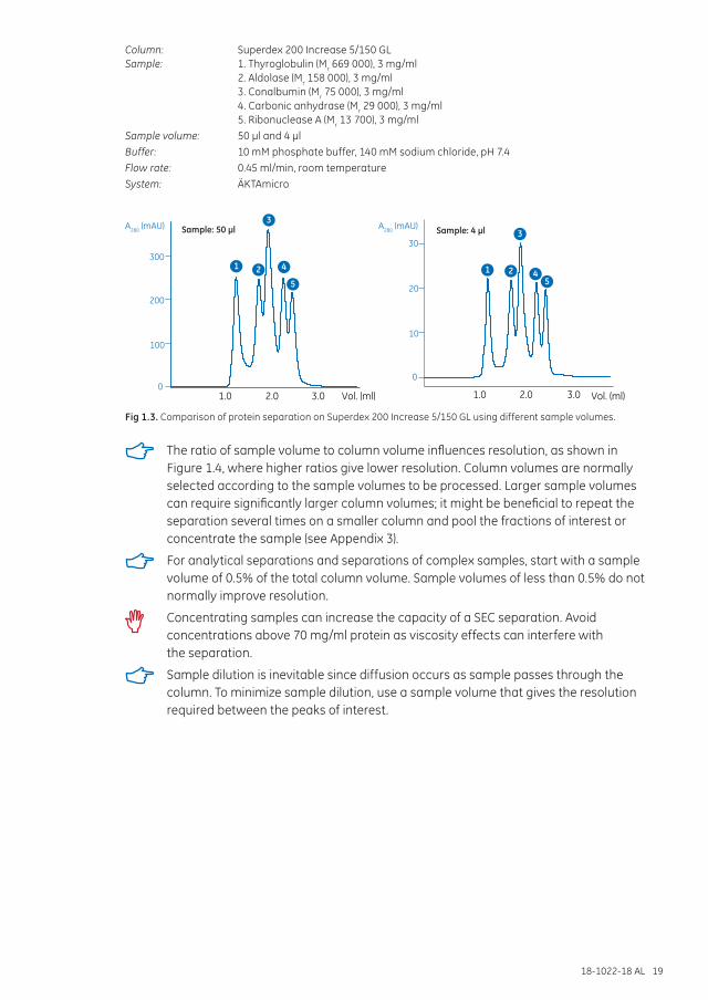
Sample volume and column dimensionsThe sample volume can be expressed as a percentage of the total column volume (packed bed). Smaller sample volumes help to avoid overlap if closely spaced peaks are eluted. Figure 1.3 illustrates how sample volume can influence a high-resolution fractionation.
For group separations, use sample volumes up to 30% of the total column volume.
For high-resolution fractionation, a sample volume from 0.5% to 4% of the total column volume is recommended, depending on the type of medium used. For most applications the sample volume should not exceed 2% to achieve maximum resolution. Depending on the nature of the specific sample, it might be possible to load larger sample volumes, particularly if the peaks of interest are well resolved. This can only be determined by experimentation.
18-1022-18 AL 19
Column: Superdex 200 Increase 5/150 GLSample: 1. Thyroglobulin (Mr 669 000), 3 mg/ml 2. Aldolase (Mr 158 000), 3 mg/ml 3. Conalbumin (Mr 75 000), 3 mg/ml 4. Carbonic anhydrase (Mr 29 000), 3 mg/ml 5. Ribonuclease A (Mr 13 700), 3 mg/mlSample volume: 50 µl and 4 µlBuffer: 10 mM phosphate buffer, 140 mM sodium chloride, pH 7.4Flow rate: 0.45 ml/min, room temperatureSystem: ÄKTAmicro
Sample: 4 µl
10
0
20
30
1.0 2.0 3.0
A280 (mAU)A280 (mAU) Sample: 50 µl
100
0
200
300
1.0 2.0 3.0 Vol. (ml) Vol. (ml)
1 2 1 2
33
44
5 5
Fig 1.3. Comparison of protein separation on Superdex 200 Increase 5/150 GL using different sample volumes.
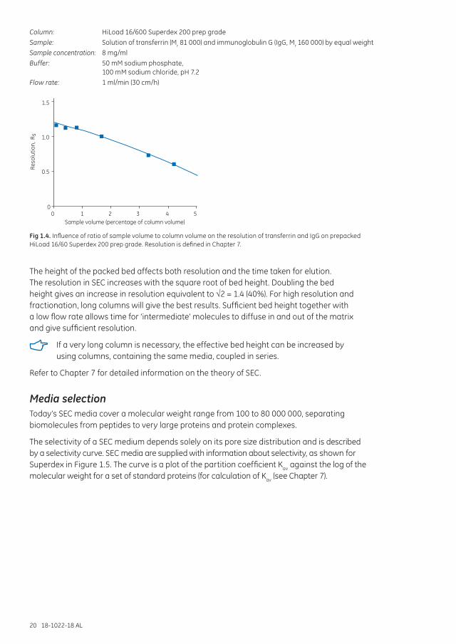
The ratio of sample volume to column volume influences resolution, as shown in Figure 1.4, where higher ratios give lower resolution. Column volumes are normally selected according to the sample volumes to be processed. Larger sample volumes can require significantly larger column volumes; it might be beneficial to repeat the separation several times on a smaller column and pool the fractions of interest or concentrate the sample (see Appendix 3).
For analytical separations and separations of complex samples, start with a sample volume of 0.5% of the total column volume. Sample volumes of less than 0.5% do not normally improve resolution.
Concentrating samples can increase the capacity of a SEC separation. Avoid concentrations above 70 mg/ml protein as viscosity effects can interfere with the separation.
Sample dilution is inevitable since diffusion occurs as sample passes through the column. To minimize sample dilution, use a sample volume that gives the resolution required between the peaks of interest.
20 18-1022-18 AL
Column: HiLoad 16/600 Superdex 200 prep gradeSample: Solution of transferrin (Mr 81 000) and immunoglobulin G (IgG, Mr 160 000) by equal weightSample concentration: 8 mg/mlBuffer: 50 mM sodium phosphate,
100 mM sodium chloride, pH 7.2Flow rate: 1 ml/min (30 cm/h)
Sample volume (percentage of column volume)0 1 2 3 4 5
0
0.5
1.5
1.0
Reso
lutio
n, R
s
Fig 1.4. Influence of ratio of sample volume to column volume on the resolution of transferrin and IgG on prepacked HiLoad 16/60 Superdex 200 prep grade. Resolution is defined in Chapter 7.
The height of the packed bed affects both resolution and the time taken for elution. The resolution in SEC increases with the square root of bed height. Doubling the bed height gives an increase in resolution equivalent to √2 = 1.4 (40%). For high resolution and fractionation, long columns will give the best results. Sufficient bed height together with a low flow rate allows time for ‘intermediate’ molecules to diffuse in and out of the matrix and give sufficient resolution.
If a very long column is necessary, the effective bed height can be increased by using columns, containing the same media, coupled in series.
Refer to Chapter 7 for detailed information on the theory of SEC.
Media selectionToday’s SEC media cover a molecular weight range from 100 to 80 000 000, separating biomolecules from peptides to very large proteins and protein complexes.
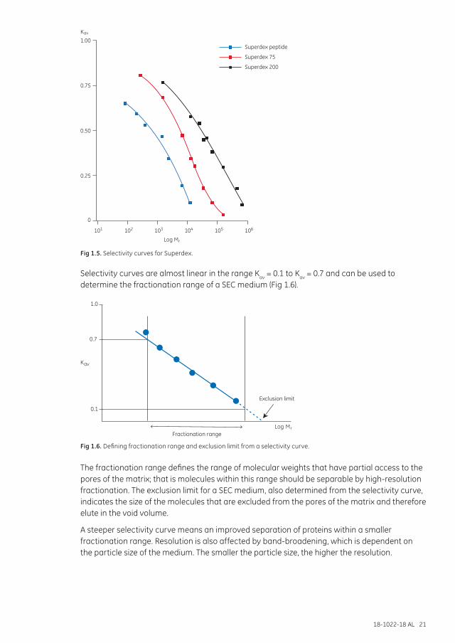
The selectivity of a SEC medium depends solely on its pore size distribution and is described by a selectivity curve. SEC media are supplied with information about selectivity, as shown for Superdex in Figure 1.5. The curve is a plot of the partition coefficient Kav against the log of the molecular weight for a set of standard proteins (for calculation of Kav (see Chapter 7).
18-1022-18 AL 21
0.25
0
0.50
0.75
1.00Kav
Superdex peptide
Superdex 200
Superdex 75
Log Mr
101 102 103 104 105 106
Fig 1.5. Selectivity curves for Superdex.
Selectivity curves are almost linear in the range Kav = 0.1 to Kav = 0.7 and can be used to determine the fractionation range of a SEC medium (Fig 1.6).
1.0
0.7
0.1
Fractionation range
Exclusion limit
Kav
Log Mr
Fig 1.6. Defining fractionation range and exclusion limit from a selectivity curve.
The fractionation range defines the range of molecular weights that have partial access to the pores of the matrix; that is molecules within this range should be separable by high-resolution fractionation. The exclusion limit for a SEC medium, also determined from the selectivity curve, indicates the size of the molecules that are excluded from the pores of the matrix and therefore elute in the void volume.
A steeper selectivity curve means an improved separation of proteins within a smaller fractionation range. Resolution is also affected by band-broadening, which is dependent on the particle size of the medium. The smaller the particle size, the higher the resolution.
22 18-1022-18 AL
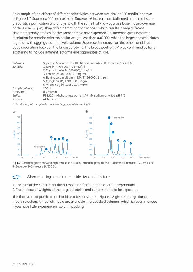
An example of the effects of different selectivities between two similar SEC media is shown in Figure 1.7. Superdex 200 Increase and Superose 6 Increase are both media for small-scale preparative purification and analysis, with the same high-flow agarose base matrix (average particle size 8.6 µm). They differ in fractionation ranges, which results in very different chromatography profiles for the same sample mix. Superdex 200 Increase gives excellent resolution for proteins with molecular weight less than 440 000, while the largest protein elutes together with aggregates in the void volume. Superose 6 Increase, on the other hand, has good separation between the largest proteins. The broad peak of IgM was confirmed by light scattering to include different isoforms and aggregates of IgM.
Columns: Superose 6 Increase 10/300 GL and Superdex 200 Increase 10/300 GLSample: 1. IgM (Mr ~ 970 000)*, 0.5 mg/ml 2. Thyroglobulin (Mr 669 000), 1 mg/ml 3. Ferritin (Mr 440 000), 0.1 mg/ml 4. Bovine serum albumin (BSA, Mr 66 000), 1 mg/ml 5. Myoglobin (Mr 17 000), 0.5 mg/ml 6. Vitamin B12 (Mr 1355), 0.05 mg/mlSample volume: 100 µlFlow rate: 0.5 ml/minBuffer: PBS, (10 mM phosphate buffer, 140 mM sodium chloride, pH 7.4)System: ÄKTAmicro
* In addition, this sample also contained aggregated forms of IgM.
0.0
10.0
20.0
30.0
40.0
50.0
60.0
70.0
0.0 5.0 10.0 15.0 20.0 25.0 Vol. (ml)
A 280 (m
AU)
1
2
3
4 56
+ aggregates
0.0
10.0
20.0
30.0
40.0
50.0
60.0
70.0
A 280 (m
AU)
0.0 5.0 10.0 15.0 20.0 Vol. (ml)
1
23 4 5
6
Aggregates
0.0
10.0
20.0
30.0
40.0
50.0
60.0
70.0
0.0 5.0 10.0 15.0 20.0 25.0 Vol. (ml)
A 280 (m
AU)
1
2
3
4 56
+ aggregates
0.0
10.0
20.0
30.0
40.0
50.0
60.0
70.0
A 280 (m
AU)
0.0 5.0 10.0 15.0 20.0 Vol. (ml)
1
23 4 5
6
Aggregates
(A) (B)
Fig 1.7. Chromatograms showing high-resolution SEC of six standard proteins on (A) Superose 6 Increase 10/300 GL and (B) Superdex 200 Increase 10/300 GL.
When choosing a medium, consider two main factors:
1. The aim of the experiment (high-resolution fractionation or group separation).2. The molecular weights of the target proteins and contaminants to be separated.
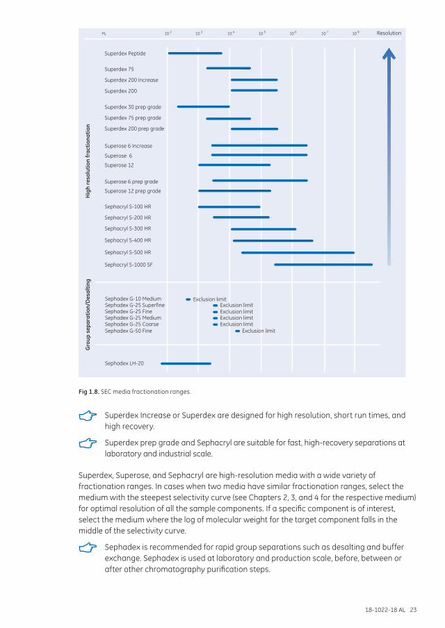
The final scale of purification should also be considered. Figure 1.8 gives some guidance to media selection. Almost all media are available in prepacked columns, which is recommended if you have little experience in column packing.
18-1022-18 AL 23
10M 2r 10 3 10 4 10 5 10 6 10 7 10 8
Superdex 30 prep grade
Superdex 75 prep grade
Superdex 200 prep grade
Superdex Peptide
Superdex 75
Superdex 200
Superose 6 prep grade
Superose 12 prep grade
Superose
Superose 6 Increase
6
Superose 12
Sephacryl S-100 HR
Sephacryl S-200 HR
Sephacryl S-300 HR
Sephacryl S-400 HR
Sephacryl S-500 HR
Sephacryl S-1000 SF
Sephadex G-10 MediumSephadex G-25 SuperfineSephadex G-25 FineSephadex G-25 MediumSephadex G-25 CoarseSephadex G-50 Fine
Exclusion limit
Exclusion limit
Exclusion limitExclusion limitExclusion limitExclusion limit
Sephadex LH-20
Hig
h re
solu
tion
frac
tiona
tion
Gro
up s
epar
atio
n/D
esal
ting
Resolution
Superdex 200 Increase
Fig 1.8. SEC media fractionation ranges.
Superdex Increase or Superdex are designed for high resolution, short run times, and high recovery.
Superdex prep grade and Sephacryl are suitable for fast, high-recovery separations at laboratory and industrial scale.
Superdex, Superose, and Sephacryl are high-resolution media with a wide variety of fractionation ranges. In cases when two media have similar fractionation ranges, select the medium with the steepest selectivity curve (see Chapters 2, 3, and 4 for the respective medium) for optimal resolution of all the sample components. If a specific component is of interest, select the medium where the log of molecular weight for the target component falls in the middle of the selectivity curve.
Sephadex is recommended for rapid group separations such as desalting and buffer exchange. Sephadex is used at laboratory and production scale, before, between or after other chromatography purification steps.
24 18-1022-18 AL
For group separations, select SEC media that elute high molecular weight-molecules at the void volume to minimize peak broadening or dilution and reduce time in the column. The lowest molecular weight substances should appear by the time one column volume of buffer has passed through the column.
Table 1.2. Sephadex media properties
Medium Cut-off Application examples
Sephadex G-10 700 Desalting of peptides
Sephadex G-25 5000 Desalting of proteins and oligonucleotides
Sephadex G-50 30 000 Removal of free labels from labeled macromolecules
Sample and buffer preparationRemoval of particles in the sample is extremely important for SEC. Clarifying a sample before applying it to a column will avoid the risk of blockage, reduce the need for stringent washing procedures and extend the life of the medium.
Samples must be clear and free from particulate matter, especially when working with bead sizes of 34 µm or less.
Appendix 3 contains an overview of sample preparation techniques. For small sample volumes, a syringe-tip filter of cellulose acetate or polyvinylidene fluoride (PVDF) can be sufficient.
Sample buffer compositionThe pH, ionic strength, and composition of the sample buffer will not significantly affect resolution as long as these parameters do not alter the size or stability of the proteins to be separated and are not outside the stability range of the SEC medium. The sample does not have to be in exactly the same buffer as that used to equilibrate and run through the column. Sample is exchanged into the running buffer during the separation, an added benefit of SEC.
Sample concentration and viscositySEC is independent of sample mass and hence sample concentration, as can be seen in Figure 1.9. High resolution can be maintained despite high sample concentration and, with the appropriate medium, high flow rates.
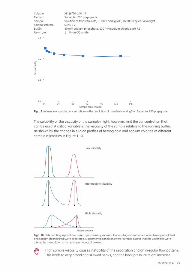
18-1022-18 AL 25
Column: XK 16/70 (140 ml)Medium: Superdex 200 prep gradeSample: Solution of transferrin (Mr 81 000) and IgG (Mr 160 000) by equal weightSample volume: 0.8% × Vt
Buffer: 50 mM sodium phosphate, 100 mM sodium chloride, pH 7.2Flow rate: 1 ml/min (30 cm/h)
0.0
Reso
lutio
n, R
S
0.5
1.0
1.5
0Sample conc. (mg/ml)
24 48 72 96 120 148
Fig 1.9. Influence of sample concentration on the resolution of transferrin and IgG on Superdex 200 prep grade.
The solubility or the viscosity of the sample might, however, limit the concentration that can be used. A critical variable is the viscosity of the sample relative to the running buffer, as shown by the change in elution profiles of hemoglobin and sodium chloride at different sample viscosities in Figure 1.10.
Elution volume
Low viscosity
Intermediate viscosity
High viscosity
Fig 1.10. Deteriorating separation caused by increasing viscosity. Elution diagrams obtained when hemoglobin (blue) and sodium chloride (red) were separated. Experimental conditions were identical except that the viscosities were altered by the addition of increasing amounts of dextran.
High sample viscosity causes instability of the separation and an irregular flow pattern. This leads to very broad and skewed peaks, and the back pressure might increase.
26 18-1022-18 AL
Samples should generally not exceed 70 mg/ml protein. Dilute viscous samples, but not more than necessary to keep the sample volume low. Remember that viscosity varies with temperature.
Sample volumeSample volume is one of the most important parameters in SEC. Refer to Sample volume and column dimensions earlier in this chapter for more information.
Buffer composition Buffer composition will generally not directly influence the resolution unless the buffer affects the shape or biological activity of the molecules. Extremes of pH and ionic strength and the presence of denaturing agents or detergents can cause conformational changes, dissociation or association of protein complexes.
Select buffer conditions that are compatible with protein stability and activity. The product of interest will be collected in this buffer. Use a buffer concentration that maintains buffering capacity and constant pH. Use up to 300 mM sodium chloride to avoid nonspecific ionic interactions with the matrix which can be seen as delays in peak elution. Note that some proteins can precipitate in low ionic strength solutions. Volatile buffers such as ammonium acetate or ammonium bicarbonate should be used if the separated product will be lyophilized.
Use high-quality water and chemicals. Solutions should be filtered through 0.45 µm or 0.22 µm filters before use. It is essential to degas buffers before any SEC separation since air bubbles can significantly affect performance. Buffers will be automatically degassed if they are filtered under vacuum.
Choose buffer conditions suitable for protein stability and activity. An increase of sodium chloride concentration up to 300 mM or addition of additives, such as detergent or organic solvents, can improve the result. See further denaturing (chaotropic) agents and detergents, below, and instructions for the product.
When working with a new sample, try these conditions first: 50 mM sodium phosphate, 150 mM sodium chloride, pH 7.0 or select the buffer into which the product should be eluted for the next step (e.g., further purification, analysis, or storage).
Avoid extreme changes in pH or other conditions that can cause inactivation or even precipitation. If the sample precipitates in the SEC column, the column will be blocked, possibly irreversibly, and the sample might be lost.
Denaturing (chaotropic) agents and detergentsDenaturing agents such as guanidine hydrochloride or urea can be used for initial solubilization of a sample as well as in SEC buffers to maintain solubility. However, since the proteins will denature, chaotropics should be avoided unless denaturation is specifically desired.
Superdex and Sephacryl are in general more suitable than classical media such as Sepharose™ or Sephadex for working under dissociating or denaturing conditions or at extreme pH values.
Detergents are useful as solubilizing agents for proteins with low aqueous solubility, such as membrane components, and will not affect the separation. Sometimes, denaturing agents or detergents are necessary to maintain the solubility of the sample. Such additives must be present all the time, both in the running buffer and the sample buffer.
If high concentrations of additives are needed, use lower flow rates to avoid excessive pressure since they can increase the viscosity of the buffer.
18-1022-18 AL 27
If proteins precipitate, elute later than expected, or are poorly resolved during SEC, add a suitable concentration of a denaturing agent or detergent to the running buffer.
Urea or guanidine hydrochloride is very useful for molecular weight determination. The presence of these denaturing agents in the running buffer maintains proteins and polypeptides in an extended configuration. For accurate molecular weight determination the calibration standards must also be run in the same buffer.
Note that selectivity curves are usually determined using globular proteins and do not reflect the behavior of denatured samples.
SEC can be used to exchange the detergent environment of a protein. For example, a protein solubilized in sodium dodecyl sulfate (SDS) could be transferred to a milder detergent such as Triton™ X-100 without losing solubility.
Column and media preparationTo perform a separation, SEC medium is packed into a column between 300 and 600 mm in height for high-resolution fractionation and up to 100 mm in height for group separations. Rapid screening experiments can be performed on 150 mm columns. The volume of the packed bed is determined by the sample volumes that will be applied.
Efficient column packing is essential, particularly for high-resolution fractionation. The efficiency of a packed column defines its ability to produce narrow symmetrical peaks during elution. Column efficiency is particularly important in SEC in which separation takes place as only a single column volume of buffer passes through the column. The uniformity of the packed bed and the particles influences the uniformity of the flow profile and hence affects the shape and width of the peaks. High-performance SEC media with high bed uniformity (smaller and more uniform particles) give decreased peak widths and improved resolution.
Efficiency is defined in terms of theoretical plates per meter (N/m).
N/m = 5.54 (Ve/W½)2/L
where
Ve = peak elution (retention) volume W½ = peak width at half peak height L = bed height (m) Ve and W½ are in same units
Refer to Chapter 7, Size exclusion chromatography in theory and Appendix 1 for further information on column efficiency and column packing.
Always perform a column efficiency test before first-time use of a column (see Appendix 1). The value from the test should be used as the baseline for the column performance. Note that the result for column efficiency is dependent on the system used, including the capillaries and dead volumes. This means that the column efficiency given in the specification for the column (tested on another system) will not be the same as your initial column efficiency result.
Prepacked columns are highly recommended for optimal performance and reproducible results.
Efficiency can be improved by using a smaller particle size. However, using a smaller particle size can create an increase in back pressure so that flow rate must be decreased and run time extended.
28 18-1022-18 AL
Buffers, media, or prepacked columns must have the same temperature before use. Rapid changes in temperature, for example removing packed columns from a cold room and applying buffer at room temperature, can cause air bubbles in the packing and affect the separation.
Storage solutions and preservatives should be washed away thoroughly before using any SEC medium. Equilibrate the column with 1 to 2 column volumes (CV) of buffer before starting a separation.
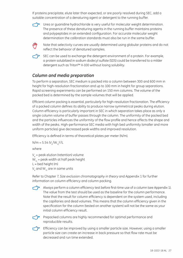
Setting column pressure limitsPressure is generated by the flow through the chromatographic system. For optimal chromatography functionality, it is important to understand the principle of the pressure drop over the different parts of a system (Fig 1.11).
poutlet = 0 MPa
ppost-cp = 1.0 MPa
ppump = 6.5 MPa
p
∆p = Ppre-cp - Ppost-cp
pre-cp = 4.5 MPa
Back pressurePressure drop
∆pafter = 1.0 MPa
∆p = 3.5 MPa
∆p before = 2.0 MPa
Pump
Fig 1.11. Example of the pressure in different parts of a system during run of a column. Note that the pressure values are only used to illustrate the principle.
∆pbefore does not affect the column.
The pressure on the column hardware is the sum of ∆pafter and ∆p. Do not exceed the column hardware limit!
∆p is individual and needs to be determined for each column.
For more information, refer to the ÄKTA™ Laboratory-scale Chromatography Systems Instrument Management Handbook, 29-0108-31.
To protect the column hardware and the packed bed of the chromatographic medium, it is important to set limits that must not be exceeded during the run. There are two important pressure limits that must be taken into consideration:
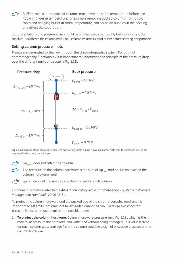
1. To protect the column hardware: Column hardware pressure limit (Fig 1.12), which is the maximum pressure the hardware can withstand without being damaged. This value is fixed for each column type. Leakage from the column could be a sign of excessive pressure on the column hardware.
18-1022-18 AL 29
Fig 1.12. Column hardware pressure limit is the maximum pressure the column can withstand without damage.
The column hardware pressure limit is included in the instructions and in UNICORN™ column list for each column type, respectively.
2. To protect the packed bed: Delta pressure (∆p) or maximum pressure over the packed bed is the maximum pressure the packed bed of chromatography medium can withstand without risking gap formation (Fig 1.13) or bed collapse. This value varies depending on conditions. A typical value for ∆p or maximum pressure drop over the packed bed is provided for each column type in the instructions and UNICORN column list. Note however that ∆p is individual for each column and needs to be determined. The procedure for doing this is described in Instructions 29-0272-71. The packed bed is best protected by controlling the flow rate. Use lower flow rates for high-viscosity solutions and/or low temperature (Table 1.3).
Table 1.3. Example of flow rate limits at different viscosity and temperature, Superose 6 Increase 10/300 GL
Temperature Flow rate (ml/min)
20ºC to 25ºC Maximum flow rate, water 1.5Maximum flow rate, 20% ethanol 0.75
4ºC to 8ºC Maximum flow rate, water 0.75Maximum flow rate, 20% ethanol 0.35
Fig 1.13. The maximum pressure over the packed bed is the maximum pressure the packed bed of chromatography medium can withstand without gap formation. This is not a fixed value.
The pressure over the packed bed is depending on a lot of parameters including:
• Flow rate• Viscosity of sample and eluent• Running temperature • Chromatography medium particle properties• Column packing
30 18-1022-18 AL
Sample applicationA liquid chromatography system should be used for high-resolution separation. For group separations it is possible to use manual purification. Samples can be applied by gravity feed to prepacked columns such as PD-10 Desalting.
Elution and flow ratesSamples are eluted isocratically from a SEC column, using a single buffer system. After sample application the entire separation takes place as one column volume of buffer (equivalent to the volume of the packed bed) passes through the column.
The goal for any separation is to achieve the highest possible resolution in the shortest possible time. Figures 1.14 to 1.16 show that resolution decreases as flow rate increases. Each separation must be optimized to provide balance between these two parameters. Put simply, maximum resolution is obtained with a long column and a low flow rate whereas the fastest run is obtained with a short column and a high flow rate. Suitable flow rates for high-resolution fractionation or group separation are supplied with each product.
The advantage of a higher flow rate (and consequently a faster separation) outweighs the loss of resolution in the separation in certain circumstances.
Column: Superdex 200 Increase 3.2/300Sample: 1. Thyroglobulin (Mr 669 000), 3 mg/ml 2. Ferritin (Mr 440 000), 0.3 mg/ml 3. Aldolase (Mr 158 000), 3 mg/ml 4. Conalbumin (Mr 75 000), 3 mg/ml 5. Ovalbumin (Mr 44 000), 3 mg/ml 6. Carbonic anhydrase (Mr 29 000), 3 mg/ml 7. Ribonuclease A (Mr 13 700), 3 mg/mlSample volume: 10 µlBuffer: 10 mM sodium phosphate, 140 mM sodium chloride, pH 7.4Flow rate: 0.15 and 0.025 ml/min, room temperatureSystem: ÄKTApurifier
0
50
100
150
200
0.0 1.0 2.0 3.0 Vol. (ml)
A280 (mAU)
1
2
3
4
5
6 7
Flow rate: 0.15 ml/minRun time: 16 min
0
50
100
150
200
0.0 1.0 2.0 3.0 Vol. (ml)
A280 (mAU)
1
2
3
4
5
67
Flow rate: 0.025 ml/minRun time: 96 min
Fig 1.14. Comparison of protein separation on Superdex 200 Increase 3.2/300 at different flow rates.
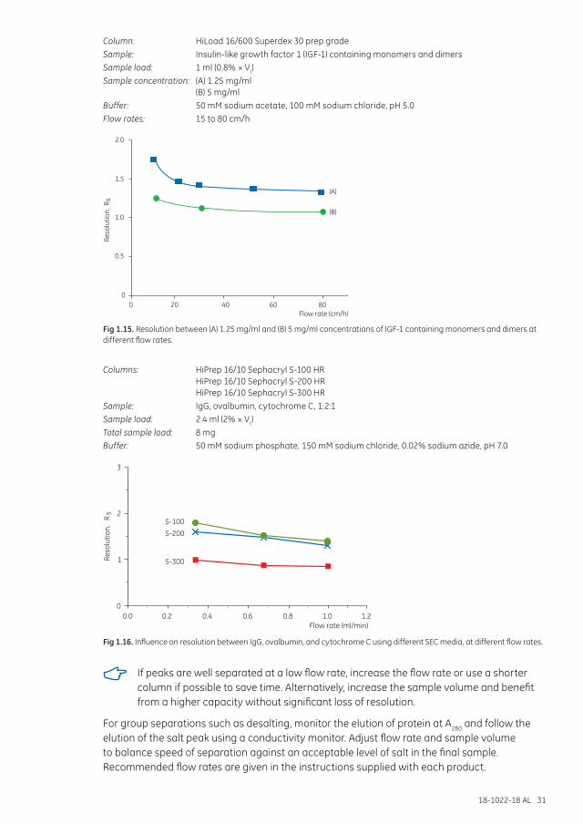
18-1022-18 AL 31
Column: HiLoad 16/600 Superdex 30 prep gradeSample: Insulin-like growth factor 1 (IGF-1) containing monomers and dimersSample load: 1 ml (0.8% × Vt)Sample concentration: (A) 1.25 mg/ml (B) 5 mg/mlBuffer: 50 mM sodium acetate, 100 mM sodium chloride, pH 5.0Flow rates: 15 to 80 cm/h
0
0.5
1.5
1.0
2.0
Reso
lutio
n, R
s
40200 60 80
(A)
(B)
Flow rate (cm/h)
Fig 1.15. Resolution between (A) 1.25 mg/ml and (B) 5 mg/ml concentrations of IGF-1 containing monomers and dimers at different flow rates.
Columns: HiPrep 16/10 Sephacryl S-100 HR HiPrep 16/10 Sephacryl S-200 HR HiPrep 16/10 Sephacryl S-300 HR
Sample: IgG, ovalbumin, cytochrome C, 1:2:1Sample load: 2.4 ml (2% × Vt)Total sample load: 8 mgBuffer: 50 mM sodium phosphate, 150 mM sodium chloride, 0.02% sodium azide, pH 7.0
Reso
lutio
n,
Rs
0
1
0.4
2
0.20.0
3
0.6 0.8 1.0 1.2Flow rate (ml/min)
S-300
S-200S-100
Fig 1.16. Influence on resolution between IgG, ovalbumin, and cytochrome C using different SEC media, at different flow rates.
If peaks are well separated at a low flow rate, increase the flow rate or use a shorter column if possible to save time. Alternatively, increase the sample volume and benefit from a higher capacity without significant loss of resolution.
For group separations such as desalting, monitor the elution of protein at A280 and follow the elution of the salt peak using a conductivity monitor. Adjust flow rate and sample volume to balance speed of separation against an acceptable level of salt in the final sample. Recommended flow rates are given in the instructions supplied with each product.
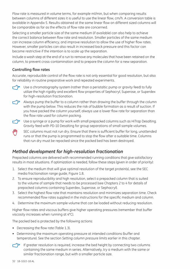
32 18-1022-18 AL
Flow rate is measured in volume terms, for example ml/min, but when comparing results between columns of different sizes it is useful to use the linear flow, cm/h. A conversion table is available in Appendix 5. Results obtained at the same linear flow on different sized columns will be comparable as far as the effects of flow rate are concerned.
Selecting a smaller particle size of the same medium (if available) can also help to achieve the correct balance between flow rate and resolution. Smaller particles of the same medium can increase column efficiency, and improve resolution to allow the use of higher flow rates. However, smaller particles can also result in increased back pressure and this factor can become restrictive if the intention is to scale up the separation.
Include a wash step at the end of a run to remove any molecules that have been retained on the column, to prevent cross-contamination and to prepare the column for a new separation.
Controlling flow ratesAccurate, reproducible control of the flow rate is not only essential for good resolution, but also for reliability in routine preparative work and repeated experiments.
Use a chromatography system (rather than a peristaltic pump or gravity feed) to fully utilize the high rigidity and excellent flow properties of Sephacryl, Superose, or Superdex for high-resolution fractionation.
Always pump the buffer to a column rather than drawing the buffer through the column with the pump below. This reduces the risk of bubble formation as a result of suction. If you have packed the column yourself, always use a lower flow rate for separation than the flow rate used for column packing.
Use a syringe or a pump for work with small prepacked columns such as HiTrap Desalting. Gravity feed with PD-10 Desalting for group separations of small sample volumes.
SEC columns must not run dry. Ensure that there is sufficient buffer for long, unattended runs or that the pump is programmed to stop the flow after a suitable time. Columns that run dry must be repacked since the packed bed has been destroyed.
Method development for high-resolution fractionationPrepacked columns are delivered with recommended running conditions that give satisfactory results in most situations. If optimization is needed, follow these steps (given in order of priority):
1. Select the medium that will give optimal resolution of the target protein(s), see the SEC media fractionation range guide, Figure 1.8.
2. To ensure reproducibility and high resolution, select a prepacked column that is suited to the volume of sample that needs to be processed (see Chapters 2 to 4 for details of prepacked columns containing Superdex, Superose, or Sephacryl).
3. Select the highest flow rate that maintains resolution and minimizes separation time. Check recommended flow rates supplied in the instructions for the specific medium and column.
4. Determine the maximum sample volume that can be loaded without reducing resolution.
Higher flow rates and viscous buffers give higher operating pressures (remember that buffer viscosity increases when running at 4°C).
The packed bed is protected by the following actions:
• Decreasing the flow rate (Table 1.3).
• Determining the maximum operating pressure at intended conditions (buffer and temperature). See the section Setting column pressure limits earlier in this chapter.
If greater resolution is required, increase the bed height by connecting two columns containing the same medium in series. Alternatively, try a medium with the same or similar fractionation range, but with a smaller particle size.
18-1022-18 AL 33
To process larger sample volumes, see scaling up below.
Maintenance of SEC columnsRoutine cleaning of the SEC column, for example every 20 runs (depending on sample), is recommended. Cleaning will prolong the column life time and is performed in order to remove any precipitated proteins or other contaminants that can build up on the column. Cleaning is necessary when a colored band can be seen at top of the column, there is loss in resolution, or a significant increase in back pressure. Detailed cleaning procedures for each SEC medium are given in Chapters 2 to 6 and in the instructions for the product.
Always use filtered buffers and samples to reduce the need for additional column maintenance. See Appendix 3 for further details on sample preparation.
In case of increased back pressure, see also Troubleshooting section in this chapter.
Always use well degassed buffers to avoid the formation of air bubbles in the packed column during a run.
Buffers and prepacked columns should be kept at the same temperature to prevent air bubbles forming in the column.
Filter cleaning solutions before use and always re-equilibrate the column with 2 to 3 CV of buffer before the next separation.
Equipment selectionAppendix 4 provides a guide to the selection of suitable systems for SEC separation.
Scaling upAfter establishing a high-resolution or group separation on a small column, larger columns can be packed to process larger sample volumes in a single step. General guidelines for scaling up are shown in Table 1.4.
Table 1.4. General guidelines for scaling up SEC separations
Maintain Increase
Bed height Column diameter
Linear flow rate Volumetric flow rate
Sample composition Sample volume
To scale up a SEC separation, follow this workflow:
1. Optimize the separation at small scale (see previous section in this chapter; Method development for high-resolution fractionation).
2. Maintain the sample concentration and the ratio of sample to column volume.3. Increase the column volume by increasing the cross-sectional area of the column.4. Maintain the bed height.5. Run the separation at the same linear flow rate as used on the smaller column (see Appendix 5).
Refer to Appendix 1 for column selection and column packing.
Different factors related to the equipment affect performance after scale-up. If a larger column has a less efficient flow distribution system, or a larger system introduces dead volumes, peak broadening can occur. This will cause extra dilution of the product fraction or even loss of resolution if the application is sensitive to variations in efficiency.
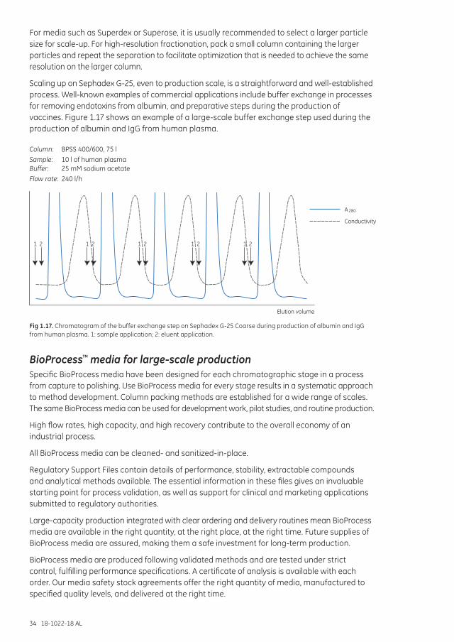
34 18-1022-18 AL
For media such as Superdex or Superose, it is usually recommended to select a larger particle size for scale-up. For high-resolution fractionation, pack a small column containing the larger particles and repeat the separation to facilitate optimization that is needed to achieve the same resolution on the larger column.
Scaling up on Sephadex G-25, even to production scale, is a straightforward and well-established process. Well-known examples of commercial applications include buffer exchange in processes for removing endotoxins from albumin, and preparative steps during the production of vaccines. Figure 1.17 shows an example of a large-scale buffer exchange step used during the production of albumin and IgG from human plasma.
Column: BPSS 400/600, 75 lSample: 10 l of human plasma Buffer: 25 mM sodium acetateFlow rate: 240 l/h
Elution volume
A 280
Conductivity
1 2 1 2 1 2 1 2 1 2
Fig 1.17. Chromatogram of the buffer exchange step on Sephadex G-25 Coarse during production of albumin and IgG from human plasma. 1: sample application; 2: eluent application.
BioProcess™ media for large-scale productionSpecific BioProcess media have been designed for each chromatographic stage in a process from capture to polishing. Use BioProcess media for every stage results in a systematic approach to method development. Column packing methods are established for a wide range of scales. The same BioProcess media can be used for development work, pilot studies, and routine production.
High flow rates, high capacity, and high recovery contribute to the overall economy of an industrial process.
All BioProcess media can be cleaned- and sanitized-in-place.
Regulatory Support Files contain details of performance, stability, extractable compounds and analytical methods available. The essential information in these files gives an invaluable starting point for process validation, as well as support for clinical and marketing applications submitted to regulatory authorities.
Large-capacity production integrated with clear ordering and delivery routines mean BioProcess media are available in the right quantity, at the right place, at the right time. Future supplies of BioProcess media are assured, making them a safe investment for long-term production.
BioProcess media are produced following validated methods and are tested under strict control, fulfilling performance specifications. A certificate of analysis is available with each order. Our media safety stock agreements offer the right quantity of media, manufactured to specified quality levels, and delivered at the right time.
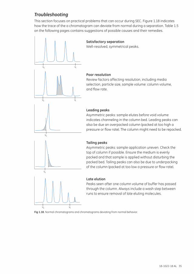
18-1022-18 AL 35
Satisfactory separation Well-resolved, symmetrical peaks.
Poor resolution Review factors affecting resolution, including media selection, particle size, sample volume: column volume, and flow rate.
Leading peaks Asymmetric peaks: sample elutes before void volume indicates channeling in the column bed. Leading peaks can also be due an overpacked column (packed at too high a pressure or flow rate). The column might need to be repacked.
Tailing peaks Asymmetric peaks: sample application uneven. Check the top of column if possible. Ensure the medium is evenly packed and that sample is applied without disturbing the packed bed. Tailing peaks can also be due to underpacking of the column (packed at too low a pressure or flow rate).
Late elution Peaks seen after one column volume of buffer has passed through the column. Always include a wash step between runs to ensure removal of late eluting molecules.
TroubleshootingThis section focuses on practical problems that can occur during SEC. Figure 1.18 indicates how the trace of the a chromatogram can deviate from normal during a separation. Table 1.5 on the following pages contains suggestions of possible causes and their remedies.
VtVo
VtVo
VtVo
Vo
Vo
Fig 1.18. Normal chromatograms and chromatograms deviating from normal behavior.
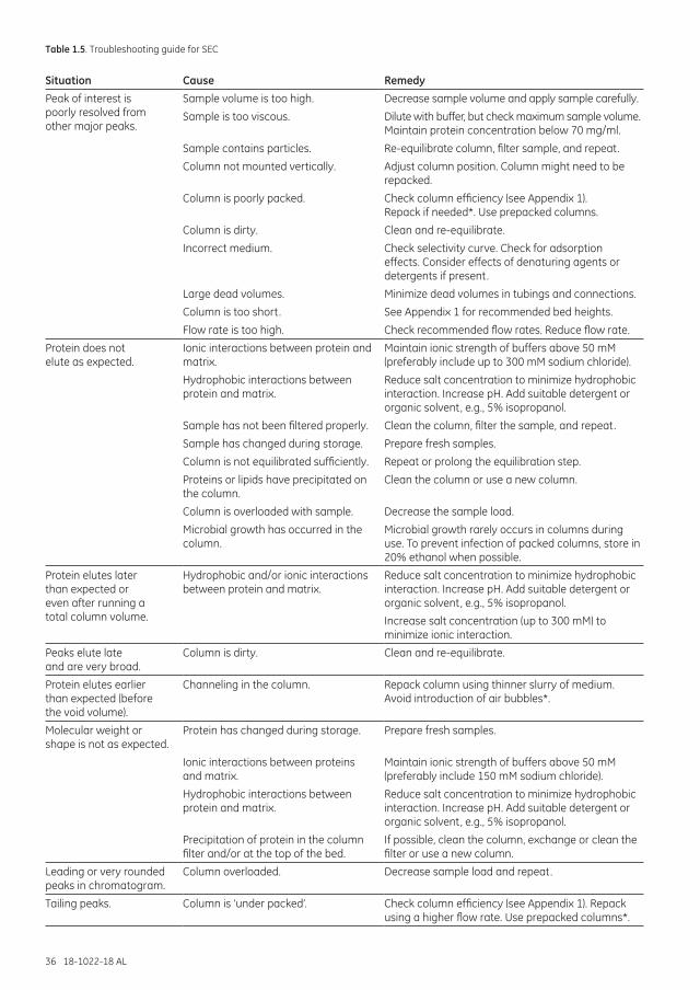
36 18-1022-18 AL
Table 1.5. Troubleshooting guide for SEC
Situation Cause RemedyPeak of interest is poorly resolved from other major peaks.
Sample volume is too high. Decrease sample volume and apply sample carefully.
Sample is too viscous. Dilute with buffer, but check maximum sample volume. Maintain protein concentration below 70 mg/ml.
Sample contains particles. Re-equilibrate column, filter sample, and repeat.
Column not mounted vertically. Adjust column position. Column might need to be repacked.
Column is poorly packed. Check column efficiency (see Appendix 1). Repack if needed*. Use prepacked columns.
Column is dirty. Clean and re-equilibrate.
Incorrect medium. Check selectivity curve. Check for adsorption effects. Consider effects of denaturing agents or detergents if present.
Large dead volumes. Minimize dead volumes in tubings and connections.
Column is too short. See Appendix 1 for recommended bed heights.
Flow rate is too high. Check recommended flow rates. Reduce flow rate.
Protein does not elute as expected.
Ionic interactions between protein and matrix.
Maintain ionic strength of buffers above 50 mM (preferably include up to 300 mM sodium chloride).
Hydrophobic interactions between protein and matrix.
Reduce salt concentration to minimize hydrophobic interaction. Increase pH. Add suitable detergent or organic solvent, e.g., 5% isopropanol.
Sample has not been filtered properly. Clean the column, filter the sample, and repeat.
Sample has changed during storage. Prepare fresh samples.
Column is not equilibrated sufficiently. Repeat or prolong the equilibration step.
Proteins or lipids have precipitated on the column.
Clean the column or use a new column.
Column is overloaded with sample. Decrease the sample load.
Microbial growth has occurred in the column.
Microbial growth rarely occurs in columns during use. To prevent infection of packed columns, store in 20% ethanol when possible.
Protein elutes later than expected or even after running a total column volume.
Hydrophobic and/or ionic interactions between protein and matrix.
Reduce salt concentration to minimize hydrophobic interaction. Increase pH. Add suitable detergent or organic solvent, e.g., 5% isopropanol.
Increase salt concentration (up to 300 mM) to minimize ionic interaction.
Peaks elute late and are very broad.
Column is dirty. Clean and re-equilibrate.
Protein elutes earlier than expected (before the void volume).
Channeling in the column. Repack column using thinner slurry of medium. Avoid introduction of air bubbles*.
Molecular weight or shape is not as expected.
Protein has changed during storage. Prepare fresh samples.
Ionic interactions between proteins and matrix.
Maintain ionic strength of buffers above 50 mM (preferably include 150 mM sodium chloride).
Hydrophobic interactions between protein and matrix.
Reduce salt concentration to minimize hydrophobic interaction. Increase pH. Add suitable detergent or organic solvent, e.g., 5% isopropanol.
Precipitation of protein in the column filter and/or at the top of the bed.
If possible, clean the column, exchange or clean the filter or use a new column.
Leading or very rounded peaks in chromatogram.
Column overloaded. Decrease sample load and repeat.
Tailing peaks. Column is ‘under packed’. Check column efficiency (see Appendix 1). Repack using a higher flow rate. Use prepacked columns*.
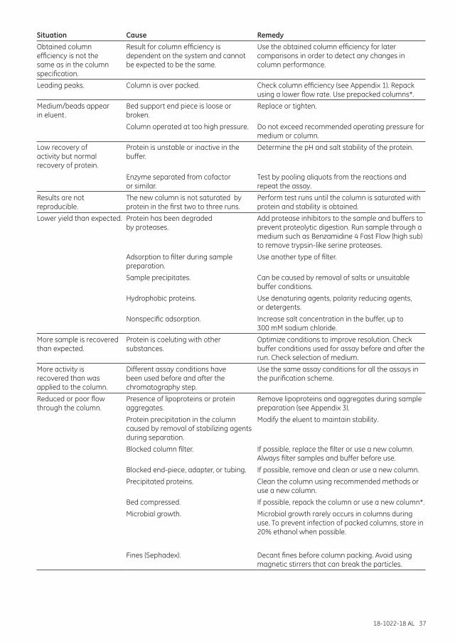
18-1022-18 AL 37
Situation Cause RemedyObtained column efficiency is not the same as in the column specification.
Result for column efficiency is dependent on the system and cannot be expected to be the same.
Use the obtained column efficiency for later comparisons in order to detect any changes in column performance.
Leading peaks. Column is over packed. Check column efficiency (see Appendix 1). Repack using a lower flow rate. Use prepacked columns*.
Medium/beads appear in eluent.
Bed support end piece is loose or broken.
Replace or tighten.
Column operated at too high pressure. Do not exceed recommended operating pressure for medium or column.
Low recovery of activity but normal recovery of protein.
Protein is unstable or inactive in the buffer.
Determine the pH and salt stability of the protein.
Enzyme separated from cofactor or similar.
Test by pooling aliquots from the reactions and repeat the assay.
Results are not reproducible.
The new column is not saturated by protein in the first two to three runs.
Perform test runs until the column is saturated with protein and stability is obtained.
Lower yield than expected. Protein has been degraded by proteases.
Add protease inhibitors to the sample and buffers to prevent proteolytic digestion. Run sample through a medium such as Benzamidine 4 Fast Flow (high sub) to remove trypsin-like serine proteases.
Adsorption to filter during sample preparation.
Use another type of filter.
Sample precipitates. Can be caused by removal of salts or unsuitable buffer conditions.
Hydrophobic proteins. Use denaturing agents, polarity reducing agents, or detergents.
Nonspecific adsorption. Increase salt concentration in the buffer, up to 300 mM sodium chloride.
More sample is recovered than expected.
Protein is coeluting with other substances.
Optimize conditions to improve resolution. Check buffer conditions used for assay before and after the run. Check selection of medium.
More activity is recovered than was applied to the column.
Different assay conditions have been used before and after the chromatography step.
Use the same assay conditions for all the assays in the purification scheme.
Reduced or poor flow through the column.
Presence of lipoproteins or protein aggregates.
Remove lipoproteins and aggregates during sample preparation (see Appendix 3).
Protein precipitation in the column caused by removal of stabilizing agents during separation.
Modify the eluent to maintain stability.
Blocked column filter. If possible, replace the filter or use a new column. Always filter samples and buffer before use.
Blocked end-piece, adapter, or tubing. If possible, remove and clean or use a new column.
Precipitated proteins. Clean the column using recommended methods or use a new column.
Bed compressed. If possible, repack the column or use a new column*.
Microbial growth. Microbial growth rarely occurs in columns during use. To prevent infection of packed columns, store in 20% ethanol when possible.
Fines (Sephadex). Decant fines before column packing. Avoid using magnetic stirrers that can break the particles.
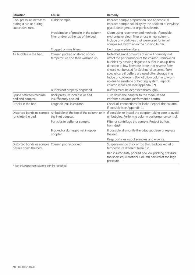
38 18-1022-18 AL
Situation Cause RemedyBack pressure increases during a run or during successive runs.
Turbid sample. Improve sample preparation (see Appendix 3). Improve sample solubility by the addition of ethylene glycol, detergents, or organic solvents.
Precipitation of protein in the column filter and/or at the top of the bed.
Clean using recommended methods. If possible, exchange or clean filter or use a new column. Include any additives that were used for initial sample solubilization in the running buffer.
Clogged on-line filters. Exchange on-line filters.
Air bubbles in the bed. Column packed or stored at cool temperature and then warmed up.
Note that small amounts of air will normally not affect the performance of the column. Remove air bubbles by passing degassed buffer in an up-flow direction at low flow rate. Note that reverse flow should not be used for Sephacryl columns. Take special care if buffers are used after storage in a fridge or cold-room. Do not allow column to warm up due to sunshine or heating system. Repack column if possible (see Appendix 1*).
Buffers not properly degassed. Buffers must be degassed thoroughly.
Space between medium bed and adapter.
Back pressure increase or bed insufficiently packed.
Turn down the adapter to the medium bed. Perform a column performance control.
Cracks in the bed. Large air leak in column. Check all connections for leaks. Repack the column if possible (see Appendix 1).
Distorted bands as sample runs into the bed.
Air bubble at the top of the column or in the inlet adapter.
If possible, re-install the adapter taking care to avoid air bubbles. Perform a column performance control.
Particles in buffer or sample. Filter or centrifuge the sample. Protect buffers from dust.
Blocked or damaged net in upper adapter.
If possible, dismantle the adapter, clean or replace the net.
Keep particles out of samples and eluents.
Distorted bands as sample passes down the bed.
Column poorly packed. Suspension too thick or too thin. Bed packed at a temperature different from run.
Bed insufficiently packed (too low packing pressure, too short equilibration). Column packed at too high pressure.
* Not all prepacked columns can be repacked.
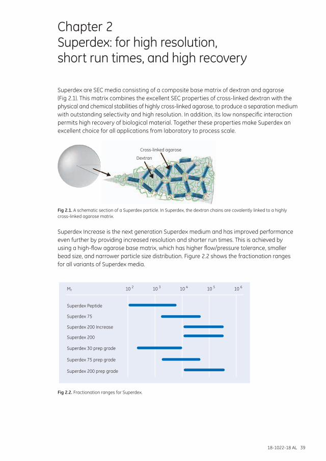
18-1022-18 AL 39
Chapter 2 Superdex: for high resolution, short run times, and high recovery
Superdex are SEC media consisting of a composite base matrix of dextran and agarose (Fig 2.1). This matrix combines the excellent SEC properties of cross-linked dextran with the physical and chemical stabilities of highly cross-linked agarose, to produce a separation medium with outstanding selectivity and high resolution. In addition, its low nonspecific interaction permits high recovery of biological material. Together these properties make Superdex an excellent choice for all applications from laboratory to process scale.
Dextran
Cross-linked agarose
Fig 2.1. A schematic section of a Superdex particle. In Superdex, the dextran chains are covalently linked to a highly cross-linked agarose matrix.
Superdex Increase is the next generation Superdex medium and has improved performance even further by providing increased resolution and shorter run times. This is achieved by using a high-flow agarose base matrix, which has higher flow/pressure tolerance, smaller bead size, and narrower particle size distribution. Figure 2.2 shows the fractionation ranges for all variants of Superdex media.
Superdex Peptide
Superdex 75
Superdex 200
Superdex 30 prep grade
Superdex 75 prep grade
Superdex 200 prep grade
Superdex 200 Increase
10M 2r 10 3 10 4 10 5 10 6
Fig 2.2. Fractionation ranges for Superdex.
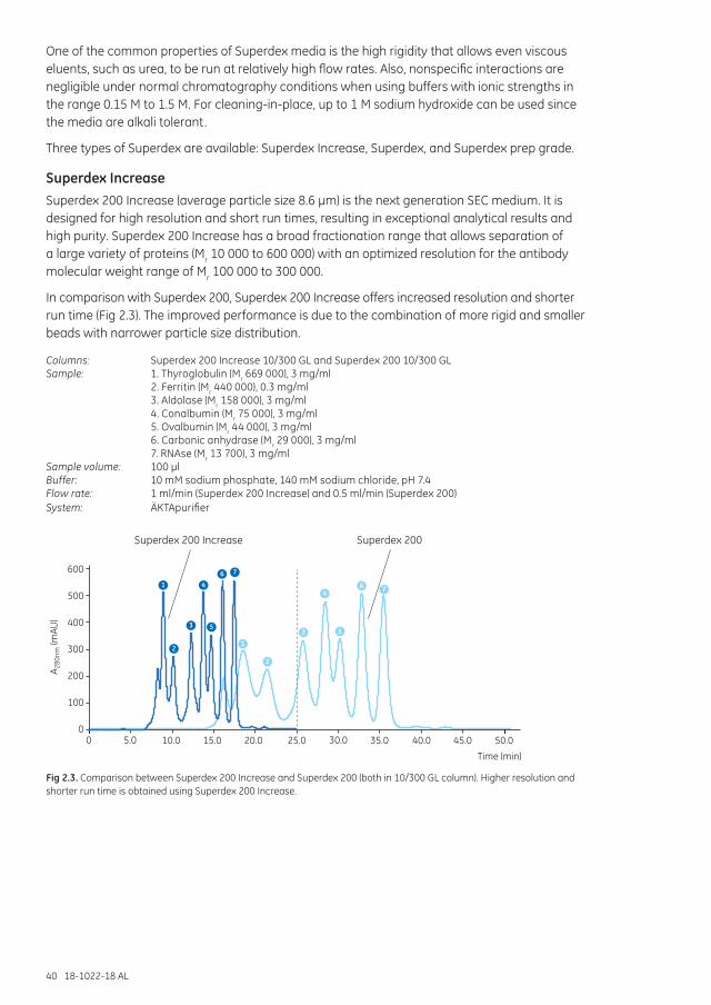
40 18-1022-18 AL
One of the common properties of Superdex media is the high rigidity that allows even viscous eluents, such as urea, to be run at relatively high flow rates. Also, nonspecific interactions are negligible under normal chromatography conditions when using buffers with ionic strengths in the range 0.15 M to 1.5 M. For cleaning-in-place, up to 1 M sodium hydroxide can be used since the media are alkali tolerant.
Three types of Superdex are available: Superdex Increase, Superdex, and Superdex prep grade.
Superdex Increase Superdex 200 Increase (average particle size 8.6 µm) is the next generation SEC medium. It is designed for high resolution and short run times, resulting in exceptional analytical results and high purity. Superdex 200 Increase has a broad fractionation range that allows separation of a large variety of proteins (Mr 10 000 to 600 000) with an optimized resolution for the antibody molecular weight range of Mr 100 000 to 300 000.
In comparison with Superdex 200, Superdex 200 Increase offers increased resolution and shorter run time (Fig 2.3). The improved performance is due to the combination of more rigid and smaller beads with narrower particle size distribution.
Columns: Superdex 200 Increase 10/300 GL and Superdex 200 10/300 GL Sample: 1. Thyroglobulin (Mr 669 000), 3 mg/ml 2. Ferritin (Mr 440 000), 0.3 mg/ml 3. Aldolase (Mr 158 000), 3 mg/ml 4. Conalbumin (Mr 75 000), 3 mg/ml 5. Ovalbumin (Mr 44 000), 3 mg/ml 6. Carbonic anhydrase (Mr 29 000), 3 mg/ml 7. RNAse (Mr 13 700), 3 mg/mlSample volume: 100 µlBuffer: 10 mM sodium phosphate, 140 mM sodium chloride, pH 7.4Flow rate: 1 ml/min (Superdex 200 Increase) and 0.5 ml/min (Superdex 200)System: ÄKTApurifier
600
500
400
300
200
100
05.0 10.0 15.0 20.0 25.0 30.0 35.0 40.0 45.0 50.00
Time (min)
A 280
nm (m
AU)
Superdex 200 Increase Superdex 200
1
2
3
4
5
6 7
1
2
3
4
5
6 7
Fig 2.3. Comparison between Superdex 200 Increase and Superdex 200 (both in 10/300 GL column). Higher resolution and shorter run time is obtained using Superdex 200 Increase.
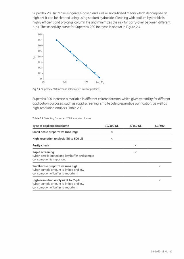
18-1022-18 AL 41
Superdex 200 Increase is agarose-based and, unlike silica-based media which decompose at high pH, it can be cleaned using using sodium hydroxide. Cleaning with sodium hydroxide is highly efficient and prolongs column life and minimizes the risk for carry-over between different runs. The selectivity curve for Superdex 200 Increase is shown in Figure 2.4.
0.8
0.7
0.6
0.5
0.4
0.3
0.2
0.1
0103 104 105
K av
Log Mr
Fig 2.4. Superdex 200 Increase selectivity curve for proteins.
Superdex 200 Increase is available in different column formats, which gives versatility for different application purposes, such as rapid screening, small-scale preparative purification, as well as high-resolution analysis (Table 2.1).
Table 2.1. Selecting Superdex 200 Increase columns
Type of application/column 10/300 GL 5/150 GL 3.2/300
Small-scale preparative runs (mg) ×
High-resolution analysis (25 to 500 µl) ×
Purity check ×
Rapid screeningWhen time is limited and low buffer and sample consumption is important
×
Small-scale preparative runs (µg)When sample amount is limited and low consumption of buffer is important
×
High-resolution analysis (4 to 25 µl)When sample amount is limited and low consumption of buffer is important
×

42 18-1022-18 AL
Superdex Superdex Peptide, Superdex 75, and Superdex 200 have an average bead size of 13 µm and are designed for different fractionation ranges. The media are available in prepacked columns for high performance. Superdex Peptide is suitable for high-resolution fractionation of peptides, oligonucleotides, and small proteins (Mr 100 to 7000). Figure 2.5 shows a separation of standard peptides on Superdex Peptide 10/300 GL.
Column: Superdex Peptide 10/300 GLSample: 1. Cytochrome C (Mr 12 384), 1 mg/ml 2. Aprotinin (Mr 6512), 2 mg/ml 3. Vitamin B12 (Mr 1355) 0.1 mg/ml 4. (Gly)3 (Mr 189), 0.1 mg/ml 5. Gly (Mr 75), 7.8 mg/mlSample load: 50 µlBuffer: 50 mM phosphate, 150 mM sodium chloride, pH 7.0Flow rate: 0.5 ml/min, room temperature
600
500
400
300
200
100
0
5.0 10.0 15.0 20.0 25.00Vol. (ml)
A 215n
m (m
AU)
12
3
4
5
VoVt
Fig 2.5. Separation of standard peptides on Superdex Peptide 10/300 GL.
Superdex 75 is designed for high-resolution fractionation of biomolecules with Mr 3000 to 70 000, such as recombinant proteins. Examples of application areas are screening of solubilization conditions for membrane proteins, study of protein-protein interactions, and rapid purity check of proteins. A dimer-monomer separation of a recombinant protein is shown in Figure 2.6.
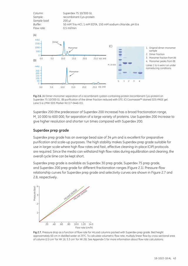
18-1022-18 AL 43
Column: Superdex 75 10/300 GLSample: recombinant Cys-proteinSample load: 200 µlBuffer: 50 mM Tris-HCl, 1 mM EDTA, 150 mM sodium chloride, pH 8.4Flow rate: 0.5 ml/min
(B)
(A)
(C)
mAU
1500
1000
500
00.0 5.0 10.0 15.0 20.0 25.0 Vol. (ml)
Dimer
Monomer
250200150100
500
Mr 20 000mAU
0.0 5.0 10.0 15.0 20.0 25.0 Vol. (ml)
Monomer
S 1 2 3 4
1. Original dimer-monomer sample
2. Dimer fraction3. Monomer fraction from (A)4. Monomer peaks from (B)
Lanes 1 to 4 were run under nonreducing conditions.
Fig 2.6. (A) Dimer-monomer separation of a recombinant cystein-containing protein (recombinant Cys-protein) on Superdex 75 10/300 GL. (B) purification of the dimer fraction reduced with DTE. (C) Coomassie™ stained SDS-PAGE gel. Lane S is LMW-SDS Marker Kit (17-0446-01).
Superdex 200 (the predecessor of Superdex 200 Increase) has a broad fractionation range, Mr 10 000 to 600 000, for separation of a large variety of proteins. Use Superdex 200 Increase to give higher resolution and shorter run times compared with Superdex 200.
Superdex prep grade
Superdex prep grade has an average bead size of 34 µm and is excellent for preparative purification and scale-up purposes. The high stability makes Superdex prep grade suitable for use in larger scale where high flow rates and fast, effective cleaning-in-place (CIP) protocols are required. Since the media can withstand high flow rates during equilibration and cleaning, the overall cycle time can be kept short.
Superdex prep grade is available as Superdex 30 prep grade, Superdex 75 prep grade, and Superdex 200 prep grade for different fractionation ranges (Figure 2.1). Pressure-flow relationship curves for Superdex prep grade and selectivity curves are shown in Figure 2.7 and 2.8, respectively.
4
3
2
1
60 80 10 0 1 20 14 0
HiLoad 26/600
HiLoad 16/600
Flow rate (cm/h)
Pres
sure
(bar
)
20 40
Fig 2.7. Pressure drop as a function of flow rate for HiLoad columns packed with Superdex prep grade. Bed height approximately 60 cm in distilled water at 25°C. To calculate volumetric flow rate, multiply linear flow by cross-sectional area of column (2.0 cm2 for XK 16, 5.3 cm2 for XK 26). See Appendix 5 for more information about flow rate calculations.
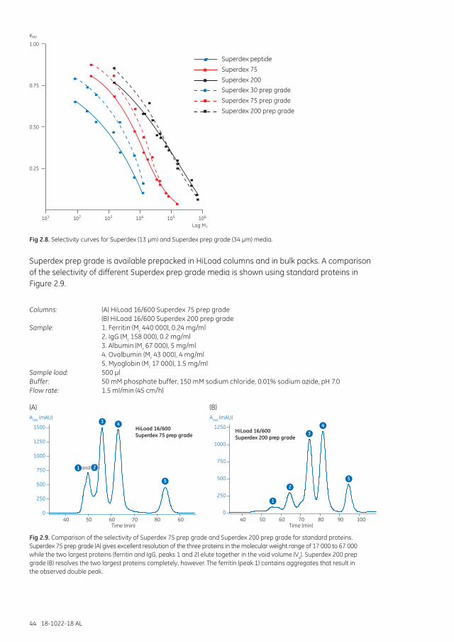
44 18-1022-18 AL
0.25
0.50
0.75
1.00
Kav
Superdex peptide
Superdex 30 prep grade
Log Mr
Superdex 200
Superdex 75
Superdex 200 prep grade
Superdex 75 prep grade
101 102 103 104 105 106
Fig 2.8. Selectivity curves for Superdex (13 µm) and Superdex prep grade (34 µm) media.
Superdex prep grade is available prepacked in HiLoad columns and in bulk packs. A comparison of the selectivity of different Superdex prep grade media is shown using standard proteins in Figure 2.9.
Columns: (A) HiLoad 16/600 Superdex 75 prep grade (B) HiLoad 16/600 Superdex 200 prep gradeSample: 1. Ferritin (Mr 440 000), 0.24 mg/ml 2. IgG (Mr 158 000), 0.2 mg/ml 3. Albumin (Mr 67 000), 5 mg/ml 4. Ovalbumin (Mr 43 000), 4 mg/ml 5. Myoglobin (Mr 17 000), 1.5 mg/mlSample load: 500 µlBuffer: 50 mM phosphate buffer, 150 mM sodium chloride, 0.01% sodium azide, pH 7.0Flow rate: 1.5 ml/min (45 cm/h)
HiLoad 16/600Superdex 75 prep grade
1500
A280 (mAU) A280 (mAU)
1250
1000
750
500
250
040 50 60 70 80 60
Time (min)
HiLoad 16/600Superdex 200 prep grade
1250
1000
750
500
250
040 50 60 70 80 90 100
Time (min)
34
3 4
5 5
1
1
2and
2
(A) (B)
Fig 2.9. Comparison of the selectivity of Superdex 75 prep grade and Superdex 200 prep grade for standard proteins. Superdex 75 prep grade (A) gives excellent resolution of the three proteins in the molecular weight range of 17 000 to 67 000 while the two largest proteins (ferritin and IgG, peaks 1 and 2) elute together in the void volume (Vo). Superdex 200 prep grade (B) resolves the two largest proteins completely, however. The ferritin (peak 1) contains aggregates that result in the observed double peak.
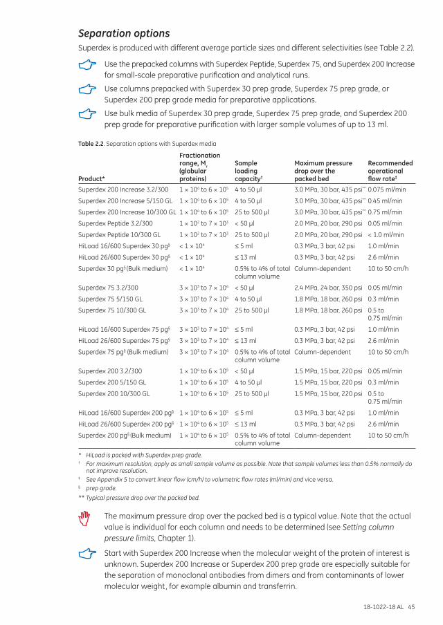
18-1022-18 AL 45
Separation optionsSuperdex is produced with different average particle sizes and different selectivities (see Table 2.2).
Use the prepacked columns with Superdex Peptide, Superdex 75, and Superdex 200 Increase for small-scale preparative purification and analytical runs.
Use columns prepacked with Superdex 30 prep grade, Superdex 75 prep grade, or Superdex 200 prep grade media for preparative applications.
Use bulk media of Superdex 30 prep grade, Superdex 75 prep grade, and Superdex 200 prep grade for preparative purification with larger sample volumes of up to 13 ml.
Table 2.2. Separation options with Superdex media
Product*
Fractionation range, Mr (globular proteins)
Sample loading capacity†
Maximum pressure drop over the packed bed
Recommended operational flow rate‡
Superdex 200 Increase 3.2/300 1 × 10⁴ to 6 × 10⁵ 4 to 50 µl 3.0 MPa, 30 bar, 435 psi** 0.075 ml/min
Superdex 200 Increase 5/150 GL 1 × 10⁴ to 6 × 10⁵ 4 to 50 µl 3.0 MPa, 30 bar, 435 psi** 0.45 ml/min
Superdex 200 Increase 10/300 GL 1 × 10⁴ to 6 × 10⁵ 25 to 500 µl 3.0 MPa, 30 bar, 435 psi** 0.75 ml/min
Superdex Peptide 3.2/300 1 × 10² to 7 × 10³ < 50 µl 2.0 MPa, 20 bar, 290 psi 0.05 ml/min
Superdex Peptide 10/300 GL 1 × 10² to 7 × 10³ 25 to 500 µl 2.0 MPa, 20 bar, 290 psi < 1.0 ml/min
HiLoad 16/600 Superdex 30 pg§ < 1 × 10⁴ ≤ 5 ml 0.3 MPa, 3 bar, 42 psi 1.0 ml/min
HiLoad 26/600 Superdex 30 pg§ < 1 × 10⁴ ≤ 13 ml 0.3 MPa, 3 bar, 42 psi 2.6 ml/min
Superdex 30 pg§ (Bulk medium) < 1 × 10⁴ 0.5% to 4% of total column volume
Column-dependent 10 to 50 cm/h
Superdex 75 3.2/300 3 × 10³ to 7 × 10⁴ < 50 µl 2.4 MPa, 24 bar, 350 psi 0.05 ml/min
Superdex 75 5/150 GL 3 × 10³ to 7 × 10⁴ 4 to 50 µl 1.8 MPa, 18 bar, 260 psi 0.3 ml/min
Superdex 75 10/300 GL 3 × 10³ to 7 × 10⁴ 25 to 500 µl 1.8 MPa, 18 bar, 260 psi 0.5 to 0.75 ml/min
HiLoad 16/600 Superdex 75 pg§ 3 × 10³ to 7 × 10⁴ ≤ 5 ml 0.3 MPa, 3 bar, 42 psi 1.0 ml/min
HiLoad 26/600 Superdex 75 pg§ 3 × 10³ to 7 × 10⁴ ≤ 13 ml 0.3 MPa, 3 bar, 42 psi 2.6 ml/min
Superdex 75 pg§ (Bulk medium) 3 × 10³ to 7 × 10⁴ 0.5% to 4% of total column volume
Column-dependent 10 to 50 cm/h
Superdex 200 3.2/300 1 × 10⁴ to 6 × 10⁵ < 50 µl 1.5 MPa, 15 bar, 220 psi 0.05 ml/min
Superdex 200 5/150 GL 1 × 10⁴ to 6 × 10⁵ 4 to 50 µl 1.5 MPa, 15 bar, 220 psi 0.3 ml/min
Superdex 200 10/300 GL 1 × 10⁴ to 6 × 10⁵ 25 to 500 µl 1.5 MPa, 15 bar, 220 psi 0.5 to 0.75 ml/min
HiLoad 16/600 Superdex 200 pg§ 1 × 10⁴ to 6 × 10⁵ ≤ 5 ml 0.3 MPa, 3 bar, 42 psi 1.0 ml/min
HiLoad 26/600 Superdex 200 pg§ 1 × 10⁴ to 6 × 10⁵ ≤ 13 ml 0.3 MPa, 3 bar, 42 psi 2.6 ml/min
Superdex 200 pg§ (Bulk medium) 1 × 10⁴ to 6 × 10⁵ 0.5% to 4% of total column volume
Column-dependent 10 to 50 cm/h
* HiLoad is packed with Superdex prep grade.† For maximum resolution, apply as small sample volume as possible. Note that sample volumes less than 0.5% normally do
not improve resolution.‡ See Appendix 5 to convert linear flow (cm/h) to volumetric flow rates (ml/min) and vice versa.§ prep grade.** Typical pressure drop over the packed bed.
The maximum pressure drop over the packed bed is a typical value. Note that the actual value is individual for each column and needs to be determined (see Setting column pressure limits, Chapter 1).
Start with Superdex 200 Increase when the molecular weight of the protein of interest is unknown. Superdex 200 Increase or Superdex 200 prep grade are especially suitable for the separation of monoclonal antibodies from dimers and from contaminants of lower molecular weight, for example albumin and transferrin.
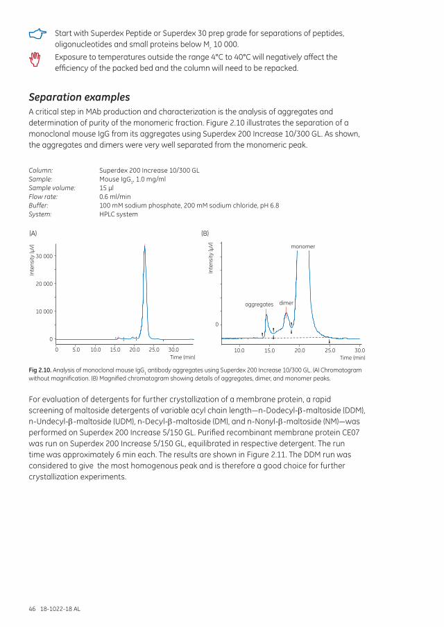
46 18-1022-18 AL
Start with Superdex Peptide or Superdex 30 prep grade for separations of peptides, oligonucleotides and small proteins below Mr 10 000.
Exposure to temperatures outside the range 4°C to 40°C will negatively affect the efficiency of the packed bed and the column will need to be repacked.
Separation examples A critical step in MAb production and characterization is the analysis of aggregates and determination of purity of the monomeric fraction. Figure 2.10 illustrates the separation of a monoclonal mouse IgG from its aggregates using Superdex 200 Increase 10/300 GL. As shown, the aggregates and dimers were very well separated from the monomeric peak.
Column: Superdex 200 Increase 10/300 GLSample: Mouse IgG1, 1.0 mg/mlSample volume: 15 µlFlow rate: 0.6 ml/minBuffer: 100 mM sodium phosphate, 200 mM sodium chloride, pH 6.8System: HPLC system
30 000
20 000
10 000
0
Inte
nsity
(µV)
Inte
nsity
(µV)
20.0 25.0 30.010.0 15.0
aggregates dimer
monomer
20.0 25.0 30.010.05.0 15.00Time (min)Time (min)
0
(B)(A)
Fig 2.10. Analysis of monoclonal mouse IgG1 antibody aggregates using Superdex 200 Increase 10/300 GL. (A) Chromatogram without magnification. (B) Magnified chromatogram showing details of aggregates, dimer, and monomer peaks.
For evaluation of detergents for further crystallization of a membrane protein, a rapid screening of maltoside detergents of variable acyl chain length—n-Dodecyl-E-maltoside (DDM), n-Undecyl-E-maltoside (UDM), n-Decyl-E-maltoside (DM), and n-Nonyl-E-maltoside (NM)—was performed on Superdex 200 Increase 5/150 GL. Purified recombinant membrane protein CE07 was run on Superdex 200 Increase 5/150 GL, equilibrated in respective detergent. The run time was approximately 6 min each. The results are shown in Figure 2.11. The DDM run was considered to give the most homogenous peak and is therefore a good choice for further crystallization experiments.
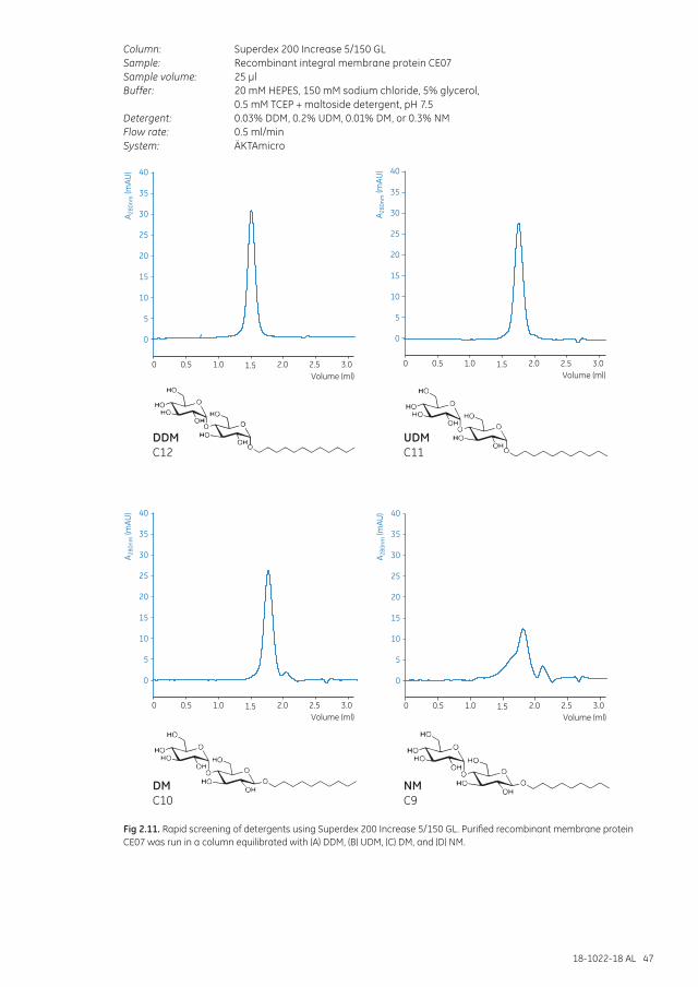
18-1022-18 AL 47
Column: Superdex 200 Increase 5/150 GLSample: Recombinant integral membrane protein CE07Sample volume: 25 µlBuffer: 20 mM HEPES, 150 mM sodium chloride, 5% glycerol,
0.5 mM TCEP + maltoside detergent, pH 7.5Detergent: 0.03% DDM, 0.2% UDM, 0.01% DM, or 0.3% NMFlow rate: 0.5 ml/minSystem: ÄKTAmicro
40
35
30
25
20
15
10
5
0
A 280
nm (m
AU)
2.0 2.5 3.01.0 1.50.50Volume (ml)
40
35
30
25
20
15
10
5
0
A 280
nm (m
AU)
2.0 2.5 3.01.0 1.50.50Volume (ml)
40
35
30
25
20
15
10
5
0
A 280
nm (m
AU)
2.0 2.5 3.01.0 1.50.50Volume (ml)
40
35
30
25
20
15
10
5
0
A 280
nm (m
AU)
2.0 2.5 3.01.0 1.50.50Volume (ml)
DDMC12
UDMC11
DMC10
NMC9
Fig 2.11. Rapid screening of detergents using Superdex 200 Increase 5/150 GL. Purified recombinant membrane protein CE07 was run in a column equilibrated with (A) DDM, (B) UDM, (C) DM, and (D) NM.
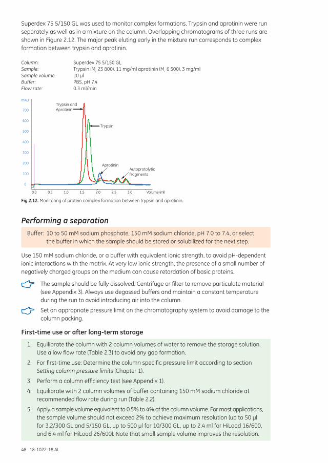
48 18-1022-18 AL
Superdex 75 5/150 GL was used to monitor complex formations. Trypsin and aprotinin were run separately as well as in a mixture on the column. Overlapping chromatograms of three runs are shown in Figure 2.12. The major peak eluting early in the mixture run corresponds to complex formation between trypsin and aprotinin.
Column: Superdex 75 5/150 GLSample: Trypsin (Mr 23 800), 11 mg/ml aprotinin (Mr 6 500), 3 mg/mlSample volume: 10 µlBuffer: PBS, pH 7.4Flow rate: 0.3 ml/min
0
100
200
300
400
500
600
700
mAU
0.0 0.5 1.0 1.5 2.0 2.5 3.0 Volume (ml)
Trypsin andAprotinin
Trypsin
AprotininAutoprotolyticfragments
Fig 2.12. Monitoring of protein complex formation between trypsin and aprotinin.
Performing a separationBuffer: 10 to 50 mM sodium phosphate, 150 mM sodium chloride, pH 7.0 to 7.4, or select
the buffer in which the sample should be stored or solubilized for the next step.
Use 150 mM sodium chloride, or a buffer with equivalent ionic strength, to avoid pH-dependent ionic interactions with the matrix. At very low ionic strength, the presence of a small number of negatively charged groups on the medium can cause retardation of basic proteins.
The sample should be fully dissolved. Centrifuge or filter to remove particulate material (see Appendix 3). Always use degassed buffers and maintain a constant temperature during the run to avoid introducing air into the column.
Set an appropriate pressure limit on the chromatography system to avoid damage to the column packing.
First-time use or after long-term storage
1. Equilibrate the column with 2 column volumes of water to remove the storage solution. Use a low flow rate (Table 2.3) to avoid any gap formation.
2. For first-time use: Determine the column specific pressure limit according to section Setting column pressure limits (Chapter 1).
3. Perform a column efficiency test (see Appendix 1).
4. Equilibrate with 2 column volumes of buffer containing 150 mM sodium chloride at recommended flow rate during run (Table 2.2).
5. Apply a sample volume equivalent to 0.5% to 4% of the column volume. For most applications, the sample volume should not exceed 2% to achieve maximum resolution (up to 50 µl for 3.2/300 GL and 5/150 GL, up to 500 µl for 10/300 GL, up to 2.4 ml for HiLoad 16/600, and 6.4 ml for HiLoad 26/600). Note that small sample volume improves the resolution.
18-1022-18 AL 49
6. Elute with 1 column volume of buffer.
7. Before applying a new sample, re-equilibrate column with buffer until the baseline monitored at A280 is stable.
Column performance should be checked at regular intervals by determining the theoretical plate number per meter and peak symmetry.
See Appendix 1 for how to check column efficiency.
See Chapter 1 for advice on optimizing the separation.
Make sure to not exceed the pressure limits of the column and consider flow limitations (Table 1.3). This is particularly important when working at low temperatures, such as in a cold room, or when the column is used with 20% ethanol or other viscous solutions.
Exposure to temperatures outside the range 4°C to 40°C will negatively affect the efficiency of a packed bed and the column will need to be repacked.
Table 2.3. Recommended flow rates at different stages using prepacked columns containing Superdex Increase, Superdex, or HiLoad columns containing Superdex prep grade media
Flow rates at room temperature
3.2/300 GL 5/150 GL 10/300 GL HiLoad 16/600 HiLoad 26/600
First-time use or after long-term storage
0.05 ml/min 0.3 ml/min 0.5 ml/min 1 ml/min* (30 cm/h)
2.6 ml/min* (30 cm/h)
First-time use or after long-term storage (Superdex 200 Increase)
0.075 ml/min 0.3 ml/min 0.75 ml/min N/A N/A
Cleaning-in-place, CIP 0.02 ml/min 0.3 ml/min† 0.5 ml/min 0.8 ml/min (25 cm/h)
2.2 ml/min (25 cm/h)
* HiLoad: To save time, higher flow rate can be used for the equilibration with buffer: 1.6 ml/min and 4.3 ml/min for HiLoad 16/600 and HiLoad 26/600, respectively. Reduce the flow rate to the recommended flow rate during run before sample application.
† 0.13 ml/min for Superdex 200 Increase 5/150 GL.
Cleaning
1. Wash with 1 column volume of 0.5 M sodium hydroxide, alternatively 0.5 M acetic acid. Use a low flow rate during the entire CIP procedure (see Table 2.3).
2. Immediately wash with 1 column volume of distilled water followed by at least 2 column volumes of buffer until the baseline monitored at A280 and the pH of the eluent are stable.
Further equilibration might be necessary if the buffer contains detergent.
Routine cleaning after every 10 to 20 separations is recommended, but the frequency of cleaning will also depend on the nature of the samples being applied.
Removing severe contamination
1. Reverse the flow.
2. Wash with 4 column volumes of 1 M NaOH (to remove hydrophobic proteins or lipoproteins) followed by 4 column volumes of distilled water.
50 18-1022-18 AL
3. Wash with 0.5 column volume of 30% isopropanol to remove lipids and very hydrophobic proteins, followed by 2 column volumes of distilled water.
4. Equilibrate the column with at least 5 column volumes of buffer, or until the baseline monitored at A280 and the pH of the eluent are stable, before beginning a new separation.
For extreme cases of contamination, check the instructions supplied with the product.
Media characteristicsMatrix: Spherical composite of cross-linked agarose and dextran.
Table 2.4. Superdex media characteristics
Product Efficiency* pH stability† Average particle size (µm)
Superdex 200 Increase‡ > 48 000 Long term: 3 to 12Short term: 1 to 14
8.6
Superdex Peptide ≥ 30 000 Long term: 1 to 14Short term: 1 to 14
13
Superdex 75‡ ≥ 30 000 Long term: 3 to 12Short term: 1 to 14
13
Superdex 200‡ ≥ 30 000 Long term: 3 to 12Short term: 1 to 14
13
Superdex 30 prep grade ≥ 13 000 Long term: 3 to 12Short term: 1 to 14
34
Superdex 75 prep grade ≥ 13 000 Long term: 3 to 12Short term: 1 to 14
34
Superdex 200 prep grade ≥ 13 000 Long term: 3 to 12Short term: 1 to 14
34
* Theoretical plates m-1 (prepacked columns only).† Long-term pH stability refers to the pH interval where the medium is stable over a long period of time without adverse side
effects on its chromatography performance. Short-term pH stability refers to the pH interval for regeneration, cleaning-in-place and sanitization procedures. All ranges are GE Healthcare estimates.
‡ Column length 300 mm.
Chemical stabilitySuperdex is stable in all commonly used aqueous buffers and additives such as detergents (1% SDS), and denaturing agents (8 M urea or 6 M guanidine hydrochloride).
The following solutions can be used for cleaning: up to 30% acetonitrile, up to 1 M sodium hydroxide, up to 70% ethanol (Superdex Increase, Superdex, Superdex 30 prep grade); up to 24% ethanol (Superdex 75 prep grade and Superdex 200 prep grade); up to 1 M acetic acid, 30% isopropanol or 0.1 M hydrochloric acid (Superdex Increase, Superdex, Superdex 30 prep grade).
StorageStore unused media 4°C to 30°C in 20% ethanol (store Superdex 30 pg and Superdex 75 pg in 200 mM sodium acetate, 20% ethanol). Do not freeze.
For long-term storage, wash with 2 column volumes of distilled water followed by 2 column volumes of 20% ethanol (or 200 mM sodium acetate, 20% ethanol for Superdex 30 pg and Superdex 75 pg). Store at 4°C to 30°C.
Degas the ethanol/water mixture thoroughly and use a low flow rate. Connect the transport tool to the capillary tubing at the column outlet to prevent air from entering the column. Fill the transport tool up to approximately 50% of the transport tool volume.
Avoid changes in temperature, which can cause formation of air bubbles in the packing.
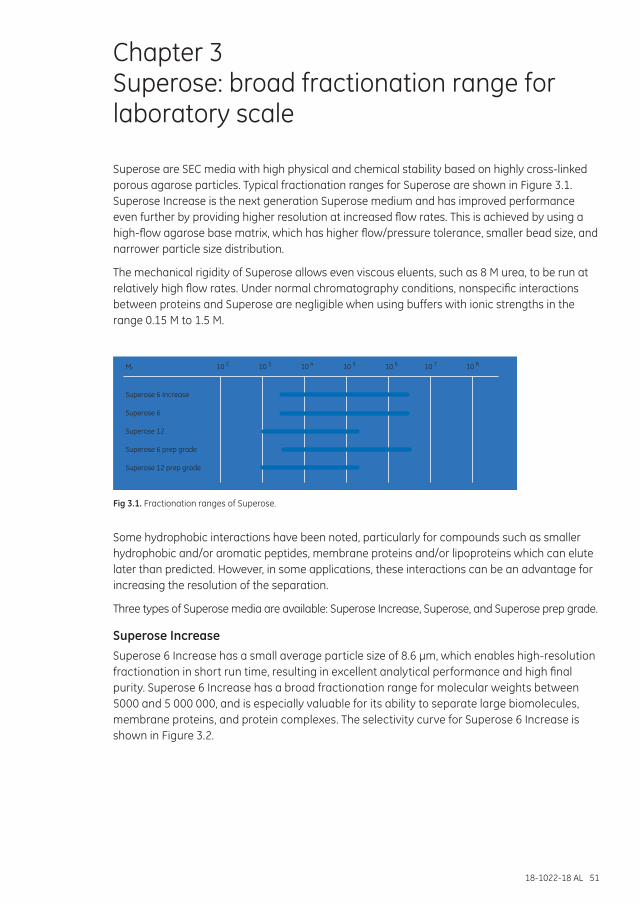
18-1022-18 AL 51
Chapter 3 Superose: broad fractionation range for laboratory scale
Superose are SEC media with high physical and chemical stability based on highly cross-linked porous agarose particles. Typical fractionation ranges for Superose are shown in Figure 3.1. Superose Increase is the next generation Superose medium and has improved performance even further by providing higher resolution at increased flow rates. This is achieved by using a high-flow agarose base matrix, which has higher flow/pressure tolerance, smaller bead size, and narrower particle size distribution.
The mechanical rigidity of Superose allows even viscous eluents, such as 8 M urea, to be run at relatively high flow rates. Under normal chromatography conditions, nonspecific interactions between proteins and Superose are negligible when using buffers with ionic strengths in the range 0.15 M to 1.5 M.
Superose 6
Superose 12
Superose 6 prep grade
Superose 12 prep grade
10M 2r 10 3 10 4 10 5 10 6 10 7 10 8
Superose 6 Increase
Fig 3.1. Fractionation ranges of Superose.
Some hydrophobic interactions have been noted, particularly for compounds such as smaller hydrophobic and/or aromatic peptides, membrane proteins and/or lipoproteins which can elute later than predicted. However, in some applications, these interactions can be an advantage for increasing the resolution of the separation.
Three types of Superose media are available: Superose Increase, Superose, and Superose prep grade.
Superose IncreaseSuperose 6 Increase has a small average particle size of 8.6 µm, which enables high-resolution fractionation in short run time, resulting in excellent analytical performance and high final purity. Superose 6 Increase has a broad fractionation range for molecular weights between 5000 and 5 000 000, and is especially valuable for its ability to separate large biomolecules, membrane proteins, and protein complexes. The selectivity curve for Superose 6 Increase is shown in Figure 3.2.
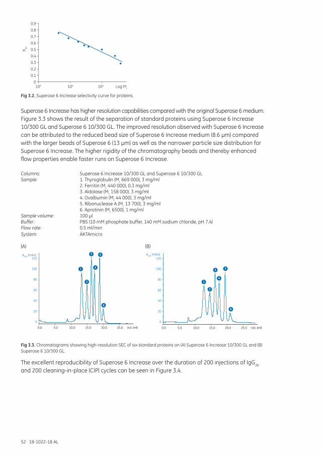
52 18-1022-18 AL
103 Log Mr
K av
105104
0.9
0.8
0.7
0.6
0.5
0.4
0.3
0.2
0.1
0
Fig 3.2. Superose 6 Increase selectivity curve for proteins.
Superose 6 Increase has higher resolution capabilities compared with the original Superose 6 medium. Figure 3.3 shows the result of the separation of standard proteins using Superose 6 Increase 10/300 GL and Superose 6 10/300 GL. The improved resolution observed with Superose 6 Increase can be attributed to the reduced bead size of Superose 6 Increase medium (8.6 µm) compared with the larger beads of Superose 6 (13 µm) as well as the narrower particle size distribution for Superose 6 Increase. The higher rigidity of the chromatography beads and thereby enhanced flow properties enable faster runs on Superose 6 Increase.
Columns: Superose 6 Increase 10/300 GL and Superose 6 10/300 GLSample: 1. Thyroglobulin (Mr 669 000), 3 mg/ml 2. Ferritin (Mr 440 000), 0.3 mg/ml 3. Aldolase (Mr 158 000), 3 mg/ml 4. Ovalbumin (Mr 44 000), 3 mg/ml 5. Ribonuclease A (Mr 13 700), 3 mg/ml 6. Aprotinin (Mr 6500), 1 mg/mlSample volume: 100 µlBuffer: PBS (10 mM phosphate buffer, 140 mM sodium chloride, pH 7.4)Flow rate: 0.5 ml/minSystem: ÄKTAmicro
0
20
40
60
80
100
120
0.0 5.0 10.0 15.0 20.0 25.0 Vol. (ml)
0
20
40
60
80
100
120
A280 (mAU)
A280 (mAU)
1
2
3
4
5
6
1
2
3
4
5
6
0.0 5.0 10.0 15.0 20.0 25.0 Vol. (ml)
0
20
40
60
80
100
120
0.0 5.0 10.0 15.0 20.0 25.0 Vol. (ml)
0
20
40
60
80
100
120
A280 (mAU)
A280 (mAU)
1
2
3
4
5
6
1
2
3
4
5
6
0.0 5.0 10.0 15.0 20.0 25.0 Vol. (ml)
(A) (B)
Fig 3.3. Chromatograms showing high-resolution SEC of six standard proteins on (A) Superose 6 Increase 10/300 GL and (B) Superose 6 10/300 GL.
The excellent reproducibility of Superose 6 Increase over the duration of 200 injections of IgG2b and 200 cleaning-in-place (CIP) cycles can be seen in Figure 3.4.
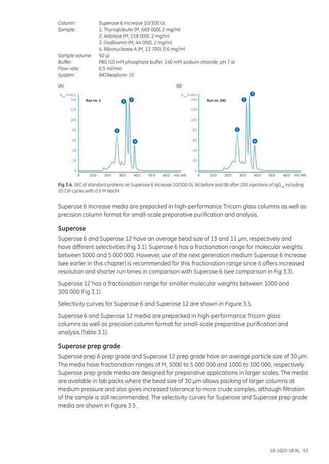
18-1022-18 AL 53
Column: Superose 6 Increase 10/300 GLSample: 1. Thyroglobulin (Mr 669 000), 2 mg/ml 2. Aldolase (Mr 158 000), 2 mg/ml 3. Ovalbumin (Mr 44 000), 2 mg/ml 4. Ribonuclease A (Mr 13 700), 0.6 mg/mlSample volume: 50 µlBuffer: PBS (10 mM phosphate buffer, 140 mM sodium chloride, pH 7.4)Flow rate: 0.5 ml/minSystem: ÄKTAexplorer 10
(A) (B)
140
120
100
80
60
40
20
00 10.0 20.0 30.5 40.0
Run no. 1 Run no. 200
60.050.0 Vol. (ml)
A280 (mAU) A280 (mAU)140
120
100
80
60
40
20
00 10.0 20.0 30.5 40.0 60.050.0 Vol. (ml)
1 1
2 233
4 4
Fig 3.4. SEC of standard proteins on Superose 6 Increase 10/300 GL (A) before and (B) after 200 injections of IgG2b including 20 CIP cycles with 0.5 M NaOH.
Superose 6 Increase media are prepacked in high-performance Tricorn glass columns as well as precision column format for small-scale preparative purification and analysis.
SuperoseSuperose 6 and Superose 12 have an average bead size of 13 and 11 µm, respectively and have different selectivities (Fig 3.1). Superose 6 has a fractionation range for molecular weights between 5000 and 5 000 000. However, use of the next generation medium Superose 6 Increase (see earlier in this chapter) is recommended for this fractionation range since it offers increased resolution and shorter run times in comparison with Superose 6 (see comparison in Fig 3.3).
Superose 12 has a fractionation range for smaller molecular weights between 1000 and 300 000 (Fig 3.1).
Selectivity curves for Superose 6 and Superose 12 are shown in Figure 3.5.
Superose 6 and Superose 12 media are prepacked in high-performance Tricorn glass columns as well as precision column format for small-scale preparative purification and analysis (Table 3.1).
Superose prep gradeSuperose prep 6 prep grade and Superose 12 prep grade have an average particle size of 30 µm. The media have fractionation ranges of Mr 5000 to 5 000 000 and 1000 to 300 000, respectively. Superose prep grade media are designed for preparative applications in larger scales. The media are available in lab packs where the bead size of 30 µm allows packing of larger columns at medium pressure and also gives increased tolerance to more crude samples, although filtration of the sample is still recommended. The selectivity curves for Superose and Superose prep grade media are shown in Figure 3.5.
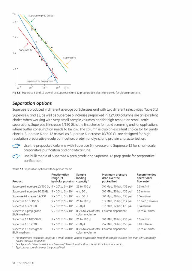
54 18-1022-18 AL
0.8
0.6
0.4
0.2
010 3 10 4 10 5 10 6
Superose 6
Superose 6 prep grade
Superose 12
Superose 12 prep grade
K av
Log Mr
Fig 3.5. Superose 6 and 12 as well as Superose 6 and 12 prep grade selectivity curves for globular proteins.
Separation optionsSuperose is produced in different average particle sizes and with two different selectivities (Table 3.1).
Superose 6 and 12, as well as Superose 6 Increase prepacked in 3.2/300 columns are an excellent choice when working with very small sample volumes and for high resolution small-scale separations. Superose 6 Increase 5/150 GL is the first choice for rapid screening and for applications where buffer consumption needs to be low. The column is also an excellent choice for for purity checks. Superose 6 and 12 as well as Superose 6 Increase 10/300 GL are designed for high-resolution preparative-scale purification, protein analysis, and protein characterization.
Use the prepacked columns with Superose 6 Increase and Superose 12 for small-scale preparative purification and analytical runs.
Use bulk media of Superose 6 prep grade and Superose 12 prep grade for preparative purification.
Table 3.1. Separation options with Superose media
Product
Fractionation range, Mr (globular proteins)
Sample loading capacity*
Maximum pressure drop over the packed bed
Recommended operational flow rate†
Superose 6 Increase 10/300 GL 5 × 10³ to 5 × 10⁶ 25 to 500 µl 3.0 Mpa, 30 bar, 435 psi‡ 0.5 ml/min
Superose 6 Increase 5/150 GL 5 × 10³ to 5 × 10⁶ 4 to 50 µ 3.0 MPa, 30 bar, 435 psi‡ 0.3 ml/min
Superose 6 Increase 3.2/300 5 × 10³ to 5 × 10⁶ 4 to 50 µl 3.0 Mpa, 30 bar, 435 psi‡ 0.04 ml/min
Superose 6 10/300 GL 5 × 10³ to 5 × 10⁶ 25 to 500 µl 1.5 MPa, 15 bar, 217 psi 0.1 to 0.5 ml/min
Superose 6 3.2/300 5 × 10³ to 5 × 10⁶ < 50 µl 1.2 MPa, 12 bar, 175 psi 0.04 ml/min
Superose 6 prep grade (Bulk medium)
5 × 10³ to 5 × 10⁶ 0.5% to 4% of total column volume
Column-dependent up to 40 cm/h
Superose 12 10/300 GL 1 × 10³ to 3 × 10⁶ 25 to 500 µl 3.0 MPa, 30 bar, 435 psi 0.5 ml/min
Superose 12 3.2/300 1 × 10³ to 3 × 10⁶ < 50 µl 2.4 MPa, 24 bar, 350 psi 0.04 ml/min
Superose 12 prep grade (Bulk medium)
1 × 10³ to 3 × 10⁶ 0.5% to 4% of total column volume
Column-dependent up to 40 cm/h
* For maximum resolution, apply as a small sample volume as possible. Note that sample volumes less than 0.5% normally do not improve resolution.
† See Appendix 5 to convert linear flow (cm/h) to volumetric flow rates (ml/min) and vice versa.‡ Typical pressure drop over the packed bed.
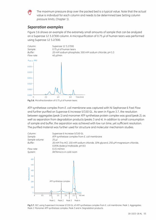
18-1022-18 AL 55
The maximum pressure drop over the packed bed is a typical value. Note that the actual value is individual for each column and needs to be determined (see Setting column pressure limits, Chapter 1).
Separation examplesFigure 3.6 shows an example of the extremely small amounts of sample that can be analyzed on a Superose 12 3.2/300 column. A micropurification of 0.75 µl of human tears was performed using Superose 12 3.2/300.
Column: Superose 12 3.2/300Sample: 0.75 µl of human tearsBuffer: 20 mM sodium phosphate, 500 mM sodium chloride, pH 5.3Flow rate: 40 µl/min
0.025
0.020
0.015
0.010
0.005
0.0000 2 0 4 0 6 0 Time (min)
A (AU)280 nm
Fig 3.6. Microfractionation of 0.75 µl of human tears.
ATP synthetase complex from E. coli membrane was captured with Ni Sepharose 6 Fast Flow and further purified on Superose 6 Increase 5/150 GL. As seen in Figure 3.7, the resolution between aggregates (peak 1) and monomer ATP synthetase protein complex was good (peak 2), as well as separation from degradation products (peaks 3 and 4). In addition to small consumption of sample and buffer, the separation was achieved with low run time, yet sufficient resolution. The purified material was further used for structure and molecular mechanism studies.
Column: Superose 6 Increase 5/150 GLSample: ATP synthetase complex from E. coli membraneSample volume: 25 µlBuffer: 20 mM Tris-HCl, 150 mM sodium chloride, 10% glycerol, 250 µM magnesium chloride,
0.05% dodecyl maltoside, pH 8.0 Flow rate: 0.15 ml/minSystem: ÄKTAmicro in cold room
600
500
400
300
200
100
0
0 0.5 1.0 1.5 2.0 2.5 Vol. (ml)
Abso
rban
ce (m
AU)
ATP synthetase complex
Peak 1 Peak 2 Peak 3 Peak 4
Fig 3.7. SEC using Superose 6 Increase 5/150 GL of ATP synthetase complex from E. coli membrane. Peak 1: Aggregates; Peak 2: Monomer ATP synthetase complex; Peak 3 and 4: Degradation products.
56 18-1022-18 AL
Performing a separation Buffer: 10 to 50 mM sodium phosphate, 150 mM sodium chloride, pH 7.0 to 7.4, or select the
buffer in which the sample should be stored or solubilized for the next step.
Use 150 mM sodium chloride or a buffer with equivalent ionic strength to avoid pH dependent nonionic interactions with the matrix. At very low ionic strength, the presence of a small number of negatively charged groups can cause retardation of basic proteins and exclusion of acidic proteins.
The sample should be fully dissolved. Centrifuge or filter to remove particulate material (see Appendix 3). Always use degassed buffers and maintain a constant temperature during the run to avoid introducing air into the column.
Set an appropriate pressure limit on the chromatography system to avoid damage to the column packing. Use lower flow rates for high viscosity solutions and low temperature (see Table 1.3).
First-time use or after long-term storage
1. Equilibrate the column with 2 column volumes of water at room temperature to remove the storage solution. Use a low flow rate (Table 3.2) to avoid any gap formation.
2. For first-time use: Determine the column specific pressure limit according to section Setting column pressure limits (Chapter 1).
3. Perform a column efficiency test (see Appendix 1).
4. Equilibrate with 2 column volumes of buffer at recommended flow rate during run (Table 3.1).
5. Apply a sample volume equivalent to between 0.5% and 4% of the column volume. For most applications the sample volume should not exceed 2% to achieve maximum resolution (up to 50 µl for 3.2/300 GL and 5/150 GL, up to 500 µl for 10/300 GL). Note that smaller sample volumes generally lead to improved resolution.
6. Elute with 1 column volume of buffer.
Before applying a new sample, re-equilibrate the column with buffer until the baseline monitored at A280 is stable.
Table 3.2. Recommended flow rates at different stages using prepacked columns containing Superose 6 Increase, Superose 6, and Superose 12 media
Flow rates (ml/min) at room temperature
3.2/300 GL 5/150 GL 10/300 GL
First-time use or after long-term storage 0.04 0.25 0.5*
Cleaning-in-place, CIP 0.02 0.1 0.5
* Start with 0.2 ml/min for 0.5 column volumes.
Column performance should be checked at regular intervals by determining the theoretical plate number m-1 and peak a symmetry factor, As. See Appendix 1 for how to check column efficiency.
Ensure that the pressure limit of the column is not exceeded. This is particularly important when working at low temperatures, such as in a cold room, or when the column is used with 20% ethanol or other viscous solutions.
Exposure to temperatures outside the range 4°C to 40°C will negatively affect the efficiency of a packed bed and the column will need to be repacked.
18-1022-18 AL 57
Cleaning
1. Wash with 1 column volume of 0.5 M sodium hydroxide, alternatively 0.5 M acetic acid. Use a low flow rate during the entire CIP procedure. Use 0.2 M sodium hydroxide for Superose prep grade media.
2. Immediately wash with 1 column volume of distilled water followed by at least 2 column volumes of buffer until the baseline monitored at A280 and the pH of the eluent are stable.
For extreme cases of contamination, check the instructions supplied with the product.
In special cases, it might be necessary to change the bottom filter or to remove and discard the top 2 to 3 mm of the gel. These operations must be done with extreme care to avoid serious loss of resolution. Note that Precision Columns (3.2/300) should not be opened.
Superose prep grade may be autoclaved repeatedly at 121°C, pH 7 for 30 min without significantly affecting its chromatographic properties. The medium must be removed from the column as autoclaving can damage column components. Note that Precision Columns (3.2/300) cannot be repacked.
Media characteristicsMatrix: Cross-linked agarose.
Table 3.2 Superose media characteristics
ProductEfficiency (theorectical plates m-1)* pH stability† Average particle size (µm)
Superose 6 Increase‡ > 48 000 Long term: 3 to 12 Short term: 1 to 14
8.6
Superose 6 ≥ 30 000 Long term: 3 to 12 Short term: 1 to 14
13
Superose 6 prep grade § Long term: 3 to 12 Short term: 1 to 14
30
Superose 12 ≥ 40 000 Long term: 3 to 12 Short term: 1 to 14
11
Superose 12 prep grade § Long term: 3 to 12 Short term: 1 to 14
30
* Prepacked columns only. † Long-term pH stability refers to the pH interval where the medium is stable over a long period of time without adverse side
effects on its chromatography performance. Short-term pH stability refers to the pH interval for regeneration, cleaning-in-place, and sanitization procedures. All ranges are GE Healthcare estimates.
‡ Column length 300 mm.§ A minimum column efficiency of 10 000 m-1 should be expected for a well-packed column.
Chemical stabilitySuperose is stable in all commonly used aqueous buffers and additives such as detergents, denaturing agents (8 M urea or 6 M guanidine hydrochloride), and 30% acetonitrile.
StorageStore unused media 4°C to 30°C in 20% ethanol. Do not freeze.
For long-term storage, wash with 2 column volumes of distilled water followed by 2 column volumes of 20% ethanol at low flow rate. Store at 4°C to 30°C.
Degas the ethanol/water mixture thoroughly and use a low flow rate. Connect the transport tool to the capillary tubing at the column outlet to prevent air from entering the column. Fill the tool up to approximately 50% of the total volume.
Avoid changes in temperature, which can cause air bubbles in the packing.

58 18-1022-18 AL
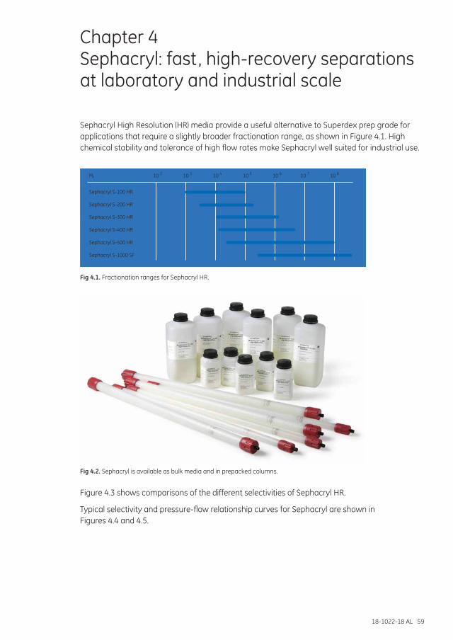
18-1022-18 AL 59
Chapter 4 Sephacryl: fast, high-recovery separations at laboratory and industrial scale
Sephacryl High Resolution (HR) media provide a useful alternative to Superdex prep grade for applications that require a slightly broader fractionation range, as shown in Figure 4.1. High chemical stability and tolerance of high flow rates make Sephacryl well suited for industrial use.
10M 2r 10 3 10 4 10 5 10 6 10 7 10 8
Sephacryl S-100 HR
Sephacryl S-200 HR
Sephacryl S-300 HR
Sephacryl S-400 HR
Sephacryl S-500 HR
Sephacryl S-1000 SF
Fig 4.1. Fractionation ranges for Sephacryl HR.
Fig 4.2. Sephacryl is available as bulk media and in prepacked columns.
Figure 4.3 shows comparisons of the different selectivities of Sephacryl HR.
Typical selectivity and pressure-flow relationship curves for Sephacryl are shown in Figures 4.4 and 4.5.
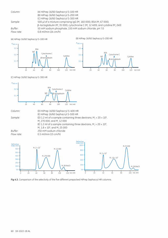
60 18-1022-18 AL
Column: (A) HiPrep 16/60 Sephacryl S-100 HR (B) HiPrep 16/60 Sephacryl S-200 HR (C) HiPrep 16/60 Sephacryl S-300 HRSample: 500 µl of a mixture comprising IgG (Mr 160 000), BSA (Mr 67 000),
E-lactoglobulin (Mr 35 000), cytochrome C (Mr 12 400), and cytidine (Mr 240)Buffer: 50 mM sodium phosphate, 150 mM sodium chloride, pH 7.0Flow rate: 0.8 ml/min (24 cm/h)
A280 nmA280 nm
0.3
0.2
0.1
20
BSA
(A) HiPrep 16/60 Sephacryl S-100 HR (B) HiPrep 16/60 Sephacryl S-200 HR
IgGCytochrome C
Cytidine
Vol. (ml)
β-lactoglobulin
40 60 80 100 120
0.3
0.2
0.1
20
BSA
IgGCytochrome C
Cytidine
Vol. (ml)
β-lactoglobulin
40 60 80 100 120
A280 nm
(C) HiPrep 16/60 Sephacryl S-300 HR
0.2
0.1
20
BSA
IgG
Cytochrome C Cytidine
Vol. (ml)
Vol. (ml) Vol. (ml)
β-lactoglobulin
40 60 80 100 120
50.0
50.0
Refractiveindex (mV)
0.0
10.0
0 20 40 60 80 100 120
20.0
40.0
30.0
Mr 2 × 10⁷
Mr 270 000
Mr 12 000
Mr 58 (NaCl)
(D) HiPrep 16/60 Sephacryl S-400 HR
60.0
80.0
100.0
Refractiveindex (mV)
0.0
0 20 40 60 80 100
20.0
40.0
Mr 2 × 10⁷
Mr 1.8 × 10⁶
Mr 25 000
Mr 58 (NaCl)
(E) HiPrep 16/60 Sephacryl S-500 HR
Column: (D) HiPrep 16/60 Sephacryl S-400 HR (E) HiPrep 16/60 Sephacryl S-500 HRSample: (D) 1.2 ml of a sample containing three dextrans; Mr > 20 × 106,
Mr 270 000, and Mr 12 000 (E) 1.2 ml of a sample containing three dextrans, Mr > 20 × 106,
Mr 1.8 × 10⁶, and Mr 25 000Buffer: 250 mM sodium chlorideFlow rate: 0.5 ml/min (15 cm/h)
A280 nmA280 nm
0.3
0.2
0.1
20
BSA
(A) HiPrep 16/60 Sephacryl S-100 HR (B) HiPrep 16/60 Sephacryl S-200 HR
IgGCytochrome C
Cytidine
Vol. (ml)
β-lactoglobulin
40 60 80 100 120
0.3
0.2
0.1
20
BSA
IgGCytochrome C
Cytidine
Vol. (ml)
β-lactoglobulin
40 60 80 100 120
A280 nm
(C) HiPrep 16/60 Sephacryl S-300 HR
0.2
0.1
20
BSA
IgG
Cytochrome C Cytidine
Vol. (ml)
Vol. (ml) Vol. (ml)
β-lactoglobulin
40 60 80 100 120
50.0
50.0
Refractiveindex (mV)
0.0
10.0
0 20 40 60 80 100 120
20.0
40.0
30.0
Mr 2 × 10⁷
Mr 270 000
Mr 12 000
Mr 58 (NaCl)
(D) HiPrep 16/60 Sephacryl S-400 HR
60.0
80.0
100.0
Refractiveindex (mV)
0.0
0 20 40 60 80 100
20.0
40.0
Mr 2 × 10⁷
Mr 1.8 × 10⁶
Mr 25 000
Mr 58 (NaCl)
(E) HiPrep 16/60 Sephacryl S-500 HR
Fig 4.3. Comparison of the selectivity of the five different prepacked HiPrep Sephacryl HR columns.
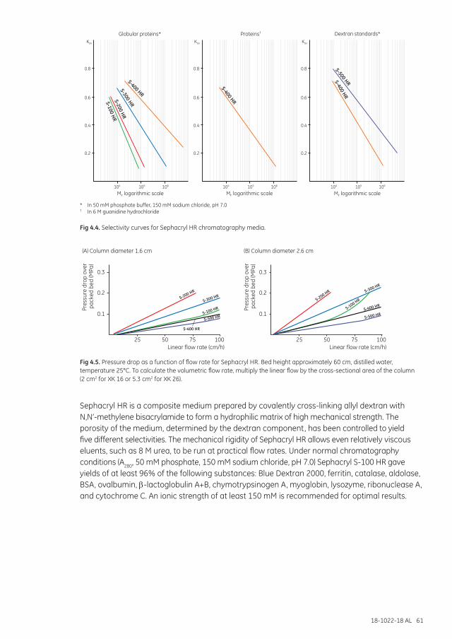
18-1022-18 AL 61
104 105
Mr logarithmic scale Mr logarithmic scale Mr logarithmic scale
Globular proteins*
S-400 HR
S-300 HR
S-200 HR
S-100 HR
106 104 105 106 104 105 106
Proteins†
S-400 HR
KavKavKav
0.8
0.6
0.4
0.2
0.8
0.6
0.4
0.2
0.8
0.6
0.4
0.2
Dextran standards*
S-400 HR
S-500 HR
* In 50 mM phosphate buffer, 150 mM sodium chloride, pH 7.0† In 6 M guanidine hydrochloride
Fig 4.4. Selectivity curves for Sephacryl HR chromatography media.
Pres
sure
dro
p ov
er p
acke
d be
d (M
Pa)
Pres
sure
dro
p ov
er p
acke
d be
d (M
Pa)
S-200 HR
S-300 HR
S-100 HR
S-500 HR
S-400 HR
Linear flow rate (cm/h)
(A) Column diameter 1.6 cm
0.3
25 50 75 100
0.2
0.1
S-200 HR S-300 HR
S-100 HR
S-500 HRS-400 HR
Linear flow rate (cm/h)
(B) Column diameter 2.6 cm
0.3
25 50 75 100
0.2
0.1
Fig 4.5. Pressure drop as a function of flow rate for Sephacryl HR. Bed height approximately 60 cm, distilled water, temperature 25°C. To calculate the volumetric flow rate, multiply the linear flow by the cross-sectional area of the column (2 cm2 for XK 16 or 5.3 cm2 for XK 26).
Sephacryl HR is a composite medium prepared by covalently cross-linking allyl dextran with N,N’-methylene bisacrylamide to form a hydrophilic matrix of high mechanical strength. The porosity of the medium, determined by the dextran component, has been controlled to yield five different selectivities. The mechanical rigidity of Sephacryl HR allows even relatively viscous eluents, such as 8 M urea, to be run at practical flow rates. Under normal chromatography conditions (A280, 50 mM phosphate, 150 mM sodium chloride, pH 7.0) Sephacryl S-100 HR gave yields of at least 96% of the following substances: Blue Dextran 2000, ferritin, catalase, aldolase, BSA, ovalbumin, E-lactoglobulin A+B, chymotrypsinogen A, myoglobin, lysozyme, ribonuclease A, and cytochrome C. An ionic strength of at least 150 mM is recommended for optimal results.
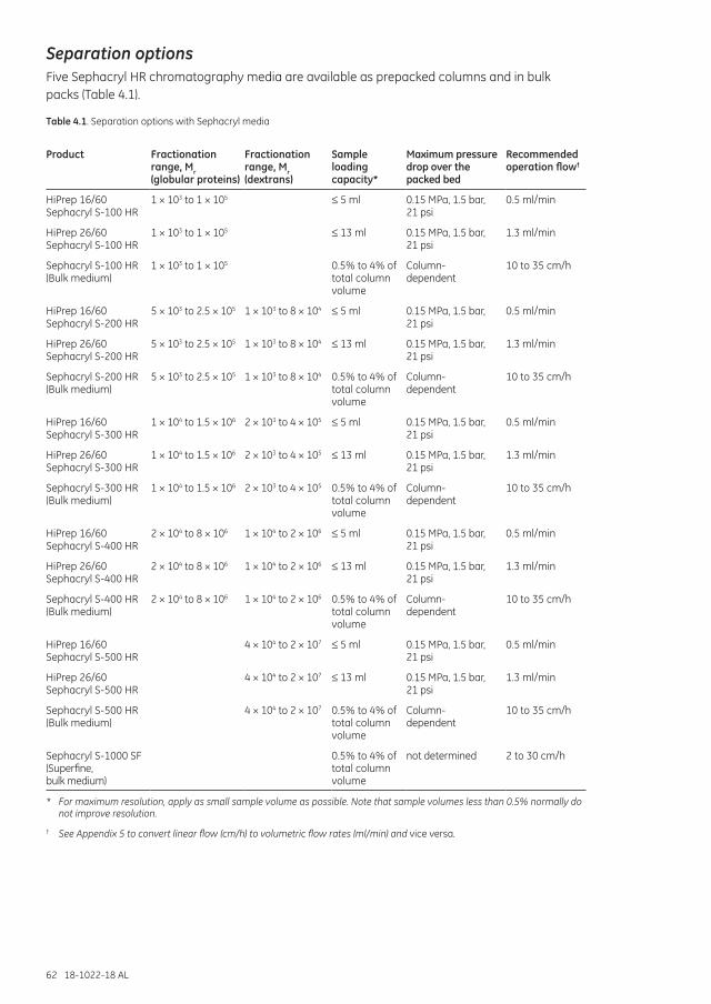
62 18-1022-18 AL
Separation optionsFive Sephacryl HR chromatography media are available as prepacked columns and in bulk packs (Table 4.1).
Table 4.1. Separation options with Sephacryl media
Product Fractionation range, Mr (globular proteins)
Fractionation range, Mr (dextrans)
Sample loading capacity*
Maximum pressure drop over the packed bed
Recommended operation flow†
HiPrep 16/60 Sephacryl S-100 HR
1 × 10³ to 1 × 10⁵ ≤ 5 ml 0.15 MPa, 1.5 bar, 21 psi
0.5 ml/min
HiPrep 26/60 Sephacryl S-100 HR
1 × 10³ to 1 × 10⁵ ≤ 13 ml 0.15 MPa, 1.5 bar, 21 psi
1.3 ml/min
Sephacryl S-100 HR (Bulk medium)
1 × 10³ to 1 × 10⁵ 0.5% to 4% of total column volume
Column- dependent
10 to 35 cm/h
HiPrep 16/60 Sephacryl S-200 HR
5 × 10³ to 2.5 × 10⁵ 1 × 10³ to 8 × 10⁴ ≤ 5 ml 0.15 MPa, 1.5 bar, 21 psi
0.5 ml/min
HiPrep 26/60 Sephacryl S-200 HR
5 × 10³ to 2.5 × 10⁵ 1 × 10³ to 8 × 10⁴ ≤ 13 ml 0.15 MPa, 1.5 bar, 21 psi
1.3 ml/min
Sephacryl S-200 HR (Bulk medium)
5 × 10³ to 2.5 × 10⁵ 1 × 10³ to 8 × 10⁴ 0.5% to 4% of total column volume
Column- dependent
10 to 35 cm/h
HiPrep 16/60 Sephacryl S-300 HR
1 × 10⁴ to 1.5 × 10⁶ 2 × 10³ to 4 × 10⁵ ≤ 5 ml 0.15 MPa, 1.5 bar, 21 psi
0.5 ml/min
HiPrep 26/60 Sephacryl S-300 HR
1 × 10⁴ to 1.5 × 10⁶ 2 × 10³ to 4 × 10⁵ ≤ 13 ml 0.15 MPa, 1.5 bar, 21 psi
1.3 ml/min
Sephacryl S-300 HR (Bulk medium)
1 × 10⁴ to 1.5 × 10⁶ 2 × 10³ to 4 × 10⁵ 0.5% to 4% of total column volume
Column- dependent
10 to 35 cm/h
HiPrep 16/60 Sephacryl S-400 HR
2 × 10⁴ to 8 × 10⁶ 1 × 10⁴ to 2 × 10⁶ ≤ 5 ml 0.15 MPa, 1.5 bar, 21 psi
0.5 ml/min
HiPrep 26/60 Sephacryl S-400 HR
2 × 10⁴ to 8 × 10⁶ 1 × 10⁴ to 2 × 10⁶ ≤ 13 ml 0.15 MPa, 1.5 bar, 21 psi
1.3 ml/min
Sephacryl S-400 HR (Bulk medium)
2 × 10⁴ to 8 × 10⁶ 1 × 10⁴ to 2 × 10⁶ 0.5% to 4% of total column volume
Column- dependent
10 to 35 cm/h
HiPrep 16/60 Sephacryl S-500 HR
4 × 10⁴ to 2 × 10⁷ ≤ 5 ml 0.15 MPa, 1.5 bar, 21 psi
0.5 ml/min
HiPrep 26/60 Sephacryl S-500 HR
4 × 10⁴ to 2 × 10⁷ ≤ 13 ml 0.15 MPa, 1.5 bar, 21 psi
1.3 ml/min
Sephacryl S-500 HR (Bulk medium)
4 × 10⁴ to 2 × 10⁷ 0.5% to 4% of total column volume
Column- dependent
10 to 35 cm/h
Sephacryl S-1000 SF (Superfine, bulk medium)
0.5% to 4% of total column volume
not determined 2 to 30 cm/h
* For maximum resolution, apply as small sample volume as possible. Note that sample volumes less than 0.5% normally do not improve resolution.
† See Appendix 5 to convert linear flow (cm/h) to volumetric flow rates (ml/min) and vice versa.
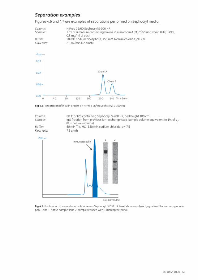
18-1022-18 AL 63
Separation examplesFigures 4.6 and 4.7 are examples of separations performed on Sephacryl media.
Column: HiPrep 26/60 Sephacryl S-100 HRSample: 1 ml of a mixture containing bovine insulin chain A (Mr 2532) and chain B (Mr 3496),
0.5 mg/ml of eachBuffer: 50 mM sodium phosphate, 150 mM sodium chloride, pH 7.0Flow rate: 2.0 ml/min (22 cm/h)
0.03
Chain A
Chain B
0.02
0.01
0.00400 80 120 160 200 240 Time (min)
A 280 nm
Fig 4.6. Separation of insulin chains on HiPrep 26/60 Sephacryl S-100 HR.
Column: BP 113/120 containing Sephacryl S-200 HR, bed height 100 cmSample: IgG fraction from previous ion-exchange step (sample volume equivalent to 1% of Vt.
(Vt = column volume)Buffer: 50 mM Tris-HCl, 150 mM sodium chloride, pH 7.5Flow rate: 7.5 cm/h
Elution volume
Immunoglobulin1 2
A280 nm
Fig 4.7. Purification of monoclonal antibodies on Sephacryl S-200 HR. Inset shows analysis by gradient the immunoglobulin pool. Lane 1, native sample; lane 2, sample reduced with 2-mercaptoethanol.
64 18-1022-18 AL
Performing a separation
Buffer: 50 mM sodium phosphate, 150 mM sodium chloride, pH 7.2 or select the buffer in which the sample should be stored or solubilized for the next step.
Use 150 mM sodium chloride, or a buffer with equivalent ionic strength, to avoid pH dependent nonionic interactions with the matrix. At very low ionic strength, the presence of a small number of negatively charged groups can cause retardation of basic proteins and exclusion of acidic proteins.
The sample should be fully dissolved. Centrifuge or filter to remove particulate material (Appendix 3). Always use degassed buffers and maintain a constant temperature during the run to avoid introducing air into the column.
Set an appropriate pressure limit on the chromatography system to avoid damage to the column packing. Use lower flow rates for high viscosity solutions and low temperature (see Table 1.3).
First-time use or after long-term storage
1. Equilibrate the column with at least 0.5 column volumes of distilled water at 15 cm/h (0.5 ml/min for 16/60 column or 1.3 ml/min for 26/60).
2. Equilibrate with 2 column volumes of buffer at 30 cm/h (1.0 ml/min for 16/60 column or 2.6 ml/min for 26/60).
3. Reduce flow to 15 cm/h and, for optimal resolution, apply a sample volume equivalent to 1% of the column volume (1.2 ml for 16/60 column or 3.2 ml for 26/60). Sample volumes between 0.5% and 4% can be applied.
4. Elute with 1 column volume of buffer.
5. Before applying a new sample-re-equilibrate column with 1 column volume of buffer at 30 cm/h until the baseline monitored at A280 is stable.
Column performance should be checked at regular intervals by determining the theoretical plate number per meter and peak symmetry. Prepacked columns are supplied with recommended values. See Appendix 1 on how to check column efficiency.
See Chapter 1 for advice on optimizing the separation.
Exposure to temperatures outside the range 4°C to 40°C will negatively affect the efficiency of a packed bed and the column will need to be repacked.
Cleaning
1. Wash with 0.5 column volumes of 0.2 M sodium hydroxide at a flow of 15 cm/h (0.5 ml/min for column 16/60 or 1.3 ml/min for 26/60) to remove most nonspecifically adsorbed proteins.
2. Re-equilibrate immediately with 2 column volumes of buffer or until the baseline monitored at A280 and the pH of the eluent are stable.
Further equilibration might be necessary if the buffer contains detergent.
Routine cleaning after every 10 to 20 separations is recommended, but the frequency of cleaning will also depend on the nature of the samples being applied.
18-1022-18 AL 65
If required, Sephacryl HR may be autoclaved repeatedly at 121°C, pH 7, for 30 min without significantly affecting its chromatographic properties. The medium must be removed from the column as autoclaving can damage column components. Note that HiPrep columns cannot be repacked.
To remove severe contamination
Reverse the flow and wash at a flow rate of 10 cm/h (0.3 ml/min for column 16/60 or 0.8 ml/min for 26/60) at room temperature using the following solutions:
1. Wash with 0.25 column volumes of 0.5 M sodium hydroxide (to remove hydrophobic proteins or lipoproteins) followed by 4 column volumes of distilled water.
2. Wash with 0.5 column volumes of 30% isopropanol (to remove lipids and very hydrophobic proteins), followed by 2 column volumes of distilled water.
For extreme cases of contamination, check the instructions supplied with the product.
Reversing flow through a column packed with Sephacryl media should only be considered under cases of severe contamination. Reversing the flow can cause channeling through the packed bed leading to poor resolution, reduced efficiency, and the need to repack the column. Professionally packed columns are less likely to be affected, but extreme care must be taken.
Media characteristicsMatrix: Cross-linked copolymer of allyl dextran and N,N’-methylene bisacrylamide.
Table 4.2 Sephacryl media characteristics
Product Efficiency: theoretical plates m-1 (prepacked columns only)
pH stability* Average particle size (µm)
Sephacryl S-100 HR ≥ 5000 Long term: 3 to 11 Short term: 2 to 13
47
Sephacryl S-200 HR ≥ 5000 Long term: 3 to 11 Short term: 2 to 13
47
Sephacryl S-300 HR ≥ 5000 Long term: 3 to 11 Short term: 2 to 13
47
Sephacryl S-400 HR 9000 Long term: 3 to 11 Short term: 2 to 13
47
Sephacryl S-500 HR 9000 Long term: 3 to 11 Short term: 2 to 13
47
Sephacryl S-1000 SF (Superfine)
† Long term: 3 to 11 Short term: 2 to 13
65
* Long-term pH stability refers to the pH interval where the medium is stable over a long period of time without adverse side effects on its chromatographic performance. Short-term pH stability refers to the pH interval for regeneration, cleaning-in-place, and sanitization procedures. All ranges are GE Healthcare estimates.
† Not determined.
Chemical stabilitySephacryl HR is stable in all commonly used aqueous buffers and additives such as detergents (1% SDS) and denaturing agents (8 M urea or 6 M guanidine hydrochloride). The medium is also stable in 30% acetonitrile, 0.5 M sodium hydroxide, up to 24% ethanol, up to 1 M acetic acid, and up to 30% isopropanol.

66 18-1022-18 AL
StorageStore unused media 4°C to 30°C in 20% ethanol. Do not freeze.
Columns can be left connected to a chromatography system with a low flow rate (0.01 ml/min) of buffer passing through the column to prevent bacterial growth or the introduction of air into the column which would negatively affect the packing.
For long-term storage, wash with 4 column volumes of distilled water followed by 4 column volumes of 20% ethanol. Store at 4°C to 30°C.
Degas the ethanol/water mixture thoroughly and use a low flow rate, checking the back pressure as the column equilibrates.
Avoid changes in temperature which can cause air bubbles in the packing.
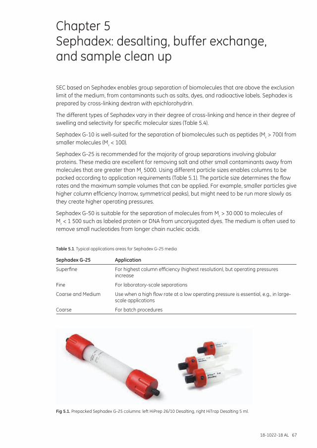
18-1022-18 AL 67
Chapter 5 Sephadex: desalting, buffer exchange, and sample clean up
SEC based on Sephadex enables group separation of biomolecules that are above the exclusion limit of the medium, from contaminants such as salts, dyes, and radioactive labels. Sephadex is prepared by cross-linking dextran with epichlorohydrin.
The different types of Sephadex vary in their degree of cross-linking and hence in their degree of swelling and selectivity for specific molecular sizes (Table 5.4).
Sephadex G-10 is well-suited for the separation of biomolecules such as peptides (Mr > 700) from smaller molecules (Mr < 100).
Sephadex G-25 is recommended for the majority of group separations involving globular proteins. These media are excellent for removing salt and other small contaminants away from molecules that are greater than Mr 5000. Using different particle sizes enables columns to be packed according to application requirements (Table 5.1). The particle size determines the flow rates and the maximum sample volumes that can be applied. For example, smaller particles give higher column efficiency (narrow, symmetrical peaks), but might need to be run more slowly as they create higher operating pressures.
Sephadex G-50 is suitable for the separation of molecules from Mr > 30 000 to molecules of Mr < 1 500 such as labeled protein or DNA from unconjugated dyes. The medium is often used to remove small nucleotides from longer chain nucleic acids.
Table 5.1. Typical applications areas for Sephadex G-25 media
Sephadex G-25 Application
Superfine For highest column efficiency (highest resolution), but operating pressures increase
Fine For laboratory-scale separations
Coarse and Medium Use when a high flow rate at a low operating pressure is essential, e.g., in large-scale applications
Coarse For batch procedures
Fig 5.1. Prepacked Sephadex G-25 columns: left HiPrep 26/10 Desalting, right HiTrap Desalting 5 ml.
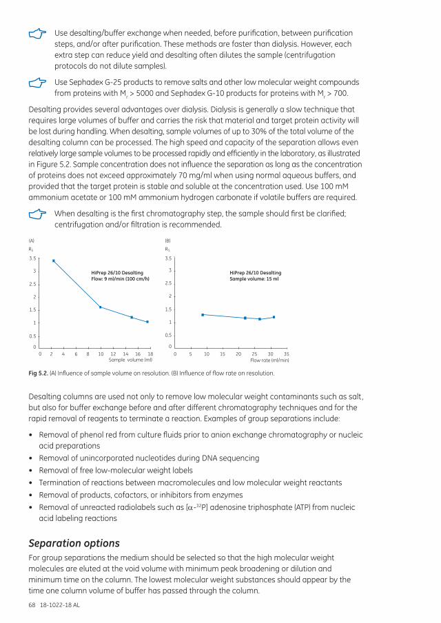
68 18-1022-18 AL
Use desalting/buffer exchange when needed, before purification, between purification steps, and/or after purification. These methods are faster than dialysis. However, each extra step can reduce yield and desalting often dilutes the sample (centrifugation protocols do not dilute samples).
Use Sephadex G-25 products to remove salts and other low molecular weight compounds from proteins with Mr > 5000 and Sephadex G-10 products for proteins with Mr > 700.
Desalting provides several advantages over dialysis. Dialysis is generally a slow technique that requires large volumes of buffer and carries the risk that material and target protein activity will be lost during handling. When desalting, sample volumes of up to 30% of the total volume of the desalting column can be processed. The high speed and capacity of the separation allows even relatively large sample volumes to be processed rapidly and efficiently in the laboratory, as illustrated in Figure 5.2. Sample concentration does not influence the separation as long as the concentration of proteins does not exceed approximately 70 mg/ml when using normal aqueous buffers, and provided that the target protein is stable and soluble at the concentration used. Use 100 mM ammonium acetate or 100 mM ammonium hydrogen carbonate if volatile buffers are required.
When desalting is the first chromatography step, the sample should first be clarified; centrifugation and/or filtration is recommended.
0.5
1
1.5
2
2.5
3
3.5
00 2 4 6 8 10 12 14 16 18
RS
Sample volume (ml)
HiPrep 26/10 DesaltingFlow: 9 ml/min (100 cm/h)
3.5
3
2.5
2
1.5
1
0.5
00 5 10 15 20 25 30 35
Flow rate (ml/min)
RS
HiPrep 26/10 DesaltingSample volume: 15 ml
(A) (B)
Fig 5.2. (A) Influence of sample volume on resolution. (B) Influence of flow rate on resolution.
Desalting columns are used not only to remove low molecular weight contaminants such as salt, but also for buffer exchange before and after different chromatography techniques and for the rapid removal of reagents to terminate a reaction. Examples of group separations include:
• Removal of phenol red from culture fluids prior to anion exchange chromatography or nucleic acid preparations
• Removal of unincorporated nucleotides during DNA sequencing• Removal of free low-molecular weight labels• Termination of reactions between macromolecules and low molecular weight reactants• Removal of products, cofactors, or inhibitors from enzymes• Removal of unreacted radiolabels such as [D-32P] adenosine triphosphate (ATP) from nucleic
acid labeling reactions
Separation optionsFor group separations the medium should be selected so that the high molecular weight molecules are eluted at the void volume with minimum peak broadening or dilution and minimum time on the column. The lowest molecular weight substances should appear by the time one column volume of buffer has passed through the column.
18-1022-18 AL 69
Sephadex media are available in prepacked columns (also spin columns), microplates, and in bulk media packs (Table 5.2).
Table 5.2. Group separation options with Sephadex media
Columns and 96-well plates
Chromatography medium
Loaded volume (ml)
Eluted volume (ml)
Dilution factor
Operation
PD SpinTrap™ G-25 Sephadex G-25 Medium
0.07 to 0.13 0.07 to 0.13* No dilution Centrifuge
PD MultiTrap™ G-25 Sephadex G-25 Medium
0.07 to 0.13 0.07 to 0.13* No dilution Centrifuge
PD MiniTrap™ G-25 Sephadex G-25 Medium
0.2 to 0.5 0.1 to 0.5 No dilution Centrifuge
0.1 to 0.5 1.0 2 to 10 Gravity flow
PD MidiTrap™ G-25 Sephadex G-25 Medium
0.75 to 1.0 0.5 to 1.0 No dilution Centrifuge
0.5 to 1.0 1.5 1.5 to 3 Gravity flow
PD-10 Desalting columns Sephadex G-25 Medium
1.75 to 2.5 1.0 to 2.5 No dilution Centrifuge
1.0 to 2.5 3.5 1.5 to 3.5 Gravity flow
PD MiniTrap G-10 Sephadex G-10 Medium
0.1 to 0.3 0.5 1.7 to 5 Gravity flow
PD MidiTrap G-10 Sephadex G-10 Medium
0.4 to 1.0 1.2 1.2 to 3.0 Gravity flow
HiTrap Desalting Sephadex G-25 Superfine
0.25 1.0 4 (approx.) Syringe/pump/system
0.5 1.5 3 (approx.) Syringe/pump/system
1.0 2.0 2 (approx.) Syringe/pump/system
1.5 (max.) 2.0 1.3 (approx.) Syringe/pump/system
2× HiTrap Desalting Sephadex G-25 Superfine
3.0 (max.) 4.0 to 5.0 1.3 to 1.7 Syringe/pump/system
3× HiTrap Desalting Sephadex G-25 Superfine
4.5 (max.) 6.0 to 7.0 1.3 to 1.7 Syringe/pump/system
HiPrep 26/10 Desalting Sephadex G-25 Fine
10 10 to 15 1.0 to 1.5 Pump/system
15 (max.) 15 to 20 1.0 to 1.3 Pump/system
2× HiPrep 26/10 Desalting Sephadex G-25 Fine
30 (max.) 30 to 40 1.0 to 1.3 Pump/system
3× HiPrep 26/10 Desalting Sephadex G-25 Fine
45 (max.) 45 to 55 1.0 to 1.2 Pump/system
4× HiPrep 26/10 Desalting Sephadex G-25 Fine
60 (max.) 60 to 70 1.0 to 1.2 Pump/system
Contains Sephadex G-25 Medium Contains Sephadex G-10 Medium Contains Sephadex G-25 Superfine Contains Sephadex G-25 Fine
* Applied volume = eluted volume; For sample volumes less than 100 µl it is recommended to apply a stacker volume of 30 µl equilibration buffer after the sample has fully absorbed.
For convenience and reliable performance, use prepacked Sephadex columns such as HiTrap Desalting 5 ml and HiPrep 26/10 Desalting.
Always use disposable columns if there is a risk of biological or radioactive contamination or when any possibility of carryover between samples is unacceptable.
The type of equipment available and the sample volume to be processed also govern the choice of prepacked column, as shown in Figure 5.3.
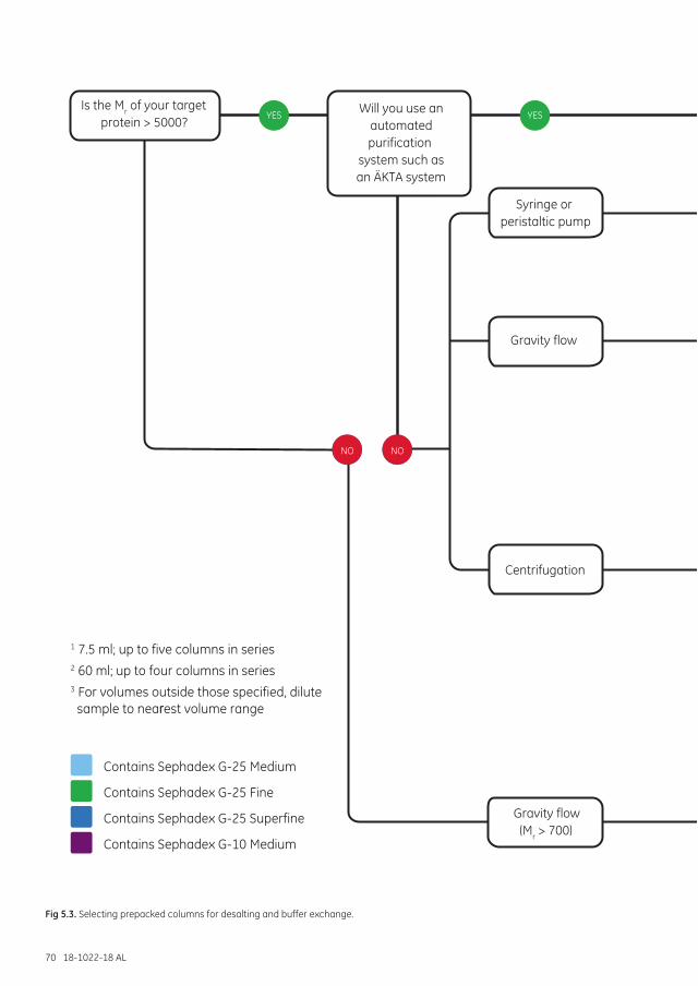
70 18-1022-18 AL
Fig 5.3. Selecting prepacked columns for desalting and buffer exchange.
Is the Mr of your target protein >5000?
NO
YES YES
Syringe or peristaltic pump
Gravity flow
Centrifugation
Will you use anautomatedpurification system such asÄKTAdesign?
NO
1 7.5 ml; up to five columns in series2 60 ml; up to four columns in series3 For volumes outside those specified, dilute sample to nearest volume range
Contains Sephadex G-25 Medium
Contains Sephadex G-25 Fine
Contains Sephadex G-25 Superfine
Contains Sephadex G-10 Medium
Is the Mr of your target protein > 5000?
NO
YES YES
Syringe or peristaltic pump
Gravity flow
Centrifugation
Will you use anautomatedpurification
system such asan ÄKTA system
NO
r
What is your sample volume?
What is your sample volume?
What is your sample volume?
What is your sample volume?
What is your sample volume?
PD MiniTrap G-25 + MiniSpin Adapter
PD MiniTrap G-10
PD MultiTrap G-25
PD-10 Desalting+ PD-10 Spin Adapter
PD MidiTrap G-25 + MidiSpin Adapter
PD SpinTrap G-25
PD-10 Desalting
PD MidiTrap G-25
PD MiniTrap G-25
HiTrap Desalting
HiPrep 26/10 Desalting
PD MidiTrap G-10
HiTrap Desalting0.1 to 1.5 ml
(7.5 ml)1
2.5 to 15 ml(60 ml)2
70 to 130 µl3
1.0 to 2.5 ml
0.5 to 1.0 ml
0.1 to 0.5 ml
0.1 to 1.5 ml(7.5 ml)1
0.2 to 0.5 ml3
0.75 to 1.0 ml3
1.75 to 2.5 ml3
70 to 130 µl(96-well plate)
100 to 300 µl3
400 µl to 1.0 ml3
Gravity flow(Mr > 700)
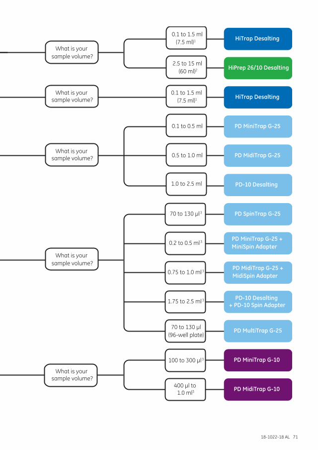
18-1022-18 AL 71
Is the Mr of your target protein >5000?
NO
YES YES
Syringe or peristaltic pump
Gravity flow
Centrifugation
Will you use anautomatedpurification system such asÄKTAdesign?
NO
1 7.5 ml; up to five columns in series2 60 ml; up to four columns in series3 For volumes outside those specified, dilute sample to nearest volume range
Contains Sephadex G-25 Medium
Contains Sephadex G-25 Fine
Contains Sephadex G-25 Superfine
Contains Sephadex G-10 Medium
Is the Mr of your target protein > 5000?
NO
YES YES
Syringe or peristaltic pump
Gravity flow
Centrifugation
Will you use anautomatedpurification
system such asan ÄKTA system
NO
r
What is your sample volume?
What is your sample volume?
What is your sample volume?
What is your sample volume?
What is your sample volume?
PD MiniTrap G-25 + MiniSpin Adapter
PD MiniTrap G-10
PD MultiTrap G-25
PD-10 Desalting+ PD-10 Spin Adapter
PD MidiTrap G-25 + MidiSpin Adapter
PD SpinTrap G-25
PD-10 Desalting
PD MidiTrap G-25
PD MiniTrap G-25
HiTrap Desalting
HiPrep 26/10 Desalting
PD MidiTrap G-10
HiTrap Desalting0.1 to 1.5 ml
(7.5 ml)1
2.5 to 15 ml(60 ml)2
70 to 130 µl3
1.0 to 2.5 ml
0.5 to 1.0 ml
0.1 to 0.5 ml
0.1 to 1.5 ml(7.5 ml)1
0.2 to 0.5 ml3
0.75 to 1.0 ml3
1.75 to 2.5 ml3
70 to 130 µl(96-well plate)
100 to 300 µl3
400 µl to 1.0 ml3
Gravity flow(Mr > 700)
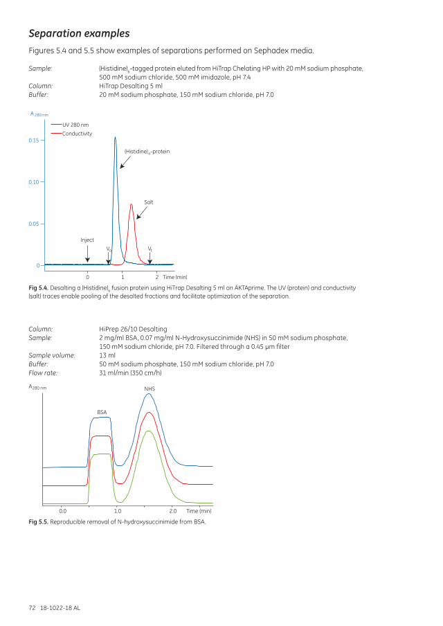
72 18-1022-18 AL
Separation examplesFigures 5.4 and 5.5 show examples of separations performed on Sephadex media.
Sample: (Histidine)6-tagged protein eluted from HiTrap Chelating HP with 20 mM sodium phosphate, 500 mM sodium chloride, 500 mM imidazole, pH 7.4
Column: HiTrap Desalting 5 ml Buffer: 20 mM sodium phosphate, 150 mM sodium chloride, pH 7.0
0
0.05
0.10
0.15
UV 280 nm
Conductivity
(Histidine) -protein
Salt
6
Inject
2 Time (min)10
VtVo
A 280 nm
Fig 5.4. Desalting a (Histidine)6 fusion protein using HiTrap Desalting 5 ml on ÄKTAprime. The UV (protein) and conductivity (salt) traces enable pooling of the desalted fractions and facilitate optimization of the separation.
Column: HiPrep 26/10 DesaltingSample: 2 mg/ml BSA, 0.07 mg/ml N-Hydroxysuccinimide (NHS) in 50 mM sodium phosphate,
150 mM sodium chloride, pH 7.0. Filtered through a 0.45 µm filterSample volume: 13 mlBuffer: 50 mM sodium phosphate, 150 mM sodium chloride, pH 7.0Flow rate: 31 ml/min (350 cm/h)
0.0 1.0 2.0 Time (min)
BSA
NHSA280 nm
Fig 5.5. Reproducible removal of N-hydroxysuccinimide from BSA.
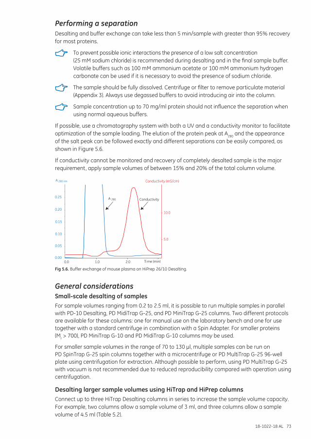
18-1022-18 AL 73
Performing a separationDesalting and buffer exchange can take less than 5 min/sample with greater than 95% recovery for most proteins.
To prevent possible ionic interactions the presence of a low salt concentration (25 mM sodium chloride) is recommended during desalting and in the final sample buffer. Volatile buffers such as 100 mM ammonium acetate or 100 mM ammonium hydrogen carbonate can be used if it is necessary to avoid the presence of sodium chloride.
The sample should be fully dissolved. Centrifuge or filter to remove particulate material (Appendix 3). Always use degassed buffers to avoid introducing air into the column.
Sample concentration up to 70 mg/ml protein should not influence the separation when using normal aqueous buffers.
If possible, use a chromatography system with both a UV and a conductivity monitor to facilitate optimization of the sample loading. The elution of the protein peak at A280 and the appearance of the salt peak can be followed exactly and different separations can be easily compared, as shown in Figure 5.6.
If conductivity cannot be monitored and recovery of completely desalted sample is the major requirement, apply sample volumes of between 15% and 20% of the total column volume.
0.0 1.0 2.00.00
0.05
0.10
0.15
0.20
0.25 A 280
5.0
10.0
Conductivity (mS/cm)
Ti me (min)
Conductivity
A 280 nm
Fig 5.6. Buffer exchange of mouse plasma on HiPrep 26/10 Desalting.
General considerationsSmall-scale desalting of samplesFor sample volumes ranging from 0.2 to 2.5 ml, it is possible to run multiple samples in parallel with PD-10 Desalting, PD MidiTrap G-25, and PD MiniTrap G-25 columns. Two different protocols are available for these columns: one for manual use on the laboratory bench and one for use together with a standard centrifuge in combination with a Spin Adapter. For smaller proteins (Mr > 700), PD MiniTrap G-10 and PD MidiTrap G-10 columns may be used.
For smaller sample volumes in the range of 70 to 130 µl, multiple samples can be run on PD SpinTrap G-25 spin columns together with a microcentrifuge or PD MultiTrap G-25 96-well plate using centrifugation for extraction. Although possible to perform, using PD MultiTrap G-25 with vacuum is not recommended due to reduced reproducibility compared with operation using centrifugation.
Desalting larger sample volumes using HiTrap and HiPrep columnsConnect up to three HiTrap Desalting columns in series to increase the sample volume capacity. For example, two columns allow a sample volume of 3 ml, and three columns allow a sample volume of 4.5 ml (Table 5.2).
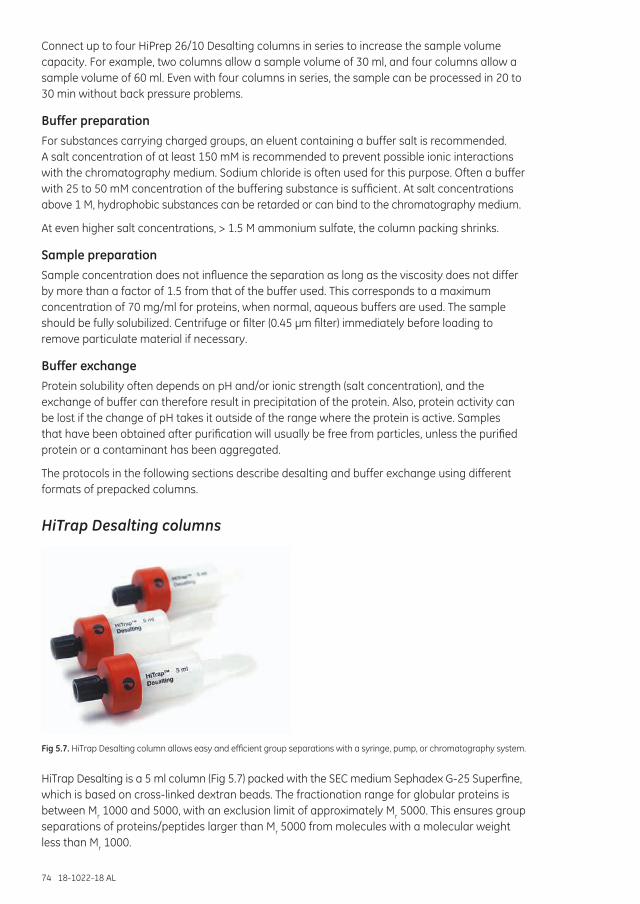
74 18-1022-18 AL
Connect up to four HiPrep 26/10 Desalting columns in series to increase the sample volume capacity. For example, two columns allow a sample volume of 30 ml, and four columns allow a sample volume of 60 ml. Even with four columns in series, the sample can be processed in 20 to 30 min without back pressure problems.
Buffer preparationFor substances carrying charged groups, an eluent containing a buffer salt is recommended. A salt concentration of at least 150 mM is recommended to prevent possible ionic interactions with the chromatography medium. Sodium chloride is often used for this purpose. Often a buffer with 25 to 50 mM concentration of the buffering substance is sufficient. At salt concentrations above 1 M, hydrophobic substances can be retarded or can bind to the chromatography medium.
At even higher salt concentrations, > 1.5 M ammonium sulfate, the column packing shrinks.
Sample preparationSample concentration does not influence the separation as long as the viscosity does not differ by more than a factor of 1.5 from that of the buffer used. This corresponds to a maximum concentration of 70 mg/ml for proteins, when normal, aqueous buffers are used. The sample should be fully solubilized. Centrifuge or filter (0.45 µm filter) immediately before loading to remove particulate material if necessary.
Buffer exchangeProtein solubility often depends on pH and/or ionic strength (salt concentration), and the exchange of buffer can therefore result in precipitation of the protein. Also, protein activity can be lost if the change of pH takes it outside of the range where the protein is active. Samples that have been obtained after purification will usually be free from particles, unless the purified protein or a contaminant has been aggregated.
The protocols in the following sections describe desalting and buffer exchange using different formats of prepacked columns.
HiTrap Desalting columns
Fig 5.7. HiTrap Desalting column allows easy and efficient group separations with a syringe, pump, or chromatography system.
HiTrap Desalting is a 5 ml column (Fig 5.7) packed with the SEC medium Sephadex G-25 Superfine, which is based on cross-linked dextran beads. The fractionation range for globular proteins is between Mr 1000 and 5000, with an exclusion limit of approximately Mr 5000. This ensures group separations of proteins/peptides larger than Mr 5000 from molecules with a molecular weight less than Mr 1000.
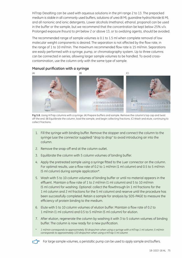
18-1022-18 AL 75
HiTrap Desalting can be used with aqueous solutions in the pH range 2 to 13. The prepacked medium is stable in all commonly used buffers, solutions of urea (8 M), guanidine hydrochloride (6 M), and all nonionic and ionic detergents. Lower alcohols (methanol, ethanol, propanol) can be used in the buffer or the sample, but we recommend that the concentration be kept below 25% v/v. Prolonged exposure (hours) to pH below 2 or above 13, or to oxidizing agents, should be avoided.
The recommended range of sample volumes is 0.1 to 1.5 ml when complete removal of low molecular weight components is desired. The separation is not affected by the flow rate, in the range of 1 to 10 ml/min. The maximum recommended flow rate is 15 ml/min. Separations are easily performed with a syringe, pump, or chromatography system. Up to three columns can be connected in series, allowing larger sample volumes to be handled. To avoid cross-contamination, use the column only with the same type of sample.
Manual purification with a syringe(A) (B) (C)
Fig 5.8. Using HiTrap columns with a syringe. (A) Prepare buffers and sample. Remove the column’s top cap and twist off the end. (B) Equilibrate the column, load the sample, and begin collecting fractions. (C) Wash and elute, continuing to collect fractions.
1. Fill the syringe with binding buffer. Remove the stopper and connect the column to the syringe (use the connector supplied) “drop to drop” to avoid introducing air into the column.
2. Remove the snap-off end at the column outlet.
3. Equilibrate the column with 5 column volumes of binding buffer.
4. Apply the pretreated sample using a syringe fitted to the Luer connector on the column. For optimal results, use a flow rate of 0.2 to 1 ml/min (1 ml column) and 0.5 to 5 ml/min (5 ml column) during sample application*.
5. Wash with 5 to 10 column volumes of binding buffer or until no material appears in the effluent. Maintain a flow rate of 1 to 2 ml/min (1 ml column) and 5 to 10 ml/min (5 ml column) for washing. Optional: collect the flowthrough (in 1 ml fractions for the 1 ml column and 2 ml fractions for the 5 ml column) and reserve until the procedure has been successfully completed. Retain a sample for analysis by SDS-PAGE to measure the efficiency of protein binding to the medium.
6. Elute with 5 to 10 column volumes of elution buffer. Maintain a flow rate of 0.2 to 1 ml/min (1 ml column) and 0.5 to 5 ml/min (5 ml column) for elution.
7. After elution, regenerate the column by washing it with 3 to 5 column volumes of binding buffer. The column is now ready for a new purification.
* 1 ml/min corresponds to approximately 30 drops/min when using a syringe with a HiTrap 1 ml column; 5 ml/min corresponds to approximately 120 drops/min when using a HiTrap 5 ml column
For large sample volumes, a peristaltic pump can be used to apply sample and buffers.
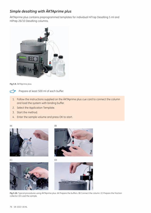
76 18-1022-18 AL
Simple desalting with ÄKTAprime plusÄKTAprime plus contains preprogrammed templates for individual HiTrap Desalting 5 ml and HiPrep 26/10 Desalting columns.
Fig 5.9. ÄKTAprime plus.
Prepare at least 500 ml of each buffer.
1. Follow the instructions supplied on the ÄKTAprime plus cue card to connect the column and load the system with binding buffer.
2. Select the Application Template.
3. Start the method.
4. Enter the sample volume and press OK to start.
(A) (B)
(D)(C)
Fig 5.10. Typical procedures using ÄKTAprime plus. (A) Prepare the buffers. (B) Connect the column. (C) Prepare the fraction collector. (D) Load the sample.
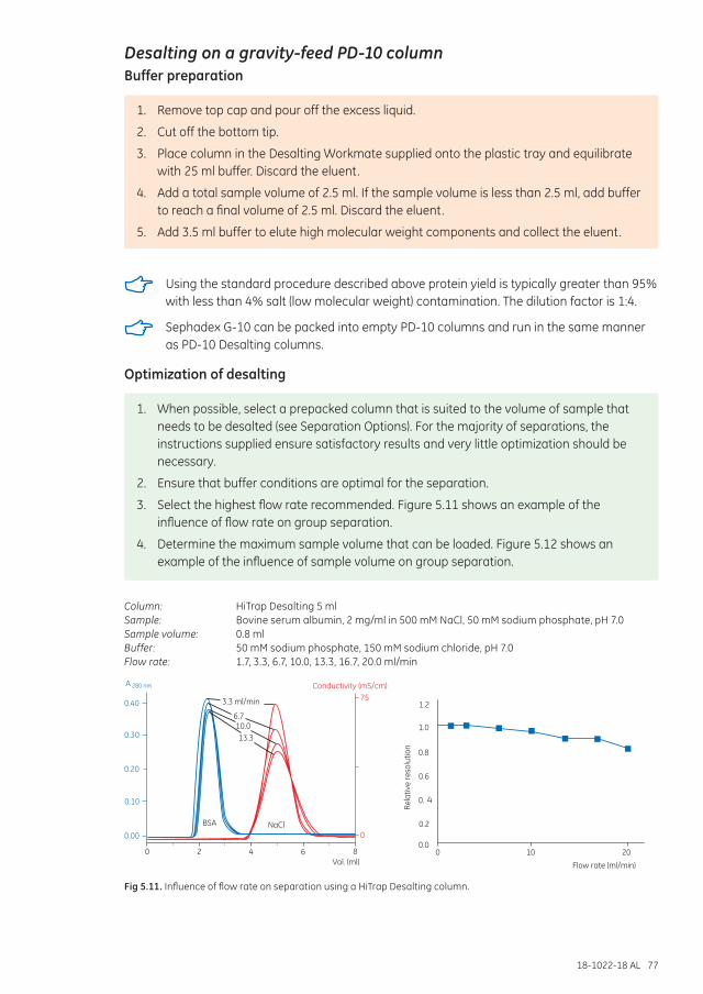
18-1022-18 AL 77
Desalting on a gravity-feed PD-10 columnBuffer preparation
1. Remove top cap and pour off the excess liquid.
2. Cut off the bottom tip.
3. Place column in the Desalting Workmate supplied onto the plastic tray and equilibrate with 25 ml buffer. Discard the eluent.
4. Add a total sample volume of 2.5 ml. If the sample volume is less than 2.5 ml, add buffer to reach a final volume of 2.5 ml. Discard the eluent.
5. Add 3.5 ml buffer to elute high molecular weight components and collect the eluent.
Using the standard procedure described above protein yield is typically greater than 95% with less than 4% salt (low molecular weight) contamination. The dilution factor is 1:4.
Sephadex G-10 can be packed into empty PD-10 columns and run in the same manner as PD-10 Desalting columns.
Optimization of desalting
1. When possible, select a prepacked column that is suited to the volume of sample that needs to be desalted (see Separation Options). For the majority of separations, the instructions supplied ensure satisfactory results and very little optimization should be necessary.
2. Ensure that buffer conditions are optimal for the separation.
3. Select the highest flow rate recommended. Figure 5.11 shows an example of the influence of flow rate on group separation.
4. Determine the maximum sample volume that can be loaded. Figure 5.12 shows an example of the influence of sample volume on group separation.
Column: HiTrap Desalting 5 mlSample: Bovine serum albumin, 2 mg/ml in 500 mM NaCl, 50 mM sodium phosphate, pH 7.0Sample volume: 0.8 mlBuffer: 50 mM sodium phosphate, 150 mM sodium chloride, pH 7.0Flow rate: 1.7, 3.3, 6.7, 10.0, 13.3, 16.7, 20.0 ml/min
Vol. (ml)0
00.00
2
BSA NaCl
0.20
4
0.10
6
0.30
8
750.40
Conductivity (mS/cm)
3.3 ml/min
6.710.013.3
A 280 nm
Flow rate (ml/min)
Rela
tive
reso
lutio
n
00.0
0.2
0. 4
0.6
0.8
1.0
1.2
10 20
Fig 5.11. Influence of flow rate on separation using a HiTrap Desalting column.
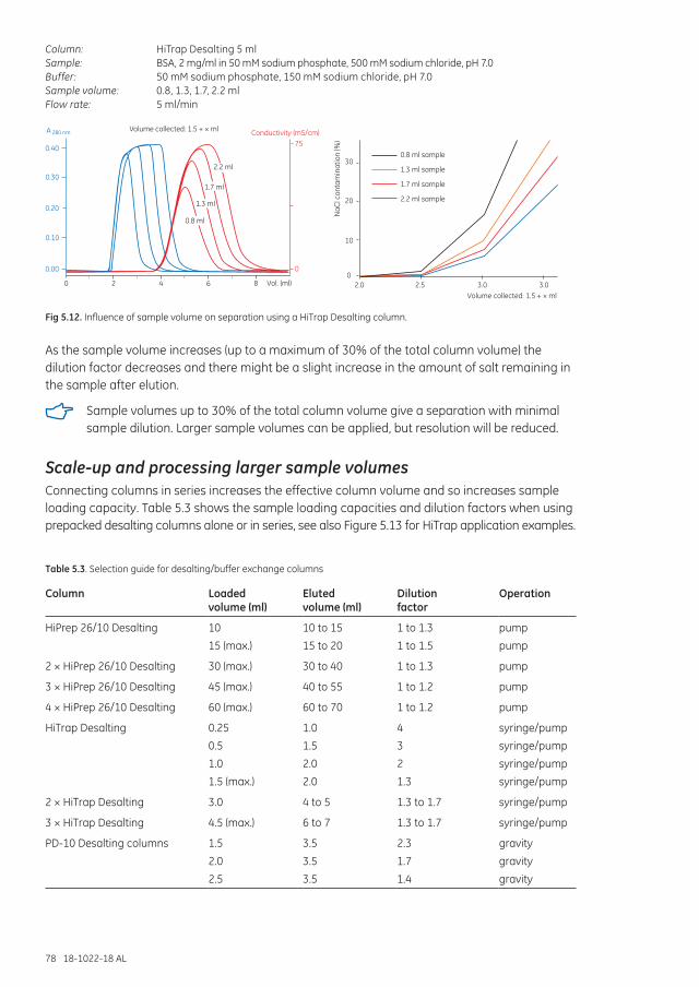
78 18-1022-18 AL
Column: HiTrap Desalting 5 mlSample: BSA, 2 mg/ml in 50 mM sodium phosphate, 500 mM sodium chloride, pH 7.0Buffer: 50 mM sodium phosphate, 150 mM sodium chloride, pH 7.0Sample volume: 0.8, 1.3, 1.7, 2.2 mlFlow rate: 5 ml/min
Vol. (ml)
Volume collected: 1.5 + × ml
0
0
0.00
2
0.20
4
0.10
6
0.30
75
8
0.40
Conductivity (mS/cm)
2.2 ml
1.7 ml
1.3 ml
0.8 ml
A 280 nm
NaC
l con
tam
inat
ion
(%)
30
2.0Volume collected: 1.5 + × ml
2.5 3.0 3.0
10
0
20
0.8 ml sample
1.3 ml sample
1.7 ml sample
2.2 ml sample
Fig 5.12. Influence of sample volume on separation using a HiTrap Desalting column.
As the sample volume increases (up to a maximum of 30% of the total column volume) the dilution factor decreases and there might be a slight increase in the amount of salt remaining in the sample after elution.
Sample volumes up to 30% of the total column volume give a separation with minimal sample dilution. Larger sample volumes can be applied, but resolution will be reduced.
Scale-up and processing larger sample volumesConnecting columns in series increases the effective column volume and so increases sample loading capacity. Table 5.3 shows the sample loading capacities and dilution factors when using prepacked desalting columns alone or in series, see also Figure 5.13 for HiTrap application examples.
Table 5.3. Selection guide for desalting/buffer exchange columns
Column Loaded volume (ml)
Eluted volume (ml)
Dilution factor
Operation
HiPrep 26/10 Desalting 10
15 (max.)
10 to 15
15 to 20
1 to 1.3
1 to 1.5
pump
pump
2 × HiPrep 26/10 Desalting 30 (max.) 30 to 40 1 to 1.3 pump
3 × HiPrep 26/10 Desalting 45 (max.) 40 to 55 1 to 1.2 pump
4 × HiPrep 26/10 Desalting 60 (max.) 60 to 70 1 to 1.2 pump
HiTrap Desalting 0.25
0.5
1.0
1.5 (max.)
1.0
1.5
2.0
2.0
4
3
2
1.3
syringe/pump
syringe/pump
syringe/pump
syringe/pump
2 × HiTrap Desalting 3.0 4 to 5 1.3 to 1.7 syringe/pump
3 × HiTrap Desalting 4.5 (max.) 6 to 7 1.3 to 1.7 syringe/pump
PD-10 Desalting columns 1.5
2.0
2.5
3.5
3.5
3.5
2.3
1.7
1.4
gravity
gravity
gravity
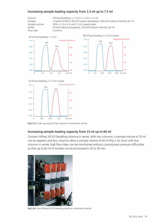
18-1022-18 AL 79
Increasing sample loading capacity from 1.5 ml up to 7.5 ml
Column: HiTrap Desalting, 1 × 5 ml, 3 × 5 ml, 5 × 5 mlSample: 2 mg/ml of BSA in 50 mM sodium phosphate, 500 mM sodium chloride, pH 7.0Sample volume: 28% × Vt (1.4, 4.3, and 7.1 ml, respectively)Buffer: 50 mM sodium phosphate, 150 mM sodium chloride, pH 7.0Flow rate: 5 ml/min
Conductivity (mS/cm)
0.40
0.00
0.30
0.20
0.10
50
40
30
20
0 10.0 20.0 30. 0 Vol. (ml)
BSANaCl
0.40
0.00
0.30
0.20
0.10
0 2 .0 4. 0 6 .0 Vol. (ml)
Conductivity (mS/cm)
50
40
30
20
BSA
NaCl 50
40
30
20
Conductivity (mS/cm)
0 5 .0 10.0 15. 0 Vol. (ml)20.0
0.40
0.00
0.30
0.20
0.10
BSANaCl
(A) HiTrap Desalting, 1 × 5 ml (B) HiTrap Desalting, 3 × 5 ml in series
(C) HiTrap Desalting, 5 × 5 ml in series
A
A
A280 nm
280 nm
280 nm
Fig 5.13. Scale-up using HiTrap columns connected in series.
Increasing sample loading capacity from 15 ml up to 60 mlConnect HiPrep 26/10 Desalting columns in series. With two columns, a sample volume of 30 ml can be applied, and four columns allow a sample volume of 60 ml (Fig 5.14). Even with four columns in series, high flow rates can be maintained without causing back pressure difficulties so that up to 60 ml of sample can be processed in 20 to 30 min.
Fig 5.14. Four HiPrep 26/10 Desalting columns connected in series.
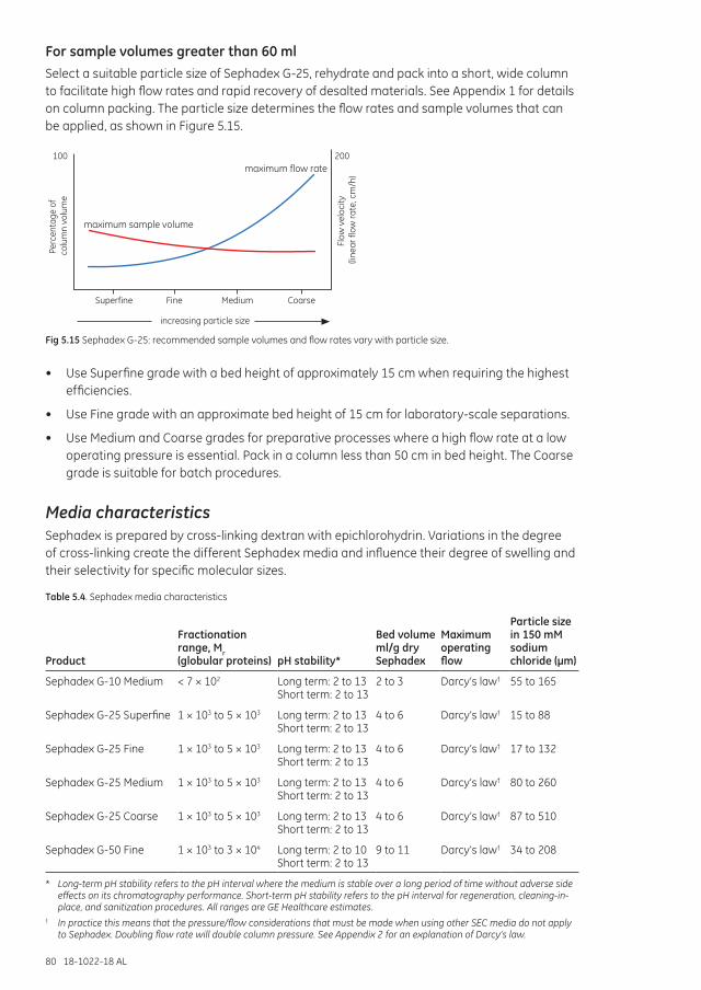
80 18-1022-18 AL
For sample volumes greater than 60 mlSelect a suitable particle size of Sephadex G-25, rehydrate and pack into a short, wide column to facilitate high flow rates and rapid recovery of desalted materials. See Appendix 1 for details on column packing. The particle size determines the flow rates and sample volumes that can be applied, as shown in Figure 5.15.
Superfine
maximum sample volume
maximum flow rate
Fine Medium Coarse
Perc
enta
ge o
f co
lum
n vo
lum
e
100
Flow
vel
ocity
(li
near
flow
rate
, cm
/h)
200
increasing particle size
Fig 5.15 Sephadex G-25: recommended sample volumes and flow rates vary with particle size.
• Use Superfine grade with a bed height of approximately 15 cm when requiring the highest efficiencies.
• Use Fine grade with an approximate bed height of 15 cm for laboratory-scale separations.
• Use Medium and Coarse grades for preparative processes where a high flow rate at a low operating pressure is essential. Pack in a column less than 50 cm in bed height. The Coarse grade is suitable for batch procedures.
Media characteristicsSephadex is prepared by cross-linking dextran with epichlorohydrin. Variations in the degree of cross-linking create the different Sephadex media and influence their degree of swelling and their selectivity for specific molecular sizes.
Table 5.4. Sephadex media characteristics
Product
Fractionation range, Mr (globular proteins) pH stability*
Bed volume ml/g dry Sephadex
Maximum operating flow
Particle size in 150 mM sodium chloride (µm)
Sephadex G-10 Medium < 7 × 10² Long term: 2 to 13 Short term: 2 to 13
2 to 3 Darcy’s law† 55 to 165
Sephadex G-25 Superfine 1 × 10³ to 5 × 10³ Long term: 2 to 13 Short term: 2 to 13
4 to 6 Darcy’s law† 15 to 88
Sephadex G-25 Fine 1 × 10³ to 5 × 10³ Long term: 2 to 13 Short term: 2 to 13
4 to 6 Darcy’s law† 17 to 132
Sephadex G-25 Medium 1 × 10³ to 5 × 10³ Long term: 2 to 13 Short term: 2 to 13
4 to 6 Darcy’s law† 80 to 260
Sephadex G-25 Coarse 1 × 10³ to 5 × 10³ Long term: 2 to 13 Short term: 2 to 13
4 to 6 Darcy’s law† 87 to 510
Sephadex G-50 Fine 1 × 10³ to 3 × 10⁴ Long term: 2 to 10 Short term: 2 to 13
9 to 11 Darcy’s law† 34 to 208
* Long-term pH stability refers to the pH interval where the medium is stable over a long period of time without adverse side effects on its chromatography performance. Short-term pH stability refers to the pH interval for regeneration, cleaning-in-place, and sanitization procedures. All ranges are GE Healthcare estimates.
† In practice this means that the pressure/flow considerations that must be made when using other SEC media do not apply to Sephadex. Doubling flow rate will double column pressure. See Appendix 2 for an explanation of Darcy’s law.
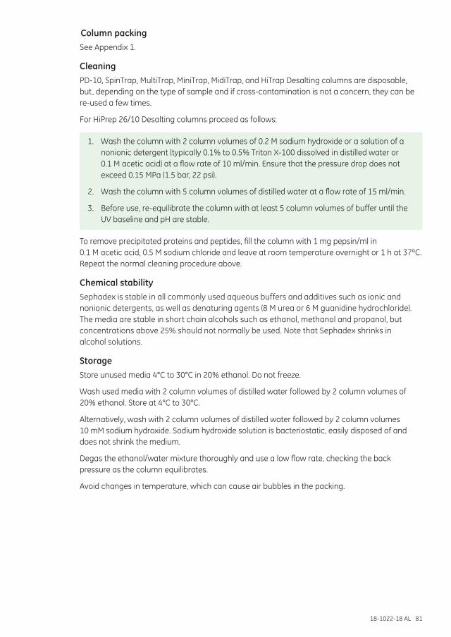
18-1022-18 AL 81
Column packingSee Appendix 1.
CleaningPD-10, SpinTrap, MultiTrap, MiniTrap, MidiTrap, and HiTrap Desalting columns are disposable, but, depending on the type of sample and if cross-contamination is not a concern, they can be re-used a few times.
For HiPrep 26/10 Desalting columns proceed as follows:
1. Wash the column with 2 column volumes of 0.2 M sodium hydroxide or a solution of a nonionic detergent (typically 0.1% to 0.5% Triton X-100 dissolved in distilled water or 0.1 M acetic acid) at a flow rate of 10 ml/min. Ensure that the pressure drop does not exceed 0.15 MPa (1.5 bar, 22 psi).
2. Wash the column with 5 column volumes of distilled water at a flow rate of 15 ml/min.
3. Before use, re-equilibrate the column with at least 5 column volumes of buffer until the UV baseline and pH are stable.
To remove precipitated proteins and peptides, fill the column with 1 mg pepsin/ml in 0.1 M acetic acid, 0.5 M sodium chloride and leave at room temperature overnight or 1 h at 37ºC. Repeat the normal cleaning procedure above.
Chemical stabilitySephadex is stable in all commonly used aqueous buffers and additives such as ionic and nonionic detergents, as well as denaturing agents (8 M urea or 6 M guanidine hydrochloride). The media are stable in short chain alcohols such as ethanol, methanol and propanol, but concentrations above 25% should not normally be used. Note that Sephadex shrinks in alcohol solutions.
StorageStore unused media 4°C to 30°C in 20% ethanol. Do not freeze.
Wash used media with 2 column volumes of distilled water followed by 2 column volumes of 20% ethanol. Store at 4°C to 30°C.
Alternatively, wash with 2 column volumes of distilled water followed by 2 column volumes 10 mM sodium hydroxide. Sodium hydroxide solution is bacteriostatic, easily disposed of and does not shrink the medium.
Degas the ethanol/water mixture thoroughly and use a low flow rate, checking the back pressure as the column equilibrates.
Avoid changes in temperature, which can cause air bubbles in the packing.

82 18-1022-18 AL
18-1022-18 AL 83
Chapter 6 Sephadex LH-20: size exclusion chromatography in organic solvents
Sephadex LH-20 is specifically designed for the separation and purification of natural products that require the presence of organic solvents to maintain their solubility, including molecules such as steroids, terpenoids, lipids and low molecular weight peptides (up to 35 amino acid residues). Compounds are usually separated by a form of liquid/liquid partitioning or absorption chromatography. Sephadex LH-20 can have a very high selectivity for aromatic compounds in certain solvents and can be used at analytical or industrial scale for the preparation of closely related species. Sephadex LH-20 has found widespread use in the isolation of active components from herb extract used in TCM (Traditional Chinese Medicine).
Sephadex LH-20 is made from hydroxypropylated dextran beads that have been cross-linked to yield a polysaccharide network. The medium can be swollen in water and organic solvents.
Sephadex LH-20 is suitable for an initial purification before polishing by IEX or RPC, or in a final polishing step, for example during the preparation of diastereoisomers.
Sephadex LH-20 exhibits both hydrophilic and hydrophobic properties, the combination of which can offer unique chromatography selectivity for certain applications.
Sephadex has been used for SEC in organic solvents, for example dimethylformamide may be used with Sephadex G-10. Mixtures of water with short-chain alcohols may be used with Sephadex G-10, G-25, and G-50.
Media characteristicsTable 6.1. Characteristics of Sephadex LH-20
Product Fractionation range (globular proteins)
Sample loading capacity*
Maximum pressure drop over the packed bed
Maximum operating flow rate
Sephadex LH-20 < 5 × 10³ (exclusion limit will depend on the solvent)
2% of column volume
Solvent-dependent 12 cm/min (720 cm/h, bed height 14 cm, 15 MPa back pressure)
* If Sephadex LH-20 is used in adsorption mode then the sample volume is unlimited until reaching the point of column saturation.
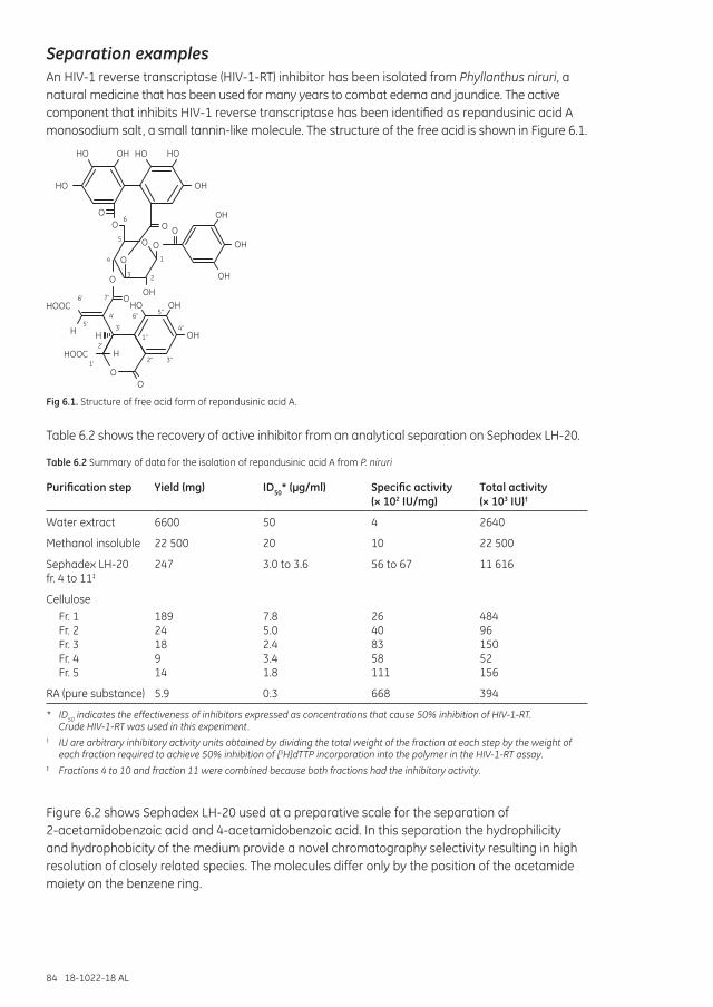
84 18-1022-18 AL
Separation examplesAn HIV-1 reverse transcriptase (HIV-1-RT) inhibitor has been isolated from Phyllanthus niruri, a natural medicine that has been used for many years to combat edema and jaundice. The active component that inhibits HIV-1 reverse transcriptase has been identified as repandusinic acid A monosodium salt, a small tannin-like molecule. The structure of the free acid is shown in Figure 6.1.
OO
O
O
O O
HO OH HO HO
HO OH
O
OHO
OHOH
OH
OO
HOOC
HOOC
H
H
H
OH
OHO
OH
6
5
4
3 2
1
6'
5'4'
3'
2'
1'
7'
6'' 5''
4''
3''2''
1''
Fig 6.1. Structure of free acid form of repandusinic acid A.
Table 6.2 shows the recovery of active inhibitor from an analytical separation on Sephadex LH-20.
Table 6.2 Summary of data for the isolation of repandusinic acid A from P. niruri
Purification step Yield (mg) ID50* (µg/ml) Specific activity (× 102 IU/mg)
Total activity (× 103 IU)†
Water extract 6600 50 4 2640
Methanol insoluble 22 500 20 10 22 500
Sephadex LH-20 fr. 4 to 11‡
247 3.0 to 3.6 56 to 67 11 616
CelluloseFr. 1 Fr. 2 Fr. 3 Fr. 4 Fr. 5
189 24 18 9 14
7.8 5.0 2.4 3.4 1.8
26 40 83 58 111
484 96 150 52 156
RA (pure substance) 5.9 0.3 668 394
* ID50 indicates the effectiveness of inhibitors expressed as concentrations that cause 50% inhibition of HIV-1-RT. Crude HIV-1-RT was used in this experiment.
† IU are arbitrary inhibitory activity units obtained by dividing the total weight of the fraction at each step by the weight of each fraction required to achieve 50% inhibition of [3H]dTTP incorporation into the polymer in the HIV-1-RT assay.
‡ Fractions 4 to 10 and fraction 11 were combined because both fractions had the inhibitory activity.
Figure 6.2 shows Sephadex LH-20 used at a preparative scale for the separation of 2-acetamidobenzoic acid and 4-acetamidobenzoic acid. In this separation the hydrophilicity and hydrophobicity of the medium provide a novel chromatography selectivity resulting in high resolution of closely related species. The molecules differ only by the position of the acetamide moiety on the benzene ring.
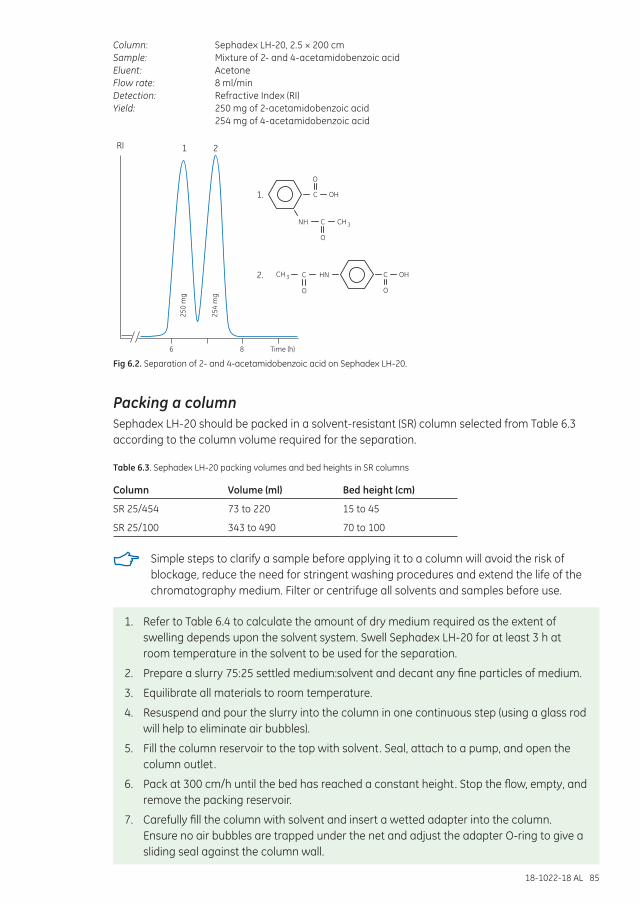
18-1022-18 AL 85
Column: Sephadex LH-20, 2.5 × 200 cmSample: Mixture of 2- and 4-acetamidobenzoic acidEluent: AcetoneFlow rate: 8 ml/minDetection: Refractive Index (RI)Yield: 250 mg of 2-acetamidobenzoic acid
254 mg of 4-acetamidobenzoic acid
250
mg
254
mg
6 8 Time (h)
RI 1 2
OO
C
C
NH
HN
3
O
O
1.
2.
OH
OH
CH
3CH
C
C
Fig 6.2. Separation of 2- and 4-acetamidobenzoic acid on Sephadex LH-20.
Packing a columnSephadex LH-20 should be packed in a solvent-resistant (SR) column selected from Table 6.3 according to the column volume required for the separation.
Table 6.3. Sephadex LH-20 packing volumes and bed heights in SR columns
Column Volume (ml) Bed height (cm)
SR 25/454 73 to 220 15 to 45
SR 25/100 343 to 490 70 to 100
Simple steps to clarify a sample before applying it to a column will avoid the risk of blockage, reduce the need for stringent washing procedures and extend the life of the chromatography medium. Filter or centrifuge all solvents and samples before use.
1. Refer to Table 6.4 to calculate the amount of dry medium required as the extent of swelling depends upon the solvent system. Swell Sephadex LH-20 for at least 3 h at room temperature in the solvent to be used for the separation.
2. Prepare a slurry 75:25 settled medium:solvent and decant any fine particles of medium.
3. Equilibrate all materials to room temperature.
4. Resuspend and pour the slurry into the column in one continuous step (using a glass rod will help to eliminate air bubbles).
5. Fill the column reservoir to the top with solvent. Seal, attach to a pump, and open the column outlet.
6. Pack at 300 cm/h until the bed has reached a constant height. Stop the flow, empty, and remove the packing reservoir.
7. Carefully fill the column with solvent and insert a wetted adapter into the column. Ensure no air bubbles are trapped under the net and adjust the adapter O-ring to give a sliding seal against the column wall.
86 18-1022-18 AL
8. Connect all tubings, ensuring that there are no air bubbles in the flow path.
9. Slowly slide down the adapter so that any air in the tubings is displaced by solvent and lock the adapter into position on the surface of the medium.
10. Open the column outlet and continue packing until the packed bed is stable and a final adjustment of the top adapter can be made.
In solvents such as chloroform, Sephadex LH-20 is less dense than the solvent and the medium will float. Pour the medium into the column and drain until the second adapter can be inserted. Lock the adapter in position at the surface of the medium and direct the flow of chloroform upwards. The bed will be packed against the top adapter and the lower adapter can be pushed slowly upwards towards the lower surface of the medium. Close the column outlet when moving the adapter to avoid compressing the bed.
Performing a separationStart at a linear flow of 1 cm/h to check resolution. Low flow rate improves the resolution.
1. Equilibrate the column with at least 2 column volumes of the solvent until a stable baseline is achieved.
2. Apply a sample volume equivalent to 1% to 2% of the total column volume.
3. Elute in 1 column volume. Re-equilibration is not needed between runs with the same solvent.
CleaningWash the column with 2 to 3 column volumes of the solvent, followed by re-equilibration in a new solvent if changing the separation conditions.
18-1022-18 AL 87
Table 6.4. Approximate values for packed bed volumes of Sephadex LH-20 swollen in different solvents
Solvent Approx. bed volume (ml/g dry Sephadex LH-20)
Dimethyl sulfoxide 4.4 to 4.6
Pyridine 4.2 to 4.4
Water 4.0 to 4.4
Dimethylformamide 4.0 to 4.4
Methanol 3.9 to 4.3
Saline 3.8 to 4.2
Ethylene dichloride 3.8 to 4.1
Chloroform* 3.8 to 4.1
Propanol 3.7 to 4.0
Ethanol† 3.6 to 3.9
Isobutanol 3.6 to 3.9
Formamide 3.6 to 3.9
Methylene dichloride 3.6 to 3.9
Butanol 3.5 to 3.8
Isopropanol 3.3 to 3.6
Tetrahydrofuran 3.3 to 3.6
Dioxane 3.2 to 3.5
Acetone 2.4 to 2.6
Acetonitrile‡ 2.2 to 2.4
Carbon tetrachloride‡ 1.8 to 2.2
Benzene‡ 1.6 to 2.0
Ethyl acetate‡ 1.6 to 1.8
Toluene‡ 1.5 to 1.6
* Containing 1% ethanol.† Containing 1% benzene.‡ Solvents that give a bed volume of less than 2.5 mg/ml dry Sephadex LH-20 are not generally useful.
Chemical stabilitySephadex LH-20 is stable in most aqueous and organic solvent systems. The medium is not stable below pH 2.0 or in strong oxidizing agents.
StorageStore dry at 4°C to 30°C. Store packed columns and used medium at 4°C to 8°C in the presence of a bacteriostatic agent.
Transferring Sephadex LH-20 from aqueous solution to organic solventsTransfer Sephadex LH-20 from an aqueous solution to the organic solvent by moving through a graded series of solvent mixtures. This will ensure efficient replacement of the water by the required solvent.
To transfer from aqueous solution or organic solvent (100% A) to a new organic solvent (100% B), proceed as follows: transfer to 70% A:30% B then to 30% A:70% B and finally to 100% B. If A and B are not mutually miscible, make the transfer via an intermediate solvent, for example from water to chloroform via acetone, as shown in Figure 6.3.
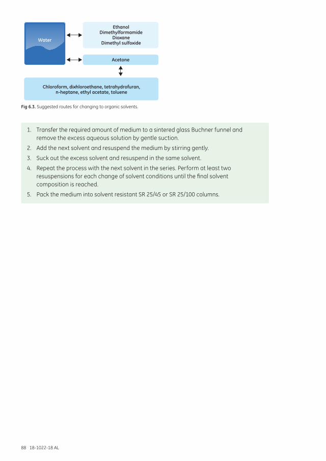
88 18-1022-18 AL
Water
Acetone
Chloroform, dixhloroethane, tetrahydrofuran, n-heptane, ethyl acetate, toluene
EthanolDimethylformamide
DioxaneDimethyl sulfoxide
Fig 6.3. Suggested routes for changing to organic solvents.
1. Transfer the required amount of medium to a sintered glass Buchner funnel and remove the excess aqueous solution by gentle suction.
2. Add the next solvent and resuspend the medium by stirring gently.
3. Suck out the excess solvent and resuspend in the same solvent.
4. Repeat the process with the next solvent in the series. Perform at least two resuspensions for each change of solvent conditions until the final solvent composition is reached.
5. Pack the medium into solvent resistant SR 25/45 or SR 25/100 columns.
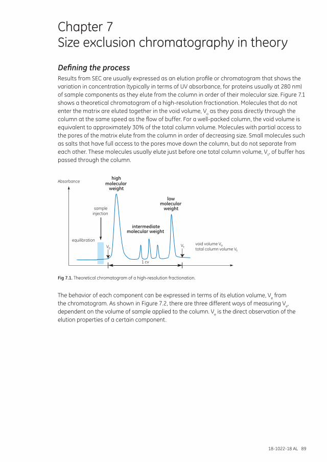
18-1022-18 AL 89
Chapter 7 Size exclusion chromatography in theory
Defining the processResults from SEC are usually expressed as an elution profile or chromatogram that shows the variation in concentration (typically in terms of UV absorbance, for proteins usually at 280 nm) of sample components as they elute from the column in order of their molecular size. Figure 7.1 shows a theoretical chromatogram of a high-resolution fractionation. Molecules that do not enter the matrix are eluted together in the void volume, Vo as they pass directly through the column at the same speed as the flow of buffer. For a well-packed column, the void volume is equivalent to approximately 30% of the total column volume. Molecules with partial access to the pores of the matrix elute from the column in order of decreasing size. Small molecules such as salts that have full access to the pores move down the column, but do not separate from each other. These molecules usually elute just before one total column volume, Vt, of buffer has passed through the column.
highmolecular
weight
intermediatemolecular weight
lowmolecular
weightsampleinjection
equilibration
Absorbance
1 cv
VtVovoid volume Vototal column volume Vt
Fig 7.1. Theoretical chromatogram of a high-resolution fractionation.
The behavior of each component can be expressed in terms of its elution volume, Ve from the chromatogram. As shown in Figure 7.2, there are three different ways of measuring Ve, dependent on the volume of sample applied to the column. Ve is the direct observation of the elution properties of a certain component.
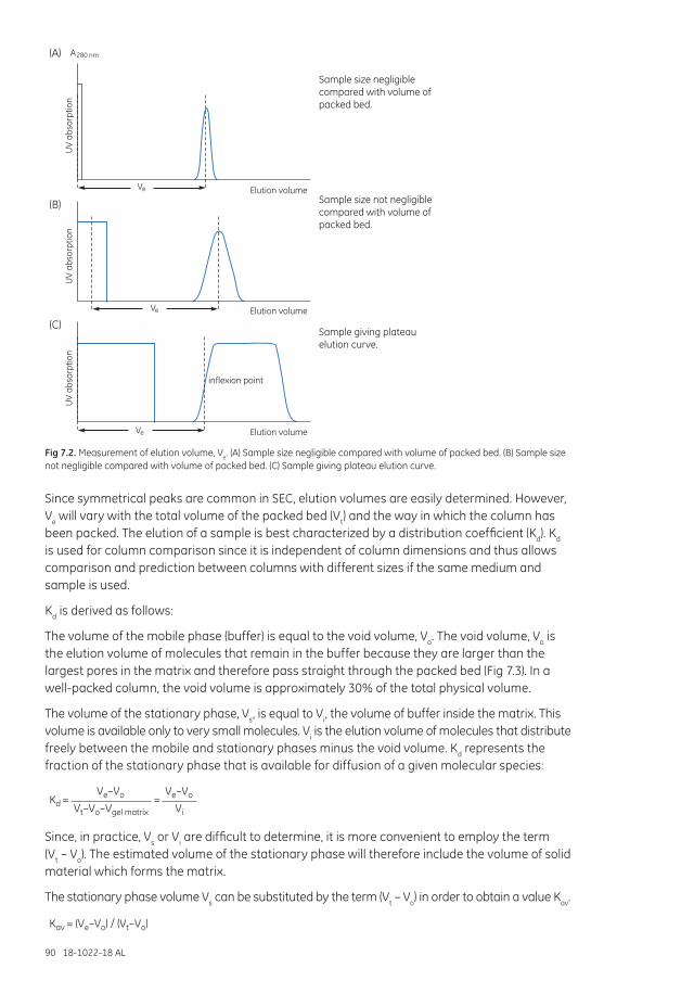
90 18-1022-18 AL
eV
eV
eV
(A) A280 nm
(B)
(C)
Elution volume
Elution volume
Elution volume
inflexion point
Sample size negligible compared with volume of packed bed.
Sample size not negligible compared with volume of packed bed.
Sample giving plateau elution curve.
UV
abso
rptio
n U
V ab
sorp
tion
UV
abso
rptio
n
Fig 7.2. Measurement of elution volume, Ve. (A) Sample size negligible compared with volume of packed bed. (B) Sample size not negligible compared with volume of packed bed. (C) Sample giving plateau elution curve.
Since symmetrical peaks are common in SEC, elution volumes are easily determined. However, Ve will vary with the total volume of the packed bed (Vt) and the way in which the column has been packed. The elution of a sample is best characterized by a distribution coefficient (Kd). Kd is used for column comparison since it is independent of column dimensions and thus allows comparison and prediction between columns with different sizes if the same medium and sample is used.
Kd is derived as follows:
The volume of the mobile phase (buffer) is equal to the void volume, Vo. The void volume, Vo is the elution volume of molecules that remain in the buffer because they are larger than the largest pores in the matrix and therefore pass straight through the packed bed (Fig 7.3). In a well-packed column, the void volume is approximately 30% of the total physical volume.
The volume of the stationary phase, Vs, is equal to Vi, the volume of buffer inside the matrix. This volume is available only to very small molecules. Vi is the elution volume of molecules that distribute freely between the mobile and stationary phases minus the void volume. Kd represents the fraction of the stationary phase that is available for diffusion of a given molecular species:
Kd = = Ve–Vo
Vt–Vo–Vgel matrix
Ve–Vo
Vi
Since, in practice, Vs or Vi are difficult to determine, it is more convenient to employ the term (Vt – Vo). The estimated volume of the stationary phase will therefore include the volume of solid material which forms the matrix.
The stationary phase volume Vs can be substituted by the term (Vt – Vo) in order to obtain a value Kav.
Kav = (Ve–Vo) / (Vt–Vo)
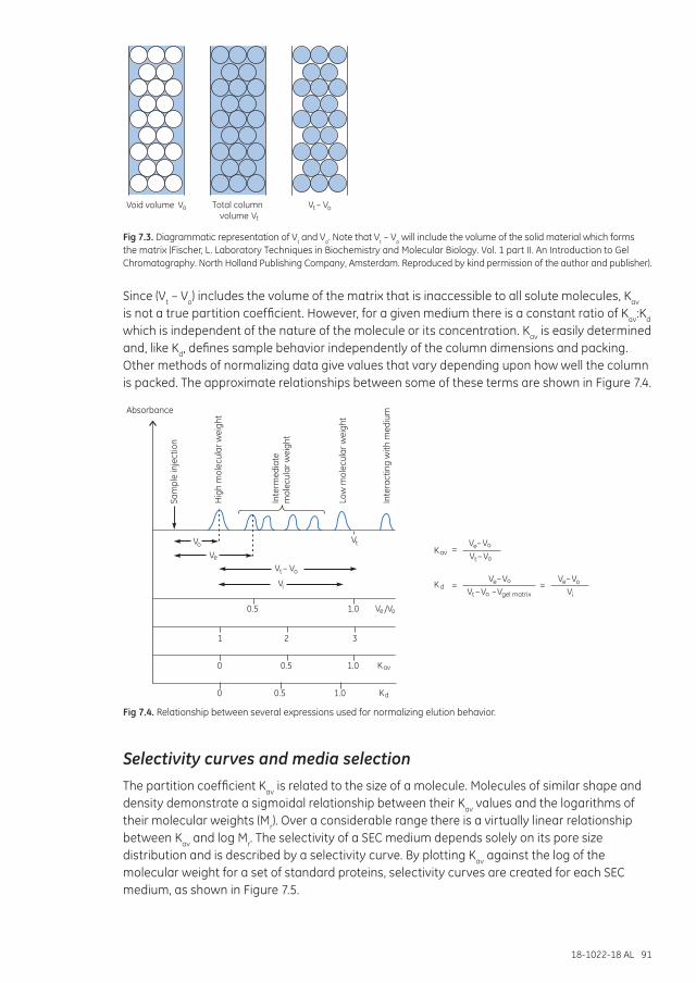
18-1022-18 AL 91
Void volume Vo V –t VoTotal columnvolume Vt
Fig 7.3. Diagrammatic representation of Vt and Vo. Note that Vt – Vo will include the volume of the solid material which forms the matrix (Fischer, L. Laboratory Techniques in Biochemistry and Molecular Biology. Vol. 1 part II. An Introduction to Gel Chromatography. North Holland Publishing Company, Amsterdam. Reproduced by kind permission of the author and publisher).
Since (Vt – Vo) includes the volume of the matrix that is inaccessible to all solute molecules, Kav is not a true partition coefficient. However, for a given medium there is a constant ratio of Kav:Kd which is independent of the nature of the molecule or its concentration. Kav is easily determined and, like Kd, defines sample behavior independently of the column dimensions and packing. Other methods of normalizing data give values that vary depending upon how well the column is packed. The approximate relationships between some of these terms are shown in Figure 7.4.
Sam
ple
inje
ctio
n
V – VotVoV –e
VoV –e
VoV –e
Vi
Kav
Kav
Kd
K dVgel matrixV – V –ot
V – Vot
Vi
VtVo
V
V
e
e
Vo/
=
= =
0.5
0.5
1 2 3
0
0 0.5
1.0
1.0
1.0
Low
mol
ecul
ar w
eigh
t
Inte
ract
ing
with
med
ium
Inte
rmed
iate
mol
ecul
ar w
eigh
t
Hig
h m
olec
ular
wei
ghtAbsorbance
Fig 7.4. Relationship between several expressions used for normalizing elution behavior.
Selectivity curves and media selectionThe partition coefficient Kav is related to the size of a molecule. Molecules of similar shape and density demonstrate a sigmoidal relationship between their Kav values and the logarithms of their molecular weights (Mr). Over a considerable range there is a virtually linear relationship between Kav and log Mr. The selectivity of a SEC medium depends solely on its pore size distribution and is described by a selectivity curve. By plotting Kav against the log of the molecular weight for a set of standard proteins, selectivity curves are created for each SEC medium, as shown in Figure 7.5.
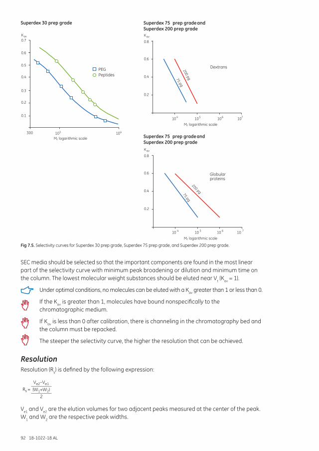
92 18-1022-18 AL
Superdex 75 andprep gradeSuperdex 200 prep grade
Globularproteins
0.4
0.2
200 pg75 pg
0.6
0.8
Kav
1010 4 10 5 10 6 7
Superdex 75 andprep gradeSuperdex 200 prep grade
Dextrans
0.4
0.2
200 pg75 pg
0.6
0.8
Kav
107104 105 106
Superdex 30 prep grade
PEGPeptides0.4
0.3
0.2
0.1
0.6
0.5
0.7Kav
104300 103
M logarithmic scaler
M logarithmic scaler
M logarithmic scaler
Fig 7.5. Selectivity curves for Superdex 30 prep grade, Superdex 75 prep grade, and Superdex 200 prep grade.
SEC media should be selected so that the important components are found in the most linear part of the selectivity curve with minimum peak broadening or dilution and minimum time on the column. The lowest molecular weight substances should be eluted near Vt (Kav = 1).
Under optimal conditions, no molecules can be eluted with a Kav greater than 1 or less than 0.
If the Kav is greater than 1, molecules have bound nonspecifically to the chromatographic medium.
If Kav is less than 0 after calibration, there is channeling in the chromatography bed and the column must be repacked.
The steeper the selectivity curve, the higher the resolution that can be achieved.
ResolutionResolution (Rs) is defined by the following expression:
Rs = Ve2–Ve1
(W1+W2)2
Ve1 and Ve2 are the elution volumes for two adjacent peaks measured at the center of the peak. W1 and W2 are the respective peak widths.
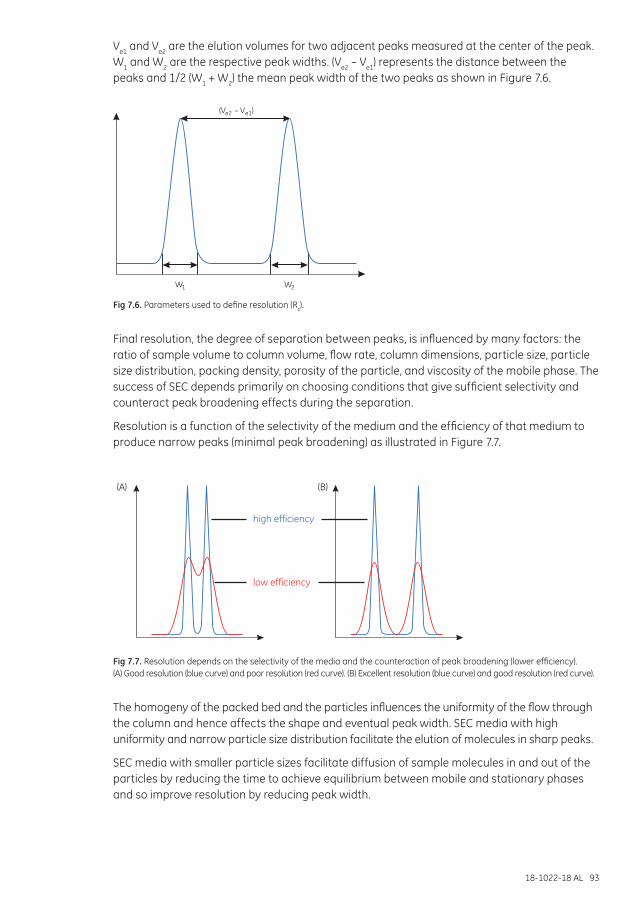
18-1022-18 AL 93
Ve1 and Ve2 are the elution volumes for two adjacent peaks measured at the center of the peak. W1 and W2 are the respective peak widths. (Ve2 – Ve1) represents the distance between the peaks and 1/2 (W1 + W2) the mean peak width of the two peaks as shown in Figure 7.6.
W1
–(V )e2 Ve1
W2
Fig 7.6. Parameters used to define resolution (Rs).
Final resolution, the degree of separation between peaks, is influenced by many factors: the ratio of sample volume to column volume, flow rate, column dimensions, particle size, particle size distribution, packing density, porosity of the particle, and viscosity of the mobile phase. The success of SEC depends primarily on choosing conditions that give sufficient selectivity and counteract peak broadening effects during the separation.
Resolution is a function of the selectivity of the medium and the efficiency of that medium to produce narrow peaks (minimal peak broadening) as illustrated in Figure 7.7.
high efficiency
low efficiency
(A) (B)
Fig 7.7. Resolution depends on the selectivity of the media and the counteraction of peak broadening (lower efficiency). (A) Good resolution (blue curve) and poor resolution (red curve). (B) Excellent resolution (blue curve) and good resolution (red curve).
The homogeny of the packed bed and the particles influences the uniformity of the flow through the column and hence affects the shape and eventual peak width. SEC media with high uniformity and narrow particle size distribution facilitate the elution of molecules in sharp peaks.
SEC media with smaller particle sizes facilitate diffusion of sample molecules in and out of the particles by reducing the time to achieve equilibrium between mobile and stationary phases and so improve resolution by reducing peak width.
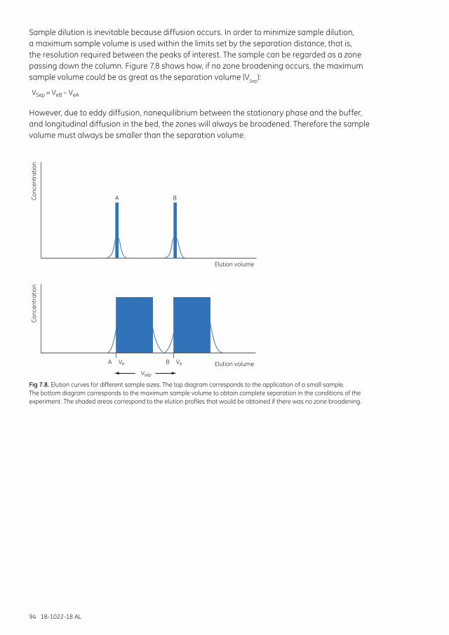
94 18-1022-18 AL
Sample dilution is inevitable because diffusion occurs. In order to minimize sample dilution, a maximum sample volume is used within the limits set by the separation distance, that is, the resolution required between the peaks of interest. The sample can be regarded as a zone passing down the column. Figure 7.8 shows how, if no zone broadening occurs, the maximum sample volume could be as great as the separation volume (VSep):
VSep = VeB – VeA
However, due to eddy diffusion, nonequilibrium between the stationary phase and the buffer, and longitudinal diffusion in the bed, the zones will always be broadened. Therefore the sample volume must always be smaller than the separation volume.
eV eVA
A
B
B
sepVElution volume
Elution volume
Conc
entr
atio
nCo
ncen
trat
ion
Fig 7.8. Elution curves for different sample sizes. The top diagram corresponds to the application of a small sample. The bottom diagram corresponds to the maximum sample volume to obtain complete separation in the conditions of the experiment. The shaded areas correspond to the elution profiles that would be obtained if there was no zone broadening.
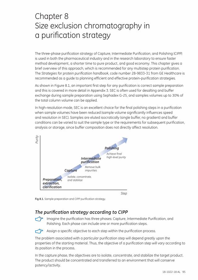
18-1022-18 AL 95
Chapter 8 Size exclusion chromatography in a purification strategy
The three-phase purification strategy of Capture, Intermediate Purification, and Polishing (CIPP) is used in both the pharmaceutical industry and in the research laboratory to ensure faster method development, a shorter time to pure product, and good economy. This chapter gives a brief overview of this approach, which is recommended for any multistep protein purification. The Strategies for protein purification handbook, code number 28-9833-31 from GE Healthcare is recommended as a guide to planning efficient and effective protein-purification strategies.
As shown in Figure 8.1, an important first step for any purification is correct sample preparation and this is covered in more detail in Appendix 3. SEC is often used for desalting and buffer exchange during sample preparation using Sephadex G-25, and samples volumes up to 30% of the total column volume can be applied.
In high-resolution mode, SEC is an excellent choice for the final polishing steps in a purification when sample volumes have been reduced (sample volume significantly influences speed and resolution in SEC). Samples are eluted isocratically (single buffer, no gradient) and buffer conditions can be varied to suit the sample type or the requirements for subsequent purification, analysis or storage, since buffer composition does not directly affect resolution.
Step
ytiruP
Capture
Intermediatepurification
Polishing
Preparation,extraction,clarification
Achieve finalhigh-level purity
Remove bulkimpurities
Isolate, concentrate,and stabilize
Fig 8.1. Sample preparation and CIPP purification strategy.
The purification strategy according to CIPP Imagine the purification has three phases: Capture, Intermediate Purification, and
Polishing. Each phase can include one or more purification steps.
Assign a specific objective to each step within the purification process.
The problem associated with a particular purification step will depend greatly upon the properties of the starting material. Thus, the objective of a purification step will vary according to its position in the process.
In the capture phase, the objectives are to isolate, concentrate, and stabilize the target product. The product should be concentrated and transferred to an environment that will conserve potency/activity.
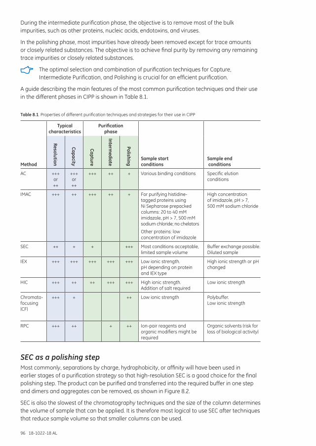
96 18-1022-18 AL
During the intermediate purification phase, the objective is to remove most of the bulk impurities, such as other proteins, nucleic acids, endotoxins, and viruses.
In the polishing phase, most impurities have already been removed except for trace amounts or closely related substances. The objective is to achieve final purity by removing any remaining trace impurities or closely related substances.
The optimal selection and combination of purification techniques for Capture, Intermediate Purification, and Polishing is crucial for an efficient purification.
A guide describing the main features of the most common purification techniques and their use in the different phases in CIPP is shown in Table 8.1.
Table 8.1. Properties of different purification techniques and strategies for their use in CIPP
Method
Typical characteristics
Purification phase
Sample start conditions
Sample end conditions
Resolution
Capacity
Capture
Intermediate
Polishing
AC +++ or ++
+++ or ++
+++ ++ + Various binding conditions Specific elution conditions
IMAC +++ ++ +++ ++ + For purifying histidine-tagged proteins using Ni Sepharose prepacked columns: 20 to 40 mM imidazole, pH > 7, 500 mM sodium chloride; no chelators
Other proteins: low concentration of imidazole
High concentration of imidazole, pH > 7, 500 mM sodium chloride
SEC ++ + + +++ Most conditions acceptable, limited sample volume
Buffer exchange possible. Diluted sample
IEX +++ +++ +++ +++ +++ Low ionic strength. pH depending on protein and IEX type
High ionic strength or pH changed
HIC +++ ++ ++ +++ +++ High ionic strength. Addition of salt required
Low ionic strength
Chromato-focusing (CF)
+++ + ++ Low ionic strength Polybuffer. Low ionic strength
RPC +++ ++ + ++ Ion-pair reagents and organic modifiers might be required
Organic solvents (risk for loss of biological activity)
SEC as a polishing stepMost commonly, separations by charge, hydrophobicity, or affinity will have been used in earlier stages of a purification strategy so that high-resolution SEC is a good choice for the final polishing step. The product can be purified and transferred into the required buffer in one step and dimers and aggregates can be removed, as shown in Figure 8.2.
SEC is also the slowest of the chromatography techniques and the size of the column determines the volume of sample that can be applied. It is therefore most logical to use SEC after techniques that reduce sample volume so that smaller columns can be used.
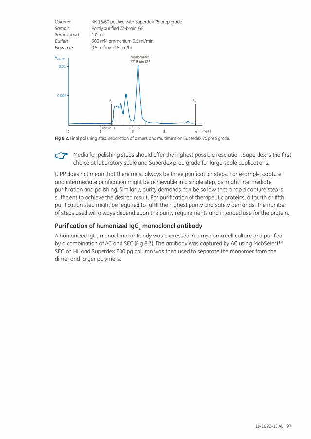
18-1022-18 AL 97
Column: XK 16/60 packed with Superdex 75 prep gradeSample: Partly purified ZZ-brain IGFSample load: 1.0 mlBuffer: 300 mM ammonium 0.5 ml/minFlow rate: 0.5 ml/min (15 cm/h)
0 1 2 3 4
0.01
0.005
monomericZZ-Brain IGF
IIIII
Time (h)Fraction
VO Vt
1 3 5
A280 nm
Fig 8.2. Final polishing step: separation of dimers and multimers on Superdex 75 prep grade.
Media for polishing steps should offer the highest possible resolution. Superdex is the first choice at laboratory scale and Superdex prep grade for large-scale applications.
CIPP does not mean that there must always be three purification steps. For example, capture and intermediate purification might be achievable in a single step, as might intermediate purification and polishing. Similarly, purity demands can be so low that a rapid capture step is sufficient to achieve the desired result. For purification of therapeutic proteins, a fourth or fifth purification step might be required to fulfill the highest purity and safety demands. The number of steps used will always depend upon the purity requirements and intended use for the protein.
Purification of humanized IgG4 monoclonal antibodyA humanized IgG4 monoclonal antibody was expressed in a myeloma cell culture and purified by a combination of AC and SEC (Fig 8.3). The antibody was captured by AC using MabSelect™. SEC on HiLoad Superdex 200 pg column was then used to separate the monomer from the dimer and larger polymers.
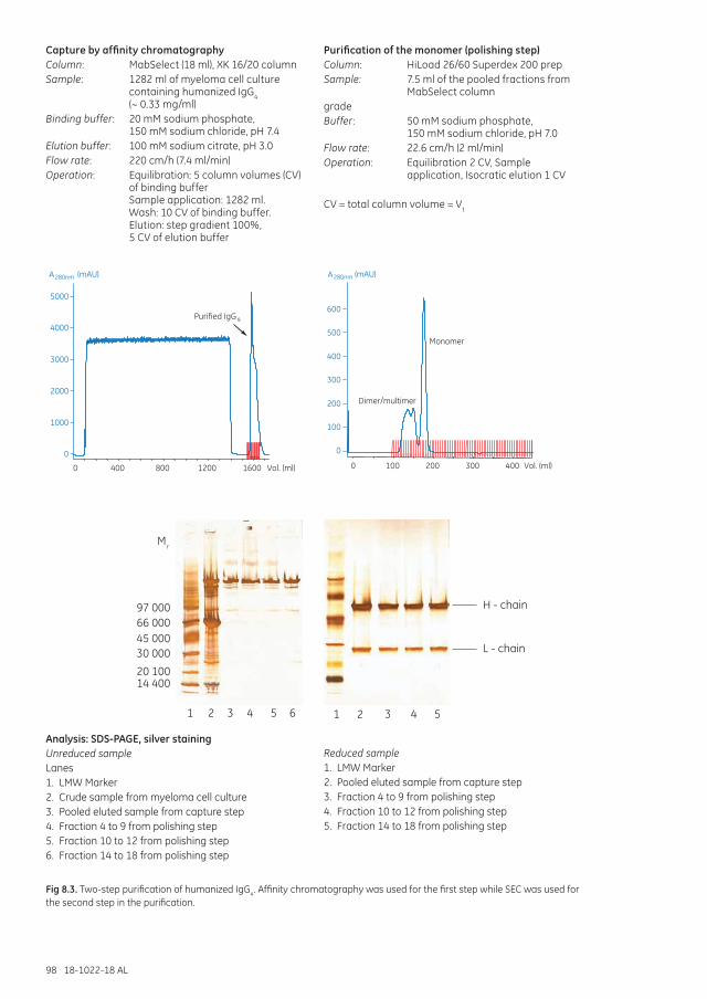
98 18-1022-18 AL
Capture by affinity chromatographyColumn: MabSelect (18 ml), XK 16/20 columnSample: 1282 ml of myeloma cell culture
containing humanized IgG4 (~ 0.33 mg/ml)
Binding buffer: 20 mM sodium phosphate, 150 mM sodium chloride, pH 7.4
Elution buffer: 100 mM sodium citrate, pH 3.0Flow rate: 220 cm/h (7.4 ml/min)Operation: Equilibration: 5 column volumes (CV)
of binding buffer Sample application: 1282 ml. Wash: 10 CV of binding buffer. Elution: step gradient 100%, 5 CV of elution buffer
0
1000
2000
3000
4000
5000
0 400 800 1200 1600 Vol. (ml)1357912 1620Waste
Purified IgG4
(mAU)A280nm
0
100
200
300
400
500
600
0 100 200 3001 2 3 4 5 6 7 8 9 10 12 14 16 18 20 22 24 26 28 30 32 34 36 38 40 42
Dimer/multimer
Monomer
44 46 48
400 Vol. (ml)50 52 54 56 58 60 62 64 66 68 70
(mAU)A280nm
Purification of the monomer (polishing step)Column: HiLoad 26/60 Superdex 200 prep Sample: 7.5 ml of the pooled fractions from
MabSelect columngradeBuffer: 50 mM sodium phosphate,
150 mM sodium chloride, pH 7.0Flow rate: 22.6 cm/h (2 ml/min)Operation: Equilibration 2 CV, Sample
application, Isocratic elution 1 CV
CV = total column volume = Vt
H - chain
L - chain
1 2 3 4 5
Mr
97 00066 00045 00030 000
20 10014 400
1 2 3 4 5 6
Analysis: SDS-PAGE, silver stainingUnreduced sampleLanes1. LMW Marker2. Crude sample from myeloma cell culture3. Pooled eluted sample from capture step4. Fraction 4 to 9 from polishing step5. Fraction 10 to 12 from polishing step6. Fraction 14 to 18 from polishing step
Reduced sample1. LMW Marker2. Pooled eluted sample from capture step3. Fraction 4 to 9 from polishing step4. Fraction 10 to 12 from polishing step5. Fraction 14 to 18 from polishing step
Fig 8.3. Two-step purification of humanized IgG4. Affinity chromatography was used for the first step while SEC was used for the second step in the purification.
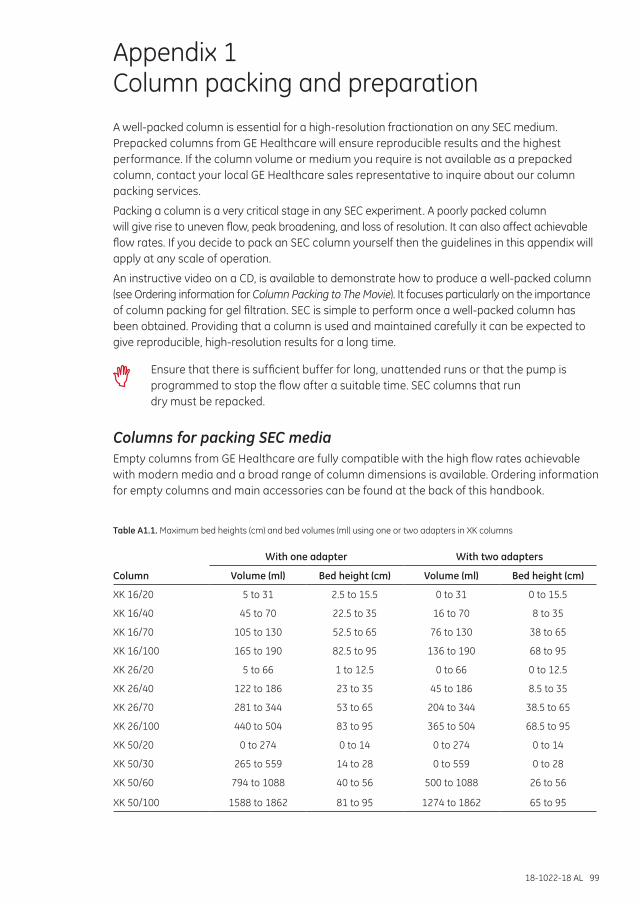
18-1022-18 AL 99
Appendix 1 Column packing and preparationA well-packed column is essential for a high-resolution fractionation on any SEC medium. Prepacked columns from GE Healthcare will ensure reproducible results and the highest performance. If the column volume or medium you require is not available as a prepacked column, contact your local GE Healthcare sales representative to inquire about our column packing services.
Packing a column is a very critical stage in any SEC experiment. A poorly packed column will give rise to uneven flow, peak broadening, and loss of resolution. It can also affect achievable flow rates. If you decide to pack an SEC column yourself then the guidelines in this appendix will apply at any scale of operation.
An instructive video on a CD, is available to demonstrate how to produce a well-packed column (see Ordering information for Column Packing to The Movie). It focuses particularly on the importance of column packing for gel filtration. SEC is simple to perform once a well-packed column has been obtained. Providing that a column is used and maintained carefully it can be expected to give reproducible, high-resolution results for a long time.
Ensure that there is sufficient buffer for long, unattended runs or that the pump is programmed to stop the flow after a suitable time. SEC columns that run dry must be repacked.
Columns for packing SEC mediaEmpty columns from GE Healthcare are fully compatible with the high flow rates achievable with modern media and a broad range of column dimensions is available. Ordering information for empty columns and main accessories can be found at the back of this handbook.
Table A1.1. Maximum bed heights (cm) and bed volumes (ml) using one or two adapters in XK columns
With one adapter With two adapters
Column Volume (ml) Bed height (cm) Volume (ml) Bed height (cm)
XK 16/20 5 to 31 2.5 to 15.5 0 to 31 0 to 15.5
XK 16/40 45 to 70 22.5 to 35 16 to 70 8 to 35
XK 16/70 105 to 130 52.5 to 65 76 to 130 38 to 65
XK 16/100 165 to 190 82.5 to 95 136 to 190 68 to 95
XK 26/20 5 to 66 1 to 12.5 0 to 66 0 to 12.5
XK 26/40 122 to 186 23 to 35 45 to 186 8.5 to 35
XK 26/70 281 to 344 53 to 65 204 to 344 38.5 to 65
XK 26/100 440 to 504 83 to 95 365 to 504 68.5 to 95
XK 50/20 0 to 274 0 to 14 0 to 274 0 to 14
XK 50/30 265 to 559 14 to 28 0 to 559 0 to 28
XK 50/60 794 to 1088 40 to 56 500 to 1088 26 to 56
XK 50/100 1588 to 1862 81 to 95 1274 to 1862 65 to 95
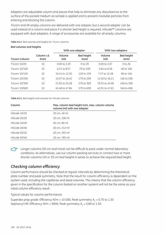
100 18-1022-18 AL
Adapters are adjustable column end pieces that help to eliminate any disturbances to the surface of the packed medium as sample is applied and to prevent insoluble particles from entering and blocking the column.
Tricorn and XK empty columns are delivered with one adapter, but a second adapter can be used instead of a column end piece if a shorter bed height is required. HiScale™ columns are equipped with dual adapters. A range of accessories are available for all empty columns.
Table A1.2. Bed volumes and heights for Tricorn columns
Bed volumes and heightsWith one adapter With two adapters
Tricorn ColumnColumn i.d.
(mm)Volume
(ml)Bed height
(mm)Volume
(ml)Bed height
(mm)
Tricorn 10/20 10 0.00 to 2.29 0 to 29 0.00 to 2.07 0 to 26
Tricorn 10/100 10 6.21 to 8.57 79 to 109 3.64 to 8.36 46 to 106
Tricorn 10/150 10 10.14 to 12.50 129 to 159 7.57 to 12.28 96 to 156
Tricorn 10/200 10 14.07 to 16.42 179 to 209 11.50 to 16.21 146 to 206
Tricorn 10/300 10 21.92 to 24.28 279 to 309 19.35 to 24.06 246 to 306
Tricorn 10/600 10 45.48 to 47.84 579 to 609 42.91 to 47.63 546 to 606
Table A1.3. Bed heights and volumes for HiScale columns
Column Max. column bed height (cm), max. column volume volume (ml) with one adapter
HiScale 16/20 20 cm, 40 ml
HiScale 26/20 20 cm, 106 ml
HiScale 16/40 40 cm, 80 ml
HiScale 26/40 20 cm, 212 ml
HiScale 50/20 20 cm, 393 ml
HiScale 50/40 20 cm, 785 ml
Longer columns (50 cm and more) can be difficult to pack under normal laboratory conditions. As alternatives, use our column packing services or connect two or more shorter columns (20 or 30 cm bed height) in series to achieve the required bed height.
Checking column efficiencyColumn performance should be checked at regular intervals by determining the theoretical plate number and peak symmetry. Note that the result for column efficiency is dependent on the system used, including the capillaries and dead volumes. This means that the column efficiency given in the specification for the column (tested on another system) will not be the same as your initial column efficiency result.
Typical values for column performance:
Superdex prep grade: Efficiency N/m > 10 000, Peak symmetry As = 0.70 to 1.30Sephacryl HR: Efficiency N/m < 9000, Peak symmetry As = 0.80 to 1.50
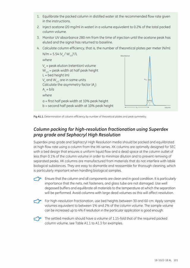
18-1022-18 AL 101
1. Equilibrate the packed column in distilled water at the recommended flow rate given in the instructions.
2. Inject acetone (20 mg/ml in water) in a volume equivalent to 0.2% of the total packed column volume.
3. Monitor UV absorbance 280 nm from the time of injection until the acetone peak has eluted and the signal has returned to baseline.
4. Calculate column efficiency, that is, the number of theoretical plates per meter (N/m):
N/m = 5.54 (Ve / W1/2)2/L
where
Ve = peak elution (retention) volume W1/2 = peak width at half peak height L = bed height (m) Ve and W1/2 are in same units Calculate the asymmetry factor (As): As = b/a
where
a = first half peak width at 10% peak height b = second half peak width at 10% peak height
Fig A1.1. Determination of column efficiency by number of theoretical plates and peak symmetry.
Column packing for high-resolution fractionation using Superdex prep grade and Sephacryl High ResolutionSuperdex prep grade and Sephacryl High Resolution media should be packed and equilibrated at high flow rate using a column from the XK-series. XK columns are optimally designed for SEC with a bed design that ensures a uniform liquid flow and a dead space at the column outlet of less than 0.1% of the column volume in order to minimize dilution and to prevent remixing of separated peaks. XK columns are manufactured from materials that do not interfere with labile biological substances. They are easy to dismantle and reassemble for thorough cleaning, which is particularly important when handling biological samples.
Ensure that the column and all components are clean and in good condition. It is particularly importance that the nets, net fasteners, and glass tube are not damaged. Use well degassed buffers and equilibrate all materials to the temperature at which the separation will be performed. Avoid columns with large dead volumes as this will affect resolution.
For high-resolution fractionation, use bed heights between 30 and 60 cm. Apply sample volumes equivalent to between 1% and 2% of the column volume. The sample volume can be increased up to 4% if resolution in the particular application is good enough.
The settled medium should have a volume of 1.15-fold that of the required packed column volume, see Table A1.1 to A1.3 for examples.
Ve
a b
w1/2
Volume
Absorbance
102 18-1022-18 AL
1. Superdex prep grade and Sephacryl HR are supplied swollen in a suspension containing 20% ethanol as a preservative. Suspend the medium by shaking gently and pour a sufficient quantity into a graduated glass cylinder or beaker.
Avoid using magnetic stirrers, spatulas or glass rods since they can damage the matrix.
2. Wash the medium with 5 to 10 column volumes of distilled water on a glass filter and resuspend in distilled water to a final concentration of 50% settled medium. The medium must be thoroughly washed to remove the 20% ethanol storage solution. Residual ethanol can interfere with subsequent procedures.
To produce a more evenly dispersed slurry of Superdex prep grade, Tween™ 20 (250 ml per 500 ml washed slurry) can be added in order to reduce surface tension.
3. Wet the bottom filter by injecting distilled water through the effluent tubing. Close the end piece outlet. Mount filter and bottom end piece onto the column.
4. Attach the packing reservoir tightly to the column.
For XK 16 and XK 26 columns using a second column instead of a packing reservoir makes it easier to obtain a well-packed column. The second column is used with Packing Connector XK 16 or XK 26 as appropriate.
5. Mount the column and packing reservoir vertically on a laboratory stand.
6. Fill the column with distilled water to a height of 2 cm above the column end piece. Avoid air bubbles.
7. Degas the suspended medium under vacuum and carefully pour the suspended medium down the wall of the column using a glass rod. Avoid introducing air bubbles. Pour everything in a single operation and fill the reservoir to the top with distilled water.
8. Connect the pump outlet to the inlet on the packing reservoir. Open the column outlet and start the flow of buffer, see Table A1.4 for flow recommendations.
To achieve satisfactory column efficiency, Superdex prep grade must be packed in two steps: Step 1 for 2 h or until the bed has reached a constant height and Step 2 for 60 min. Table A1.4 shows the flow rates for each step.
Sephacryl HR can usually be packed satisfactorily using only the higher flow rate given in Step 2 of Table A1.4. Use the two step process if the column efficiency was unsatisfactory after the first attempt.
9. Stop the pump and remove the packing reservoir. Carefully fill the column with distilled water to form an upward meniscus at the top and insert the adapter. Adjust the adapter to the surface of the packed bed.
10. Continue packing the column at the flow rate used in Step 2 for approximately 10 min. If the recommended flow rate cannot be obtained, use the maximum flow rate the pump can deliver. Mark the position of the top of the packed medium, stop the pump, close the column outlet, move the adapter down onto to the surface of the medium and then push the adapter a further 3 mm into the medium. The column is now ready to use. See Table A1.4 for maximum recommended flow rate and operating pressure for Sephacryl HR and Superdex prep grade media.
Do not exceed maximum pressures during packing: 0.2 to 0.4 MPa, 2 to 4 bar for Sephacryl S-300 HR and S-100 HR respectively, 0.15 MPa, 1.5 bar for Sephacryl S-400 HR and S-500 HR, and 0.4 to 0.5 MPa, 4 to 5 bar for Superdex prep grade (75 and 200).
Always check the specific storage instructions supplied with the product.
18-1022-18 AL 103
Table A1.4. Recommended flow rates during column packing
ColumnBed height
(cm)
Step 1 Sephacryl HR
(ml/min)
Step 2 Sephacryl HR
(ml/min)
Step 1 Superdex prep grade (ml/min)
Step 2 Superdex prep grade (ml/min)
XK 16/40 35 1 to 2 2 to 4 1 to 2 4 to 6
XK 16/70 65 1 to 2 2 to 4 1 to 2 4 to 6
XK16/100 95 1 to 2 2 to 4 1 to 2 4 to 6
XK 26/40 35 2 to 4 4 to 8 2 to 4 10 to 14
XK 26/70 65 2 to 4 4 to 8 2 to 4 10 to 14
XK 26/100 95 2 to 4 4 to 8 2 to 4 10 to 14
XK 50/20 10 to 15 8 to 10 4 to 8 9 to 11 19 to 21
XK 50/30 20 to 25 8 to 10 4 to 8 9 to 11 19 to 21
XK 50/60 55 8 to 10 4 to 8 9 to 11 19 to 21
XK 50/100 95 8 to 10 4 to 8 9 to 11 19 to 21
Column packing for group separations using SephadexSephadex is supplied as a dry powder and must be allowed to swell in excess buffer before use. After swelling, adjust with buffer to form a thick slurry from which air bubbles are removed under vacuum. Approximately 75% settled medium is suitable. Fine particles can be decanted.
Accelerate the swelling process by using a boiling water bath (Table A1.5). This also serves to degas the suspension. Allow the suspension to cool before use.
Table A1.5. Bed volume and swelling times for Sephadex
MediumApprox. bed volume
(ml/1 g medium) Swelling time (h), 20°C Swelling time (h), 90°C
Sephadex G-10 Medium 2 to 3 3 1
Sephadex G-25 (all grades) 4 to 6 3 1
Sephadex G-50 Fine 9 to 11 3 1
Ensure that the column and all components are clean and in good condition. It is particularly important that the nets, net fasteners, and glass tube are not damaged. Use well-degassed buffers and equilibrate all materials to the temperature at which the separation will be performed. Keep a packed column away from locations that are exposed to drafts or direct sunlight that can cause temperature changes and the formation of bubbles.
For group separations, use up to 10 cm bed height. Sample volumes can be up to 30% of the column volume. Pack a quantity of medium up to five-fold the volume of the sample to be desalted.
Note: These instructions assume that a column with two adapters is used for packing.
1. Weigh out the correct amount of dry Sephadex and allow the medium to swell according to the instructions above. Avoid using magnetic stirrers, spatulas, or glass rods since they can damage the medium.
2. Wet the bottom filter by injecting distilled water through the effluent tubing. Close the end piece outlet. Mount filter and bottom end piece onto the column.
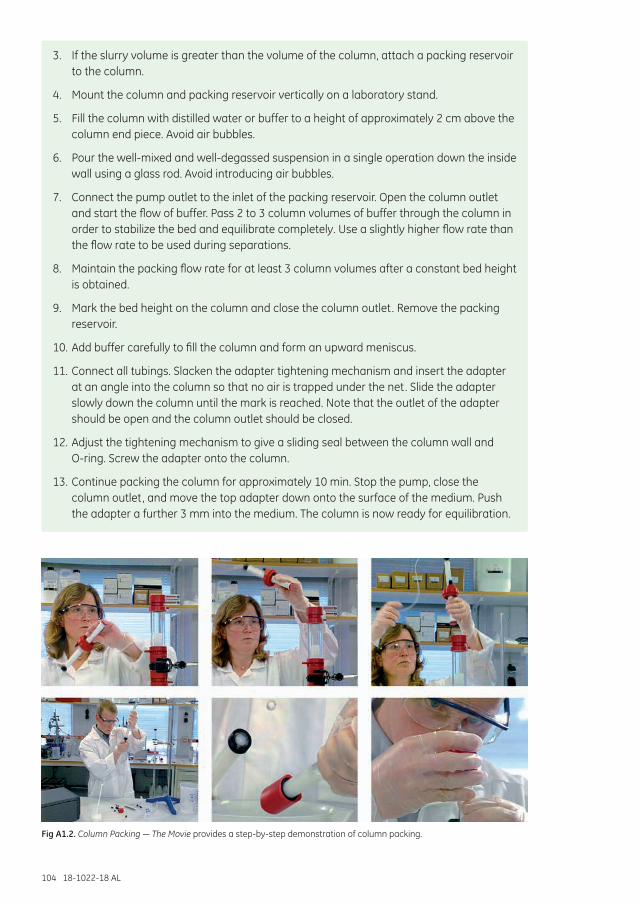
104 18-1022-18 AL
3. If the slurry volume is greater than the volume of the column, attach a packing reservoir to the column.
4. Mount the column and packing reservoir vertically on a laboratory stand.
5. Fill the column with distilled water or buffer to a height of approximately 2 cm above the column end piece. Avoid air bubbles.
6. Pour the well-mixed and well-degassed suspension in a single operation down the inside wall using a glass rod. Avoid introducing air bubbles.
7. Connect the pump outlet to the inlet of the packing reservoir. Open the column outlet and start the flow of buffer. Pass 2 to 3 column volumes of buffer through the column in order to stabilize the bed and equilibrate completely. Use a slightly higher flow rate than the flow rate to be used during separations.
8. Maintain the packing flow rate for at least 3 column volumes after a constant bed height is obtained.
9. Mark the bed height on the column and close the column outlet. Remove the packing reservoir.
10. Add buffer carefully to fill the column and form an upward meniscus.
11. Connect all tubings. Slacken the adapter tightening mechanism and insert the adapter at an angle into the column so that no air is trapped under the net. Slide the adapter slowly down the column until the mark is reached. Note that the outlet of the adapter should be open and the column outlet should be closed.
12. Adjust the tightening mechanism to give a sliding seal between the column wall and O-ring. Screw the adapter onto the column.
13. Continue packing the column for approximately 10 min. Stop the pump, close the column outlet, and move the top adapter down onto the surface of the medium. Push the adapter a further 3 mm into the medium. The column is now ready for equilibration.
Fig A1.2. Column Packing — The Movie provides a step-by-step demonstration of column packing.
18-1022-18 AL 105
Sephadex G-10, G-25, and G-50 obey Darcy’s law, that is, if the flow rate is doubled then the column pressure will double, hence maximum values for flow rates or operating pressures do not need to be considered (see Appendix 2 for an explanation of Darcy’s law).
Controlling flow ratesThe safest and easiest way in which to control flow rates during column packing and chromatography separation is to use a pump controlled within an ÄKTA chromatography system. Accurate and reproducible flow control is particularly important for efficient column packing and when repeating experiments or performing routine preparative work. A peristaltic pump can be used with Sephadex packed in smaller columns.
The maximum flow rate achievable will depend on column diameter and buffer viscosity. Narrow columns allow a higher pressure and higher linear flow (cm/h) than wide columns.
Always connect a pump so that buffer is pumped onto the column (rather than connecting the pump after the column and drawing buffer through the column). This reduces the risk of bubble formation due to suction effects.
Always use a flow rate for column packing that is higher than the flow rate used for separation.
Do not exceed the maximum recommended values for pressure or linear flow for the medium. Exceeding these values might cause the medium to compress and reduce the flow rate and resolution during the separation.
Do not exceed 75% of the packing flow rate during any separation.
Do not use a peristaltic pump when packing Superdex or Sephacryl media in larger columns since a flow rate high enough to obtain high-resolution fractionation cannot be achieved.
106 18-1022-18 AL
Appendix 2 Sephadex and Darcy’s lawSephadex G-10, G-25, and G-50 can be assumed to behave as rigid spheres in SEC and therefore obey Darcy’s Law. This Law describes a general relationship for flow in porous media:
U = K × ∆P × L-1 Equation (1)
U = linear flow rate expressed in cm/h (see Appendix 5) ∆P = pressure drop over the packed bed expressed in cm waterL = bed height expressed in cm K = constant of proportionality depending on the properties of the bed material and the buffer
Assuming a buffer with viscosity of 1 cP: U = Ko × ∆P × L-1 Equation (2)
Ko = the “specific permeability” depending on the particle size of the medium and the water regain
Note that flow is proportional to the pressure drop over the bed and, assuming a constant pressure head, inversely proportional to the bed height. In practice this means that the pressure/flow considerations that must be made when using other SEC media do not apply to Sephadex and that a doubling of flow rate leads to a doubling in column pressure. To a good approximation, flow rate is independent of the column diameter.
Flow at viscosities greater than 1 cP can be obtained by using the relationship: flow rate is inversely proportional to viscosity. High buffer viscosities can be compensated for by increasing the operating pressure to maintain a high flow rate.
Theoretical flow (not maximum) can be calculated from equation (2) by inserting values for ∆P and L. Specific permeabilities (K) are given in Table A2.1.
Table A2.1. Specific permeabilities of Sephadex
Sephadex type Permeability K
Sephadex G-10 19
Sephadex G-25 Superfine 9
Sephadex G-25 Fine 30
Sephadex G-25 Medium 80
Sephadex G-25 Coarse 290
Sephadex G-50 Fine 36
18-1022-18 AL 107
Appendix 3 Sample preparationSamples for chromatographic purification should be clear and free from particulate matter. Simple steps to clarify a sample before beginning purification will avoid clogging the column, can reduce the need for stringent washing procedures, and can extend the life of the chromatographic medium.
Sample extraction procedures and the selection of buffers, additives, and detergents are determined largely by the source of the material, the stability of the target molecule, the chromatographic techniques that will be employed, and the intended use of the product. These subjects are dealt with in general terms in the Protein Purification Handbook and more specifically according to target molecule in the Recombinant Protein Handbook, and Antibody Purification Handbook, available from GE Healthcare.
Sample clarificationCentrifugation and filtration are standard laboratory techniques for sample clarification and are used routinely when handling small samples.
It is highly recommended to centrifuge and filter any sample immediately before chromatographic purification.
CentrifugationCentrifugation removes lipids and particulate matter, such as cell debris. If the sample is still not clear after centrifugation, use filter paper or a 5 µm filter as a first step and one of the filters below as a second step filter.
For small sample volumes or proteins that adsorb to filters, centrifuge at 10 000 × g for 15 min.
For cell lysates, centrifuge at 40 000 to 50 000 × g for 30 min.
Serum samples can be filtered through glass wool after centrifugation to remove any remaining lipids.
FiltrationFiltration removes particulate matter. Membrane filters that give the least amount of nonspecific binding of proteins are composed of cellulose acetate or PVDF.
For sample preparation before chromatography, select a filter pore size in relation to the bead size of the chromatographic medium (Table A3.1).
Table A3.1. Filter pore size recommendations
Nominal pore size of filter Particle size of chromatography medium1 µm 90 µm and upwards
0.45 µm 34 µm
0.22 µm 3, 10, 15 µm or when extra clean samples or sterile filtration is required
Check the recovery of the target protein in a test run. Some proteins can adsorb nonspecifically to filter surfaces.
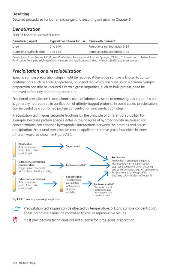
108 18-1022-18 AL
DesaltingDetailed procedures for buffer exchange and desalting are given in Chapter 5.
DenaturationTable A3.2. Common denaturing agents
Denaturing agent Typical conditions for use Removal/comment
Urea 2 to 6 M Remove using Sephadex G-25
Guanidine hydrochloride 3 to 6 M Remove using Sephadex G-25
Details taken from: Scopes R.K., Protein Purification, Principles and Practice, Springer, (1994), J.C. Janson and L. Rydén, Protein Purification, Principles, High Resolution Methods and Applications, 2nd ed. Wiley Inc, (1998) and other sources.
Precipitation and resolubilizationSpecific sample preparation steps might be required if the crude sample is known to contain contaminants such as lipids, lipoproteins, or phenol red, which can build up on a column. Sample preparation can also be required if certain gross impurities, such as bulk protein, need be removed before any chromatographic step.
Fractional precipitation is occasionally used at laboratory scale to remove gross impurities but is generally not required in purification of affinity-tagged proteins. In some cases, precipitation can be useful as a combined protein concentration and purification step.
Precipitation techniques separate fractions by the principle of differential solubility. For example, because protein species differ in their degree of hydrophobicity, increased salt concentrations can enhance hydrophobic interactions between the proteins and cause precipitation. Fractional precipitation can be applied to remove gross impurities in three different ways, as shown in Figure A3.1.
ClarificationBulk proteins andparticulate matterprecipitated
Extraction, clarification,concentrationTarget protein precipitatedwith proteins of similar solubility
Extraction, clarificationBulk proteins andparticulate matterprecipitated
Supernatant
Redissolve pellet*
Redissolve pellet*
Purification
ConcentrationTarget proteinprecipitatedwith proteinsof similarsolubility
Remember: if precipitating agent isincompatible with next purificationstep, use Sephadex G-25 for desaltingand buffer exchange, e.g., HiTrap DesaltingPD-10 columns, or HiPrep 26/10
,Desalting column (refer to Chapter 5)
*Remember: not all proteins are easy
to redissolve, yield can be reduced
Fig A3.1. Three ways to use precipitation.
Precipitation techniques can be affected by temperature, pH, and sample concentration. These parameters must be controlled to ensure reproducible results.
Most precipitation techniques are not suitable for large-scale preparation.
18-1022-18 AL 109
Examples of precipitation agents are reviewed in Table A3.3. The most common precipitation method using ammonium sulfate is described in more detail below.
Table A3.3. Examples of precipitation techniques
Precipitation agent Typical conditions for use Sample type Comment
Ammonium sulfate As described below. > 1 mg/ml proteins, especially immunoglobulins.
Stabilizes proteins, no denaturation; supernatant can go directly to HIC. Helps to reduce lipid content.
Dextran sulfate Add 0.04 ml of 10% dextran sulfate and 1 ml of 1 M CaCl2 per ml of sample, mix 15 min, centrifuge at 10 000 × g, discard pellet.
Samples with high levels of lipoprotein, e.g., ascites.
Precipitates lipoprotein.
Polyvinylpyrrolidine Add 3% (w/v), stir 4 h, centrifuge at 17 000 × g, discard pellet.
Samples with high levels of lipoprotein, e.g., ascites.
Alternative to dextran sulfate.
Polyethylene glycol (PEG, Mr > 4000)
Up to 20% (w/v). Plasma proteins. No denaturation, supernatant goes directly to IEX or AC, complete removal can be difficult. Stabilizes proteins.
Acetone (cold) Up to 80% (v/v) at 0°C. Collect pellet after centrifugation at full speed in microcentrifuge.
Can denature protein irreversibly. Useful for peptide precipitation or concentration of sample for electrophoresis.
Polyethyleneimine 0.1% (w/v). Precipitates aggregated nucleoproteins.
Protamine sulfate 1% (w/v). Precipitates aggregated nucleoproteins.
Streptomycin sulfate 1% (w/v). Precipitates nucleic acids.
Caprylic acid (X/15) g where X = volume of sample.
Antibody concentration should be > 1 mg/ml.
Precipitates bulk of proteins from sera or ascites, leaving immunoglobulins in solution.
Details taken from: Scopes R.K., Protein Purification, Principles and Practice, Springer, (1994), J.C. Janson and L. Rydén, Protein Purification, Principles, High Resolution Methods and Applications, 2nd ed. Wiley Inc, (1998).
Ammonium sulfate precipitationAmmonium sulfate precipitation is frequently used for initial sample concentration and clean up. As the concentration of the salt is increased, proteins will begin to “salt out.” Different proteins salt out at different concentrations, a process that can be taken advantage of to remove contaminating proteins from the crude extract. The salt concentration needs to be optimized to remove contaminants and not the desired protein. An additional step with increased salt concentration should then precipitate the target protein. If the target protein cannot be safely precipitated and redissolved, only the first step should be employed. HIC is often an excellent next purification step, as the sample already contains a high salt concentration and can be applied directly to the HIC column with little or no additional preparation. The elevated salt level enhances the interaction between the hydrophobic components of the sample and the chromatography medium.
110 18-1022-18 AL
Solutions needed for precipitation:
Saturated ammonium sulfate solution (add 100 g ammonium sulfate to 100 ml distilled water, stir to dissolve).
1 M Tris-HCl, pH 8.0.
Buffer for first purification step.
Some proteins can be damaged by ammonium sulfate. Take care when adding crystalline ammonium sulfate: high local concentrations can cause contamination of the precipitate with unwanted proteins.
For routine, reproducible purification, precipitation with ammonium sulfate should be avoided in favor of chromatography.
In general, precipitation is rarely effective for protein concentrations below 1 mg/ml.
1. Filter (0.45 µm) or centrifuge the sample (10 000 × g at 4°C).
2. Add 1 part 1 M Tris-HCl, pH 8.0 to 10 parts sample volume to maintain pH.
3. Stir gently. Add ammonium sulfate solution, drop by drop. Add up to 50% saturation*. Stir for 1 h.
4. Centrifuge for 20 min at 10 000 × g.
5. Remove supernatant. Wash the pellet twice by resuspension in an equal volume of ammonium sulfate solution of the same concentration (i.e., a solution that will not redissolve the precipitated protein or cause further precipitation). Centrifuge again.
6. Dissolve the pellet in a small volume of the buffer to be used for the next step.
7 Ammonium sulfate is removed during clarification/buffer exchange steps with Sephadex G-25 using desalting columns (see Chapter 5).
* The percentage saturation can be adjusted either to precipitate a target molecule or to precipitate contaminants.
The quantity of ammonium sulfate required to reach a given degree of saturation varies according to temperature. Table A3.4 shows the quantities required at 20°C.
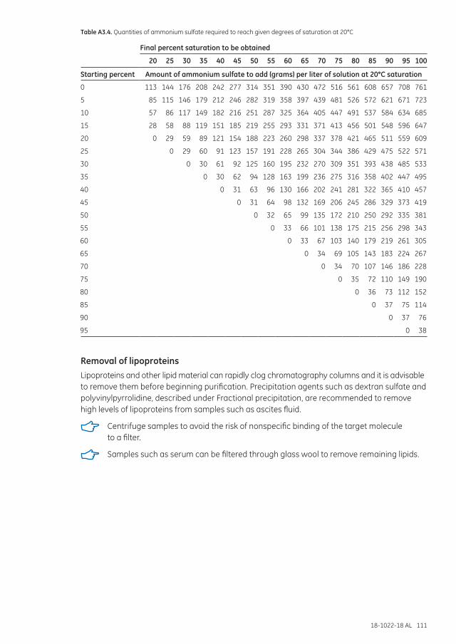
18-1022-18 AL 111
Table A3.4. Quantities of ammonium sulfate required to reach given degrees of saturation at 20°C
Final percent saturation to be obtained
20 25 30 35 40 45 50 55 60 65 70 75 80 85 90 95 100
Starting percent Amount of ammonium sulfate to add (grams) per liter of solution at 20°C saturation
0 113 144 176 208 242 277 314 351 390 430 472 516 561 608 657 708 761
5 85 115 146 179 212 246 282 319 358 397 439 481 526 572 621 671 723
10 57 86 117 149 182 216 251 287 325 364 405 447 491 537 584 634 685
15 28 58 88 119 151 185 219 255 293 331 371 413 456 501 548 596 647
20 0 29 59 89 121 154 188 223 260 298 337 378 421 465 511 559 609
25 0 29 60 91 123 157 191 228 265 304 344 386 429 475 522 571
30 0 30 61 92 125 160 195 232 270 309 351 393 438 485 533
35 0 30 62 94 128 163 199 236 275 316 358 402 447 495
40 0 31 63 96 130 166 202 241 281 322 365 410 457
45 0 31 64 98 132 169 206 245 286 329 373 419
50 0 32 65 99 135 172 210 250 292 335 381
55 0 33 66 101 138 175 215 256 298 343
60 0 33 67 103 140 179 219 261 305
65 0 34 69 105 143 183 224 267
70 0 34 70 107 146 186 228
75 0 35 72 110 149 190
80 0 36 73 112 152
85 0 37 75 114
90 0 37 76
95 0 38
Removal of lipoproteinsLipoproteins and other lipid material can rapidly clog chromatography columns and it is advisable to remove them before beginning purification. Precipitation agents such as dextran sulfate and polyvinylpyrrolidine, described under Fractional precipitation, are recommended to remove high levels of lipoproteins from samples such as ascites fluid.
Centrifuge samples to avoid the risk of nonspecific binding of the target molecule to a filter.
Samples such as serum can be filtered through glass wool to remove remaining lipids.
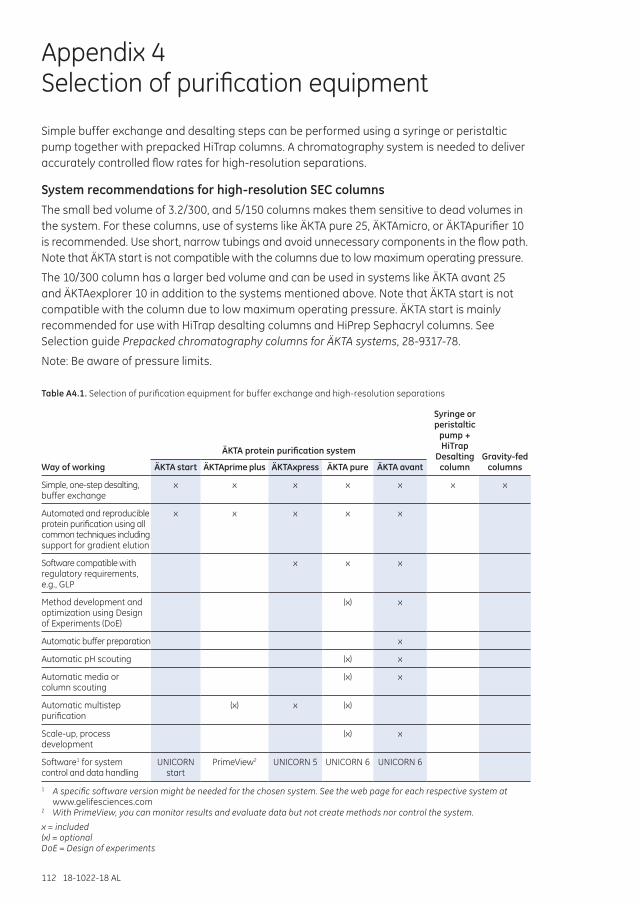
112 18-1022-18 AL
Appendix 4 Selection of purification equipment
Simple buffer exchange and desalting steps can be performed using a syringe or peristaltic pump together with prepacked HiTrap columns. A chromatography system is needed to deliver accurately controlled flow rates for high-resolution separations.
System recommendations for high-resolution SEC columnsThe small bed volume of 3.2/300, and 5/150 columns makes them sensitive to dead volumes in the system. For these columns, use of systems like ÄKTA pure 25, ÄKTAmicro, or ÄKTApurifier 10 is recommended. Use short, narrow tubings and avoid unnecessary components in the flow path. Note that ÄKTA start is not compatible with the columns due to low maximum operating pressure.
The 10/300 column has a larger bed volume and can be used in systems like ÄKTA avant 25 and ÄKTAexplorer 10 in addition to the systems mentioned above. Note that ÄKTA start is not compatible with the column due to low maximum operating pressure. ÄKTA start is mainly recommended for use with HiTrap desalting columns and HiPrep Sephacryl columns. See Selection guide Prepacked chromatography columns for ÄKTA systems, 28-9317-78.
Note: Be aware of pressure limits.
Table A4.1. Selection of purification equipment for buffer exchange and high-resolution separations
Way of working
Syringe or peristaltic
pump + HiTrap
Desalting column
Gravity-fed columns
ÄKTA protein purification system
ÄKTA start ÄKTAprime plus ÄKTAxpress ÄKTA pure ÄKTA avant
Simple, one-step desalting, buffer exchange
x x x x x x x
Automated and reproducible protein purification using all common techniques including support for gradient elution
x x x x x
Software compatible with regulatory requirements, e.g., GLP
x x x
Method development and optimization using Design of Experiments (DoE)
(x) x
Automatic buffer preparation x
Automatic pH scouting (x) x
Automatic media or column scouting
(x) x
Automatic multistep purification
(x) x (x)
Scale-up, process development
(x) x
Software1 for system control and data handling
UNICORN start
PrimeView2 UNICORN 5 UNICORN 6 UNICORN 6
1 A specific software version might be needed for the chosen system. See the web page for each respective system at www.gelifesciences.com
2 With PrimeView, you can monitor results and evaluate data but not create methods nor control the system.
x = included(x) = optionalDoE = Design of experiments
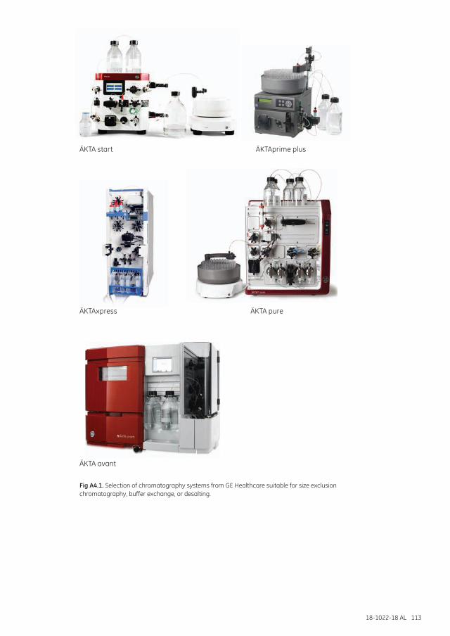
18-1022-18 AL 113
ÄKTA start
ÄKTA pure
ÄKTA avant
ÄKTAprime plus
ÄKTAxpress
Fig A4.1. Selection of chromatography systems from GE Healthcare suitable for size exclusion chromatography, buffer exchange, or desalting.
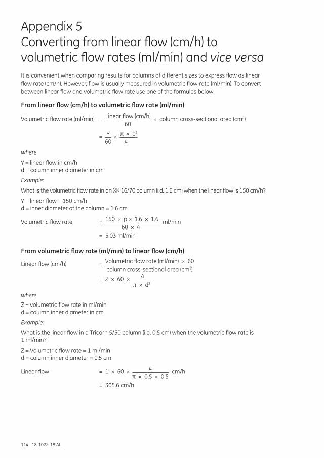
114 18-1022-18 AL
Appendix 5 Converting from linear flow (cm/h) to volumetric flow rates (ml/min) and vice versaIt is convenient when comparing results for columns of different sizes to express flow as linear flow rate (cm/h). However, flow is usually measured in volumetric flow rate (ml/min). To convert between linear flow and volumetric flow rate use one of the formulas below:
From linear flow (cm/h) to volumetric flow rate (ml/min)
Volumetric flow rate (ml/min) = Linear flow (cm/h) × column cross-sectional area (cm2)
60
= Y × π × d2
60 4
where
Y = linear flow in cm/h d = column inner diameter in cm
Example:
What is the volumetric flow rate in an XK 16/70 column (i.d. 1.6 cm) when the linear flow is 150 cm/h?
Y = linear flow = 150 cm/h d = inner diameter of the column = 1.6 cm
Volumetric flow rate = 150 × p × 1.6 × 1.6 ml/min
60 × 4 = 5.03 ml/min
From volumetric flow rate (ml/min) to linear flow (cm/h)
Linear flow (cm/h) = Volumetric flow rate (ml/min) × 60 column cross-sectional area (cm2)
= Z × 60 × 4
π × d2
whereZ = volumetric flow rate in ml/min d = column inner diameter in cm
Example:
What is the linear flow in a Tricorn 5/50 column (i.d. 0.5 cm) when the volumetric flow rate is 1 ml/min?
Z = Volumetric flow rate = 1 ml/min d = column inner diameter = 0.5 cm
Linear flow = 1 × 60 × 4 cm/h π × 0.5 × 0.5 = 305.6 cm/h
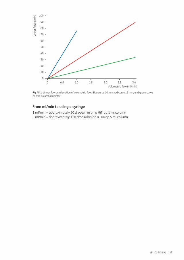
18-1022-18 AL 115
100
90
80
70
60
50
40
30
20
10
0
Line
ar fl
ow (c
m/h
)
0 0.5 1.0 1.5 2.0 2.5 3.0Volumetric flow (ml/min)
Fig A5.1. Linear flow as a function of volumetric flow: Blue curve 10 mm, red curve 16 mm, and green curve 26 mm column diameter.
From ml/min to using a syringe1 ml/min = approximately 30 drops/min on a HiTrap 1 ml column 5 ml/min = approximately 120 drops/min on a HiTrap 5 ml column
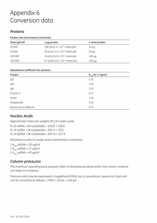
116 18-1022-18 AL
Appendix 6 Conversion data
ProteinsProtein size and amount conversion
Mass (g/mol) 1 µg protein 1 nmol protein
10 000 100 pmol; 6 × 10¹³ molecules 10 µg
50 000 20 pmol; 1.2 × 10¹³ molecules 50 µg
100 000 10 pmol; 6.0 × 10¹² molecules 100 µg
150 000 6.7 pmol; 4.0 × 10¹² molecules 150 µg
Absorbance coefficient for proteins
Protein A280 for 1 mg/ml
IgG 1.35
IgM 1.20
IgA 1.30
Protein A 0.17
Avidin 1.50
Streptavidin 3.40
Bovine serum albumin 0.70
Nucleic AcidsApproximate molecular weights (Mr) of nucleic acids
Mr of ssRNA = (# nucleotides × 320.5) + 159.0 Mr of ssDNA = (# nucleotides × 303.7) + 79.0 Mr of dsDNA = (# nucleotides × 607.4) + 157.9
Absorbance units to nucleic acid concentration conversion
1 A280 dsDNA = 50 µg/ml 1 A280 ssDNA = 37 µg/ml 1 A280 ssRNA = 40 µg/ml
Column pressuresThe maximum operating back pressure refers to the pressure above which the column contents can begin to compress.
Pressure units may be expressed in megaPascal (MPa), bar, or pounds per square inch (psi) and can be converted as follows: 1 MPa = 10 bar = 145 psi.

18-1022-18 AL 117
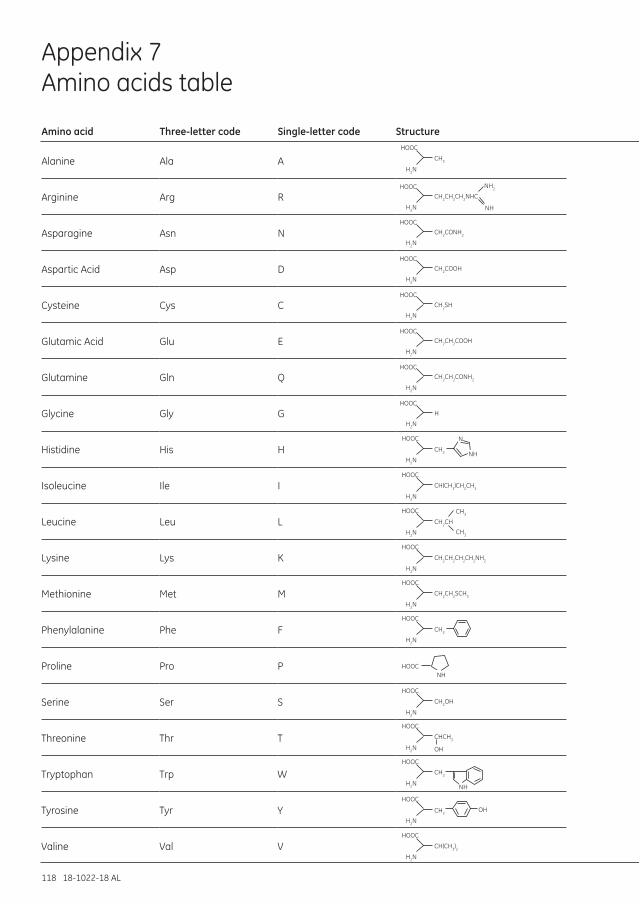
118 18-1022-18 AL
CH3
HOOC
H2N
HOOC
H2N
HOOC
H2N
HOOC
H2N
HOOC
H2N
HOOC
H2N
HOOC
H2N
HOOC
H2N
HOOC
H2N
HOOC
H2N
HOOC
H2N
HOOC
H2N
HOOC
H2N
HOOC
H2N
HOOC
HOOC
H2N
CH2CH2CH2NHC
NH2
NH
CH2CONH2
CH2COOH
CH2SH
CH2CH2COOH
CH2CH2CONH2
H
CH2
CH(CH3)CH2CH3
CH2CH
CH3
CH3
CH2CH2CH2CH2NH2
CH2CH2SCH3
CH2
HOOC
H2N
HOOC
H2N
HOOC
H2N
CH2OH
CHCH3
OH
CH2
CH(CH3)2
HOOC
H2N
OHCH2
NH
NH
N
NH
Appendix 7 Amino acids table
Amino acid Three-letter code Single-letter code Structure
Alanine Ala A
Arginine Arg R
Asparagine Asn N
Aspartic Acid Asp D
Cysteine Cys C
Glutamic Acid Glu E
Glutamine Gln Q
Glycine Gly G
Histidine His H
Isoleucine Ile I
Leucine Leu L
Lysine Lys K
Methionine Met M
Phenylalanine Phe F
Proline Pro P
Serine Ser S
Threonine Thr T
Tryptophan Trp W
Tyrosine Tyr Y
Valine Val V
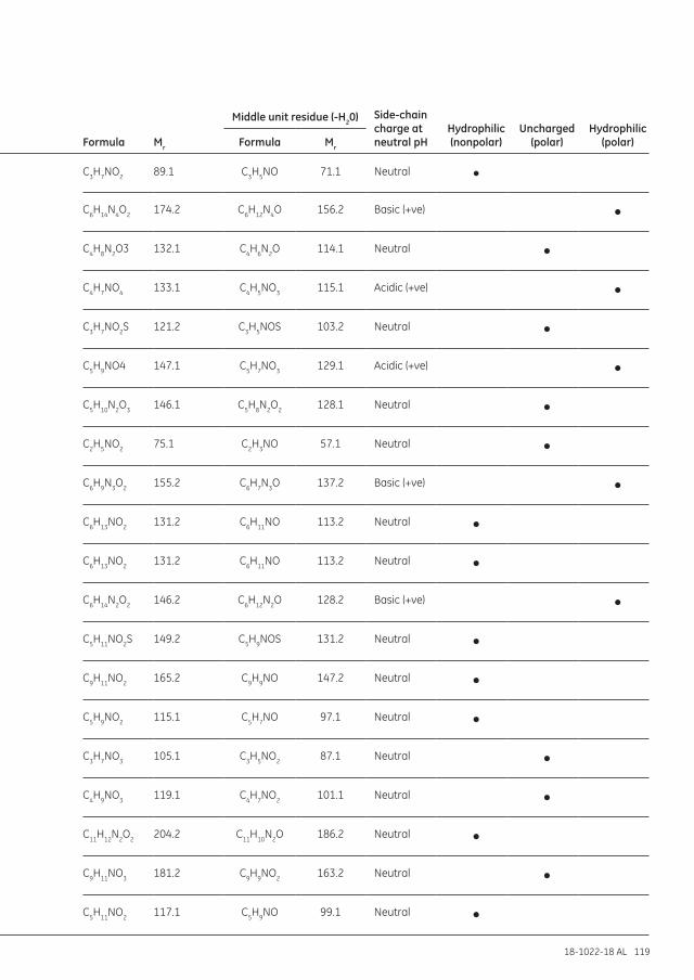
18-1022-18 AL 119
Formula Mr
Middle unit residue (-H20) Side-chain charge at neutral pH
Hydrophilic (nonpolar)
Uncharged (polar)
Hydrophilic (polar)Formula Mr
C3H7NO2 89.1 C3H5NO 71.1 Neutral •
C6H14N4O2 174.2 C6H12N4O 156.2 Basic (+ve) •
C4H8N2O3 132.1 C4H6N2O 114.1 Neutral •
C4H7NO4 133.1 C4H5NO3 115.1 Acidic (+ve) •
C3H7NO2S 121.2 C3H5NOS 103.2 Neutral •
C5H9NO4 147.1 C5H7NO3 129.1 Acidic (+ve) •
C5H10N2O3 146.1 C5H8N2O2 128.1 Neutral •
C2H5NO2 75.1 C2H3NO 57.1 Neutral •
C6H9N3O2 155.2 C6H7N3O 137.2 Basic (+ve) •
C6H13NO2 131.2 C6H11NO 113.2 Neutral •
C6H13NO2 131.2 C6H11NO 113.2 Neutral •
C6H14N2O2 146.2 C6H12N2O 128.2 Basic (+ve) •
C5H11NO2S 149.2 C5H9NOS 131.2 Neutral •
C9H11NO2 165.2 C9H9NO 147.2 Neutral •
C5H9NO2 115.1 C5H7NO 97.1 Neutral •
C3H7NO3 105.1 C3H5NO2 87.1 Neutral •
C4H9NO3 119.1 C4H7NO2 101.1 Neutral •
C11H12N2O2 204.2 C11H10N2O 186.2 Neutral •
C9H11NO3 181.2 C9H9NO2 163.2 Neutral •
C5H11NO2 117.1 C5H9NO 99.1 Neutral •
120 18-1022-18 AL
Appendix 8 Analysis and characterization
Analytical assays are essential to follow the progress of purification. They are used to assess the effectiveness of the purification in terms of yield, biological activity, recovery, and degree of purification. The importance of a reliable assay for the target molecule cannot be overemphasized.
Protein detection and quantitationDetection and quantitation of the target protein are needed when optimizing purification protocols. For overexpressed proteins, the high concentration in itself can be used for detection of the target protein fraction in a chromatogram, but in such a case verification of the identity of the protein in the final preparation is needed. Specific detection of tagged proteins can often be accomplished by analyzing the presence of the tag by activity or immunoassay, or simply by the spectral properties of the tag. Specific detection of the target protein can be obtained by functional assays, immunodetection, and mass spectrometry. SDS-polyacrylamide gel electrophoresis (SDS-PAGE) is the key method for checking purity of proteins. The target protein band can often be identified using the apparent molecular weight obtained by including standard molecular weight markers in the analysis. Subsequent verification of protein identity should always be obtained. Optimizing purification protocols can require functional assays to assess the intactness of the target protein. In general:
• The relative yield of tagged protein can often be determined by measuring the absorbance at 280 nm because the purity after a single purification step is high, that is, most of the eluted material may be considered to be the target protein. The extinction coefficient of the target protein will be needed. A good estimation can be obtained by theoretical calculation from the amino acid composition of the protein.
• The yield of protein may also be determined by standard chromogenic methods (e.g., Lowry, BCA™ protein assay, and Bradford total protein assay).
• Immunoassays (Western blot, ELISA, and immunoprecipitation) can be used for quantitation if a suitable standard curve can be produced. In this case, it is not necessary to purify the tagged protein so long as a purified standard is available. Therefore, these techniques may be used for quantitation during protocol development. The immunoassay technique is also particularly suitable for screening large numbers of samples when a simple yes/no answer is required (e.g., when testing fractions from a chromatographic run).
Purity check and protein characterizationPurityPurity of the target protein is most often estimated by SDS-PAGE, capillary electrophoresis, RPC, or mass spectrometry. The absorbance at 280 nm gives a rough but fast and nondestructive estimate of total protein content in crude samples. Lowry or Bradford assays are also frequently used to determine the total protein. The Bradford assay is particularly suited to samples where there is a high lipid content that can interfere with the Lowry assay. Absorbance data can be calibrated to high accuracy for pure proteins.
Obtaining a single band in electrophoresis is indicative of a pure protein, although potentially other proteins can comigrate. At this stage, additional methods based on orthogonal separation principles, such as chromatography in analytical scale should be used to confirm homogeneity/purity.
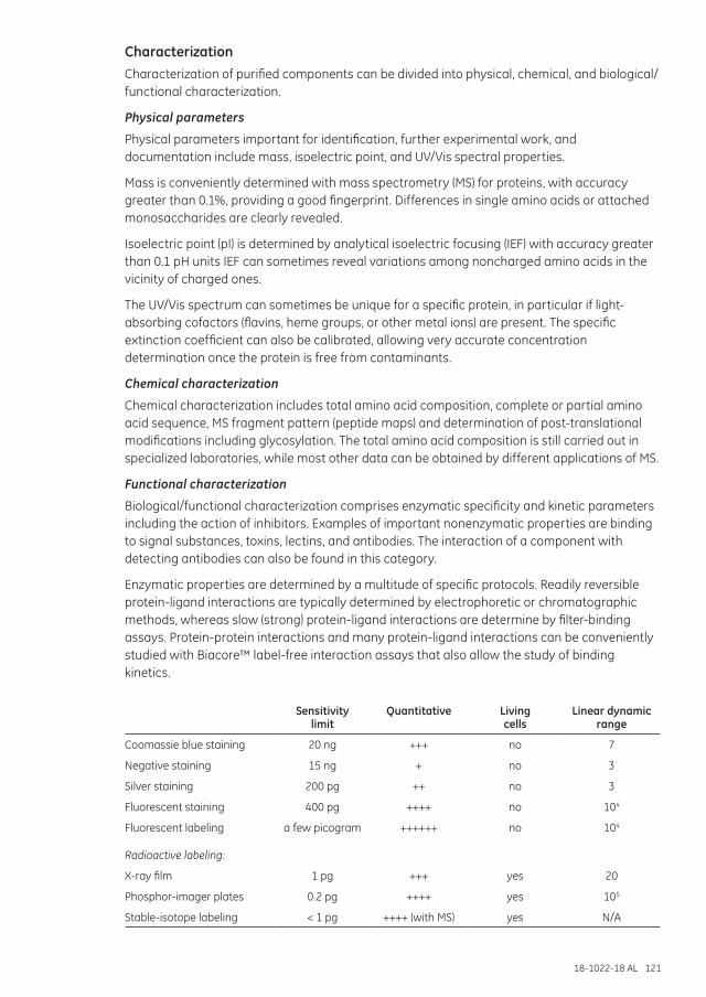
18-1022-18 AL 121
CharacterizationCharacterization of purified components can be divided into physical, chemical, and biological/functional characterization.
Physical parameters
Physical parameters important for identification, further experimental work, and documentation include mass, isoelectric point, and UV/Vis spectral properties.
Mass is conveniently determined with mass spectrometry (MS) for proteins, with accuracy greater than 0.1%, providing a good fingerprint. Differences in single amino acids or attached monosaccharides are clearly revealed.
Isoelectric point (pI) is determined by analytical isoelectric focusing (IEF) with accuracy greater than 0.1 pH units IEF can sometimes reveal variations among noncharged amino acids in the vicinity of charged ones.
The UV/Vis spectrum can sometimes be unique for a specific protein, in particular if light-absorbing cofactors (flavins, heme groups, or other metal ions) are present. The specific extinction coefficient can also be calibrated, allowing very accurate concentration determination once the protein is free from contaminants.
Chemical characterization
Chemical characterization includes total amino acid composition, complete or partial amino acid sequence, MS fragment pattern (peptide maps) and determination of post-translational modifications including glycosylation. The total amino acid composition is still carried out in specialized laboratories, while most other data can be obtained by different applications of MS.
Functional characterization
Biological/functional characterization comprises enzymatic specificity and kinetic parameters including the action of inhibitors. Examples of important nonenzymatic properties are binding to signal substances, toxins, lectins, and antibodies. The interaction of a component with detecting antibodies can also be found in this category.
Enzymatic properties are determined by a multitude of specific protocols. Readily reversible protein-ligand interactions are typically determined by electrophoretic or chromatographic methods, whereas slow (strong) protein-ligand interactions are determine by filter-binding assays. Protein-protein interactions and many protein-ligand interactions can be conveniently studied with Biacore™ label-free interaction assays that also allow the study of binding kinetics.
Sensitivity limit
Quantitative Living cells
Linear dynamic range
Coomassie blue staining 20 ng +++ no 7
Negative staining 15 ng + no 3
Silver staining 200 pg ++ no 3
Fluorescent staining 400 pg ++++ no 104
Fluorescent labeling a few picogram ++++++ no 104
Radioactive labeling:
X-ray film 1 pg +++ yes 20
Phosphor-imager plates 0.2 pg ++++ yes 105
Stable-isotope labeling < 1 pg ++++ (with MS) yes N/A
122 18-1022-18 AL
Appendix 9 Storage of biological samples
The advice given here is of a general nature and cannot be applied to every biological sample. Always consider the properties of the specific sample and its intended use before following any of these recommendations.
General recommendations Add stabilizing agents when necessary. Stabilizing agents are often required for storage
of purified proteins.
Serum, culture supernatants, and ascites should be kept frozen at -20°C or -70°C, in small aliquots.
Avoid repeated freeze/thawing or freeze drying/redissolving that can reduce biological activity.
Avoid conditions close to stability limits, in terms of for example pH or salt concentrations, reducing, or chelating agents.
Keep refrigerated at 4°C in a closed vessel to minimize bacterial growth and protease activity. For prolonged storage at 4°C (more than 24 h), add a preserving agent (e.g., merthiolate 0.01%).
Sodium azide can interfere with coupling methods, and some biological assays, and can be a health hazard. It can be removed by using a desalting column (see Chapter 5).
Common storage conditions for purified proteins Store as a precipitate in a high concentration of, for example, 4 M ammonium sulfate.
Freeze in 50% glycerol, especially suitable for enzymes.
Avoid using preserving agents if the product is to be used for a biological assay. Preserving agents should not be added if in vivo experiments are to be performed. Store samples in small aliquots and keep frozen.
Sterile filter to prolong storage time.
Add stabilizing agents, for example, glycerol (5% to 20%), serum albumin (10 mg/ml), ligand (concentration is selected based on concentration of active protein) to maintain biological activity. Remember that any additive will reduce the purity of the protein and might need to be removed at a later stage.
Avoid repeated freeze/thawing or freeze drying/redissolving that can reduce biological activity.
Certain proteins, including some mouse antibodies of the IgG3 subclass, should not be stored at 4°C as they precipitate at this temperature (cryoproteins). Store at room temperature in the presence of a preserving agent.
18-1022-18 AL 123
Appendix 10 Molecular weight estimation and molecular weight distribution analysis
Unlike electrophoretic techniques, SEC provides a means of determining the molecular weight or size (Stokes radius) of native or denatured proteins under a wide variety of conditions of pH, ionic strength and temperature, free from the constraints imposed by the charge state of the molecules. In order to understand and follow the procedures outlined, it is important to have read Chapter 7, SEC in theory.
For molecular weight determination, several theoretical models have been proposed to describe the behavior of solutes during SEC. Most models assume that the partition of solute molecules between the particles and surrounding liquid is an entirely steric effect. However, in practice a homologous series of compounds demonstrate a sigmodial relationship between their various elution volume parameters and the logarithm of their molecular weights. Thus, molecular weight determination by SEC can be made by comparing an elution volume parameter, such as Kav of the substance of interest, with the values obtained for several known calibration standards.
A calibration curve is prepared by measuring the elution volumes of several standards, calculating their corresponding Kav values (or similar parameter), and plotting their Kav values versus the logarithm of their molecular weight. The molecular weight of an unknown substance can be determined from the calibration curve once its Kav value is calculated from its measured elution volume. Various elution parameters, such as Ve, Ve/Vo, Kd, and Kav have been used in the literature for the preparation of calibration curves but the use of Kav is recommended since: 1) it is less sensitive to errors which can be introduced as a result of variations in column preparation and column dimensions, 2) it does not require the unreliable determination of the internal volume (Vi) as is required with Kd.
For good estimation of molecular weight, the calibration standards must have the same relationship between molecular weight and molecular size as the substance of interest. Calibration Kits from GE Healthcare provide well-characterized, globular protein standards for protein molecular weight estimation. The Low Molecular Weight (LMW) Gel Filtration Calibration Kit contains five individually lyophilized proteins with molecular weights in the range 6500 to 75 000 and Blue Dextran 2000 (Table A10.1). The High Molecular Weight (HMW) Gel Filtration Calibration Kit contains five individually lyophilized proteins with molecular weights in the range 43 000 to 669 000 and Blue Dextran 2000 (Table A10.2). Blue Dextran 2000 determines the void fraction in the column.
These well-defined protein standards show excellent behavior in SEC and enable simple, reliable calibration of SEC columns.
Table A10.1. Characteristics of Gel Filtration Calibration Kit LMW
Protein (weight per vial) Molecular weight (Mr) Source
Aprotinin (10 mg) 6500 Bovine lung
Ribonuclease A (50 mg) 13 700 Bovine pancreas
Carbonic anhydrase (15 mg) 29 000 Bovine erythrocytes
Ovalbumin (50 mg) 43 000 Hen egg
Conalbumin (50 mg) 75 000 Chicken egg white
Blue Dextran 2000 (50 mg) 2 000 000
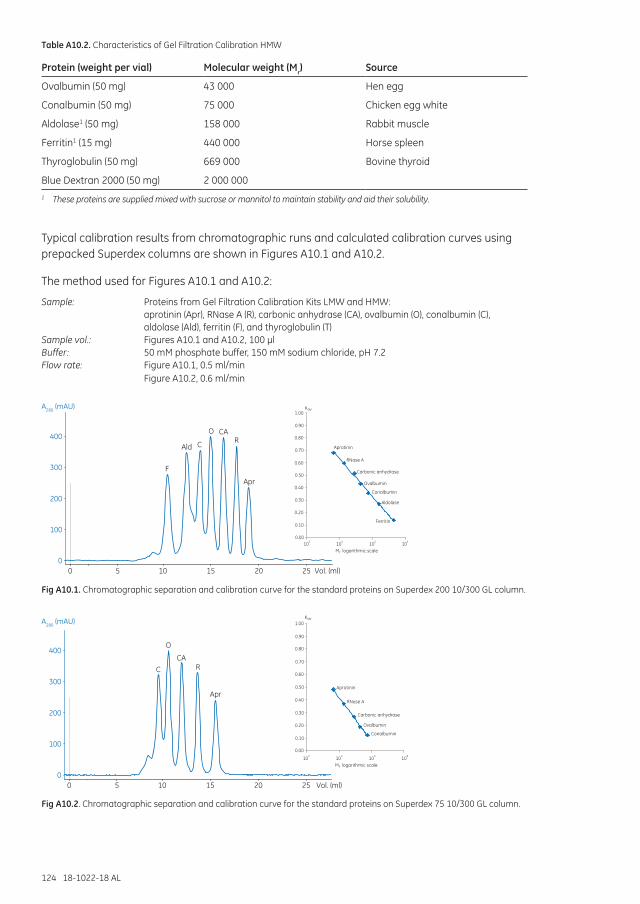
124 18-1022-18 AL
Table A10.2. Characteristics of Gel Filtration Calibration HMW
Protein (weight per vial) Molecular weight (Mr) Source
Ovalbumin (50 mg) 43 000 Hen egg
Conalbumin (50 mg) 75 000 Chicken egg white
Aldolase1 (50 mg) 158 000 Rabbit muscle
Ferritin1 (15 mg) 440 000 Horse spleen
Thyroglobulin (50 mg) 669 000 Bovine thyroid
Blue Dextran 2000 (50 mg) 2 000 0001 These proteins are supplied mixed with sucrose or mannitol to maintain stability and aid their solubility.
Typical calibration results from chromatographic runs and calculated calibration curves using prepacked Superdex columns are shown in Figures A10.1 and A10.2.
The method used for Figures A10.1 and A10.2:
Sample: Proteins from Gel Filtration Calibration Kits LMW and HMW: aprotinin (Apr), RNase A (R), carbonic anhydrase (CA), ovalbumin (O), conalbumin (C), aldolase (Ald), ferritin (F), and thyroglobulin (T)Sample vol.: Figures A10.1 and A10.2, 100 µlBuffer: 50 mM phosphate buffer, 150 mM sodium chloride, pH 7.2Flow rate: Figure A10.1, 0.5 ml/min Figure A10.2, 0.6 ml/min
0
100
200
300
400
A280 (mAU)
0 5 10 15 20 25 Vol. (ml)
0.00
0.20
0.40
0.60
0.80
1.00
Mr logarithmic scale
Kav
0.10
0.30
0.50
0.70
0.90
Ferritin
Aldolase
Conalbumin
Ovalbumin
Carbonic anhydrase
RNase A
Aprotinin
F
Ald C
O CA R
Apr
103 104 105 106
Fig A10.1. Chromatographic separation and calibration curve for the standard proteins on Superdex 200 10/300 GL column.
0
100
200
300
400
A280 (mAU)
0 5 10 15 20 25 Vol. (ml)
0.00
0.20
0.40
0.60
0.80
1.00 Kav
0.10
0.30
0.50
0.70
0.90
Conalbumin
Ovalbumin
Carbonic anhydrase
RNase A
Aprotinin
C
O
CA R
Apr
Mr logarithmic scale103 104 105 106
Fig A10.2. Chromatographic separation and calibration curve for the standard proteins on Superdex 75 10/300 GL column.
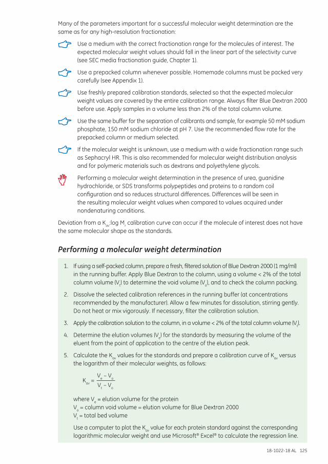
18-1022-18 AL 125
Many of the parameters important for a successful molecular weight determination are the same as for any high-resolution fractionation:
Use a medium with the correct fractionation range for the molecules of interest. The expected molecular weight values should fall in the linear part of the selectivity curve (see SEC media fractionation guide, Chapter 1).
Use a prepacked column whenever possible. Homemade columns must be packed very carefully (see Appendix 1).
Use freshly prepared calibration standards, selected so that the expected molecular weight values are covered by the entire calibration range. Always filter Blue Dextran 2000 before use. Apply samples in a volume less than 2% of the total column volume.
Use the same buffer for the separation of calibrants and sample, for example 50 mM sodium phosphate, 150 mM sodium chloride at pH 7. Use the recommended flow rate for the prepacked column or medium selected.
If the molecular weight is unknown, use a medium with a wide fractionation range such as Sephacryl HR. This is also recommended for molecular weight distribution analysis and for polymeric materials such as dextrans and polyethylene glycols.
Performing a molecular weight determination in the presence of urea, guanidine hydrochloride, or SDS transforms polypeptides and proteins to a random coil configuration and so reduces structural differences. Differences will be seen in the resulting molecular weight values when compared to values acquired under nondenaturing conditions.
Deviation from a Kav:log Mr calibration curve can occur if the molecule of interest does not have the same molecular shape as the standards.
Performing a molecular weight determination
1. If using a self-packed column, prepare a fresh, filtered solution of Blue Dextran 2000 (1 mg/ml) in the running buffer. Apply Blue Dextran to the column, using a volume < 2% of the total column volume (Vt) to determine the void volume (Vo), and to check the column packing.
2. Dissolve the selected calibration references in the running buffer (at concentrations recommended by the manufacturer). Allow a few minutes for dissolution, stirring gently. Do not heat or mix vigorously. If necessary, filter the calibration solution.
3. Apply the calibration solution to the column, in a volume < 2% of the total column volume (Vt).
4. Determine the elution volumes (Ve) for the standards by measuring the volume of the eluent from the point of application to the centre of the elution peak.
5. Calculate the Kav values for the standards and prepare a calibration curve of Kav versus the logarithm of their molecular weights, as follows:
Kav =
Ve – Vo
Vt – Vo
where Ve = elution volume for the protein Vo = column void volume = elution volume for Blue Dextran 2000 Vt = total bed volume
Use a computer to plot the Kav value for each protein standard against the corresponding logarithmic molecular weight and use Microsoft® Excel® to calculate the regression line.
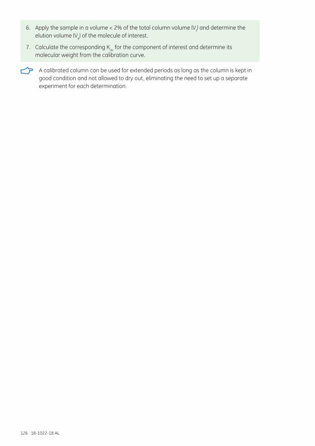
126 18-1022-18 AL
6. Apply the sample in a volume < 2% of the total column volume (Vt) and determine the elution volume (Ve) of the molecule of interest.
7. Calculate the corresponding Kav for the component of interest and determine its molecular weight from the calibration curve.
A calibrated column can be used for extended periods as long as the column is kept in good condition and not allowed to dry out, eliminating the need to set up a separate experiment for each determination.
18-1022-18 AL 127
Product indexÄKTA avant chromatography system 112–113
ÄKTAexplorer chromatography system 53, 112
ÄKTAmicro chromatography system 19, 22, 47, 52, 55, 112
ÄKTAprime chromatography system 72
ÄKTAprime plus chromatography system 76, 113
ÄKTApurifier chromatography system 30, 40, 112
ÄKTA start chromatography system 112–113
ÄKTAxpress chromatography system 112–113
Blue Dextran 2000 61, 123, 124, 125
Gel Filtration Calibration Kit LMW 123–124
Gel Filtration Calibration Kit HMW 123–124
HiLoad 16/600 Superdex 30 prep grade 31
HiLoad 16/600 Superdex 75 prep grade 44
HiLoad 16/600 Superdex 200 prep grade 20, 44
HiPrep 26/10 Desalting 67–69, 71–74, 76, 78–79, 81, 108
HiPrep 16/60 Sephacryl S-100 HR 60, 62
HiPrep 26/60 Sephacryl S-100 HR 62–63
HiPrep 16/60 Sephacryl S-200 HR 60, 62
HiPrep 26/60 Sephacryl S-200 HR 62
HiPrep 16/60 Sephacryl S-300 HR 60, 62
HiPrep 26/60 Sephacryl S-300 HR 62
HiPrep 16/60 Sephacryl S-400 HR 60, 62
HiPrep 26/60 Sephacryl S-400 HR 62
HiPrep 16/60 Sephacryl S-500 HR 60, 62
HiPrep 26/60 Sephacryl S-500 HR 62
HiScale empty columns 100
HiTrap Chelating HP 17, 72
HiTrap Desalting Column 32, 67, 69, 71–79, 81, 108, 112
MabSelect 97, 98
MidiSpin Adapter 71
MiniSpin Adapter 71
Packing Connectors 102
PD-10 Desalting Column 69, 77–78
PD-10 Spin Adapter 71
PD MidiTrap G-25 69, 71, 73
PD MiniTrap G-10 69, 71, 73
PD MiniTrap G-25 69, 71, 73
PD MultiTrap G-25 69, 71, 73
PD SpinTrap G-25 69, 71, 73
PrimeView software 112
Sephacryl S-100 HR 23, 59, 61–62, 65, 102
Sephacryl S-200 HR 23, 59, 61–62, 65, 102
Sephacryl S-300 HR 23, 59, 62, 65, 102
Sephacryl S-400 HR 23, 59, 61–62, 65, 102
Sephacryl S-500 HR 23, 59, 62, 65, 102
Sephacryl S-1000 SF 23, 59, 62, 65
Sephadex G-10 16, 23–24, 67–69, 77, 80, 83, 103, 105–106
Sephadex G-25 Superfine 23–24, 67, 69–70, 74, 80, 103, 106
Sephadex G-25 Fine 23, 67, 69–70, 80, 103, 106
Sephadex G-25 Medium 67, 69–70, 80, 103, 106
Sephadex G-25 Coarse 23, 34 67, 80, 103, 106
Sephadex LH-20 83–87
Solvent resistant (SR) empty columns 85, 88
Superdex 75 3.2/300 45
Superdex 75 5/150 GL 18, 45, 48
Superdex 75 10/300 GL 43, 45, 124
Superdex 200 Increase 3.2/300 30, 45
Superdex 200 Increase 5/150 GL 18–19, 45–47, 49
Superdex 200 Increase 10/300 GL 22, 40, 45–46
Superdex 200 3.2/300 45
Superdex 200 10/300 GL 40, 45, 124
Superdex Peptide 3.2/300 45
Superdex Peptide 10/300 GL 42, 45,
Superdex 30 prep grade 23, 31, 39, 43, 44, 45, 46, 50, 92
Superdex 75 prep grade 23, 39, 43, 44, 45, 50, 92, 97
Superdex 200 prep grade 20, 23, 25, 39, 43–44, 45, 50, 92
Superose 6 Increase 3.2/300 54
Superose 6 Increase 5/150 GL 18, 54–55
Superose 6 Increase 10/300 GL 22, 29, 52–53, 54
Superose 6 3.2/300 54
Superose 6 10/300 GL 52, 54
Superose 12 3.2/300 54, 55
Superose 12 10/300 GL 54
Superose 6 prep grade 23, 51, 54, 57,
Superose 12 prep grade 23, 53-54, 57,
Tricorn columns 18, 53, 100, 114
UNICORN control software 29, 112
XK empty columns 25, 43, 61, 97, 98, 99, 100–103, 110, 114
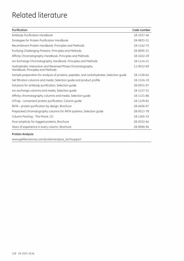
128 18-1022-18 AL
Related literature
Purification Code number
Antibody Purification Handbook 18-1037-46
Strategies for Protein Purification Handbook 28-9833-31
Recombinant Protein Handbook, Principles and Methods 18-1142-75
Purifying Challenging Proteins, Principles and Methods 28-9095-31
Affinity Chromatography Handbook, Principles and Methods 18-1022-29
Ion Exchange Chromatography Handbook, Principles and Methods 18-1114-21
Hydrophobic Interaction and Reversed Phase Chromatography Handbook, Principles and Methods
11-0012-69
Sample preparation for analysis of proteins, peptides, and carbohydrates, Selection guide 18-1128-62
Gel filtration columns and media, Selection guide and product profile 18-1124-19
Solutions for antibody purification, Selection guide 28-9351-97
Ion exchange columns and media, Selection guide 18-1127-31
Affinity chromatography columns and media, Selection guide 18-1121-86
HiTrap - convenient protein purification, Column guide 18-1129-81
ÄKTA - protein purification by design, Brochure 28-4026-97
Prepacked chromatography columns for ÄKTA systems, Selection guide 28-9317-78
Column Packing - The Movie, CD 18-1165-33
Pure simplicity for tagged proteins, Brochure 28-9353-64
Years of experience in every column, Brochure 28-9090-94
Protein Analysis
www.gelifesciences.com/proteinanalysis_techsupport
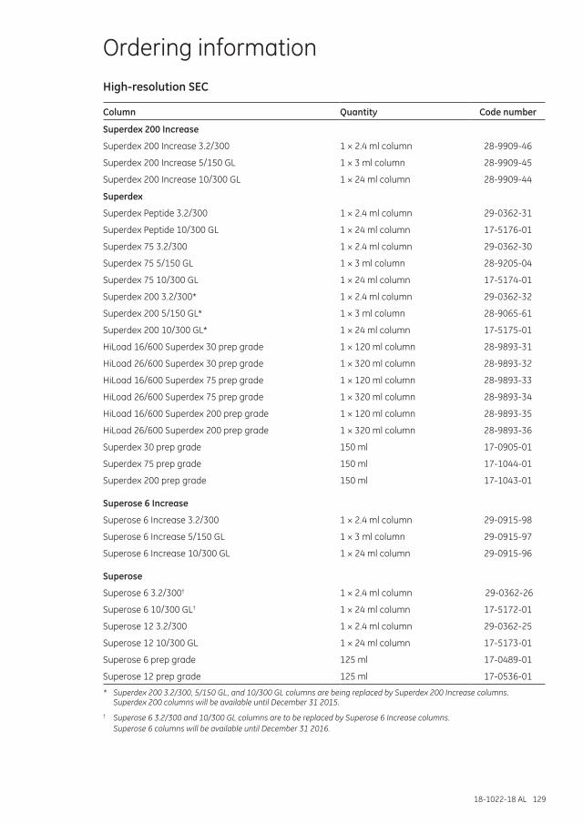
18-1022-18 AL 129
Ordering informationHigh-resolution SEC
Column Quantity Code number
Superdex 200 Increase
Superdex 200 Increase 3.2/300 1 × 2.4 ml column 28-9909-46
Superdex 200 Increase 5/150 GL 1 × 3 ml column 28-9909-45
Superdex 200 Increase 10/300 GL 1 × 24 ml column 28-9909-44
Superdex
Superdex Peptide 3.2/300 1 × 2.4 ml column 29-0362-31
Superdex Peptide 10/300 GL 1 × 24 ml column 17-5176-01
Superdex 75 3.2/300 1 × 2.4 ml column 29-0362-30
Superdex 75 5/150 GL 1 × 3 ml column 28-9205-04
Superdex 75 10/300 GL 1 × 24 ml column 17-5174-01
Superdex 200 3.2/300* 1 × 2.4 ml column 29-0362-32
Superdex 200 5/150 GL* 1 × 3 ml column 28-9065-61
Superdex 200 10/300 GL* 1 × 24 ml column 17-5175-01
HiLoad 16/600 Superdex 30 prep grade 1 × 120 ml column 28-9893-31
HiLoad 26/600 Superdex 30 prep grade 1 × 320 ml column 28-9893-32
HiLoad 16/600 Superdex 75 prep grade 1 × 120 ml column 28-9893-33
HiLoad 26/600 Superdex 75 prep grade 1 × 320 ml column 28-9893-34
HiLoad 16/600 Superdex 200 prep grade 1 × 120 ml column 28-9893-35
HiLoad 26/600 Superdex 200 prep grade 1 × 320 ml column 28-9893-36
Superdex 30 prep grade 150 ml 17-0905-01
Superdex 75 prep grade 150 ml 17-1044-01
Superdex 200 prep grade 150 ml 17-1043-01
Superose 6 Increase
Superose 6 Increase 3.2/300 1 × 2.4 ml column 29-0915-98
Superose 6 Increase 5/150 GL 1 × 3 ml column 29-0915-97
Superose 6 Increase 10/300 GL 1 × 24 ml column 29-0915-96
Superose
Superose 6 3.2/300† 1 × 2.4 ml column 29-0362-26
Superose 6 10/300 GL† 1 × 24 ml column 17-5172-01
Superose 12 3.2/300 1 × 2.4 ml column 29-0362-25
Superose 12 10/300 GL 1 × 24 ml column 17-5173-01
Superose 6 prep grade 125 ml 17-0489-01
Superose 12 prep grade 125 ml 17-0536-01
* Superdex 200 3.2/300, 5/150 GL, and 10/300 GL columns are being replaced by Superdex 200 Increase columns. Superdex 200 columns will be available until December 31 2015.
† Superose 6 3.2/300 and 10/300 GL columns are to be replaced by Superose 6 Increase columns. Superose 6 columns will be available until December 31 2016.
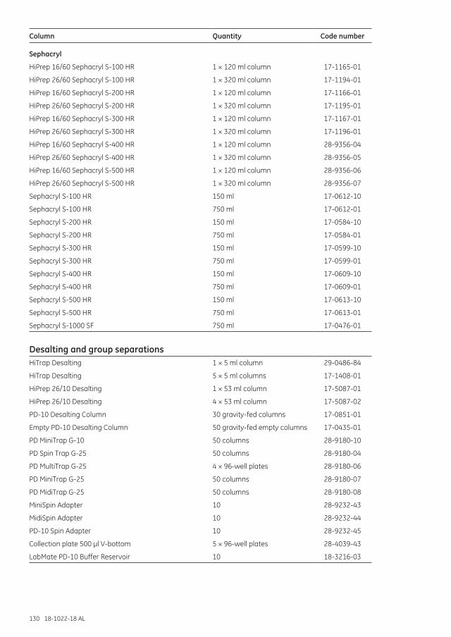
130 18-1022-18 AL
Column Quantity Code number
Sephacryl
HiPrep 16/60 Sephacryl S-100 HR 1 × 120 ml column 17-1165-01
HiPrep 26/60 Sephacryl S-100 HR 1 × 320 ml column 17-1194-01
HiPrep 16/60 Sephacryl S-200 HR 1 × 120 ml column 17-1166-01
HiPrep 26/60 Sephacryl S-200 HR 1 × 320 ml column 17-1195-01
HiPrep 16/60 Sephacryl S-300 HR 1 × 120 ml column 17-1167-01
HiPrep 26/60 Sephacryl S-300 HR 1 × 320 ml column 17-1196-01
HiPrep 16/60 Sephacryl S-400 HR 1 × 120 ml column 28-9356-04
HiPrep 26/60 Sephacryl S-400 HR 1 × 320 ml column 28-9356-05
HiPrep 16/60 Sephacryl S-500 HR 1 × 120 ml column 28-9356-06
HiPrep 26/60 Sephacryl S-500 HR 1 × 320 ml column 28-9356-07
Sephacryl S-100 HR 150 ml 17-0612-10
Sephacryl S-100 HR 750 ml 17-0612-01
Sephacryl S-200 HR 150 ml 17-0584-10
Sephacryl S-200 HR 750 ml 17-0584-01
Sephacryl S-300 HR 150 ml 17-0599-10
Sephacryl S-300 HR 750 ml 17-0599-01
Sephacryl S-400 HR 150 ml 17-0609-10
Sephacryl S-400 HR 750 ml 17-0609-01
Sephacryl S-500 HR 150 ml 17-0613-10
Sephacryl S-500 HR 750 ml 17-0613-01
Sephacryl S-1000 SF 750 ml 17-0476-01
Desalting and group separationsHiTrap Desalting 1 × 5 ml column 29-0486-84
HiTrap Desalting 5 × 5 ml columns 17-1408-01
HiPrep 26/10 Desalting 1 × 53 ml column 17-5087-01
HiPrep 26/10 Desalting 4 × 53 ml column 17-5087-02
PD-10 Desalting Column 30 gravity-fed columns 17-0851-01
Empty PD-10 Desalting Column 50 gravity-fed empty columns 17-0435-01
PD MiniTrap G-10 50 columns 28-9180-10
PD Spin Trap G-25 50 columns 28-9180-04
PD MultiTrap G-25 4 × 96-well plates 28-9180-06
PD MiniTrap G-25 50 columns 28-9180-07
PD MidiTrap G-25 50 columns 28-9180-08
MiniSpin Adapter 10 28-9232-43
MidiSpin Adapter 10 28-9232-44
PD-10 Spin Adapter 10 28-9232-45
Collection plate 500 µl V-bottom 5 × 96-well plates 28-4039-43
LabMate PD-10 Buffer Reservoir 10 18-3216-03
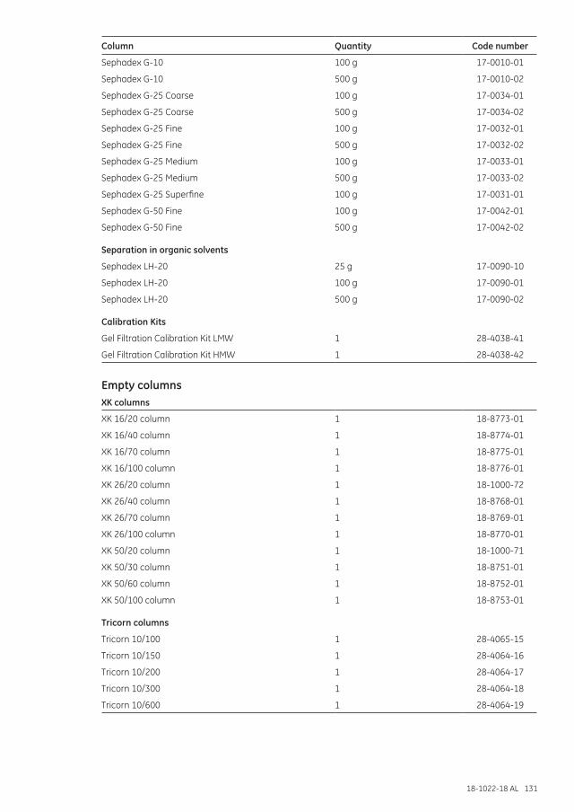
18-1022-18 AL 131
Column Quantity Code number
Sephadex G-10 100 g 17-0010-01
Sephadex G-10 500 g 17-0010-02
Sephadex G-25 Coarse 100 g 17-0034-01
Sephadex G-25 Coarse 500 g 17-0034-02
Sephadex G-25 Fine 100 g 17-0032-01
Sephadex G-25 Fine 500 g 17-0032-02
Sephadex G-25 Medium 100 g 17-0033-01
Sephadex G-25 Medium 500 g 17-0033-02
Sephadex G-25 Superfine 100 g 17-0031-01
Sephadex G-50 Fine 100 g 17-0042-01
Sephadex G-50 Fine 500 g 17-0042-02
Separation in organic solvents
Sephadex LH-20 25 g 17-0090-10
Sephadex LH-20 100 g 17-0090-01
Sephadex LH-20 500 g 17-0090-02
Calibration Kits
Gel Filtration Calibration Kit LMW 1 28-4038-41
Gel Filtration Calibration Kit HMW 1 28-4038-42
Empty columnsXK columns
XK 16/20 column 1 18-8773-01
XK 16/40 column 1 18-8774-01
XK 16/70 column 1 18-8775-01
XK 16/100 column 1 18-8776-01
XK 26/20 column 1 18-1000-72
XK 26/40 column 1 18-8768-01
XK 26/70 column 1 18-8769-01
XK 26/100 column 1 18-8770-01
XK 50/20 column 1 18-1000-71
XK 50/30 column 1 18-8751-01
XK 50/60 column 1 18-8752-01
XK 50/100 column 1 18-8753-01
Tricorn columns
Tricorn 10/100 1 28-4065-15
Tricorn 10/150 1 28-4064-16
Tricorn 10/200 1 28-4064-17
Tricorn 10/300 1 28-4064-18
Tricorn 10/600 1 28-4064-19
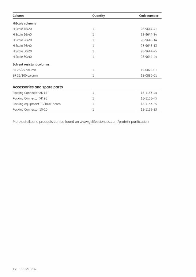
132 18-1022-18 AL
Column Quantity Code number
HiScale columns
HiScale 16/20 1 28-9644-41
HiScale 16/40 1 28-9644-24
HiScale 26/20 1 28-9645-14
HiScale 26/40 1 28-9645-13
HiScale 50/20 1 28-9644-45
HiScale 50/40 1 28-9644-44
Solvent resistant columns
SR 25/45 column 1 19-0879-01
SR 25/100 column 1 19-0880-01
Accessories and spare partsPacking Connector XK 16 1 18-1153-44
Packing Connector XK 26 1 18-1153-45
Packing equipment 10/100 (Tricorn) 1 18-1153-25
Packing Connector 10-10 1 18-1153-23
More details and products can be found on www.gelifesciences.com/protein-purification

18-1022-18 AL 133

134 18-1022-18 AL

18-1022-18 AL 135

Size Exclusion Chromatography – Principles and M
ethods
18-1022-18 AL 11/2014
GE and GE monogram are trademarks of General Electric Company.
ÄKTA, Biacore, BioProcess, HiLoad, HiPrep, HiScale, HiTrap, MabSelect, MidiTrap, MiniTrap, MultiTrap, Sephacryl, Sephadex, Sepharose, SpinTrap, Superdex, Superose, Tricorn, and UNICORN are trademarks of GE Healthcare Company or one of its subsidiaries.
BCA is a trademark of Thermo Fisher Scientific LLC. Coomassie is a trademark of Imperial Chemical Industries Limited. Microsoft and Excel are registered trademarks of the Microsoft Corporation. Triton is a trademark of Union Carbide Chemicals and Plastic Company Inc. Tween is a trademark of Uniqema Americas LLC.
All other third-party trademarks are the property of their respective owners.
IMAC Sepharose products, Ni Sepharose products and Fe Sepharose products: These products are sold under a license from Sigma-Aldrich under patent number EP 1276716 (Metal chelating compositions) and equivalent patents and patent applications in other countries.
© 2000–2014 General Electric Company—All rights reserved. First published Dec. 2000.
All goods and services are sold subject to the terms and conditions of sale of the company within GE Healthcare which supplies them. A copy of these terms and conditions is available on request. Contact your local GE Healthcare representative for the most current information.
GE Healthcare UK Limited Amersham Place Little Chalfont Buckinghamshire, HP7 9NA UK
GE Healthcare Europe, GmbH Munzinger Strasse 5 D-79111 Freiburg Germany
GE Healthcare Bio-Sciences Corp. 800 Centennial Avenue, P.O. Box 1327 Piscataway, NJ 08855-1327 USA
GE Healthcare Bio-Sciences KK Sanken Bldg., 3-25-1, Hyakunincho Shinjuku-ku, Tokyo 169-0073 Japan
For local office contact information,please visit www.gelifesciences.com/contact
www.gelifesciences.com/sizeexclusion
GE Healthcare Bio-Sciences ABBjörkgatan 30751 84 UppsalaSweden
Size Exclusion ChromatographyPrinciples and Methods
GE HealthcareLife Sciences
imagination at workimagination at work