
Size- and Shape-Dependent Antibacterial Studies of Silver ... · nanomaterials Article Size- and...
15
nanomaterials Article Size- and Shape-Dependent Antibacterial Studies of Silver Nanoparticles Synthesized by Wet Chemical Routes Muhammad Akram Raza 1, * ,† , Zakia Kanwal 2,† , Anum Rauf 1 , Anjum Nasim Sabri 3 , Saira Riaz 1 and Shahzad Naseem 1 1 Centre of Excellence in Solid State Physics, University of the Punjab, QAC, Lahore 54590, Pakistan; [email protected] (A.R.); [email protected] (S.R.); [email protected] (S.N.) 2 Department of Zoology, Lahore College for Women University, Jail Road, Lahore 54000, Pakistan; [email protected] 3 Department of MicroBiology & Molecular Genetics, University of the Punjab, QAC, Lahore 54590, Pakistan; [email protected] * Correspondance: [email protected]; Tel.: +92-42-992-31136 † These authors contributed equally to this work. Academic Editor: Thomas Nann Received: 5 January 2016; Accepted: 2 March 2016; Published: 15 April 2016 Abstract: Silver nanoparticles (AgNPs) of different shapes and sizes were prepared by solution-based chemical reduction routes. Silver nitrate was used as a precursor, tri-sodium citrate (TSC) and sodium borohydride as reducing agents, while polyvinylpyrrolidone (PVP) was used as a stabilizing agent. The morphology, size, and structural properties of obtained nanoparticles were characterized by scanning electron microscopy (SEM), UV-visible spectroscopy (UV-VIS), and X-ray diffraction (XRD) techniques. Spherical AgNPs, as depicted by SEM, were found to have diameters in the range of 15 to 90 nm while lengths of the edges of the triangular particles were about 150 nm. The characteristic surface plasmon resonance (SPR) peaks of different spherical silver colloids occurring in the wavelength range of 397 to 504 nm, whereas triangular particles showed two peaks, first at 392 nm and second at 789 nm as measured by UV-VIS. The XRD spectra of the prepared samples indicated the face-centered cubic crystalline structure of metallic AgNPs. The in vitro antibacterial properties of all synthesized AgNPs against two types of Gram-negative bacteria, Pseudomonas aeruginosa and Escherichia coli were examined by Kirby–Bauer disk diffusion susceptibility method. It was noticed that the smallest-sized spherical AgNPs demonstrated a better antibacterial activity against both bacterial strains as compared to the triangular and larger spherical shaped AgNPs. Keywords: AgNPs; reduction method; antibacterial activity; Pseudomonas aeruginosa (P. aeruginosa); Escherichia coli (E. coli) 1. Introduction Nanoparticles are of tremendous importance in the field of nanotechnology by serving as basic building blocks in nanodevices for various practical applications. By virtue of a large surface-to-volume ratio and quantum confinement effect, nanoparticles exhibit unique and dramatically different physical, chemical, and biological properties relative to bulk materials [1]. Remarkable attraction and utilization of metallic nanoparticles in modern technologies results from their interesting surface plasmon characteristics, exciting physicochemical properties, and fascinating unique morphologies [2]. Metallic nanoparticles, therefore, are successfully playing their vital role in a variety of fields, such as information storage devices, photography, biological labeling, catalysis, photonics, optoelectronics, construction of magnetic ferrofluids, and surface enhanced Raman scattering [3]. Furthermore, they can Nanomaterials 2016, 6, 74; doi:10.3390/nano6040074 www.mdpi.com/journal/nanomaterials
Transcript of Size- and Shape-Dependent Antibacterial Studies of Silver ... · nanomaterials Article Size- and...

nanomaterials
Article
Size- and Shape-Dependent Antibacterial Studiesof Silver Nanoparticles Synthesized by WetChemical RoutesMuhammad Akram Raza 1,*,†, Zakia Kanwal 2,†, Anum Rauf 1, Anjum Nasim Sabri 3, Saira Riaz 1
and Shahzad Naseem 1
1 Centre of Excellence in Solid State Physics, University of the Punjab, QAC, Lahore 54590, Pakistan;[email protected] (A.R.); [email protected] (S.R.); [email protected] (S.N.)
2 Department of Zoology, Lahore College for Women University, Jail Road, Lahore 54000, Pakistan;[email protected]
3 Department of MicroBiology & Molecular Genetics, University of the Punjab, QAC, Lahore 54590, Pakistan;[email protected]
* Correspondance: [email protected]; Tel.: +92-42-992-31136† These authors contributed equally to this work.
Academic Editor: Thomas NannReceived: 5 January 2016; Accepted: 2 March 2016; Published: 15 April 2016
Abstract: Silver nanoparticles (AgNPs) of different shapes and sizes were prepared by solution-basedchemical reduction routes. Silver nitrate was used as a precursor, tri-sodium citrate (TSC) and sodiumborohydride as reducing agents, while polyvinylpyrrolidone (PVP) was used as a stabilizing agent.The morphology, size, and structural properties of obtained nanoparticles were characterized byscanning electron microscopy (SEM), UV-visible spectroscopy (UV-VIS), and X-ray diffraction (XRD)techniques. Spherical AgNPs, as depicted by SEM, were found to have diameters in the range of 15to 90 nm while lengths of the edges of the triangular particles were about 150 nm. The characteristicsurface plasmon resonance (SPR) peaks of different spherical silver colloids occurring in thewavelength range of 397 to 504 nm, whereas triangular particles showed two peaks, first at 392 nmand second at 789 nm as measured by UV-VIS. The XRD spectra of the prepared samples indicatedthe face-centered cubic crystalline structure of metallic AgNPs. The in vitro antibacterial propertiesof all synthesized AgNPs against two types of Gram-negative bacteria, Pseudomonas aeruginosa andEscherichia coli were examined by Kirby–Bauer disk diffusion susceptibility method. It was noticedthat the smallest-sized spherical AgNPs demonstrated a better antibacterial activity against bothbacterial strains as compared to the triangular and larger spherical shaped AgNPs.
Keywords: AgNPs; reduction method; antibacterial activity; Pseudomonas aeruginosa (P. aeruginosa);Escherichia coli (E. coli)
1. Introduction
Nanoparticles are of tremendous importance in the field of nanotechnology by serving as basicbuilding blocks in nanodevices for various practical applications. By virtue of a large surface-to-volumeratio and quantum confinement effect, nanoparticles exhibit unique and dramatically differentphysical, chemical, and biological properties relative to bulk materials [1]. Remarkable attractionand utilization of metallic nanoparticles in modern technologies results from their interesting surfaceplasmon characteristics, exciting physicochemical properties, and fascinating unique morphologies [2].Metallic nanoparticles, therefore, are successfully playing their vital role in a variety of fields, such asinformation storage devices, photography, biological labeling, catalysis, photonics, optoelectronics,construction of magnetic ferrofluids, and surface enhanced Raman scattering [3]. Furthermore, they can
Nanomaterials 2016, 6, 74; doi:10.3390/nano6040074 www.mdpi.com/journal/nanomaterials

Nanomaterials 2016, 6, 74 2 of 15
also be used as a model system to experimentally investigate the effects of quantum confinementon magnetic, electronic and other related properties [4]. The fundamental characteristics of metallicnanoparticles strongly depend on their shapes, sizes, configurations, crystallinity, and structurewhether they are in solid form or in hollow geometries [5]. Thus, by controlling such parameters onecan achieve the desired properties of the nanoparticles.
Since ancient times silver has been used extensively for many applications such asjewelry, metalcraft, vessels or containers for liquid, coins, shavings, foils, and photography(where photosensitive Ag halides are reduced). Additionally, silver has been part of many medicalapplications throughout known history [6]. In the modern era, nano-sized silver particles arealso a vibrant part of nanotechnology because of growing demands in various field; for example,chemical and biological sensing due to surface-enhanced Raman scattering properties, electronics andoptoelectronics applications owing to the highest electrical and thermal conductivity among all othermetals, energy harvesting, catalysis, imaging, and biomedicine [7,8].
One of the most important properties of AgNPS is their antimicrobial action against severalbacteria, fungi, and viruses [9,10] and, thus, they are widely used as antimicrobial agents indifferent products, including clothes, plasters, bandages, toothbrushes, catheters, scalpels, cosmetics,needles, refrigerators, and mobile phones [11]. In fact, when AgNPs interact with microorganisms(bacteria, fungi, and viruses), silver ions (Ag+) are released and these ions may affect and damage themicroorganism in different ways; for example, they attack the negatively-charged cell walls of themicrobes to deactivate cellular enzymes and disrupt membrane permeability; consequently, cell lysisand cell death occurs [12,13]. Furthermore, the broad spectrum killing, oligodynamic action andlesser possibility for development of microbial resistance against AgNPs make them advantageousantibacterial agents. Surprisingly, AgNPs are safe and non-toxic to human and animal cells at lowconcentrations because the possible toxicity of AgNPs to the environment is considered extremelylow as compared to other materials [14,15]. Perhaps, that is why AgNPs achieved the highest levelof commercialization and account for 55.4% of the total nanomaterial-based consumer productsavailable in the market (313 out of 565 products) [16,17]. Interestingly, the antimicrobial activity ofnano-sized silver particles was found size- and shape-dependent, one of the reasons could be thatdifferent morphologies provide different areas to interact with microbes and thus results in differentantibacterial efficiency [15,18,19].
AgNPs are prepared by various physical, chemical, and biological methods [8]. The mostsignificant physical methods include arc-discharge, physical vapor condensation, and laserablation [20,21]. The chemical methods frequently used for synthesis of nano-sized silver particlesinclude chemical reduction, microemulsion, photoinduced reduction, UV-initiated photoreduction,photoinduced reduction, electrochemical synthetic approach, microwave-assisted synthesis,and irradiation methods [2,22,23]. The bio-based methods mostly consist of green synthesis approaches,where extracts of different plants, prokaryotic bacterial cells, and/or eukaryotic fungi are used asreducing agents to reduce the metallic silver precursor for the preparation of AgNPs [24].
In aforementioned techniques, physical methods normally demand sophisticated equipmentso they may not be cost effective. For biological methods, in spite of their environment-friendlyadvantages, many critical aspects need to be considered; for instance, the nature of organisms,inheritable and genetic characteristics of organisms, suitable circumstances for cell growth and enzymeactivity, optimum reaction environments, and choice of the biocatalyst to obtain the desired stablenanoparticles [2]. However, chemical methods, especially wet-chemical reduction techniques, can beconsidered the best approach owing to cost-effectiveness, simpler handling, low impurity factors,thermal stability, availability of chemicals for wide range nanoparticles synthesis, defined controlover growth rate, and convenience to use different stabilizer to enhance the stability of preparedparticles [1].
Although the antibacterial properties of silver nanomaterials are well accepted and documented,however, the debate on the topic that how the size and shape of nano-sized entities influence the

Nanomaterials 2016, 6, 74 3 of 15
antimicrobial performance is still ongoing. Since some of the researchers reported that anisotropicshapes of silver particles, such as nanoplates or triangular nanoprisms, played a key role to achievehigh biocidal activity. For example, Pal, et al. [15] showed that truncated triangular AgNPs exhibitedbetter antibacterial efficiency than that of the spherical and rod-shaped silver particles. Dong, et al. [19]presented their results claiming that sharp edge and sharp vertex triangular silver nanoprisms showedthe best antiseptic performance as compared to spherical and near spherical particles. Similarly,Sadeghi, et al. [25] also described that the silver nanoplates demonstrated the higher antibacterialeffectiveness than silver nanopsheres or nanorods. One of the reasons given for such great antibacterialactivity of these anisotropic-shaped AgNPs was the basal plane with high-atom-density {111} facetswhich acted as the maximum reactivity sites leading to the strongest antibacterial activity [26].
On the other hand, some reports [2,27,28] indicated that isotropic geometries such as sphericalparticles also demonstrated high antibacterial effectiveness. Their main argument was large surfaceto volume ratio of spherical shapes, which provided the maximum reactivity to obtain the highestantibacterial activity. Therefore, the investigations to understand the influence of nanoparticles withdifferent geometry size, chemical functionality, and surface charge on biological systems is of greatimportance [29].
The aim of the present study was to explore the effect of AgNPs having different shapes andsizes against two Gram-negative bacteria. We synthesized the triangle- and sphere-shaped AgNPs bywet chemical routes and characterized them by standard characterizing techniques as UV-VIS, XRD,and SEM. The antibacterial properties of produced AgNPs were studied against Pseudomonas aeruginosa(P. aeruginosa) and Escherichia coli (E. coli) by disk diffusion methods. The antibacterial effects ofprepared silver particles were determined by zone of inhibition (ZOI) for both bacterial strains.The obtained results were analyzed and discussed in light of available literature.
2. Results
2.1. Silver Nanoparticles Production
Figure 1 shows the beautiful colors of prepared five different AgNPs samples labeled as S1, S2, S3,S4, and S5; different colors depict the different characteristics of silver nanoentities. The preparationsummary for each sample is listed in Table 1.
Nanomaterials 2016, 6, 74 3 of 16
shapes of silver particles, such as nanoplates or triangular nanoprisms, played a key role to achieve
high biocidal activity. For example, Pal, et al. [15] showed that truncated triangular AgNPs exhibited
better antibacterial efficiency than that of the spherical and rod-shaped silver particles. Dong, et al.
[19] presented their results claiming that sharp edge and sharp vertex triangular silver nanoprisms
showed the best antiseptic performance as compared to spherical and near spherical particles.
Similarly, Sadeghi, et al. [25] also described that the silver nanoplates demonstrated the higher
antibacterial effectiveness than silver nanopsheres or nanorods. One of the reasons given for such
great antibacterial activity of these anisotropic-shaped AgNPs was the basal plane with high-atom-
density {111} facets which acted as the maximum reactivity sites leading to the strongest antibacterial
activity [26].
On the other hand, some reports [2,27,28] indicated that isotropic geometries such as spherical
particles also demonstrated high antibacterial effectiveness. Their main argument was large surface
to volume ratio of spherical shapes, which provided the maximum reactivity to obtain the highest
antibacterial activity. Therefore, the investigations to understand the influence of nanoparticles with
different geometry size, chemical functionality, and surface charge on biological systems is of great
importance [29].
The aim of the present study was to explore the effect of AgNPs having different shapes and
sizes against two Gram-negative bacteria. We synthesized the triangle- and sphere-shaped AgNPs
by wet chemical routes and characterized them by standard characterizing techniques as UV-VIS,
XRD, and SEM. The antibacterial properties of produced AgNPs were studied against Pseudomonas
aeruginosa (P. aeruginosa) and Escherichia coli (E. coli) by disk diffusion methods. The antibacterial
effects of prepared silver particles were determined by zone of inhibition (ZOI) for both bacterial
strains. The obtained results were analyzed and discussed in light of available literature.
2. Results
2.1. Silver Nanoparticles Production
Figure 1 shows the beautiful colors of prepared five different AgNPs samples labeled as S1, S2,
S3, S4, and S5; different colors depict the different characteristics of silver nanoentities. The
preparation summary for each sample is listed in Table 1.
Figure 1. The manifestation of different colors of silver nanoparticle samples, the type of sample is
written on the lid of each sample bottle.
The mechanism reduction method to prepared different types AgNPs can briefly be described
as silver nitrate (precursor) was dissolved into the water, silver ions (Ag+) were produced. The
addition of reducing agents, such as sodium citrate and/or sodium borohydride, created free metallic
silver (Ag0) atoms by the reduction of silver ions. As the reaction proceeded, under the influence of
reaction conditions, such as constant stirring and temperature, these silver atoms (Ag0) accumulated
into the oligomeric clusters and finally these clusters led to the foundation of silver colloids [30]. The
role of the PVP (polyvinylpyrrolidone, a stabilizing agent) was to stabilize the formed AgNPs and to
Figure 1. The manifestation of different colors of silver nanoparticle samples, the type of sample iswritten on the lid of each sample bottle.
The mechanism reduction method to prepared different types AgNPs can briefly be described assilver nitrate (precursor) was dissolved into the water, silver ions (Ag+) were produced. The additionof reducing agents, such as sodium citrate and/or sodium borohydride, created free metallic silver(Ag0) atoms by the reduction of silver ions. As the reaction proceeded, under the influence of reactionconditions, such as constant stirring and temperature, these silver atoms (Ag0) accumulated into theoligomeric clusters and finally these clusters led to the foundation of silver colloids [30]. The role of

Nanomaterials 2016, 6, 74 4 of 15
the PVP (polyvinylpyrrolidone, a stabilizing agent) was to stabilize the formed AgNPs and to preventthem from absorbing or attaching with each other’s surfaces and, thus, to avoid the agglomeration ofnanoparticles during synthesis. The hydrogen peroxide provided the required assistance to inducefurther oxidation of small particles into Ag+ for the formation of different sizes and shapes [19,28].
Table 1. Summary of experimental work for the preparation of silver nanoparticles (AgNPs). Silvernitrate (AgNO3), tri-sodium citrate (TSC, Na3C6H5O7), sodium borohydride (NaBH4), hydrogenperoxide (H2O2) and polyvinylpyrrolidone (PVP) were used for the synthesis of silver nanoparticles.
Sample Precursor Reducing Agent StabilizingAgent
OxidizingAgent Reaction Conditions
S1 AgNO3(1 mM, 50 mL)
Na3C6H5O7(1%, 5 mL),
added dropwiseTSC -
At boiling temp, vigorouscontinuous stirring, finally
cooled at room temp
S2 AgNO3(5 mM, 1 mL)
NaBH4(50 mM, 0.5 mL),
Na3C6H5O7(30 mM, 3 mL)
PVP(0.5 mL, 1 mM),
TSC
H2O2(0.2 mL)
At room temp, continuousstirring (400 rpm) through
the experiment
S3AgNO3
(1 mM, 2 mL),Added dropwise
NaBH4(2 mM, 50 mL) - -
At ice cooling, stirring(400 rpm) until all AgNO3
was added
S4 AgNO3(5 mM, 1 mL)
NaBH4(50 mM, 0.5 mL),
Na3C6H5O7(30 mM, 0.5 mL)
PVP(0.5 mL, 1 mM),
TSC-
At room temp, NaBH4 wasadded quickly, Stirring(400 rpm) was stoppedbefore adding NaBH4
S5 AgNO3(5 mM, 1 mL)
NaBH4(50 mM, 0.5 mL),
Na3C6H5O7(30 mM, 3 mL)
PVP(0.5 mL, 1 mM),
TSC
H2O2(0.2 mL)
At room temp, in dark,stirring (400 rpm) was
stopped beforeadding NaBH4
2.2. UV-VIS Spectroscopy Investigations
The optical and structural properties of all samples were determined by UV-VIS and obtainedabsorption spectra are shown in Figure 2. The absorption spectra of tri-sodium citrate (TSC) andPVP showed no absorption peak in entire visible region which is according to the already reportedresults [31,32]. Sample 1 (S1, greenish yellow) showed a single surface plasmon resonance peak at thevalue of 426 nm, the full width at half maximum (FWHM) value of this peak measured as 107 nm.This indicated that particles were spherical with a somewhat wide range of size distribution.
Nanomaterials 2016, 6, 74 4 of 16
prevent them from absorbing or attaching with each other’s surfaces and, thus, to avoid the
agglomeration of nanoparticles during synthesis. The hydrogen peroxide provided the required
assistance to induce further oxidation of small particles into Ag+ for the formation of different sizes
and shapes [19,28].
Table 1. Summary of experimental work for the preparation of silver nanoparticles (AgNPs). Silver
nitrate (AgNO3), tri-sodium citrate (TSC, Na3C6H5O7), sodium borohydride (NaBH4), hydrogen
peroxide (H2O2) and polyvinylpyrrolidone (PVP) were used for the synthesis of silver nanoparticles.
Sample Precursor Reducing Agent Stabilizing
Agent
Oxidizing
Agent Reaction Conditions
S1 AgNO3 (1 mM,
50 mL)
Na3C6H5O7 (1%, 5
mL), added dropwise TSC -
At boiling temp, vigorous
continuous stirring, finally
cooled at room temp
S2 AgNO3 (5 mM,
1 mL)
NaBH4 (50 mM, 0.5
mL), Na3C6H5O7
(30 mM, 3 mL)
PVP (0.5 mL,
1 mM), TSC H2O2 (0.2 mL)
At room temp, continuous
stirring (400 rpm) through the
experiment
S3
AgNO3 (1 mM,
2 mL), Added
dropwise
NaBH4
(2 mM, 50 mL) - -
At ice cooling, stirring (400
rpm) until all AgNO3 was
added
S4 AgNO3 (5 mM,
1 mL)
NaBH4 ( 50 mM,
0.5 mL), Na3C6H5O7
(30 mM, 0.5 mL)
PVP (0.5 mL,
1 mM), TSC -
At room temp, NaBH4 was
added quickly, Stirring (400
rpm) was stopped before
adding NaBH4
S5 AgNO3 (5 mM,
1 mL)
NaBH4 (50 mM,
0.5 mL), Na3C6H5O7
(30 mM, 3 mL)
PVP (0.5 mL,
1 mM), TSC H2O2 (0.2 mL)
At room temp, in dark,
stirring (400 rpm) was stopped
before adding NaBH4
2.2. UV-VIS Spectroscopy Investigations
The optical and structural properties of all samples were determined by UV-VIS and obtained
absorption spectra are shown in Figure 2. The absorption spectra of tri-sodium citrate (TSC) and PVP
showed no absorption peak in entire visible region which is according to the already reported results
[31,32]. Sample 1 (S1, greenish yellow) showed a single surface plasmon resonance peak at the value
of 426 nm, the full width at half maximum (FWHM) value of this peak measured as 107 nm. This
indicated that particles were spherical with a somewhat wide range of size distribution.
Figure 2. Ultraviolet visible (UV-VIS) absorption spectra of all samples showing different surface
plasmon resonance (SPR) peaks.
Figure 2. Ultraviolet visible (UV-VIS) absorption spectra of all samples showing different surfaceplasmon resonance (SPR) peaks.
Nanomaterials 2016, 6, 74 5 of 15
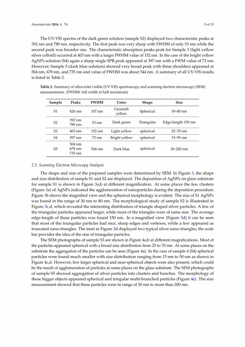
The UV-VIS spectra of the dark green solution (sample S2) displayed two characteristic peaks at392 nm and 789 nm, respectively. The first peak was very sharp with FWHM of only 53 nm while thesecond peak was broader one. The characteristic absorption peaks peak for Sample 3 (light yellowsilver colloid) occurred at 403 nm with a larger FWHM value of 152 nm. In the case of the bright yellowAgNPs solution (S4) again a sharp single SPR peak appeared at 397 nm with a FWM value of 73 nm.However, Sample 5 (dark blue solution) showed very broad peak with three shoulders appeared at504 nm, 678 nm, and 735 nm and value of FWHM was about 544 nm. A summary of all UV-VIS resultsis listed in Table 2.
Table 2. Summary of ultraviolet visible (UV-VIS) spectroscopy and scanning electron microscopy (SEM)measurements. (FWHM: full width at half maximum)
Sample Peaks FWHM Color Shape Size
S1 426 nm 107 nm Greenishyellow Spherical 30–80 nm
S2392 nm
53 nm Dark green Triangular Edge-length 150 nm789 nm
S3 403 nm 152 nm Light yellow spherical 25–70 nm
S4 397 nm 73 nm Bright yellow spherical 15–50 nm
S5504 nm
544 nm Dark blue spherical 30–200 nm678 nm735 nm
2.3. Scanning Electron Micrscopy Analysis
The shape and size of the prepared samples were determined by SEM. In Figure 3, the shapeand size distribution of sample S1 and S2 are displayed. The deposition of AgNPs on glass substratefor sample S1 is shown in Figure 3a,b at different magnification. At some places the few clusters(Figure 3a) of AgNPs indicated the agglomeration of nanoparticles during the deposition procedure.Figure 3b shows the magnified view and the spherical morphology is evident. The size of S1 AgNPswas found in the range of 30 nm to 80 nm. The morphological study of sample S2 is illustrated inFigure 3c,d, which revealed the interesting distribution of triangle shaped silver particles. A few ofthe triangular particles appeared larger, while most of the triangles were of same size. The averageedge-length of these particles was found 150 nm. In a magnified view (Figure 3d) it can be seenthat most of the triangular particles had nice, sharp edges and vertexes, while a few appeared astruncated nano-triangles. The inset in Figure 3d displayed two typical silver nano-triangles; the scalebar provides the idea of the size of triangular particles.
The SEM photographs of sample S3 are shown in Figure 4a,b at different magnifications. Most ofthe particles appeared spherical with a broad size distribution from 25 to 70 nm. At some places on thesubstrate the aggregation of the particles can be seen (Figure 4a). In the case of sample 4 (S4) sphericalparticles were found much smaller with size distribution ranging from 15 nm to 50 nm as shown inFigure 4c,d. However, few larger spherical and near-spherical objects were also present, which couldbe the result of agglomeration of particles at some places on the glass substrate. The SEM photographsof sample S5 showed aggregation of silver particles into clusters and bunches. The morphology ofthese bigger objects appeared spherical and irregular multi-branched particles (Figure 4e). The sizemeasurement showed that these particles were in range of 30 nm to more than 200 nm.

Nanomaterials 2016, 6, 74 6 of 15
Nanomaterials 2016, 6, 74 6 of 16
Figure 3. SEM images of Sample 1 at lower (a) and higher (b) magnification showing the spherical
morphology of the particles. The triangle-shaped silver particles are represented in (c) and (d) at
different magnifications. The inset showed the color of sample S1 and sample S2 solution the beaker.
The SEM photographs of sample S3 are shown in Figure 4a,b at different magnifications. Most
of the particles appeared spherical with a broad size distribution from 25 to 70 nm. At some places
on the substrate the aggregation of the particles can be seen (Figure 4a). In the case of sample 4 (S4)
spherical particles were found much smaller with size distribution ranging from 15 nm to 50 nm as
shown in Figure 4c,d. However, few larger spherical and near-spherical objects were also present,
which could be the result of agglomeration of particles at some places on the glass substrate. The SEM
photographs of sample S5 showed aggregation of silver particles into clusters and bunches. The
morphology of these bigger objects appeared spherical and irregular multi-branched particles (Figure 4e).
The size measurement showed that these particles were in range of 30 nm to more than 200 nm.
Figure 3. SEM images of Sample 1 at lower (a) and higher (b) magnification showing the sphericalmorphology of the particles. The triangle-shaped silver particles are represented in (c) and (d) atdifferent magnifications. The inset showed the color of sample S1 and sample S2 solution the beaker.
Nanomaterials 2016, 6, 74 7 of 16
Figure 4. SEM micrographs of sample S3 (a,b), S4 (c,d), and S5 (e,f) presenting the shape and size of
preapred AgNPs. The inset showed the the color of correspoding colloidal sample.
2.4. X-Ray Diffraction Pattern
In order to obtain the crystalline information of synthesized AgNPs, X-ray powder diffraction
analysis was carried out. The XRD pattern of sample (S1) is shown in Figure 5. Four sharp peaks
appeared at 2θ = 38.5°, 44.7°, 64.7°, and 77.6°, which can be assigned to the (111), (200), (220), and (311)
planes of the face centered cubic (FCC) structure of metallic silver, respectively, according to the
JCPDS File No. 04-0783 [33]. The average crystal size as calculated by the Debye–Scherrer formula
[34] was found d = 40 nm. Furthermore, the most intensive peak located at 2θ = 38.5° corresponding
to the diffractions of spherical nanoparticles crystallized in the FCC structure with basal {111} lattice
plane.
Figure 4. SEM micrographs of sample S3 (a,b), S4 (c,d), and S5 (e,f) presenting the shape and size ofpreapred AgNPs. The inset showed the the color of correspoding colloidal sample.

Nanomaterials 2016, 6, 74 7 of 15
2.4. X-Ray Diffraction Pattern
In order to obtain the crystalline information of synthesized AgNPs, X-ray powder diffractionanalysis was carried out. The XRD pattern of sample (S1) is shown in Figure 5. Four sharp peaksappeared at 2θ = 38.5˝, 44.7˝, 64.7˝, and 77.6˝, which can be assigned to the (111), (200), (220),and (311) planes of the face centered cubic (FCC) structure of metallic silver, respectively, accordingto the JCPDS File No. 04-0783 [33]. The average crystal size as calculated by the Debye–Scherrerformula [34] was found d = 40 nm. Furthermore, the most intensive peak located at 2θ = 38.5˝
corresponding to the diffractions of spherical nanoparticles crystallized in the FCC structure with basal{111} lattice plane.Nanomaterials 2016, 6, 74 8 of 16
Figure 5. Xray difraction (XRD) pattern of sample 1 (S1), showing the face centered cubic (FCC)
crystalline metallic silver nanoparticles (AgNPs). The intesity in vertical axis is mearred in counts per
second (CPS) and diffraction angle (2 theta) measred is taken along horizental axis. The value of
wavelngth (WL in angstrom) is also mentioned in the figure.
2.5. Antibacterial Activity Study
To evaluate the shape and size dependent bactericidal action of produced AgNPs on P.
aeruginosa and E. coli, the disk diffusion method was followed. Sterile paper disks impregnated in
different AgNPs were placed on the nutrient agar plates on which bacteria were spread. After 24 h
of incubating the plates at 37 °C, the resulting growth of bacteria was detected. The obtained results
for bacterial growth on the agar plates in the presence of AgNPs impregnated disks are illustrated in
Figure 6 for P. aeruginosa and in Figure 7 for E. coli. Probably the phase of the growth, in the case of
P. aeruginosa in the first 24 h of incubation, was in the late log phase as reported by Wu, et al. [35]. In
the case of E. coli, the growth phase in the first 24 h of incubation could be a stationary phase [36,37].
In the case of P. aeruginosa the distance (mm) of the first colony formed from the disk was noted and
taken as Zone of Inhibition (ZOI). It can be seen from Figure 6 that in case of negative control (C)
which was pure water impregnated disk, there was a huge growth of bacterial colonies all around
the disk and no distance (0 mm) between disk and the colonies can be seen. In case of antibiotic
Ciprofloxcin (Ab) the distance between the Ab disk and the first bacterial colony was 11.3 mm. The
ZOI for all the AgNPs, S1, S2, S3, S4, and S5 were found to be 1 mm, 3 mm, 1.6 mm, 8 mm, and 0.8
mm, respectively. Thus, the maximum antibacterial activity shown in our assay was that of antibiotic
(11.3 mm) and second highest efficiency was of sample S4 (8 mm), however all types of AgNPs
showed antibacterial action against P. aeruginosa. All the measurements are listed in Table 3. It is
evident from Figure 6 and Table 3 that sample S4 (smaller spherical AgNPs) demonstrated the
strongest bactericidal activity while sample S5 (larger spherical and near spherical AgNPs) showed
the minimum activity. Interestingly, antibacterial performance of triangular AgNPs (S2) against P.
aeruginosa was found to be lesser than that of smallest spherical AgNPs (S4).
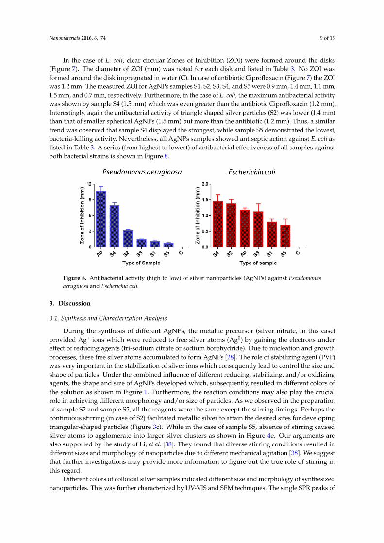
In the case of E. coli, clear circular Zones of Inhibition (ZOI) were formed around the disks
(Figure 7). The diameter of ZOI (mm) was noted for each disk and listed in Table 3. No ZOI was
formed around the disk impregnated in water (C). In case of antibiotic Ciprofloxacin (Figure 7) the
ZOI was 1.2 mm. The measured ZOI for AgNPs samples S1, S2, S3, S4, and S5 were 0.9 mm, 1.4 mm,
1.1 mm, 1.5 mm, and 0.7 mm, respectively. Furthermore, in the case of E. coli, the maximum
antibacterial activity was shown by sample S4 (1.5 mm) which was even greater than the antibiotic
Ciprofloxacin (1.2 mm). Interestingly, again the antibacterial activity of triangle shaped silver
particles (S2) was lower (1.4 mm) than that of smaller spherical AgNPs (1.5 mm) but more than the
antibiotic (1.2 mm). Thus, a similar trend was observed that sample S4 displayed the strongest, while
Figure 5. Xray difraction (XRD) pattern of sample 1 (S1), showing the face centered cubic (FCC)crystalline metallic silver nanoparticles (AgNPs). The intesity in vertical axis is mearred in countsper second (CPS) and diffraction angle (2 theta) measred is taken along horizental axis. The value ofwavelngth (WL in angstrom) is also mentioned in the figure.
2.5. Antibacterial Activity Study
To evaluate the shape and size dependent bactericidal action of produced AgNPs on P. aeruginosaand E. coli, the disk diffusion method was followed. Sterile paper disks impregnated in differentAgNPs were placed on the nutrient agar plates on which bacteria were spread. After 24 h of incubatingthe plates at 37 ˝C, the resulting growth of bacteria was detected. The obtained results for bacterialgrowth on the agar plates in the presence of AgNPs impregnated disks are illustrated in Figure 6 forP. aeruginosa and in Figure 7 for E. coli. Probably the phase of the growth, in the case of P. aeruginosain the first 24 h of incubation, was in the late log phase as reported by Wu, et al. [35]. In the caseof E. coli, the growth phase in the first 24 h of incubation could be a stationary phase [36,37]. In thecase of P. aeruginosa the distance (mm) of the first colony formed from the disk was noted and takenas Zone of Inhibition (ZOI). It can be seen from Figure 6 that in case of negative control (C) whichwas pure water impregnated disk, there was a huge growth of bacterial colonies all around the diskand no distance (0 mm) between disk and the colonies can be seen. In case of antibiotic Ciprofloxcin(Ab) the distance between the Ab disk and the first bacterial colony was 11.3 mm. The ZOI for all theAgNPs, S1, S2, S3, S4, and S5 were found to be 1 mm, 3 mm, 1.6 mm, 8 mm, and 0.8 mm, respectively.Thus, the maximum antibacterial activity shown in our assay was that of antibiotic (11.3 mm) andsecond highest efficiency was of sample S4 (8 mm), however all types of AgNPs showed antibacterialaction against P. aeruginosa. All the measurements are listed in Table 3. It is evident from Figure 6 and

Nanomaterials 2016, 6, 74 8 of 15
Table 3 that sample S4 (smaller spherical AgNPs) demonstrated the strongest bactericidal activity whilesample S5 (larger spherical and near spherical AgNPs) showed the minimum activity. Interestingly,antibacterial performance of triangular AgNPs (S2) against P. aeruginosa was found to be lesser thanthat of smallest spherical AgNPs (S4).
Nanomaterials 2016, 6, 74 9 of 16
sample S5 demonstrated the lowest, bacteria-killing activity. Nevertheless, all AgNPs samples
showed antiseptic action against E. coli as listed in Table 3. A series (from highest to lowest) of
antibacterial effectiveness of all samples against both bacterial strains is shown in Figure 8.
Figure 6. Pseudomonas aeruginosa zones of inhibition (ZOI) around silver nanoparticles (AgNPs)
impregnated disks. The distance of the first colony from the disk/ZOI is demonstrated by arrow
headed lines.
Figure 7. Escherichia coli zones of inhibition (ZOI) around silver nanoparticles (AgNPs) impregnated
disks. The distance of the bacterial lawn from disk/ZOI is demonstrated by red lines.
Table 3. Average zone of inhibition (mm) of silver nanoparticles (AgNPs) against Pseudomonas
aeruginosa (P. aeruginosa) and Escherichia coli (E. coli).
Sample P. aeruginosa E. coli
C 0 0
Ab 11.3 ± 0.8 1.2 ± 0.1
S1 1 ± 0.2 0.9 ± 0.15
S2 3 ± 0.2 1.4 ± 0.2
S3 1.6 ± 0.1 1.1 ± 0.35
S4 8 ± 0.5 1.5 ± 0.3
S5 0.8 ± 0.1 0.7 ± 0.3
Figure 6. Pseudomonas aeruginosa zones of inhibition (ZOI) around silver nanoparticles (AgNPs)impregnated disks. The distance of the first colony from the disk/ZOI is demonstrated by arrowheaded lines.
Nanomaterials 2016, 6, 74 9 of 16
sample S5 demonstrated the lowest, bacteria-killing activity. Nevertheless, all AgNPs samples
showed antiseptic action against E. coli as listed in Table 3. A series (from highest to lowest) of
antibacterial effectiveness of all samples against both bacterial strains is shown in Figure 8.
Figure 6. Pseudomonas aeruginosa zones of inhibition (ZOI) around silver nanoparticles (AgNPs)
impregnated disks. The distance of the first colony from the disk/ZOI is demonstrated by arrow
headed lines.
Figure 7. Escherichia coli zones of inhibition (ZOI) around silver nanoparticles (AgNPs) impregnated
disks. The distance of the bacterial lawn from disk/ZOI is demonstrated by red lines.
Table 3. Average zone of inhibition (mm) of silver nanoparticles (AgNPs) against Pseudomonas
aeruginosa (P. aeruginosa) and Escherichia coli (E. coli).
Sample P. aeruginosa E. coli
C 0 0
Ab 11.3 ± 0.8 1.2 ± 0.1
S1 1 ± 0.2 0.9 ± 0.15
S2 3 ± 0.2 1.4 ± 0.2
S3 1.6 ± 0.1 1.1 ± 0.35
S4 8 ± 0.5 1.5 ± 0.3
S5 0.8 ± 0.1 0.7 ± 0.3
Figure 7. Escherichia coli zones of inhibition (ZOI) around silver nanoparticles (AgNPs) impregnateddisks. The distance of the bacterial lawn from disk/ZOI is demonstrated by red lines.
Table 3. Average zone of inhibition (mm) of silver nanoparticles (AgNPs) against Pseudomonasaeruginosa (P. aeruginosa) and Escherichia coli (E. coli).
Sample P. aeruginosa E. coli
C 0 0Ab 11.3 ˘ 0.8 1.2 ˘ 0.1S1 1 ˘ 0.2 0.9 ˘ 0.15S2 3 ˘ 0.2 1.4 ˘ 0.2S3 1.6 ˘ 0.1 1.1 ˘ 0.35S4 8 ˘ 0.5 1.5 ˘ 0.3S5 0.8 ˘ 0.1 0.7 ˘ 0.3
Nanomaterials 2016, 6, 74 9 of 15
In the case of E. coli, clear circular Zones of Inhibition (ZOI) were formed around the disks(Figure 7). The diameter of ZOI (mm) was noted for each disk and listed in Table 3. No ZOI wasformed around the disk impregnated in water (C). In case of antibiotic Ciprofloxacin (Figure 7) the ZOIwas 1.2 mm. The measured ZOI for AgNPs samples S1, S2, S3, S4, and S5 were 0.9 mm, 1.4 mm, 1.1 mm,1.5 mm, and 0.7 mm, respectively. Furthermore, in the case of E. coli, the maximum antibacterial activitywas shown by sample S4 (1.5 mm) which was even greater than the antibiotic Ciprofloxacin (1.2 mm).Interestingly, again the antibacterial activity of triangle shaped silver particles (S2) was lower (1.4 mm)than that of smaller spherical AgNPs (1.5 mm) but more than the antibiotic (1.2 mm). Thus, a similartrend was observed that sample S4 displayed the strongest, while sample S5 demonstrated the lowest,bacteria-killing activity. Nevertheless, all AgNPs samples showed antiseptic action against E. coli aslisted in Table 3. A series (from highest to lowest) of antibacterial effectiveness of all samples againstboth bacterial strains is shown in Figure 8.Nanomaterials 2016, 6, 74 10 of 16
Figure 8. Antibacterial activity (high to low) of silver nanoparticles (AgNPs) against Pseudomonas
aeruginosa and Escherichia coli.
3. Discussion
3.1. Synthesis and Characterization Analysis
During the synthesis of different AgNPs, the metallic precursor (silver nitrate, in this case)
provided Ag+ ions which were reduced to free silver atoms (Ag0) by gaining the electrons under effect
of reducing agents (tri-sodium citrate or sodium borohydride). Due to nucleation and growth
processes, these free silver atoms accumulated to form AgNPs [28]. The role of stabilizing agent (PVP)
was very important in the stabilization of silver ions which consequently lead to control the size and
shape of particles. Under the combined influence of different reducing, stabilizing, and/or oxidizing
agents, the shape and size of AgNPs developed which, subsequently, resulted in different colors of
the solution as shown in Figure 1. Furthermore, the reaction conditions may also play the crucial role
in achieving different morphology and/or size of particles. As we observed in the preparation of
sample S2 and sample S5, all the reagents were the same except the stirring timings. Perhaps the
continuous stirring (in case of S2) facilitated metallic silver to attain the desired sites for developing
triangular-shaped particles (Figure 3c). While in the case of sample S5, absence of stirring caused
silver atoms to agglomerate into larger silver clusters as shown in Figure 4e. Our arguments are also
supported by the study of Li, et al. [38]. They found that diverse stirring conditions resulted in
different sizes and morphology of nanoparticles due to different mechanical agitation [38]. We
suggest that further investigations may provide more information to figure out the true role of
stirring in this regard.
Different colors of colloidal silver samples indicated different size and morphology of
synthesized nanoparticles. This was further characterized by UV-VIS and SEM techniques. The single
SPR peaks of sample S1, S3, and S4 (Figure 2) at 426 nm, 403 nm, and 397 nm, respectively, depicted
the spherical AgNPs with sizes S1 > S3 > S4. This was confirmed by SEM analysis of the samples that
all three samples were almost spherical morphology with size distribution slightly different as
suggested by UV-VIS spectra. Apparently the size measured from the SEM images seems larger than
predicted by UV-VIS spectra. For example in the case of sample S1 and sample S4, the characteristics
peaks at 426 nm and 397 nm predicted the size of AgNPs 45 nm and 10 nm, respectively, as reported
by Bastus, et al. [7]. In our case the larger size of AgNPs appeared in SEM photographs could be due
to the possible agglomeration of the particles on the glass substrate during the deposition process.
In case of sample S2 (dark green solution), instead of a single peak, two characteristics peaks
appeared at 397 nm and 789 nm indicating the anisotropic morphology of the AgNPs. These peaks
suggested the induced polarizations; peak at 397 nm indicated the out-of-plane dipole resonance
while peak at 789 nm designated in-plane dipole plasmon resonance. These peaks suggested the
triangular shaped structure formation. Moreover the peak at around 789 nm indicated the perfect
sharpness of vertexes of our silver triangle shaped particles [39]. The SEM results confirmed the UV-
VIS interpretation for sample S2 revealing the triangular silver particles with sharp tips (Figure 3c,d).
The sample S5, showed a very wide peak with contours appearing at 504 nm, 678 nm, and 735 nm.
Figure 8. Antibacterial activity (high to low) of silver nanoparticles (AgNPs) against Pseudomonasaeruginosa and Escherichia coli.
3. Discussion
3.1. Synthesis and Characterization Analysis
During the synthesis of different AgNPs, the metallic precursor (silver nitrate, in this case)provided Ag+ ions which were reduced to free silver atoms (Ag0) by gaining the electrons undereffect of reducing agents (tri-sodium citrate or sodium borohydride). Due to nucleation and growthprocesses, these free silver atoms accumulated to form AgNPs [28]. The role of stabilizing agent (PVP)was very important in the stabilization of silver ions which consequently lead to control the size andshape of particles. Under the combined influence of different reducing, stabilizing, and/or oxidizingagents, the shape and size of AgNPs developed which, subsequently, resulted in different colors ofthe solution as shown in Figure 1. Furthermore, the reaction conditions may also play the crucialrole in achieving different morphology and/or size of particles. As we observed in the preparationof sample S2 and sample S5, all the reagents were the same except the stirring timings. Perhaps thecontinuous stirring (in case of S2) facilitated metallic silver to attain the desired sites for developingtriangular-shaped particles (Figure 3c). While in the case of sample S5, absence of stirring causedsilver atoms to agglomerate into larger silver clusters as shown in Figure 4e. Our arguments arealso supported by the study of Li, et al. [38]. They found that diverse stirring conditions resulted indifferent sizes and morphology of nanoparticles due to different mechanical agitation [38]. We suggestthat further investigations may provide more information to figure out the true role of stirring inthis regard.
Different colors of colloidal silver samples indicated different size and morphology of synthesizednanoparticles. This was further characterized by UV-VIS and SEM techniques. The single SPR peaks of
Nanomaterials 2016, 6, 74 10 of 15
sample S1, S3, and S4 (Figure 2) at 426 nm, 403 nm, and 397 nm, respectively, depicted the sphericalAgNPs with sizes S1 > S3 > S4. This was confirmed by SEM analysis of the samples that all threesamples were almost spherical morphology with size distribution slightly different as suggested byUV-VIS spectra. Apparently the size measured from the SEM images seems larger than predictedby UV-VIS spectra. For example in the case of sample S1 and sample S4, the characteristics peaksat 426 nm and 397 nm predicted the size of AgNPs 45 nm and 10 nm, respectively, as reported byBastus, et al. [7]. In our case the larger size of AgNPs appeared in SEM photographs could be due tothe possible agglomeration of the particles on the glass substrate during the deposition process.
In case of sample S2 (dark green solution), instead of a single peak, two characteristics peaksappeared at 397 nm and 789 nm indicating the anisotropic morphology of the AgNPs. These peakssuggested the induced polarizations; peak at 397 nm indicated the out-of-plane dipole resonancewhile peak at 789 nm designated in-plane dipole plasmon resonance. These peaks suggested thetriangular shaped structure formation. Moreover the peak at around 789 nm indicated the perfectsharpness of vertexes of our silver triangle shaped particles [39]. The SEM results confirmed the UV-VISinterpretation for sample S2 revealing the triangular silver particles with sharp tips (Figure 3c,d).The sample S5, showed a very wide peak with contours appearing at 504 nm, 678 nm, and 735 nm.The lack of sharpness in the peaks suggested the irregular larger silver objects as was seen in SEMimages (Figure 4e).
The XRD results of sample 1 showed the lattice parameter 4.06 Å which is close to the literaturevalue 4.086 Å [30]. The crystallite size of S1 was found 40 nm and SEM results also confirmed thataverage size measured was 45 nm.
3.2. Effect of Shape and Size on Antibacterial Activity
We evaluated the antibacterial performance of AgNPs samples against P. aeruginosa and E. coli.It can be seen from the antibacterial activity analysis that all AgNPs demonstrated the bactericidalfunction against both bacterial strains (Figures 6 and 7). However, it is evident as illustrated in Figure 8that the antibacterial efficiency of all silver samples against E. coli was low as compared to P. aeruginosa.This indicated that the P. aeruginosa was more susceptible than E. coli. Sample S4, the smallest-sizedspherical AgNPs, exhibited the maximum bactericidal efficacy against both bacterial strains whilesample S2, the triangular AgNPs, showed the second highest antibacterial activity in both bacterialstudies. Furthermore, antibacterial performance of samples S4 and S2 were observed to be evenbetter than Ciprofloxacin (Ab), which suggests that silver nanoparticles can be a good alternative forantibiotics which have resulted in greater bacterial resistance. Interestingly, the overall antibacterialefficiency trend of all five samples against P. aeruginosa and E. coli was found similar and can be listedas S4 > S2 > S3 > S1 > S5 (Table 3, Figure 8). This indicated that the smallest-sized spherical AgNPs (S4)were more efficient to kill and destroy both types of bacteria as compared to larger spherical AgNPs(S3, S1, and S5). When paper disks were impregnated with colloidal silver particles of different sizeand shape, the rate of dissolution of silver cations for various particles was different. Due to the highsurface to volume ratio, the smaller-sized nanoparticles released more silver cations and, thus, provedmore effective to kill the bacteria as compared to larger-sized particles. These results are in accordancewith already reported outcomes [2,27].
AgNPs may interact with microorganisms in many ways to damage them. For example, AgNPsmay release silver ions when come in contact with bacterial cells. These ions may affect the bacterialDNA replication functions; deactivating the production of some enzymes and cellular proteinsnecessary for adenosine tri-phosphate (ATP) synthesis [2,15]. Furthermore, silver ions may disrupt therespiratory chain by disturbing the working of membrane-bound enzymes [40]. The smaller sphericalAgNPs showed better inhibitory action because a significantly large surface area was in contact withthe bacterial effluent owing to the larger surface to volume ratio as compared to larger spherical AgNPs.Thus, smaller particles released more silver ions than larger particles to kill more bacteria [15,41].

Nanomaterials 2016, 6, 74 11 of 15
Surprisingly, the triangle shaped silver particles (S2) demonstrated less antibacterial activity thanthat of the smaller spherical particles (S4). Apparently, our results seem different than those reportedby Pal, et al. [15] and Dong, et al. [19] who described the strongest antibacterial activity of triangularshaped silver nonparties as compared to spherical ones. They argued that high reactivity of triangularAgNPs was due to their geometrical structure and {111} crystal planes. These high-atomic-density{111} facets lead to maximum antibacterial productivity.
The higher biocidal efficacy of our smaller spherical AgNPs (S4) as compared to our triangularAgNPs (S2) can be explained in various ways. For example, in the XRD pattern (Figure 5) of sphericalAgNPs, the most intense diffraction peak appeared at 2θ = 38.5˝ from the {111} lattice plane indicatedthat spherical AgNPs had the top basal plane with {111} facets. This suggested that the smallerspherical AgNPs (S4) might also have the high-atomic-density {111} facets which acted as activesites. Thus, large surface to volume ratio and high-atomic-density {111} facets perhaps enhanced thebacterial killing efficiency of S4 as compared to S2. Moreover, the smaller-sized spherical AgNPs(S4) were more effective to penetrate inside the bacteria as compared to the larger triangular-shapedAgNPs [42]. Inside the bacteria, the spherical AgNPs, being a soft acid, probably interacted anddestroyed the sulfur- and phosphorus-containing complexes (soft bases) like DNA, and also disruptedthe morphology of the membrane, finally leading to the cell death [2,15,26,40].
Although our results demonstrated that smallest sized spherical AgNPs were the best antibacterialagents among triangular and larger spherical AgNPs, nevertheless, we suggest more investigations tofully explore the shape- and size-dependent biocidal activity of AgNPs because the role of effectivesurface areas of different geometries is still not fully understood [15]. We hope that our study onsize- and shape-dependent bactericidal efficacy could facilitate a new paradigm for considering thetrue role of AgNPs as antimicrobial agents in drug formulation.
4. Materials and Methods
4.1. Materials
Silver Nitrate (AgNO3, Molecular weight (Mw): 169.87 g/mol), tri-sodium citrate (Na3C6H5O7,Mw: 294.10 g/mol), sodium borohydride (NaBH4, Mw: 37.83 g/mol), hydrogen peroxide (H2O2 30%),and polyvinylpyrrolidone (PVP; Mw: 1,300,000) were of analytical grade from Merck (Darmstadt,Germany). Ciprofloxacin (Bayer-Leverkusen, Germany) was purchased from a local medical store.Highly-purified deionized water was used throughout the experiment. The bacterial strains used forantibacterial activity was obtained from Department of Microbiology and Molecular Genetics (MMG),University of the Punjab, Lahore, Pakistan.
4.2. Preparation of Silver Nanoparticles
Five different samples of AgNPs were synthesized by wet chemical reduction methods followingthe procedure of Dong, et al. [19] with some modifications. A summary of experimental detailsincluding reagents with quantities are listed in Table 1.
4.2.1. Sample 1 (S1)
50 mL of 1 mM solution of AgNO3 prepared in water was heated to the boiling temperatureunder vigorous stirring to dissolve completely. With the help of dropper, 5 mL of 1% tri-sodium citrate(Na3C6H5O7) aqueous solution was added dropwise into the boiling AgNO3 solution. The reaction wascompleted at boiling point under constant stirring and refluxing condition. The color of the solutionchanged at different stages during the reaction (as shown in Figure 9) from transparent (Figure 9a),to pale yellow (Figure 9b), to bright yellow (Figure 9c), and finally greenish yellow (Figure 9d) whichindicated the completion of the reaction. The solution was allowed to cool at room temperature understirring. Since the tri-sodium citrate after oxidation became the stabilizer these particles (S1) can alsobe known as citrate-stabilized silver nanoparticles.

Nanomaterials 2016, 6, 74 12 of 15
Nanomaterials 2016, 6, 74 12 of 16
Although our results demonstrated that smallest sized spherical AgNPs were the best
antibacterial agents among triangular and larger spherical AgNPs, nevertheless, we suggest more
investigations to fully explore the shape- and size-dependent biocidal activity of AgNPs because the
role of effective surface areas of different geometries is still not fully understood [15]. We hope that
our study on size- and shape-dependent bactericidal efficacy could facilitate a new paradigm for
considering the true role of AgNPs as antimicrobial agents in drug formulation.
4. Materials and Methods
4.1. Materials
Silver Nitrate (AgNO3, Molecular weight (Mw): 169.87 g/mol), tri-sodium citrate (Na3C6H5O7,
Mw: 294.10 g/mol), sodium borohydride (NaBH4, Mw: 37.83 g/mol), hydrogen peroxide (H2O2 30%),
and polyvinylpyrrolidone (PVP; Mw: 1,300,000) were of analytical grade from Merck (Darmstadt,
Germany). Ciprofloxacin (Bayer-Leverkusen, Germany) was purchased from a local medical store.
Highly-purified deionized water was used throughout the experiment. The bacterial strains used for
antibacterial activity was obtained from Department of Microbiology and Molecular Genetics (MMG),
University of the Punjab, Lahore, Pakistan.
4.2. Preparation of Silver Nanoparticles
Five different samples of AgNPs were synthesized by wet chemical reduction methods
following the procedure of Dong, et al. [19] with some modifications. A summary of experimental
details including reagents with quantities are listed in Table 1.
4.2.1. Sample 1 (S1)
50 mL of 1 mM solution of AgNO3 prepared in water was heated to the boiling temperature
under vigorous stirring to dissolve completely. With the help of dropper, 5 mL of 1% tri-sodium
citrate (Na3C6H5O7) aqueous solution was added dropwise into the boiling AgNO3 solution. The
reaction was completed at boiling point under constant stirring and refluxing condition. The color of
the solution changed at different stages during the reaction (as shown in Figure 9) from transparent
(Figure 9a), to pale yellow (Figure 9b), to bright yellow (Figure 9c), and finally greenish yellow (Figure 9d)
which indicated the completion of the reaction. The solution was allowed to cool at room temperature
under stirring. Since the tri-sodium citrate after oxidation became the stabilizer these particles (S1)
can also be known as citrate-stabilized silver nanoparticles.
Figure 9. Color changing of sample 1 at different phases of the reaction. (a) Transparent color
appeared on dissolving silver nitrate into the water to form silver ions at boiling temperature under
continuous stirring; (b) light yellow color indicated the reduction of silver ions into very small silver
particles after the addition tri-sodium citrate; (c) bright yellow color depicted the formation of larger
silver particles from the smaller ones; (d) finally greenish yellow color revealed the completion of the
reaction when all silver ions had been reduced into the elemental silver nanoparticles by the tri-sodium
citrate.
4.2.2. Sample 2 (S2)
Figure 9. Color changing of sample 1 at different phases of the reaction. (a) Transparent color appearedon dissolving silver nitrate into the water to form silver ions at boiling temperature under continuousstirring; (b) light yellow color indicated the reduction of silver ions into very small silver particles afterthe addition tri-sodium citrate; (c) bright yellow color depicted the formation of larger silver particlesfrom the smaller ones; (d) finally greenish yellow color revealed the completion of the reaction whenall silver ions had been reduced into the elemental silver nanoparticles by the tri-sodium citrate.
4.2.2. Sample 2 (S2)
First of all 1 mL of 5 mM AgNO3 solution was added in 50 mL deionized water during stirring(400 rpm), then 0.5 mL of 1 mM PVP (Mw: 1,300,000) was added in above solution at room temperature.After 10 min 3 mL of 30 mM tri-sodium citrate and 0.2 mL of hydrogen peroxide was added underconstant stirring. After 30 s 0.5 mL of 50 mM NaBH4 was added. After about 30 min, the solutionchanged from faint yellow to dark green color. The reaction was continued for 5 h under constantstirring (400 rpm).
4.2.3. Sample 3 (S3)
50 mL of 2 mM NaBH4 aqueous solution was prepared and ice cooled under constant stirring(400 rpm) for 30 min. 2 mL of 1 mM AgNO3 solution was then added dropwise with the help ofdropper at the rate of one drop per second. Stirring was stopped as soon as all the AgNO3 was addedin the solution. The whole reaction was carried out at room temp.
4.2.4. Sample 4 (S4)
0.5 mL of 30 mM tri-sodium citrate was added into 50 mL deionized water under constant stirring(400 rpm) at room temp and allowed to dissolve it completely. Afterwards 1 mL of 5 mM AgNO3 wasadded to above solution. Before adding freshly prepared 0.5 mL of 50 mM NaBH4 aqueous solutionquickly, the stirring was stopped. The color of the solution changed to light yellow. After 30 s, 0.5 mLof 1 mM PVP (Mw: 1,300,000) aqueous solution was added and reaction continued for another 30 min.The color turned into bright yellow at the completion of reaction.
4.2.5. Sample 5 (S5)
All procedures and reagents were the same as in the case of sample 2 synthesis. Only reactionconditions were changed, the whole experiment was carried out in the dark, and at the stage whenNaBH4 was added the stirring was stopped before adding NaBH4. After 30 min, the color of thesolution changed from faint yellow to dark blue. The reaction was allowed to continue for 5 h.
4.3. Characterization of Prepared Silver Nanoparticle Samples
To determine various characteristic of formulated AgNPs, different techniques were used.To study the optical absorption properties of different silver colloids, ultraviolet–visible spectroscopy(Nicolet, Evolution 300, Thermo Electron Corporation, Waltham, MA, USA) was used at roomtemperature in air. We used the X-ray powder diffractometer (Model: D-maxIIA, Rigaku, Tokyo, Japan)

Nanomaterials 2016, 6, 74 13 of 15
to analyze the structural properties and crystallite size. For the XRD sample, a few droplets of a typicalsample (S1) were dried on the glass substrate to form a thick film. The size and shape analysis ofprepared AgNPs was carried out by scanning electron microscope (Model: S3400N, Hitachi, Tokyo,Japan). For the SEM samples, different approaches were practiced to deposit silver nanoparticles onthe glass substrate, such as the drop casting method and substrate immersion method.
4.4. Antibacterial Activity Tests
The antibacterial susceptibility of prepared AgNPs against two Gram-negative bacterial strains;P. aeruginosa and E. coli was evaluated by disk diffusion/Kibry–Bauer method (17). Briefly, a 100 µLsample of freshly-grown bacterial suspension (with a concentration of ~104 and ~106 colony formingunit (CFU)/mL of P. aeruginosa and E. coli, respectively) cultured in LB (Luria Bertani) was spread onthe nutrient agar plates. Small sterile paper disks of uniform size (10 mm) were impregnated with asprepared AgNPs colloidal samples and then placed on the nutrient agar plates. Disks impregnatedwith Ciprofloxacin and pure water were also placed on nutrient agar for positive (Ab) and negative (C)controls, respectively. Plates were then incubated at 37 ˝C for 24 h. The resulting bacterial colonies’distance/inhibition zones around the disks were then recorded.
5. Conclusions
We successfully prepared triangular AgNPs and spherical nanoparticles of different sizes.The characterization of the prepared nanoparticles was carried out by SEM, UV-VIS. and XRD. UV-VISprovided the morphological and size information by absorption spectra. SEM images confirmedspherical and triangular shapes of AgNPs. XRD indicated the FCC crystalline structure of preparedAgNPs. The antibacterial inhibition tests showed that all of our AgNPs were toxic to both P. aeruginosaand E. coli and their antibacterial efficacy was found size and shape dependent. The smaller-sizedspherical AgNPs demonstrated higher antiseptic efficacy than that of triangular AgNPs, whereas largerspherical AgNPs were found less efficient in bactericidal action than triangle shaped AgNPs againstboth bacterial strains. Two of our samples, S2 and S4, showed more bactericidal activity against E. colithan Ciprofloxacin, which suggests that AgNPs with optimized size and shape could be a potentialalternative for antibiotics which have encountered more bacterial resistance.
Acknowledgments: We highly acknowledged Centre of Excellence in Solid State Physics, University of the Punjab,Lahore-54590, Pakistan for the financial support. The authors are grateful to Department of Environmental Science,Lahore College for Women University, Lahore-54000, Pakistan for providing the UV-VIS Spectroscopy facility.The authors also acknowledge the Department of MicroBiology & Molecular Genetics, University of the Punjab,QAC, Lahore-54590, Pakistan for providing the bacterial stains.
Author Contributions: All the authors contributed in the preparation of this manuscript. Shahzad Naseem,and Saira Riaz, provided the required chemicals, conducted the SEM and XRD of the samples and contributed indata analysis and discussion. Anjum Nasim Sabri provided the bacterial strains and helped in antibacterialactivity analysis. Anum Rauf and Muhammad Akram Raza prepared the AgNPs. Zakia Kanwal andAnum Rauf performed the antibacterial activity experiments. Muhammad Akram Raza and Zakia Knawalwrote the manuscript.
Conflicts of Interest: The authors declare no conflict of interest.
Abbreviations
The following abbreviations are used in this manuscript:
AgNPs Silver nanoparticlesTSC Tri-sodium citrateSEM Scanning electron microscopyXRD X-ray DiffractionZOI Zone of InhibitionFCC Face Centered Cubic

Nanomaterials 2016, 6, 74 14 of 15
References
1. Takeshima, T.; Tada, Y.; Sakaguchi, N.; Watari, F.; Fugetsu, B. DNA/Ag nanoparticles as antibacterial agentsagainst gram-negative bacteria. Nanomaterials 2015, 5, 284–297. [CrossRef]
2. Agnihotri, S.; Mukherji, S.; Mukherji, S. Size-controlled silver nanoparticles synthesized over the range5–100 nm using the same protocol and their antibacterial efficacy. RSC Adv. 2014, 4, 3974–3983. [CrossRef]
3. Xu, R.; Wang, D.; Zhang, J.; Li, Y. Size-dependent catalytic activity of silver nanoparticles for the oxidation ofstyrene. Chem. Asian J. 2006, 1, 888–893. [CrossRef] [PubMed]
4. Sun, Y.; Xia, Y. Shape-Controlled Synthesis of Gold and Silver Nanoparticles. Science 2002, 298, 2176–2178.[CrossRef] [PubMed]
5. Pileni, M.-P. Magnetic Fluids: Fabrication, Magnetic Properties, and Organization of Nanocrystals.Adv. Funct. Mater. 2001, 11, 323–336. [CrossRef]
6. Alexander, J.W. History of the medical use of silver. Surg. Infect. 2009, 10, 289–292. [CrossRef] [PubMed]7. Bastus, N.G.; Merkoci, F.; Piella, J.; Puntes, V. Synthesis of highly monodisperse citrate-stabilized silver
nanoparticles of up to 200 nm: Kinetic control and catalytic properties. Chem. Mater. 2014, 26, 2836–2846.[CrossRef]
8. Iravani, S.; Korbekandi, H.; Mirmohammadi, S.V.; Zolfaghari, B. Synthesis of silver nanoparticles: Chemical,physical and biological methods. Res. Pharm. Sci. 2014, 9, 385–406. [PubMed]
9. Panacek, A.; Kolar, M.; Vecerova, R.; Prucek, R.; Soukupova, J.; Krystof, V.; Hamal, P.; Zboril, R.; Kvitek, L.Antifungal activity of silver nanoparticles against Candida spp. Biomaterials 2009, 30, 6333–6340. [CrossRef][PubMed]
10. Galdiero, S.; Falanga, A.; Vitiello, M.; Cantisani, M.; Marra, V.; Galdiero, M. Silver Nanoparticles as PotentialAntiviral Agents. Molecules 2011, 16, 8894–8918. [CrossRef] [PubMed]
11. Chernousova, S.; Epple, M. Silver as antibacterial agent: Ion, nanoparticle, and metal. Angew. Chem. Int. Ed.2012, 51, 2–20. [CrossRef] [PubMed]
12. Feng, Q.L.; Wu, J.; Chen, G.Q.; Cui, F.Z.; Kim, T.N.; Kim, J.O. A mechanistic study of the antibacterial effectof silver ions on Escherichia coli and Staphylococcus aureus. J. Biomed. Mater. Res. 2000, 52, 662–668. [CrossRef]
13. Choi, O.; Deng, K.K.; Kim, N.J.; Ross, L.; Surampalli, R.Y.; Hu, Z.Q. The inhibitory effects of silvernanoparticles, silver ions, and silver chloride colloids on microbial growth. Water Res. 2008, 42, 3066–3074.[CrossRef] [PubMed]
14. Duran, N.; Marcato, P.D.; De Souza, G.I.H.; Alves, O.L.; Esposito, E. Antibacterial effect of silver nanoparticlesproduced by fungal process on textile fabrics and their effluent treatment. J. Biomed. Nanotechnol. 2007,3, 203–208. [CrossRef]
15. Pal, S.; Tak, Y.K.; Song, J.M. Does the Antibacterial Activity of Silver Nanoparticles Depend on the Shape ofthe Nanoparticle? A Study of the Gram-Negative Bacterium Escherichia coli. Appl. Environ. Microbiol. 2007,73, 1712–1720. [CrossRef] [PubMed]
16. Woodrow Wilson Database. An Inventory of Nanotechnology based Consumer Products Currently on theMarket. 2011. Available online: http://www.nanotechproject.org/inventories/consumer/analysis_draft/(accessed on 25 December 2015).
17. Asghari, S.; Johari, S.A.; Lee, J.H.; Kim, Y.S.; Jeon, Y.B.; Choi, H.J.; Moon, M.C.; Yu, I.J. Toxicity of varioussilver nanoparticles compared to silver ions in Daphnia magna. J. Nanobiotechnol. 2012, 10, 1–14. [CrossRef][PubMed]
18. Martinez-Castanon, G.A.; Nino-Martinez, N.; Martinez-Gutierrez, F.; Martinez-Mendoza, J.R.; Ruiz, F.Synthesis and antibacterial activity of silver nanoparticles with different sizes. J. Nanopart. Res. 2008,10, 1343–1348. [CrossRef]
19. Dong, P.V.; Ha, C.H.; Binh, L.T.; Kasbohm, J. Chemical synthesis and antibacterial activity of novel-shapedsilver nanoparticles. Int. Nano Lett. 2012, 2, 1–9.
20. Kruis, F.; Fissan, H.; Rellinghaus, B. Sintering and evaporation characteristics of gas-phase synthesis ofsize-selected PbS nanoparticles. Mater. Sci. Eng. B 2000, 69, 329–334. [CrossRef]
21. Mafune, F.; Kohno, J.; Takeda, Y.; Kondow, T.; Sawabe, H. Structure and stability of silver nanoparticles inaqueous solution produced by laser ablation. J. Phys. Chem. B 2000, 104, 8333–8337. [CrossRef]
22. Abid, J.P.; Wark, A.W.; Brevet, P.F.; Girault, H.H. Preparation of silver nanoparticles in solution from a silversalt by laser irradiation. Chem. Commun. 2002, 7, 792–793. [CrossRef]
Nanomaterials 2016, 6, 74 15 of 15
23. Swathy, B. A Review on Metallic Silver Nanoparticles. IOSR J. Pharm. 2014, 4, 38–44. [CrossRef]24. Mohanpuria, P.; Rana, N.K.; Yadav, S.K. Biosynthesis of nanoparticles: Technological concepts and future
applications. J. Nanopart. Res. 2008, 10, 507–517. [CrossRef]25. Sadeghi, B.; Garmaroudi, F.S.; Hashemi, M.; Nezhad, H.R.; Nasrollahi, A.; Ardalan, S.; Ardalan, S.
Comparison of the anti-bacterial activity on the nanosilver shapes: Nanoparticles, nanorods and nanoplates.Adv. Powder Technol. 2012, 23, 22–26. [CrossRef]
26. Morones, J.R.; Elechiguerra, J.L.; Camacho, A.; Holt, K.; Kouri, J.B.; Ramirez, J.T.; Yacaman, M.J.The bactericidal effect of silver nanoparticles. Nanotechnology 2005, 16, 2346–2353. [CrossRef] [PubMed]
27. Torres, L.A.; Gmez-Quintero, T.J.R.; Padron, G.H.; Santana, F.B.; Hernandez, J.F.; Castano, V.M. Silvernanoprisms and nanospheres for prosthetic biomaterials, IADR/AADR/CADR General Session andExhibition 2013; 03/2013. Available online: https://www.researchgate.net/publication/266784565_Silver_nanoprisms_and_nanospheres_for_prosthetic_biomaterials (accessed on 25 December 2015).
28. El-Kheshen, A.A.; El-Rab, S.F.G. Effect of reducing and protecting agents on size of silver nanoparticles andtheir anti-bacterial activity. Pharma Chem. 2012, 4, 53–65.
29. Albanese, A.; Tang, P.S.; Chan, W.C.W. The effect of nanoparticle size, shape, and surface chemistry onbiological systems. Annu. Rev. Biomed. Eng. 2012, 14, 1–16. [CrossRef] [PubMed]
30. Wiley, B.; Sun, Y.; Mayers, B.; Xi, Y. Shape-controlled synthesis of metal nanostructures: The case of silver.Chem. Eur. J. 2005, 11, 454–463. [CrossRef] [PubMed]
31. Mishra, G.; Singh, D.; Yadawa, P.K. Study of Copper/Palladium Nanoclusters Using Acoustic Particle Sizer.Platin. Met. Rev. 2013, 57, 186–191. [CrossRef]
32. Veerakumar, P.; Lu, Z.Z.; Velayudham, M.; Lu, K.L.; Rajagopal, S. Alumina supported nanorutheniumas efficient heterogeneous catalyst for the selective H2O2 oxidation of aliphatic and aromatic sulfides tosulfoxides. J. Mol. Catal. A Chem. 2010, 332, 128–137. [CrossRef]
33. Agasti, N.; Kaushik, N.K. One pot synthesis of crystalline silver nanoparticles. Am. J. Nanomater. 2014, 2, 4–7.34. Ashraf, R.; Bashir, M.; Raza, M.A.; Riaz, S.; Naseem, S. Effect of calcination on structural and magnetic
properties of Co doped ZnO nanostructures. IEEE Trans. Magn. 2015, 51, 1–4.35. Wu, H.; Lee, B.; Yang, L.; Wang, H.; Givskov, M.; Molin, S.; Høiby, N.; Song, Z. Effects of ginseng on
Pseudomonas aeruginosa motility and biofilm formation. FEMS Immunol. Med. Microbiol. 2011, 62, 49–56.[CrossRef] [PubMed]
36. Cloning & Expression of sMMO. Available online: http://2014.igem.org/Team:Braunschweig/Results-content(accessed on 16 February 2016).
37. Registery of Standard Bilogical Parts. Available online: http://parts.igem.org/File:2014SDUGrowth_curve_%28WT,_OneProt, _Empty_vector%29.png (accessed on 16 February 2016).
38. Li, D.; Kaner, R.B. Shape and Aggregation Control of Nanoparticles: Not Shaken, Not Stirred. J. Am. Chem. Soc.2006, 128, 968–975. [CrossRef] [PubMed]
39. Jin, R.; Cao, Y.W.; Mirkin, C.A.; Kelly, K.L.; Schatz, G.C.; Zheng, J.G. Photoinduced Conversion of SilverNanospheres to Nanoprisms. Science 2001, 294, 1901–1903. [CrossRef] [PubMed]
40. Bragg, P.D.; Rainnie, D.J. The effect of silver ions on the respiratory chains of Escherichia coli. Can. J. Microbiol.1974, 20, 883–889. [CrossRef] [PubMed]
41. Xiu, Z.; Zhang, Q.; Puppala, H.L.; Colvin, V.L.; Alvarez, P.J.J. Negligible Particle-Specific AntibacterialActivity of Silver Nanoparticles. Nano Lett. 2012, 12, 4271–4275. [CrossRef] [PubMed]
42. Tak, Y.K.; Pal, S.; Naoghare, P.K.; Rangasamy, S.; Song, J.M. Shape-dependent skin penetration of silvernanoparticles: Does it really matter. Sci. Rep. 2015, 5. [CrossRef] [PubMed]
© 2016 by the authors; licensee MDPI, Basel, Switzerland. This article is an open accessarticle distributed under the terms and conditions of the Creative Commons Attribution(CC-BY) license (http://creativecommons.org/licenses/by/4.0/).


![Research Article Antibacterial Activity of pH-Dependent ...media recording, optics, catalysis, and environmental reme-diation [ ]. Traditionally silver has been used in customary medicine](https://static.fdocuments.in/doc/165x107/60d1f2389de17d1b7e6bfb11/research-article-antibacterial-activity-of-ph-dependent-media-recording-optics.jpg)