Six new taxa of subarctic Parmales (Chrysophyceae)ina.tmsoc.org/JNR/online/29/Konno et al 2007 JNR...
21
1. Introduction Probably due to their small size (2-5µm), the Parmales escaped detection for most of the 20th Century. However, with the advent of the electron microscope, scientists began to record the presence of ‘siliceous cysts’ in marine water samples (Iwai & Nishida, 1976; Nishida, 1979, 1986; Booth et al., 1980, 1981, 1982; Silver et al., 1980; Takahashi et al., 1986) and of incomplete cysts and isolat- ed siliceous plates in zooplankton faecal pellets (Urban et al., 1993), sediment traps (Ohyama & Jordan, 2002, unpubl. data) and marine sediments (Stradner & Allram, 1982; Franklin & Marchant, 1995; Zielinski, 1997; Thorn, 2004). Booth & Marchant (1987) formally identified these siliceous cysts as belonging to a new order, the Parmales, and assigned them to the Class Chrysophyceae. The previous year, Marchant & McEldowney (1986) had sectioned several of these siliceous cysts and demonstrat- ed that they were algae. However, their assignment to the Chrysophyceae remains speculative, since no one has suc- ceeded in culturing them or conducting genetic studies on them directly. Recently, Lovejoy et al. (2006) analysed the genetic diversity of Arctic waters, using 18S rRNA, and noticed several novel sequences. One such cluster was a sister group to the Bolidophyceae, a group closely related to the diatoms (Guillou et al., 1999). Lovejoy et al. (2006) speculated that this cluster represented the Parmales, although clearly more evidence is needed before we can be sure. But if it is true, then Mann & Marchant (1989) may have been right to suggest that a Parmales-like ancestor gave rise to the diatoms, presum- ably some time after the Permian/Triassic mass extinction event (Medlin et al., 1997). However, some of the oldest Parmales fossils are reportedly those in Late Eocene to Early Oligocene (ODP Leg 188, Hole 1166A, 148.11mbsf) and Middle to latest Miocene (ODP Leg 188, Hole 1165B, 169.58-291.28mbsf) sediments from Prydz Bay, Antarctica (Thorn, 2004). Unfortunately, the author did not provide photographs to support the findings, and so micrographs of Parmales specimens from the Middle America Trench slope (DSDP Leg 66, Site 490-1-4, 25- 26cm), thought to be Middle to Late Quaternary (NN20) in age, are currently the oldest piece of evidence from the fossil record (Stradner & Allram, 1982). The exclusively marine Parmales are now well known from polar waters, however, their occasional presence in tropical waters (Silver et al., 1980; Kosman et al., 1993; Bravo-Sierra & Hernández-Becerril, 2003) suggests that their distribution may be worldwide, albeit in restricted habitats. Despite this, the ecology of the Parmales is very poorly known. Several workers had noticed that the Parmales remained covered in plates all year round (e.g. Booth & Marchant, 1987), but Komuro et al. (2005) were perhaps the first to show this clearly, and to mention that they behaved like diatoms, ‘blooming’ in spring, sinking to the pycnocline/nutricline in the summer. A few years earlier, Tanimoto et al. (2003) had shown that, where sub- arctic Pacific waters entered the Bering Sea (i.e. through 108 J. Nannoplankton Res. 29 (2), 2007, pp.108-128 © 2007 International Nannoplankton Association ISSN 1210-8049 Printed by Cambridge University Press, UK Six new taxa of subarctic Parmales (Chrysophyceae) Susumu Konno, Ryoko Ohira Graduate School of Science & Engineering, Yamagata University, 1-4-12 Kojirakawa-machi, Yamagata 990-8560, Japan Chika Komuro Department of Earth & Environmental Sciences, Faculty of Science, Yamagata University, 1-4-12 Kojirakawa-machi, Yamagata 990-8560, Japan Naomi Harada Institute of Observational Research for Global Change, Japan Agency for Marine-Earth Science & Technology, 2-15 Natsushima-cho, Yokosuka 237-0061, Japan Richard W. Jordan* Department of Earth & Environmental Sciences, Faculty of Science, Yamagata University, 1-4-12 Kojirakawa-machi, Yamagata 990-8560, Japan; *[email protected] Manuscript received 12th April, 2007; revised manuscript accepted 14th November, 2007 Abstract The Parmales are an enigmatic group of marine phytoplankton, with siliceous plates of varying mor- phology surrounding their 2-5µm-diameter cells. Although rarely found in sediment traps or underlying sediments, in oceanic surface-waters they often outnumber even the diatoms during the spring months. Since their discovery, several decades ago, in the Antarctic and subarctic Pacific, the Parmales have been found in temperate and tropical regions. Despite the publication of several significant taxonomic papers, a number of Parmales remain without for- mal description. Here we provide descriptions of six new taxa from subarctic waters: Tetraparma catinifera sp. nov., T. gracilis sp. nov., Triparma columacea f. convexa f. nov., T. columacea f. fimbriata f. nov., T. laevis f. inornata f. nov. and T. laevis f. longispina f. nov. In addition, an annotated checklist of all Parmales, including undescribed taxa, is presented for the first time. Distribution maps of all subarctic taxa are also provided for the North Pacific and its marginal seas. Keywords Bering Sea, Chukchi Sea, Japan Sea, Parmales, Sea of Okhotsk, subarctic Pacific, taxonomy
Transcript of Six new taxa of subarctic Parmales (Chrysophyceae)ina.tmsoc.org/JNR/online/29/Konno et al 2007 JNR...

1. IntroductionProbably due to their small size (2-5µm), the Parmalesescaped detection for most of the 20th Century. However,with the advent of the electron microscope, scientistsbegan to record the presence of ‘siliceous cysts’ in marinewater samples (Iwai & Nishida, 1976; Nishida, 1979,1986; Booth et al., 1980, 1981, 1982; Silver et al., 1980;Takahashi et al., 1986) and of incomplete cysts and isolat-ed siliceous plates in zooplankton faecal pellets (Urban etal., 1993), sediment traps (Ohyama & Jordan, 2002,unpubl. data) and marine sediments (Stradner & Allram,1982; Franklin & Marchant, 1995; Zielinski, 1997; Thorn,2004). Booth & Marchant (1987) formally identifiedthese siliceous cysts as belonging to a new order, theParmales, and assigned them to the Class Chrysophyceae.The previous year, Marchant & McEldowney (1986) hadsectioned several of these siliceous cysts and demonstrat-ed that they were algae. However, their assignment to theChrysophyceae remains speculative, since no one has suc-ceeded in culturing them or conducting genetic studies onthem directly. Recently, Lovejoy et al. (2006) analysedthe genetic diversity of Arctic waters, using 18S rRNA,and noticed several novel sequences. One such clusterwas a sister group to the Bolidophyceae, a group closelyrelated to the diatoms (Guillou et al., 1999). Lovejoy et al.(2006) speculated that this cluster represented theParmales, although clearly more evidence is neededbefore we can be sure. But if it is true, then Mann &
Marchant (1989) may have been right to suggest that aParmales-like ancestor gave rise to the diatoms, presum-ably some time after the Permian/Triassic mass extinctionevent (Medlin et al., 1997). However, some of the oldestParmales fossils are reportedly those in Late Eocene toEarly Oligocene (ODP Leg 188, Hole 1166A,148.11mbsf) and Middle to latest Miocene (ODP Leg 188,Hole 1165B, 169.58-291.28mbsf) sediments from PrydzBay, Antarctica (Thorn, 2004). Unfortunately, the authordid not provide photographs to support the findings, andso micrographs of Parmales specimens from the MiddleAmerica Trench slope (DSDP Leg 66, Site 490-1-4, 25-26cm), thought to be Middle to Late Quaternary (NN20)in age, are currently the oldest piece of evidence from thefossil record (Stradner & Allram, 1982).
The exclusively marine Parmales are now well knownfrom polar waters, however, their occasional presence intropical waters (Silver et al., 1980; Kosman et al., 1993;Bravo-Sierra & Hernández-Becerril, 2003) suggests thattheir distribution may be worldwide, albeit in restrictedhabitats. Despite this, the ecology of the Parmales is verypoorly known. Several workers had noticed that theParmales remained covered in plates all year round (e.g.Booth & Marchant, 1987), but Komuro et al. (2005) wereperhaps the first to show this clearly, and to mention thatthey behaved like diatoms, ‘blooming’ in spring, sinkingto the pycnocline/nutricline in the summer. A few yearsearlier, Tanimoto et al. (2003) had shown that, where sub-arctic Pacific waters entered the Bering Sea (i.e. through
108J. Nannoplankton Res. 29 (2), 2007, pp.108-128 © 2007 International Nannoplankton AssociationISSN 1210-8049 Printed by Cambridge University Press, UK
Six new taxa of subarctic Parmales (Chrysophyceae)
Susumu Konno, Ryoko OhiraGraduate School of Science & Engineering, Yamagata University, 1-4-12 Kojirakawa-machi, Yamagata 990-8560, Japan
Chika KomuroDepartment of Earth & Environmental Sciences, Faculty of Science, Yamagata University, 1-4-12 Kojirakawa-machi, Yamagata 990-8560, Japan
Naomi HaradaInstitute of Observational Research for Global Change, Japan Agency for Marine-Earth Science & Technology, 2-15 Natsushima-cho, Yokosuka237-0061, Japan
Richard W. Jordan*Department of Earth & Environmental Sciences, Faculty of Science, Yamagata University, 1-4-12 Kojirakawa-machi, Yamagata 990-8560, Japan;
Manuscript received 12th April, 2007; revised manuscript accepted 14th November, 2007
Abstract The Parmales are an enigmatic group of marine phytoplankton, with siliceous plates of varying mor-phology surrounding their 2-5µm-diameter cells. Although rarely found in sediment traps or underlying sediments,in oceanic surface-waters they often outnumber even the diatoms during the spring months. Since their discovery,several decades ago, in the Antarctic and subarctic Pacific, the Parmales have been found in temperate and tropicalregions. Despite the publication of several significant taxonomic papers, a number of Parmales remain without for-mal description. Here we provide descriptions of six new taxa from subarctic waters: Tetraparma catinifera sp. nov.,T. gracilis sp. nov., Triparma columacea f. convexa f. nov., T. columacea f. fimbriata f. nov., T. laevis f. inornata f.nov. and T. laevis f. longispina f. nov. In addition, an annotated checklist of all Parmales, including undescribed taxa,is presented for the first time. Distribution maps of all subarctic taxa are also provided for the North Pacific and itsmarginal seas.
Keywords Bering Sea, Chukchi Sea, Japan Sea, Parmales, Sea of Okhotsk, subarctic Pacific, taxonomy

the shallow straits of the Aleutian Islands), the Parmaleswere present at the surface during the summer, but awayfrom the Aleutian Islands the Parmales were almostabsent from the subarctic surface-waters. The data ofKomuro et al. (2005) clearly explains why some of theearlier workers found them at deeper depths, while othersfound them at the surface. Their seasonal dataset also con-firms Marchant & McEldowney’s (1986) observation thatthe Parmales are not cysts at all, but the vegetative stageof an alga with a presumably high growth-rate (i.e. high-er than that reported by Taniguchi et al. (1995) from theirbag experiments) and high silica uptake rate.
Bravo-Sierra & Hernández-Becerril (2003) provided alist of extant Parmales taxa, including three genera, eightspecies, four subspecies and one forma. They proposedthat, in the future, the status of the four subspecies bechanged to forma or variety, since phytoplankton workerson other algal groups rarely used subspecies in this way -that is, when only morphological features are used as sep-aration criteria, and life-cycle or interbreeding informa-tion is not known. Although we support this proposal, wedid not encounter any of the four subspecies in this study,and so hesitate to make the changes here. However, someof the new taxa herein are assigned forma status, in recog-nition of their suggestion.
It is now known that the Parmales are a significantcomponent of the phytoplankton community, especially inhigh latitudes, however their enumeration at the species orsubspecific level is currently hampered by an underesti-mation of their diversity. Until this problem is addressed,ecological and biogeographic studies will be difficult toundertake with any confidence. Therefore, in this first tax-onomic paper, we describe most of the subarctic taxa thatare presently without formal names.
2. Material and methodsSamples were collected on three cruises undertaken atvarious times of the year and in different years: KH99-3(July-August, 1999) of the R/V Hakuho Maru, MR00K01(January, 2000) and MR06-04 (August-September, 2006)of the R/V Mirai. Figure 1 shows the station locationsfrom which water-samples were collected and specimensphotographed. The Kyodo North Pacific Ocean Time-Series (KNOT; Kyodo = cooperative in Japanese) stationwas established in 1997 as part of the Joint Global OceanFlux Study, and was visited on 16th August, 1999 duringKH99-3 and 17th January, 2000 during MR00K01.During the first visit, several hydrocasts were carried outover a short time-interval. Samples from two of thesehydrocasts, referred to in the plate captions simply asStations 1 and 2, have been analysed for this study.Samples were also collected from hydrocasts at Stations16 and 17 during KH99-3, and from Stations 3, 4, 6, 7 and12ex on MR06-04. In addition, two surface-water sam-ples (Stations 23 and 24) were obtained while underwayon MR06-04. Vertical water-samples were acquired onshallow hydrocasts (5-300m) using a Conductivity
Temperature Depth (CTD) rig equipped with a rosette ofwater bottles. Surface-waters were collected either bybucket, or using the onboard continuous sea-water supply.For each CTD sample, a suite of hydrographic parameterswas normally measured, while for those taken with theonboard sea-water supply, only temperature and salinitymeasurements were available from instruments connectedto the continuous flow of sea-water.
Water-samples were filtered, prepared for scanningelectron microscopy (SEM), and photographed as detailedin Konno & Jordan (2006). All filter samples, SEM stubs,negatives and scanned images used in this study (includ-ing those associated with the holotypes) are presentlycurated in the Department of Earth & EnvironmentalSciences, Faculty of Science, Yamagata University,Yamagata, Japan.
3. Results3.1 TerminologyIn general, the terminology used in this paper follows thatof Booth et al. (1981) and Booth & Marchant (1987), butalso incorporates the recent findings and recommenda-tions of Konno & Jordan (2007). The latter authorsrevealed that the cell-wall structures of Triparma andTetraparma are more closely related than previouslythought, with the same plate configuration; that is, threegirdle plates, three shield plates, one dorsal plate and oneventral plate. The major differences between the two gen-era are the shape of the dorsal and girdle plates and thesize of the ventral plate. In Tetraparma, the triradiate dor-sal plate is notched, the girdle plates are also triradiate,and the ventral plate is smaller than the shield plates. In
Konno, Ohira, Komuro, Harada, Jordan109
Figure 1: Map showing locations of the sampling stations used in thepresent study (closed circles), and those (open circles) of Tanimoto et al.(2003). Map created using M. Weinelt’s ‘Online Map Creation’ site atwww.aquarius.ifm-geomar.de

Triparma, the triradiate dorsal plate has rounded ends, thegirdle plates are oblong, and the ventral plate is largerthan the shield plates.
3.2 Systematic taxonomyIn this section, six new taxa are described from subarcticwaters. To avoid taxonomic complications in the future,the bibliography for each taxon has been restricted to thesubarctic Pacific and its marginal seas, although it is pre-sumed that specimens occurring in the subarctic NorthAtlantic belong to one or more of the taxa below. Forthose species which are seemingly bipolar (i.e. morpho-logically identical), it is presumed that future studies willdiscover that they are actually cryptic species, as has beenfound in other plankton groups. An annotated checklist ofthe Parmales, complete with dates and authorities, isgiven in the discussion.
The cell and plate dimensions given in the descrip-tions below are of specimens photographed in the presentstudy, while biogeographic references to water-sampleswith ‘NP’ and ‘B’ notations are from Tanimoto et al.(2003). Only one paper provides ultrastructural informa-tion on the Parmales. Marchant & McEldowney (1986)showed that the parmalian cell contains a large chloro-plast and very little storage material, indicating its photo-synthetic tendencies. Furthermore, they showed that itpossesses a chloroplast endoplasmic reticulum, as foundin diatoms and chrysophytes. Thus, it should be noted thatthe inclusion of the Parmales in the Class Chrysophyceaesensu lato is still tentative, pending detailed ultrastructur-al and genetic work.
Class CHRYSOPHYCEAE PascherOrder PARMALES Booth & Marchant emend.
Konno & JordanFamily TRIPARMACEAE Booth & Marchant emend.
Konno & Jordan
Genus Tetraparma Booth in Booth & Marchant emend.Konno & Jordan
Cells planktonic, solitary, non-motile, spherical to sub-spherical. Cells possess eight plates: three shield plates,three triradiate girdle plates, one triradiate dorsal plateand one circular ventral plate. Plate boundaries distinct.All plates are slightly to strongly convex in the centralarea, with or without papillae, and are radially veined withveins dichotomously branching and anastomosingincreasingly toward the margin, forming a wide inner ringof elongate areolae. Arms of triradiate dorsal plate withnotched ends. Marine.
Tetraparma catinifera sp. nov.Pl.1, figs 1-9; Pl.2, figs 1-2
1976 Sp. indet. A Iwai & Nishida: pl.II, fig.1.1980 Siliceous cyst Booth et al.: fig.1(8).1981 Cyst VIII Booth et al.: figs 57-59.
1987 Tetraparma pelagica Booth & Marchant: fig.4.2003 Tetraparma pelagica Booth & Marchant: Tanimoto et al.,
pl.3, fig.6.
Etymology: Catina (L.) meaning bowl, fero (L.) meaningto carry, in reference to the bowl-shaped shield plates.
Cellula solitaria, sphaerica, 2.7-3.3µm diametro.Laminae papillis praesentes, circa 6-12/µm. Papillaepraesentes in seribus radiantibus atque vel concentricis.Laminae processus centralis, sine umbonatae. Laminaeparmae 1.7-2.4µm diametro, margines elevatos. Laminaetriradiatae, brachii 1.3-1.5µm longitudo. Lamina dorsalis1.0-1.4µm longitudo. Lamina ventralis, circa 1.6µmdiametro. Species planctonica marina, ad 44˚N, 155˚E(Statio KNOT). Holotypus, hic designatus: EM StubKNOTJ0030. Iconotypus: Lamina 1, Figura 2.
Description: Cells solitary, spherical, 2.7-3.3µm in diam-eter. Papillae usually present, ca.6-12 papillae/µm. Platesusually with radially arranged papillae between the slit-like areolae, and one or more concentric rows of papillaealong the top of a raised marginal rim. Plates with smallcentral papilla, but no central mound. Shield platesca.1.7-2.4µm in diameter, with a wall-like plate margin.Triradiate girdle plates, arms ca.1.3-1.5µm long (meas-ured along the dorso-ventral plane, from end to centralpapilla). Arms of dorsal plate, ca.1.0-1.4µm long. Ventralplate ca.1.6µm in diameter. Marine, in plankton at 44˚N,155˚E (Station KNOT), 17th January, 2000 (30m). Holotype: EM Stub KNOTJ0030 (specimen in Pl.1,fig.2).Note: In Booth et al. (1981), this form was originally dis-tinguished from T. pelagica (as Cysts VIII and IX, respec-tively), and when the latter species was formallydescribed in Booth & Marchant (1987), the two formswere again distinguished in the text, but both appeared inthe figure captions under the same name. Apart from anotable size difference, T. catinifera differs from T. pelag-ica by having wall-like shield-plate margins of variableheight, lacking the triangular spines at the plate centre,and having a somewhat flattened central area. However,Booth & Marchant (1987, fig.5) showed an intriguingspecimen (collected in May from surface-waters at56˚59’N, 141˚27’W) that seemingly possesses a rimmedshield-plate in addition to typical T. pelagica plates. In thepresent study though, no such specimens have been seen.T. catinifera specimens show a significant amount of mor-phological variation, not just in wall height (compare Pl.1,fig.1 with Pl.2, fig.1), but also in the distribution anddegree of papillation (compare Pl.1, figs 3, 4 with Pl.1,figs 6, 7), and the number of radiating slits in the centralarea (compare Pl.1, fig.5 with Pl.1, fig.7). It should benoted that the girdle and dorsal plates have less prominentwalls than the shield and ventral plates (Pl.1, fig.2), andthat, due to the curvature of the cell, all of the plates areactually convex (Pl.1, fig.9). This species has mostly been
110New taxa of subarctic Parmales

Konno, Ohira, Komuro, Harada, Jordan111
Plate 1
Tetraparma catinifera
Specimen with less prominentpapillae. Ventral plate at bot-tom of photo. Bering Sea,KH99-3, St.16, 50m
Note small ventralplate at bottom ofphoto (arrowed). Sea ofOkhotsk, St.6, 30m
Collapsed cell. Note curvature ofplates. Sea of Okhotsk, St.7, 30m
Collapsed cell. Sea of Okhotsk, St.6, 30m
Cell wall, showing shield (s), girdle(g), dorsal (d) and ventral (v) plates.N Pacific, KNOT St.1, 100m
Specimen clearly showing juncturebetween girdle and dorsal (arrowed)plates. N Pacific, KNOT MR00K01,30m. Holotype
Dorsal plate interlocking with three gir-dle plates. Note plates strongly papillate.Sea of Okhotsk, St.7, 30m
Specimen with clear plate junctures.Note papillae on inside of plate rims.Sea of Okhotsk, St.7, 30m
Specimen with moreradiating slits, lackingpapillae. N Pacific, KH99-3St.17, 50m

recorded in the subarctic Pacific, but also occurs in theGulf of St. Lawrence, Canada (Bérard-Therriault et al.,1999, p.246, pl.113, figs a, e) and in the Chukchi Sea (thisstudy). Biogeography (this study): Sea of Okhotsk - MR06-04St.4, MR06-04 St.6, MR06-04 St.7; NW Pacific - St.KNOT (August, 1999, KH99-3; January, 2000,MR00K01), KH99-3 St.17; NE Pacific - KH99-3 NP15;Bering Sea - MR06-04 St.24, KH99-3 St.16; ChukchiSea - MR060-04 St.12ex.
Tetraparma gracilis sp. nov.Pl.2, figs 3-7
1981 Cyst IX Booth et al.: fig.68?2003 Tetraparma pelagica Booth & Marchant: Tanimoto et al.,
pl.3, fig.7.
Etymology: Gracilis (L.) meaning slender, in reference tothe shape of the central process.
Cellula solitaria, sphaerica, 2.3-2.5µm diametro.Laminae papillis carentes. Processus centralis, 0.7-0.8µmlongitudo, in laminae sine umbonatae. Laminae margineslaevi et leviter elevatiae. Laminae parmae 1.3-1.8µmdiametro. Laminae triradiatae circa 1µm longitudo.Lamina dorsalis circa 1µm longitudo. Lamina ventralis,circa 1.2µm diametro. Species planctonica marina, ad44˚N, 155˚E (Statio KNOT). Holotypus, hic designatus:EM Stub KNOTA9960. Iconotypus: Lamina 2, Figura 4.
Description: Cells solitary, spherical, 2.3-2.5µm in diam-eter. Plates without papillae. All plates lack a centralmound, but have a central process 0.7-0.8µm long.Processes on girdle and dorsal plates slender, those onshield and ventral plates appear bifurcate. Plates with asmooth, slightly raised margin. Shield plates ca.1.3-1.8µm in diameter. Triradiate girdle plates ca.1µm long(measured along the dorso-ventral plane, from end to cen-tral structure). Arms of dorsal plate, ca.1µm long. Ventralplate ca.1.2µm in diameter. Marine, in plankton at 44˚N,155˚E (Station KNOT), 16th August, 1999 (60m). Holotype: EM Stub KNOTA9960 (specimen in Pl.2,fig.4).Note: T. gracilis differs from T. pelagica by seeminglylacking papillae and having a long central process. A sim-ilar specimen was illustrated by Iwai & Nishida (1976,pl.II, fig.7), but the central process on at least some of theplates was cruciate. The processes on the shield and ven-tral plates of some of our specimens appear bifurcate,whilst those on the girdle and dorsal plates are slender(Pl.2, fig.4). Furthermore, in broken specimens, the cen-tral process cross-section appears circular (Pl.2, figs 6, 7),whilst that of T. pelagica (subarctic forms) is elongate(Pl.3, figs 12, 13). Another specimen featured by Nishida(1986, pl.1, fig.4), from the Southern Ocean, has anangled short spine on each plate. Both of these forms
clearly belong to Tetraparma, but are in need of furtherobservations and formal descriptions. T. gracilis has onlybeen recorded in the subarctic Pacific. Biogeography (this study): Sea of Okhotsk - MR06-04St.7; NW Pacific - St. KNOT (August, 1999, KH99-3),KH99-3 NP30, KH99-3 St.17; Bering Sea - MR06-04St.23.
Tetraparma pelagica Booth & MarchantPl.3, figs 1-13
1987 Tetraparma pelagica Booth & Marchant: p.248, figs 2(holotype, from 64˚59.8’S, 83˚02’E; January, surface-water), 3, 5, non fig.4.
1976 Sp. indet. B Iwai & Nishida: pl.II, fig.2.1976 Sp. indet. C Iwai & Nishida: pl.II, fig.3?1979 Genus & species indeterminable Nishida: pl.1, fig.4.1980 Siliceous cyst Booth et al.: fig.1(9).1981 Cyst IX Booth et al.: figs 60-62, 69, non fig.68.2003 Tetraparma pelagica Booth & Marchant: Tanimoto et al.,
pl.3, fig.5, non figs 6, 7.
Description: Cells 1.9-2.5µm in diameter. All plates withor without papillae, ca.12-15 papillae/µm, are radiallyveined, with veins dichotomously branching and anasto-mosing increasingly toward the margin, forming a wideinner ring of elongate areolae. Plates with radially-arranged papillae between the slit-like areolae, and twoconcentric rows of papillae on top of a low marginal rim.Small triangular spine covered by papillae usually presentat plate centre. Shield plates ca.1.3-1.6µm in diameter.Triradiate girdle plates ca.0.8-1.3µm long (measuredalong the dorso-ventral plane from end to central struc-ture). Arms of dorsal plate, ca.0.8µm long. Ventral plateca.1.1-1.8µm in diameter. Marine.Note: T. pelagica was originally described from theAntarctic (Booth & Marchant, 1987), but smaller, seem-ingly indistinguishable, cells are also found in the subarc-tic Pacific and Bering Sea (compare with Findlay, 1998,pl.3, fig.6; Marchant & Scott, 2005, figs 7.5a, b). Our ownunpublished data on Antarctic T. pelagica specimens alsoconform to the original dimensions given by Booth &Marchant (1987). A closer examination of specimensfrom both polar regions may reveal them to be two cryp-tic taxa in the future. In addition, some specimens lackingplate spines, but possessing the other characteristic fea-tures of this species, have been found exclusively in thesubarctic Pacific (e.g. Iwai & Nishida, 1976, pl.II, fig.3,as Sp. indet. C; Nishida, 1979, pl.1, fig.4, as Genus &species indeterminable, and in text-figs 4 and 6 as Indet.B) and the Sea of Okhotsk (Pl.3, figs 10, 11). These non-spiny forms are rare, and presumed to represent the samespecies as the spiny forms. The large ventral plate(1.8µm) mentioned in the above description was meas-ured in ventral view, and so one assumes that shield plateslarger than 1.6µm were present on the other side of thecell. A disarticulated specimen of T. pelagica from DiskoBay, Greenland, clearly shows the size difference between
112New taxa of subarctic Parmales

Konno, Ohira, Komuro, Harada, Jordan113
Plate 2
1-2: Tetraparma catinifera; 3-7: Tetraparma gracilis
Note low-walled rims and less prominent papillae.Bering Sea, St.24, 75m
Note low-walled rims and plates with moreradiating slits. N Pacific, KNOT St.1, 100m
Cell wall showing shield (s), dorsal (d) andgirdle (g) plates. N Pacific, KH99-3 St.17,50m
Cell wall showing all the plate types, including the ventralplate (v). N Pacific, KNOT St.1, 60m. Holotype
Note dorsal/girdle plate juncture(arrowed). Bering Sea, St.23,10m
Note strong curvature of shieldplate in upper left corner. Sea ofOkhotsk, St.7, 30m
Note circular hole left by brokenspine, and ventral plate on left-hand side. Sea of Okhotsk, St.7,30m

114
Plate 3
Tetraparma pelagica
Specimen clearly showing dorsalplates (centre). N Pacific, KH99-3St.17, 50m
Bering Sea, St.23, 0m Bering Sea, St.23, 30m Bering Sea, St.23, 10m
Bering Sea, St.24, 0m
Note plates seemingly lackspines. Sea of Okhotsk,St.7, 30m
Note shape of aperture left bybroken spine on girdle plate, andsmall ventral plate on left. Sea ofOkhotsk, St.7, 30m
Note shape of aperture left by brokenspine on shield plate. Sea of Okhotsk,St.7, 30m
Sea of Okhotsk, St.7,30m
N Pacific, KH99-3 St.17,50m
Note greater degree ofornamentation. Sea ofOkhotsk, St.4, 30m
Note plates seeminglylack spines. Sea ofOkhotsk, St.7, 30m
New taxa of subarctic Parmales
Cell wall, showing shield (s),girdle (g), dorsal (d) and ventral(v) plates. Bering Sea, St.24, 0m

Konno, Ohira, Komuro, Harada, Jordan115
Plate 4
Triparma columacea f. fimbriata
Girdle view. Bering Sea, St.23, 0m
Cell wall with one shield-plate missing. Note ventral platein bottom right corner. Bering Sea, St.23, 30m
Ventral view showing ventral plate. Bering Sea,St.23, 30m
Cell wall showing girdle (g), shield (s) anddorsal (d) plates (ventral plate not visible). NPacific, KNOT St.1, 125m. Holotype
Ventral view showing ventral plate (v). Bering Sea, St.24, 10m
Ventral view. Note upper girdleplates appear interlocked. BeringSea, St.24, 30m
Ventral view showing ventral plate.Bering Sea, St.24, 50m

the shield plates and the ventral plate (Kosman et al.,1993, fig.31).Biogeography (this study): Sea of Okhotsk - MR06-04St.4, MR06-04 St.7; NW Pacific - St. KNOT (August,1999, KH99-3), KH99-3 St.17; Bering Sea - KH99-3St.B5, MR06-04 St.23, MR06-04 St.24.
Genus Triparma Booth & Marchant emend. Konno &Jordan
Cells planktonic, solitary, non-motile, spherical to sub-spherical (excluding extensions). Cells possess eightplates: three shield plates, three oblong girdle plates, onetriradiate dorsal plate, and one circular ventral plate. Plateboundaries distinct. Arms of triradiate dorsal plate withrounded or slightly squarish ends. Marine.
Triparma columacea f. convexa f. nov.Pl.5, figs 1-4
1976 Sp. indet. E Iwai & Nishida: pl.II, fig.5.1979 Genus & species indeterminable (Indet. A) Nishida: text-
figs 4, 6, pl.1, fig.3.1981 Cyst VI Booth et al.: pp.71, 74, figs 47-49.2003 Triparma columacea Booth: Tanimoto et al., pl.3, fig.4.
Etymology: Convexa (L.) meaning convex, in referenceto the shape of the shield plates.
Cellula 1.8-3.3µm diametro. Laminae parmae 1.1-1.7µmdiametro, convexae. Laminae oblongae 1.6-2.1µm longi-tudo, carina fere conspicua et undulata in aspectu cingu-laris, seriebus unabus areolae elongatae in utraque lateroet perpendicularibus carinam. Lamina dorsalis brachii1.0-1.5µm longitudo, fere rotundis extremis. Lamina dor-salis cum foramina centrica, carina triradiata, areolaeelongatae in unibus serie perpendicularibus ad quemquemarginem et formantes latera carinam, atque seribuspluribus ex areolae parvae in extremum utraque bracho.Lamina ventralis incognita. Planctonica marina, ad60˚N, 179˚W (Mare Bering). Holotypus, hic designatus:EM Stub BS062410. Iconotypus: Lamina 5, Figura 1.
Description: Cells 1.8-3.3µm in diameter. All plates withcoarse venation, without ornamentation. Shield platesca.1.1-1.7µm in diameter, convex; radially veined, withveins dichotomously branching and anastomosingincreasingly toward the margin forming a wide inner ringof elongate areolae and a narrow outer ring of compactareolae. Oblong girdle plates ca.1.6-2.1µm long, withkeel more or less pronounced, undulating in girdle view,with a single row of elongate areolae on each side of thekeel and perpendicular to it. Arms of dorsal plate, ca.1.0-1.5µm long with slightly rounded ends. Dorsal plate withcentral indistinct hole, triradiate keel with a single row ofelongate areolae perpendicular to each margin formingthe sides of the keel, and with several rows of small areo-lae at the end of each arm. Ventral plate unknown. Marine,
in plankton at 60˚N, 179˚W, Bering Sea (St.24, 10m). Holotype: EM Stub BS062410 (specimen in Pl.5, fig.1).Note: Kosman et al. (1993) have already suggested that itwould be reasonable to describe this taxon as a new form(their p.119). T. columacea f. convexa is clearly differentfrom the type (and its currently described forms) as it pos-sesses convex shield-plates rather than flattened ones(Pl.5, fig.1). Also, there appears to be a difference in theshape of the ends of the dorsal plate (more squarish in f.convexa, distinctly rounded in the type: Pl.5, fig.2). In theoriginal description of T. columacea, the dorsal plate isdescribed as having a hole in the middle, and with smallholes at the end of each arm. Although not clear from ourmicrographs, it is possible to see these structures in somespecimens. Furthermore, f. convexa appears to have onlyone row of areolae either side of the keel (as in f. alataMarchant and f. fimbriata), while there are two in thetype. Booth & Marchant (1987, p.251) mentioned thatsimilar forms had been seen in the Bay of Bothnia inwaters of 5-6 PSU (H. Thomsen, pers. comm. in Booth &Marchant, 1987). In a later paper, Kosman et al. (1993)showed photographs of specimens of T. cf. T. columaceafrom Denmark and Finland, but this Baltic Sea form,although possessing convex shield-plates, had a muchmore bulky and less spherical cell shape, due to thestrongly-keeled girdle-plates. In our opinion, the twoforms are different, and both clearly deserve to be separat-ed from the type.Biogeography (this study): Sea of Okhotsk - MR06-04St.7; NW Pacific - St. KNOT (January, 2000, MR00K01),KH99-3 NP30; Bering Sea - MR06-04 St.24.
Triparma columacea f. fimbriata f. nov.Pl.4, figs 1-7
1980 Siliceous cyst Booth et al.: fig.4.1981 Cyst IV Booth et al.: pp.68, 71, figs 31-38.1987 Triparma columacea subsp. alata Marchant in Booth &
Marchant: p.251, figs 17, 18 (only N Pacific form).
Etymology: Fimbria (L.) meaning fringe, in reference todistal extension on the girdle plates.
Cellula 2.3-2.7µm diametro. Laminae parmae non altae,1.3-1.5µm diametro. Laminae oblongae 2.7-3.0µm longi-tudo, carina cum fimbri ala fere undulata, una serie are-olae elongatae in quoque latere et perpendicularis cari-nam. Lamina dorsali brachii 1.2-1.5µm longitudo, diluterotundis extremis. Lamina dorsalis cum foramine in cen-tro obscurus, carina triradiate, striis elongatis in serieuna perpendicularibus ad quemque marginem et forman-tibus latera carinum, atque seribus pluribus areolarumparvarum in extremum utraque bracho. Lamina ventralisconvexa, 2.3-2.7µm diametro, laminae parmae venaesimilis, sed annulum angustum exterior areolarum com-pactarum. Planctonica marina, ad 44˚N, 155˚E, StatioKNOT. Holotypus, hic designatus: EM Stub
116New taxa of subarctic Parmales

Konno, Ohira, Komuro, Harada, Jordan117
Plate 5
1-4: Triparma columacea f. convexa; 5-9: Triparma laevis f. laevis
Cell wall showing girdle (g), shield (s)and dorsal (d) plates (ventral plate notvisible). Bering Sea, St.24, 10m.Holotype
Cell wall with clear radial markings onplates. Bering Sea, St.24, 30m
Dorso-ventral view with ventral plate justvisible (black arrow). Note notch of girdleplate (white arrow). Sea of Okhotsk, St.7, 30m
Dorsal view with strong keel ondorsal plate (arrowed). Sea ofOkhotsk, St.7, 30m
Ventral plate (v). BeringSea, St.24, 0m
Ventral view. Bering Sea,St.24, 30m
Japan Sea, St.3, 50m Ventral view of ventral plate. BeringSea, St.23, 0m
Cell wall showing girdle (g),shield (s) and dorsal (d) plates(ventral plate not visible). JapanSea, St.3, 50m

KNOTA99125. Iconotypus: Lamina 4, Figura 1.Description: Cells 2.3-2.7µm in diameter. All plates withcoarse venation. Shield plates ca.1.3-1.5µm in diameter,flattened; radially veined, with veins dichotomouslybranching and anastomosing increasingly toward the mar-gin, forming a wide inner ring of elongate areolae. Oblonggirdle-plates ca.2.7-3.0µm long, with a keel bearing anundulating fringe-like extension, with a single row ofelongate areolae on each side of the keel and perpendicu-lar to it. Arms of dorsal plate ca.1.2-1.5µm long withslightly rounded ends. Dorsal plate with central, indistincthole, triradiate keel, with a single row of elongate areolaeperpendicular to each margin forming the sides of thekeel. Ventral plate somewhat domed, ca.2.3-2.7µm indiameter, similar venation to shield plates, but with a nar-row outer ring of compact areolae. Marine, in plankton at44˚N, 155˚E, Station KNOT (16th August, 1999, 125m). Holotype: EM Stub KNOTA99125 (specimen in Pl.4,fig.1).Note: T. columacea f. fimbriata differs from the type byhaving a projection on the girdle plates, rather than a merekeel (compare Pl.4, figs 1, 4-7 with Booth & Marchant,1987, figs 8, 11), and from f. alata by the shape of the pro-jection. In f. fimbriata, the projection extends along theentire length of the girdle-plate (Pl.4, figs 1, 4-7), where-as in f. alata it is located centrally (see Booth &Marchant, 1987, figs 15, 16). Two other forms of T.columacea are more similar to the type and lack exten-sions on the girdle plates (see above note on T. columaceaf. convexa).Biogeography (this study): NW Pacific - St. KNOT(August, 1999, KH99-3); Bering Sea - MR06-04 St.23,MR06-04 St.24.
Triparma laevis Booth in Booth & Marchant f. laevisPl.5, figs 5-9
1980 Siliceous cyst Booth et al.: fig.1-1.1981 Cyst V Booth et al.: p.71, figs 39-46.1987 Triparma laevis Booth in Booth & Marchant: p.255, figs
31 (holotype, from 56˚45’N, 137˚27’W; May, surface-water), 32.
2003 Triparma laevis Booth subsp. laevis Tanimoto et al., 2003:pl.3, fig.2.
Description: Cells 2.4-2.8µm in diameter. Central area ofplates smooth, without papillae or conspicuous areolation.Shield plates 1.3-1.6µm in diameter, with raised marginalrim and inverted cone or small arch at centre. Largest ofthe girdle plates about 1.8-3.7µm in length, with wingextending distally 2.0-2.5µm. Each girdle plate has a cen-tral spine or two spines, one near each end of the plate.Spines are buttressed on the dorsal side. Girdle platesinterlock. Arms of dorsal plate, 1.4-1.5µm long, withslightly rounded or somewhat squarish ends and with atriradiate keel, bifurcate at one end. Ventral plate convex,ca.2.1-2.4µm in diameter, with an incomplete mid-radiuscircular ridge and a slightly raised central mound. Marine.
Note: This taxon has been recorded in various studies andit always appears to have one girdle wing longer than theothers. However, whether the bifurcation at one end of thedorsal plate keel is always aligned in the same way is notknown as yet. The type, f. laevis, has also been recordedfrom the Gulf of St. Lawrence, as T. aff. T. laevis (Bérard-Therriault et al., 1999, pp.246-247, pl.113f). T. laevis nowhas a number of subspecific taxa affiliated to it, whichshare several key characters, but studies have shown thatthe ventral plate ornamentation is a good separation char-acteristic for this group.Biogeography (this study): NW Pacific - St. KNOT(August, 1999, KH99-3; January, 2000, MR00K01); NEPacific - NP16; Japan Sea - MR06-04 St.3; Bering Sea- MR06-04 St.23, MR06-04 St.24.
Triparma laevis Booth in Booth & Marchant f. inornataf. nov.
Pl.6, figs 1-7
1987 “Cell like Triparma laevis” Booth & Marchant: fig.39.
Etymology: Inornatus (L.) meaning unadorned, in refer-ence to the relatively plain girdle plates and lack of platespines.
Cellula 2.2-2.7µm diametro. Laminae expolitae, areolaevel papillis carentes. Laminae parmae 1.4-1.8µmdiametro, convexae, conicae, habens flabella alta centri-ca. Laminae oblongae 1.9-2.3µm longitudo, carinam alasimilis, spinae carente. Lamina dorsalis brachii 1.5-1.6µm longitudo, fere rotundis extremis, carina triradiatanon furcata. Lamina ventralis convexa, circa 2.1µmdiametro, flabella alta centrica praesente. Planctonicamarina, ad 60˚N, 179˚W (Mare Bering). Holotypus, hicdesignatus: EM Stub BS062430. Iconotypus: Lamina 6,Figura 1.
Description: Cells 2.2-2.7µm in diameter. All platessmooth, without areolae or papillae. Shield plates ca.1.4-1.8µm in diameter, convex, conical with a raised flap atthe centre. Oblong girdle plates ca.1.9-2.3µm long, with asingle keel-like wing, lacking spines. Arms of dorsal plateca.1.5-1.6µm long, with slightly rounded ends, triradiatekeel not forked. Ventral plate somewhat domed, ca.2.1µmin diameter, with raised flaps in the centre. Marine, inplankton at 60˚N, 179˚W, Bering Sea (St.24, 30m).Holotype: EM Stub BS062430 (specimen in Pl.6, fig.1).Note: Until now, the other described subspecific taxa of T.laevis have all possessed some sort of ornamentation; f.laevis, f. mexicana (Kosman) Hernández-Becerril &Bravo-Sierra and f. longispina have wings, subsp. pin-natilobata Marchant and subsp. ramispina Marchant haveplate spines. However, a somewhat similar form was seenin Antarctica, but the specimen had “a slightly taller trira-diate plate keel and heavier wings on the girdle plates”(Booth & Marchant, 1987, p.255, fig.33).
118New taxa of subarctic Parmales

Konno, Ohira, Komuro, Harada, Jordan119
Plate 6
Triparma laevis f. inornata
Close-up of dorsal plate. Bering Sea, St.23, 30m Note plate junctions. Sea of Okhotsk, St.4, 30m
Cell wall showing girdle (g), shield (s) anddorsal (d) plates (ventral plate not visible).Bering Sea, St.24, 30m. Holotype
Ventral view showing ventral plate (v). Sea ofOkhotsk, St.7, 30m
Note unusual shield plate. N Pacific,KH99-3 St.17, 50m
Note plate junctions. Bering Sea,St.23, 10m
Note plate junctions. Sea of Okhotsk, St.4,30m

Biogeography (this study): Sea of Okhotsk - MR06-04St.4, MR06-04 St.7; NW Pacific - St. KNOT (August,1999, KH99-3), KH99-3 St.17; Bering Sea - MR06-04St.23, MR06-04 St.24.
Triparma laevis Booth in Booth & Marchant f.longispina f. nov.
Pl.7, figs 1-9
1981 Cyst similar to Cyst V Booth et al.: p.71, figs 66-67.
Etymology: Longus (L.) meaning long, spina (L.) mean-ing spine, in reference to the long spine on one of the gir-dle plates.
Cellula 2.8-3.2µm diametro. Laminae expolitae, areolaevel papillis carente. Laminae parmae 1.6-2.2µmdiametro, convexae, conicae, habens crater centrico.Laminae oblongae 2.8-3.2µm longitudo, cum ala 0.6-1.0µm latitudo, margines alam irregularis. Utraque alacum spina anterides et longa, 11µm longitudo, extremisbifurcata. Lamina dorsalis brachii 1.3-2.2µm longitudo,fere rotundis extremis, carina triradiata et furcata.Lamina ventralis convexa, 2.5-3.2µm diametro, habensuna flabella centrica et alter flabellae in annulus.Planctonica marina, ad 59˚N, 179˚W (Mare Bering).Holotypus, hic designatus: EM Stub BS991650.Iconotypus: Lamina 7, Figura 3.
Description: Cells 2.8-3.2µm in diameter. All platessmooth, without areolae or papillae. Shield plates ca.1.6-2.2µm in diameter, convex, conical with a crater at thecentre. Oblong girdle-plates ca.2.8-3.2µm long, with asingle wing 0.6-1.0µm wide, wing margin irregular. Eachwing with a buttressed spine, one of which is very long(up to 11µm), bifurcated at the end. Arms of dorsal plateca.1.3-2.2µm long, with slightly rounded ends, triradiatekeel forked at each end. Ventral plate somewhat domed,ca.2.5-3.2µm in diameter, with raised flaps in a mid-radius ring, and one flap in the centre. Marine, in plank-ton at 59˚N, 179˚W, St.16 (50m).Holotype: EM Stub BS991650 (specimen in Pl.7, fig.3).Note: This form was mentioned in Booth & Marchant(1987, p.256) as resembling the Antarctic taxon T. laevissubsp. pinnatilobata, and in Booth et al. (1981) as beingsimilar to Cyst V (= T. laevis). Whilst f. longispina isclearly related to T. laevis, it is quite distinct from theother subspecific taxa, with its single, long girdle-spineand unique ventral-plate ornamentation.Biogeography (this study): Sea of Okhotsk - MR06-04St.7; NW Pacific - St. KNOT (August, 1999, KH99-3),KH99-3 St.17; Bering Sea - KH99-3 St.16, MR06-04St.23, MR06-04 St.24.
Triparma strigata Booth in Booth & MarchantPl.8, figs 1-10
1980 Siliceous cyst Booth et al.: figs 1-7.1981 Cyst III Booth et al.: p.68, figs 24-30, 65.1987 Triparma strigata Booth in Booth & Marchant: pp.256,
258, figs 40-42 (holotype = fig.40, from 63˚S, 93˚E;January, surface-water), ?fig.43
2003 Triparma strigata Booth: Tanimoto et al., pl.3, fig.3.
Description: Cells 2.2-2.8µm in diameter. All platesslightly convex with central area characterised by tubularprocesses, 0.2-0.3µm long, sometimes straight or forked,ca.6-7 processes/µm (Pl.8, figs 4, 6). Plates lack central-area structure. Shield plates ca.1.3-1.9µm in diameter.Each girdle plate ca.1.5µm in length, with two straightishspines, one at each end of the plate, directed at a diverg-ing angle. Spines ca.0.8-6.2µm long and bifurcate at theends. Junction between adjacent girdle plates uncertain.Arms of dorsal plate ca.1.1-1.5µm long, with squared offends. Dorsal plate without a keel, although some of theprocesses are aligned in an identical position to the keelsof other species (Pl.8, figs 1, 7, 9, 10). Other processes onthe dorsal plate are arranged along the plate margin (Pl.8,figs 7, 9, 10). Ventral plate ca.1.9-2.1µm in diameter.Plate boundaries sometimes indistinct. Marine.Note: Although their cell and plate dimensions are small-er, the subarctic Pacific and Bering Sea specimens areotherwise indistinguishable from those illustrated fromthe Antarctic. However, our specimens exhibit a range ofmorphologies. In particular, the length of the girdlespines; some being very short (Pl.8, fig.3), others muchlonger (Pl.8, fig.5). In this study, all of the specimensappear to bear straight spines, although the spines wereusually broken and so we did not see any spines withbifurcate ends (see Booth et al., 1981, fig.27). In contrast,specimens from the Gulf of St. Lawrence, Canada, exhib-ited either straight or twisted spines (Bérard-Therriault etal., 1999, p.246, pl.113b, c, respectively), while Kosmanet al. (1993, figs 33-37, as T. cf. T. strigata) showed anAntarctic form, which bore shorter girdle-plate spines andshorter plate processes than those specimens attributableto T. strigata from both polar regions. Furthermore, anintriguing specimen from the subarctic lacked girdle-platespines altogether (fig.42 in Booth & Marchant, 1987),while a specimen supposedly with mixed Tetraparma/Triparma features has also been illustrated (fig.30 ofBooth et al., 1981; also shown as fig.43 in Booth &Marchant, 1987). Another area of variability is the centralarea. The holotype specimen (Booth & Marchant, 1987)has a shield plate with a centrally raised structure, as com-pared to our specimens, which have a more gradually-sloped central area (Pl.8, figs 9, 10). The plate junctionsof this form were clearly visible and showed that the endsof the dorsal plate were slightly rounded and fitted into acorrespondingly-shaped notch on the girdle plate(Kosman et al., fig.33). On the other hand, the girdleplates seemingly abutted with each other (Kosman et al.,fig.35).Biogeography (this study): Sea of Okhotsk - MR06-04
120New taxa of subarctic Parmales

Konno, Ohira, Komuro, Harada, Jordan121
Plate 7
Triparma laevis f. longispina
Note one long, bifurcate girdle spine. Bering Sea,KH99-3 St.16, 50m. Holotype
Bering Sea, St.23, 30m
Note girdle plate with long spine. N Pacific,KNOT St.1, 125m
Bering Sea, St.24, 0m
Ventral view. Bering Sea,St.23, 0m
Ventral plate, girdle view. NPacific, KNOT St.1, 100m
Ventral view showing ventral plate (v).Bering Sea, St.23, 0m
Cell wall showing girdle (g), shield (s) anddorsal (d) plates (ventral plate not visible).Bering Sea, St.24, 30m
Ventral plate and junctions with girdleplates. Bering Sea, St.24, 100m

122New taxa of subarctic Parmales
Plate 8
Triparma strigata
Cell wall showing girdle (g), shield (s) and dorsal (d)plates (ventral plate not visible). N Pacific, KNOTSt.1, 100m
Ventral view showing ventral plate(v). Bering Sea, St.24, 30m
Ventral view showing ventral plate. Sea ofOkhotsk, St.4, 30m
Cell wall with two long girdle spines (arrowed).Bering Sea, St.23, 30m
Cell wall with one shield platemissing. Bering Sea, St.24, 0m
Cell wall with two girdle platesvisible (arrowed). Bering Sea,St.24, 30m
Cell wall with twolong spines (arrowed).Bering Sea, St.24,30m
Close-up of Fig.5. Note plateprocesses are bifurcated
Cell wall with one shieldplate displaced. Note thinnessof plates. Bering Sea, St.23,30m
Cell wall with one girdle platevisible (arrowed). Bering Sea,St.24, 50m

St.4; NW Pacific - St. KNOT (August, 1999, KH99-3),KH99-3 NP30; Bering Sea - KH99-3 St.16, MR06-04St.23, MR06-04 St.24.
Triparma verrucosa Booth in Booth & MarchantPl.9, figs 1-6
1980 Siliceous cyst Booth et al.: fig.1(5).1981 Cyst I Booth et al.: p.63, figs 3-8, 63-64.1987 Triparma verrucosa Booth in Booth & Marchant: p.258,
figs 44 (= holotype, from 57˚N, 141˚W; May, surface-water), 45.
Description: Cells 2.7-3.1µm in diameter. All platesslightly convex with radiating papillae, ca.10-15 papil-lae/µm (Pl.9, fig.1). On the shield and ventral plates, anumber of papillae are enlarged, some of which form acomplete or incomplete mid-radius ring. The ventral platemay have another ring near the margin (Pl.9, fig.2).Circular plates lack central-area structure. Shield platesca.1.7-1.8µm in diameter. Each girdle plate 1.7-2.1µm inlength, with two straightish spines, up to ca.7µm long,one at each end of the plate, directed at a diverging angle.Narrow wing margin usually straight, occasionally crenu-late. Girdle-plate papillae in rows perpendicular to thewings, adjacent plates interlock by alternating roundedand notched ends. Arms of dorsal plate ca.1.4-1.5µmlong, with squared off ends. Dorsal plate with keel, bothends of keel forked (Pl.9, figs 1, 6). Dorsal plate papillaein rows perpendicular to plate arms. Ventral plate ca.2.3-2.8µm in diameter. Marine.Note: In the original description of this species, Booth &Marchant (1987) mentioned that the spines on the girdleplates are ca.5µm long, however, those on one of ourspecimens shown here are considerably longer (see Pl.9,fig.3). Furthermore, a specimen from St. KNOT (collect-ed in August, 1999), not shown here, had a girdle spinewith a bifurcate end. However, this specimen pho-tographed in ventral view had a different ventral platemorphology to those shown here. Our observationsappear to be the first record of this species on the westernside of the Pacific and in the Sea of Okhotsk.Biogeography (this study): Sea of Okhotsk - MR06-04St.6, MR06-04 St.7; NW Pacific - St. KNOT (August,1999, KH99-3).
4. Discussion4.1 New taxaIn this study, a total of 10 taxa were encountered in oursamples, six of which are formally described as new. As aresult, most Parmales in the subarctic Pacific region cannow be identified with a higher degree of confidence.However, some of these described taxa are in need of fur-ther observations (e.g. the appearance of the ventral plateof Triparma columacea f. convexa is currently unknown).Also, several subarctic taxa previously featured by otherworkers were absent from our samples (e.g. Triparma
columacea f. columacea, T. retinervis and T. retinervissubsp. crenata). Despite this, we believe that greatprogress is being made on the taxonomy of this under-studied, yet significant, group of phytoplankton, and weare now in a better position to carry out detailed seasonaland biogeographic studies using our extensive samplecollection. Additional studies are currently being under-taken in the Antarctic, where similar taxonomic uncertain-ty exists, and so it is hoped that the true diversity of thisgroup may be realised in the near future. In an attempt tohasten this process, an annotated checklist of Parmalestaxa is presented below, which includes undescribed taxaand those potentially cryptic taxa which are currently con-sidered to have a bipolar distribution.
4.2 Checklist of extant and fossilParmalesClass Chrysophyceae Pascher, 19141
Order Parmales Booth & Marchant, 1987 emend. Konno& Jordan, 2007
Family Pentalaminaceae Marchant in Booth & Marchant,1987 emend. Konno & Jordan, 2007Pentalamina Marchant in Booth & Marchant, 1987P. corona Marchant in Booth & Marchant, 19872
Family Triparmaceae Booth & Marchant, 1988 emend.Konno & Jordan, 20073
Tetraparma Booth in Booth & Marchant, 1987 emend.Konno & Jordan, 2007T. catinifera Konno et al., 2007T. gracilis Konno et al., 2007T. insecta Bravo-Sierra & Hernández-Becerril, 20034
T. pelagica Booth & Marchant, 19875
Sp. indet. G sensu Iwai & Nishida, 19766
Siliceous microorganism sensu Nishida, 19867
Siliceous cyst sensu Silver et al., 1980, fig.38
Enigmatic siliceous cyst sensu Stradner & Allram, 19829
Triparma Booth & Marchant, 1987 emend. Konno &Jordan, 2007T. columacea subsp. alata Marchant in Booth &Marchant, 1987T. columacea Booth in Booth & Marchant, 1987 f.columaceaT. columacea f. convexa Konno et al., 2007T. columacea f. fimbriata Konno et al., 2007T. cf. T. columacea sensu Kosman et al. 1993, figs 17-2310
T. laevis f. inornata Konno et al., 2007T. laevis Booth in Booth & Marchant, 1987 f. laevisT. laevis subsp. laevis (Antarctic form) sensu Booth &Marchant, 198711
T. laevis f. longispina Konno et al., 2007T. laevis f. mexicana (Kosman in Kosman et al., 1993)Hernández-Becerril in Bravo-Sierra & Hernández-Becerril, 200312
T. laevis subsp. pinnatilobata Marchant in Booth &Marchant, 1987
Konno, Ohira, Komuro, Harada, Jordan123

124New taxa of subarctic Parmales
Plate 9
Triparma verrucosa
Cell wall showing girdle (g), shield (s) and dorsal (d)plates (ventral plate not visible). Sea of Okhotsk, St.6,30m
Ventral view showing ventral plate (v). Sea of Okhotsk, St.6,30m
Specimen with long girdle spines. N Pacific, KNOTSt.2, 60/70m
Ventral view with ventral plate inside collapsed cell. Note girdle-plate junctures (arrowed). Sea of Okhotsk, St.7, 30m
Collapsed cell in ventral view. Sea of Okhotsk,St.7, 30m
Cell in dorsal view. Note notch in girdle plate (arrowed). Sea ofOkhotsk, St.7, 30m

T. laevis subsp. ramispina Marchant in Booth &Marchant, 1987T. retinervis subsp. crenata Booth in Booth & Marchant,1987T. retinervis Booth in Booth & Marchant, 1987 subsp.retinervisT. strigata Booth in Booth & Marchant, 198713
T. verrucosa Booth in Booth & Marchant, 1987Sp. indet. F sensu Iwai & Nishida, 197614
Spined forms of Triparma spp. sensu Kosman et al.,199315
Cysts of uncertain affiliationUnidentified species of Parmales sensu Tanimoto et al.,200316
Cyst 3D sensu Takahashi et al., 198617
Cyst 3A sensu Takahashi et al., 198618
Cyst 3B sensu Takahashi et al., 198619
Cyst 4C sensu Takahashi et al., 198620
Taxonomic notes1 Pascher (1914) was the first person to erect the ClassChrysophyceae, and since then it has been emended manytimes as groups of taxa were removed or added (e.g.Christensen, 1962; Hibberd, 1976). Here, we follow thestance taken by Kristiansen & Preisig (2001), in whichChrysophyceae sensu lato is retained, and in which theOrder Parmales can be included.2 Previously recorded as ‘Cyst 4A’ by Takahashi et al.(1986, figs 17, 18), and also illustrated by Silver et al.(1980, figs 1C, E) and Buck & Garrison (1983, fig.35)from Antarctic waters.3 Replaced original family name, Octolaminaceae Booth& Marchant, 1987, which was invalid since it was notbased on a generic name (ICBN Art.18.17: Voss et al.,1983 Edition).4 Only reported from the Gulf of Tehuantepec, MexicanPacific (Bravo-Sierra & Hernández-Becerril, 2003).5 Presently considered to be bipolar, however, some vari-ation has been noted (Booth & Marchant, 1987). Theholotype from the Antarctic has spines, as do most speci-mens in the subarctic Pacific (e.g. Iwai & Nishida, 1976,pl.II, fig.2, as Sp. indet. B; Tanimoto et al., 2003, pl.3,fig.5), while some specimens in the subarctic Pacific lackthem (e.g. Iwai & Nishida, 1976, pl.II, fig.3, as Sp. indet.C; Nishida, 1979, pl.1, fig.4, as Genus & species indeter-minable and Indet. B). Future studies may show that spec-imens from the subarctic belong to a cryptic species, butin the meantime the name T. pelagica should be used.6 This form, illustrated by Iwai & Nishida (1976, pl.II,fig.7), resembles T. gracilis, but differs in possessingcross-shaped central spines. Clearly, more specimens areneeded before this taxon can be formally described.7 This form, illustrated by Nishida (1986, pl.1, fig.4) andKonno & Jordan (2007, fig.4), also resembles T. gracilis,but differs in having shorter, more pointed central spines.8 This form slightly resembles T. insecta, but has highly-
domed circular shields, with a small papilla on top. It wasrecorded in a towed net sample from 2800m water-depthin the eastern equatorial Pacific (Silver et al. 1980, fig.3).It appears to have a cell wall composed of shield and tri-radiate plates, and so belongs in the genus Tetraparma.9 Specimens of an ‘enigmatic siliceous cyst’ found inMiddle-Late Quaternary sediments off Mexico (Stradner& Allram, pl.1, figs 1-4) resemble the form found bySilver et al. (1980), but differ in having a raised structurewith straighter edges, on top of which there is a papillatehump. This form appears to have a cell wall composed ofshield and triradiate plates, and so should be placed in thegenus Tetraparma.10 The holotype specimen of T. columacea, and otherspecimens from the type material (subarctic Pacific), haveflattened shield plates, while those from Finland andDenmark (Kosman et al., 1993, figs 17-23) have convexcentres. Specimens of the latter differ from T. columaceaf. convexa by having a much more bulky and less spheri-cal cell-shape due to strongly keeled girdle-plates.11 Booth & Marchant (1987, fig.33) showed a specimen ofT. laevis subsp. laevis from the Antarctic. However, itsgirdle plates lacked the extended wings which charac-terise the holotype. Konno & Jordan (2007, figs 5, 6)showed two more specimens from the Antarctic, as T. lae-vis f., one of which possessed a different ventral plate thanthat of T. laevis f. laevis. This taxon (or taxa) needs to beexamined more thoroughly.12 Originally described from the Sea of Cortez as T. laevissubsp. mexicana Kosman (Kosman et al., 1993, figs 1-10).13 T. strigata was originally described from the Antarctic(Booth & Marchant, 1987, figs 40, 41), although similarspecimens had previously been recorded from the subarc-tic (Booth et al., 1980, 1981). A spineless form was alsofound in the subarctic (Booth & Marchant, 1987, fig.42),and a specimen with twisted spines was illustrated fromthe Gulf of St. Lawrence (Bérard-Therriault et al., 1999,pl.113c), but these are not considered here to be separatetaxa. Also, it is not possible to separate the Northern andSouthern Hemisphere specimens on purely morphologicalgrounds, and so for the time being the name T. strigatashould be used.14 Iwai & Nishida (1976, pl.II, fig.6, as Sp. indet. F) illus-trated a specimen from 50˚20’N, 178˚56’E that had anelongate central process on each shield plate.15 Cyst II of Booth et al. (1981, figs 9-16, 21) includedspecimens with and without long girdle spines. Later,Booth & Marchant (1987, figs 23, 24, 27, 28) described T.retinervis subsp. crenata, based on the spineless forms,but noted that a “form of Triparma retinervis subsp. cre-nata with spines” also existed (p.253, fig.29). Kosman etal. (1993, figs 24-28) illustrated spined forms of Triparmaspp. from California and Antarctica, while isolated scales,called ‘pulvinate siliceous structures’, previously featuredby Norris (1971, pl.2, figs 10-13; pl.3, fig.14) from theIndian Ocean, may belong to a similar taxon.
Konno, Ohira, Komuro, Harada, Jordan125

16 A spiny form first illustrated by Tanimoto et al. (2003,pl.3, fig.1) from NP38 (east of the Tsugaru Strait), wasfound in the surface-waters of the Soya Strait (MR06-04St.3) in this study. Although Tanimoto et al. (2003, p.102,pl.3, fig.1) referred to it as an “unidentified species ofParmales”, it has overlapping monomorphic scales, andso lacks the dimorphic or polymorphic interlocking platescharacteristic of all Parmales genera.17 Cyst 3D, from the Kita-no-seto Strait near SyowaStation in Antarctica (Takahashi et al., 1986, pl.3, fig.14),appears to be related to the Parmales (Booth & Marchant,1987). In particular, the bottom left specimen has lost oneof its ‘shield’ plates, and the resulting view of the flangelooks similar to that seen in specimens of Parmales.18, 19 Cyst 3A and Cyst 3B, from the Kita-no-seto Straitnear Syowa Station in Antarctica (Takahashi et al., 1986,pl.2, figs 9, 10 and 11, 12 respectively) may or may not berelated to the Parmales (Booth & Marchant, 1987). Thisuncertainty is due to the fact that the ‘cysts’ comprise twohemispherical parts, supposedly with flattened bottoms(only their pl.2, fig.12 shows what appears to be a dim-pled apex), and a narrow flange. Cyst 3B also has long,tapering spines associated with it. Clearly, more speci-mens are needed to elucidate the true nature of these‘cysts’, but all the Parmales known so far have five oreight plates, with two to four plate types that are morpho-logically different.20 Booth & Marchant (1987) believed that Cyst 4C ofTakahashi et al. (1986, figs 22, 23) belongs to an unde-scribed species of Pentalamina, but since then no one hasreinvestigated this form. The cell wall appears to be com-posed of three round plates and two triradiate plates.
4.3 Biogeographic distribution ofsubarctic taxaPhotographic data from the subarctic Atlantic is rathersparse, limited to isolated sampling points in the Gulf ofSt. Lawrence (Bérard-Therriault et al., 1999), Gulf ofBothnia in Finland (Thomsen, 1986; Kosman et al.,1993), Great Belt in Denmark, and Disko Bay inGreenland (Kosman et al., 1993). Consequently, biogeo-graphic maps encompassing only the subarctic Pacificand its marginal seas were compiled for each taxon, usingdata from the literature as well as that generated duringthis study (Figure 2). Although this dataset is also limited,it is clear that many of the taxa are distributed across thesubarctic zone, whilst others appear poorly distributeddue to their rarity and the paucity of samples analysed forParmales.
5. ConclusionsIn this study, ten taxa of Parmales have been recorded andillustrated from the subarctic Pacific and its marginalseas, including the descriptions of six new taxa. As aresult of these additions, a checklist of all Parmales hasbeen presented, with the aim of providing some stimulusfor further taxonomic research. Using the data collected
thus far, biogeographic maps of each taxon have beencompiled for the study area. Perhaps from now on,detailed ecological studies can be carried out with greatertaxonomic certainty.
AcknowledgementsThe authors would like to thank the captains, crews and scien-tists involved in the various cruises mentioned above for theirhelp and cooperation in acquiring the CTD data and water-sam-ples used in this paper. We are also grateful to former students,Chieko Aizawa, Yumiko Iwasaki and Maiko Tanimoto whosethesis datasets, negative collections and SEM stubs have provid-ed us with additional information. This research was partly sup-ported by a Grants-in-Aid for Scientific Research (No.13440152) awarded to RWJ and Kozo Takahashi by theJapanese Society for the Promotion of Science (JSPS). Finally,we would like to thank the reviewers, Harvey Marchant andDavid Hernández-Becerril, and the Editor, Jackie Lees, for theirvaluable comments and suggestions on our manuscript, espe-cially David Hernández-Becerril who greatly improved theLatin diagnoses.
ReferencesBérard-Therriault, L., Poulin, M. & Bossé, L. 1999. Guide d’i-
dentification du phytoplancton marin de l’estuaire et duGolfe du Saint-Laurent – incluant également certains proto-zoaires. Publication spéciale canadienne des scienceshalieutiques et aquatiques, 128: 1-387.
Booth, B.C., Lewin, J. & Norris, R.E. 1980. Siliceousnanoplankton. I. Newly discovered cysts from the Gulf ofAlaska. Marine Biology, 58: 205-209.
Booth, B.C., Lewin, J. & Norris, R.E. 1981. Silicified cysts inNorth Pacific nanoplankton. Biological Oceanography,1(1): 57-80.
Booth, B.C., Lewin, J. & Norris, R.E. 1982. Nanoplanktonspecies predominant in the subarctic Pacific in May andJune 1978. Deep-Sea Research, 29: 185-200.
Booth, B.C. & Marchant, H.J. 1987. Parmales, a new order ofmarine chrysophytes, with descriptions of three new generaand seven new species. Journal of Phycology, 23(2): 245-260.
Booth, B.C. & Marchant, H.J. 1988. Triparmaceae, a substitutename for a family in the order Parmales (Chrysophyceae).Journal of Phycology, 24(1): 124.
Bravo-Sierra, E. & Hernández-Becerril, D.U. 2003. Parmales(Chrysophyceae) from the Gulf of Tehuantepec, Mexico,including the description of a new species, Tetraparmainsecta sp. nov., and a proposal to the taxonomy of thegroup. Journal of Phycology, 39(3): 577-583.
Buck, K.R. & Garrison, D.C. 1983. Protists from the ice-edgeregion of the Weddell Sea. Deep-Sea Research, 30: 1261-1277.
Christensen, T. 1962. Alger. In: T.W. Böcher, M. Lange & T.Sørensen (Eds). Botanik Bd.2, Systematisk Botanik, Nr.2.Munksgaard, Copenhagen: 1-178.
Findlay, C.S. 1998. Living and fossil calcareous nannoplanktonfrom the Australian Sector of the Southern Ocean:Implications for paleoceanography. Unpublished PhD the-sis, University of Tasmania, Australia: 148pp.
Franklin, D.C. & Marchant, H.J. 1995. Parmales in sediments ofPrydz Bay, East Antarctica: a new biofacies and paleoenvi-ronmental indicator of cold water deposition?
126New taxa of subarctic Parmales

Konno, Ohira, Komuro, Harada, Jordan127
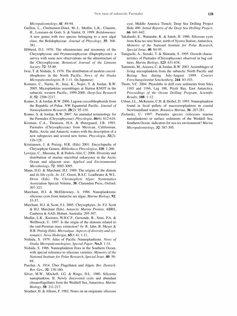
Figure 2: Biogeographic distribution of subarctic Parmales taxa in the North Pacific and its marginal seas, including taxa not reported in this study.Maps created using M. Weinelt’s ‘Online Map Creation’ site at www.aquarius.ifm-geomar.de
Micropaleontology, 41: 89-94.Guillou, L., Chrétiennot-Dinet, M.-J., Medlin, L.K., Claustre,
H., Loiseaux-de Goër, S. & Vaulot, D. 1999. Bolidomonas:A new genus with two species belonging to a new algalclass, the Bolidophyceae. Journal of Phycology, 35: 368-381.
Hibberd, D.J. 1976. The ultrastructure and taxonomy of theChrysophyceae and Prymnesiophyceae (Haptophyceae): asurvey with some new observations on the ultrastructure ofthe Chrysophyceae. Botanical Journal of the LinneanSociety, 72: 55-80.
Iwai, T. & Nishida, S. 1976. The distribution of modern coccol-ithophores in the North Pacific. News of the OsakaMicropaleontologists, 5: 1-11. (In Japanese)
Komuro, C., Narita, H., Imai, K., Nojiri, Y. & Jordan, R.W.2005. Microplankton assemblages at Station KNOT in thesubarctic western Pacific, 1999-2000. Deep-Sea ResearchII, 52: 2206-2217.
Konno, S. & Jordan, R.W. 2006. Lagoon coccolithophorids fromthe Republic of Palau, NW Equatorial Pacific. Journal ofNannoplankton Research, 28(2): 95-110.
Konno, S. & Jordan, R.W. 2007. An amended terminology forthe Parmales (Chrysophyceae). Phycologia, 46(6): 612-616.
Kosman, C.A., Thomsen, H.A. & Østergaard, J.B. 1993.Parmales (Chrysophyceae) from Mexican, Californian,Baltic, Arctic and Antarctic waters with the description of anew subspecies and several new forms. Phycologia, 32(2):116-128.
Kristiansen, J. & Preisig, H.R. (Eds) 2001. Encyclopedia ofChrysophyte Genera. Bibliotheca Phycologica, 110: 1-260.
Lovejoy, C., Massana, R. & Pedrós-Alió, C. 2006. Diversity anddistribution of marine microbial eukaryotes in the ArcticOcean and adjacent seas. Applied and EnvironmentalMicrobiology, 72: 3085-3095.
Mann, D.G. & Marchant, H.J. 1989. The origins of the diatomand its life cycle. In: J.C. Green, B.S.C. Leadbeater & W.L.Diver (Eds). The Chromophyte Algae. SystematicsAssociation Special Volume, 38. Clarendon Press, Oxford:307-323.
Marchant, H.J. & McEldowney, A. 1986. Nanoplanktonicsiliceous cysts from Antarctic are algae. Marine Biology, 92:53-57.
Marchant, H.J. & Scott, F.J. 2005. Chrysophytes. In: F.J. Scott& H.J. Marchant (Eds). Antarctic Marine Protists. ABRS,Canberra & AAD, Hobart, Australia: 295-307.
Medlin, L.K., Kooistra, W.H.C.F., Gersonde, R., Sims, P.A. &Wellbrock, U. 1997. Is the origin of the diatoms related tothe end-Permian mass extinction? In: R. Jahn, B. Meyer &H.R. Preisig (Eds). Microalgae. Aspects of diversity and sys-tematics. Nova Hedwigia, 65(1-4): 1-11.
Nishida, S. 1979. Atlas of Pacific Nannoplanktons. News ofOsaka Micropaleontologists, Special Paper, No.3: 1-31.
Nishida, S. 1986. Nannoplankton flora in the Southern Ocean,with special reference to siliceous varieties. Memoirs of theNational Institute for Polar Research, Special Issue, 40: 56-68.
Pascher, A. 1914. Über Flagellaten und Algen. Ber. Deutsch.Bot. Ges., 32: 136-160.
Silver, M.W., Mitchell, J.G. & Ringo, D.L. 1980. Siliceousnanoplankton. II. Newly discovered cysts and abundantchoanoflagellates from the Weddell Sea, Antarctica. MarineBiology, 58: 211-217.
Stradner, H. & Allram, F. 1982. Notes on an enigmatic siliceous
cyst, Middle America Trench, Deep Sea Drilling ProjectHole 490. Initial Reports of the Deep Sea Drilling Project,66: 641-642.
Takahashi, E., Watanabe, K. & Satoh, H. 1986. Siliceous cystsfrom Kita-no-seto Strait, north of Syowa Station, Antarctica.Memoirs of the National Institute for Polar Research,Special Issue, 40: 84-95.
Taniguchi, A., Suzuki, T. & Shimada, S. 1995. Growth charac-teristics of Parmales (Chrysophyceae) observed in bag cul-tures. Marine Biology, 123: 631-638.
Tanimoto, M., Aizawa, C. & Jordan, R.W. 2003. Assemblages ofliving microplankton from the subarctic North Pacific andBering Sea during July-August 1999. CourierForschungsinstitut Senckenberg, 244: 83-103.
Thorn, V.C. 2004. Phytoliths in drill core sediments from Sites1165 and 1166, Leg 188, Prydz Bay, East Antarctica.Proceedings of the Ocean Drilling Program, ScientificResults, 188: 1-12.
Urban, J.L., McKenzie, C.H. & Deibel, D. 1993. Nannoplanktonfound in fecal pellets of macrozooplankton in coastalNewfoundland waters. Botanica Marina, 36: 267-281.
Zielinski, U. 1997. Parmales species (siliceous marinenanoplankton) in surface sediments of the Weddell Sea,Southern Ocean: indicators for sea-ice environment? MarineMicropaleontology, 32: 387-395.
128New taxa of subarctic Parmales