Six Chains of the Human T Cell Antigen ReceptoreCD3 ... · anti-Leu-4 (CD3) or AM92.3 (Identi-T@...
5
THE JOURNAL OF BIOLOGICAL CHEMISTRY 0 1991 by The American Society for Biochemistry and Molecular Biology, Inc. Vol. 266, No. 12, Issue of April 25, pp. 7883-7887, 1991 Printed in U.S.A. Six Chains ofthe Human T Cell Antigen ReceptoreCD3 Complex Are Necessary and Sufficient for Processing the Receptor Heterodimer to the Cell Surface* (Received for publication, July 10, 1990) Gerald R. Carson$, Rolf E. Kuestner, Asma Ahmed,Carolyn L. Pettey, and Michael F. Concinoe From the Protein Expression Group, T Cell Sciences, Inc., Cambridge, Massachusetts 02139 The T cell antigen receptor (TCR) plays a key role in the process of antigen recognition. It is a complex of at least seven peptide chains (&y&r-r). It is found on the surface of mature T cells and functions in antigen binding in the presence of the major histocompatibility complex. It has been known for some time that physical associations between the CD3 proteins and the TCR chains are essential for efficient transport of either component to the surface of T cells. For example, T cells that lack either the (Y, 8, or 6 chains synthesize partial complexes that are eventuallydegraded. cDNAs encoding the six chains of receptor have be- come availablerecently. We have used transfection techniques to generate a panel of Chinese hamster ovary cells that contain partial receptor complexes of known composition and also cells that express all six subunits of the TCR*CD3complex. Cells in this panel were analyzed for the ability to form a-/3 heterodimers and also an ability to transport the synthesized chains to the plasma membrane. These studies have allowed us to define the minimum requirements for TCR-CD3 expression on the cell surface. The T cell antigen receptor.CD3 complex (TCReCD3)’ is a multisubunit protein composed of a variable glycoprotein heterodimer and an invariable complex of at least five chains (y,6, t, and a 1 homodimer) (1). The complex is found on the surface of functional T cells and confers antigenic specificity. Several studies have shown the close physical association between TCR and CD3 as well as the T cell’s requirement for co-expression of the two protein complexes in order to display receptor on the cell surface (2, 3). CD3 has a central role in the assembly of the T cell receptor within the cell. Pulse-chase experiments on T cells and T cell mutants have been used to demonstrate that the T cell recep- tor chains, once associated with components of the CD3 complex, were less susceptible to degradation (1,4). Establishing the requirements and mechanism of TCR. CD3 assembly remains a formidable task. Mutant T cells have been helpful in gaining insight into the assembly process but *This work was supported by T Cell Sciences Inc. and Central Research, Pfizer Inc. The costs of publication of this article were defrayed in part by the payment of page charges. This article must therefore be hereby marked “aduertisement” in accordance with 18 U.S.C. Section 1734 solely to indicate this fact. Inc., 38 Sidney St., Cambridge, MA 02139. 4 To whom correspondence should be addressed T Cell Sciences 5 Present address: Procept Inc., Cambridge, MA 02139. The abbreviations used are: TCR, T cell antigen receptor; CHO, Chinese hamster ovary cells; HEPES, 4-(2-hydroxyethyl)-l-pipera- zineethanesulfonic acid. studies with such cells are subject to a degree of uncertainty. Often the cells are not completely devoid of certain subunits, but rather, express these at very low levels. Their effects on the assembly and transport of the predominant chains cannot be known with certainty. Several T cell variants selected for low expression of one chain have been found to produce little or no detectable amounts of a second chain as well (5). Also, in a cell expressing the full complement of T cell proteins, it is difficult to isolate and study processes such as assembly, antigen binding, and signal transduction. Because the TCR e CD3 complex is a key component in the primary function of T cells, ensuring its proper assembly is crucial to the operation of the immune response. In this study we have generated a heterologous system to facilitate the study of these processes. A panel of transfected Chinese hamster ovary (CHO) cells, expressing both the T cell receptor heterodimer and the chains of the CD3 complex, have been established. We have used these cells to determine the minimal requirements for TCR. CD3 expression on the surface of cells. EXPERIMENTAL PROCEDURES Construction of Expression Plasmids-The pTCS series of expres- sion vectors consisted of the adenovirus 2 major late promotor in tandem with the simian virus 40 early promotor, both of which were located upstream of the adenovirus tripartite leader (6). An XhoI restriction site at the 3’ end of the tripartite leader sequence was used as theunique cloning site. cDNA inserts transcribed from these promoters utilized the murine Ig K polyadenylation signal (7) located 3’ of the XhoI site to terminate transcription. The plasmids carried various selection markers useful in mammalian cells as well as the 8- lactamase gene and bacterial origin of replication found on plasmid pSV2gpt (8). The original version of pTCS carried the xanthine- guanine phosphoribosyltransferase gene from pTCSgpt. Plasmids pTCSdhfr, pTCSneo, and pTCShygro were constructed by exchang- ing portions of the pTCSgpt plasmid with the corresponding portions of pSV2dhfr (9), pSV2neo (lo), and pSV2hygro (11). Strategy for the Construction of TCR. CD3-expressing CHO CelLs- Each of the full-length cDNAs of the (Y and (3 chains of TCR, the three chains of the CD3 complex, and TCRC were inserted into one of several expression vectors developed at T Cell Sciences Inc. pJPFL/ TCSgpt, the expression plasmid for TCRp chain, was constructed as follows. The 1.3-kilobase EcoRI fragment from pYT35, carrying the full-length cDNA for TCR(3 chain from the human T cell hybridoma Jurkat (12) was filled in to a blunt end with the Klenow fragment of Escherichia coli polymerase I and ligated to pTCSgpt at the XhoI site, which was also filled in to a blunt end. JaFL/TCSgpt, was constructed in a similar manner using pY14 as the source of cDNA. Plasmids T3y/TCSdhfr, T38/TCSneo, and T3c/TCSdhfr were prepared by the same strategy using the EcoRI fragment from pJT3y-2 (13) the KpnI/ XbaI fragment from pPGBC9 (14) and the Ban1 fragment from pDJ4 (151, respectively. pTCR{/TCShygro was constructed from pTCShygro and the BbuI fragment of the human TCR cDNA in All transfections were performed according to the calcium phos- phate co-precipitation method of Graham and van der Erb (17). Cultures transfected with the pTCSgpt vector were selected using pGEM-3Z (16). 7883
Transcript of Six Chains of the Human T Cell Antigen ReceptoreCD3 ... · anti-Leu-4 (CD3) or AM92.3 (Identi-T@...
THE JOURNAL OF BIOLOGICAL CHEMISTRY 0 1991 by The American Society for Biochemistry and Molecular Biology, Inc.
Vol. 266, No. 12, Issue of April 25, pp. 7883-7887, 1991 Printed in U.S.A.
Six Chains of the Human T Cell Antigen ReceptoreCD3 Complex Are Necessary and Sufficient for Processing the Receptor Heterodimer to the Cell Surface*
(Received for publication, July 10, 1990)
Gerald R. Carson$, Rolf E. Kuestner, Asma Ahmed, Carolyn L. Pettey, and Michael F. Concinoe From the Protein Expression Group, T Cell Sciences, Inc., Cambridge, Massachusetts 02139
The T cell antigen receptor (TCR) plays a key role in the process of antigen recognition. It is a complex of at least seven peptide chains (&y&r-r). It is found on the surface of mature T cells and functions in antigen binding in the presence of the major histocompatibility complex. It has been known for some time that physical associations between the CD3 proteins and the TCR chains are essential for efficient transport of either component to the surface of T cells. For example, T cells that lack either the (Y, 8, or 6 chains synthesize partial complexes that are eventually degraded.
cDNAs encoding the six chains of receptor have be- come available recently. We have used transfection techniques to generate a panel of Chinese hamster ovary cells that contain partial receptor complexes of known composition and also cells that express all six subunits of the TCR*CD3 complex. Cells in this panel were analyzed for the ability to form a-/3 heterodimers and also an ability to transport the synthesized chains to the plasma membrane. These studies have allowed us to define the minimum requirements for TCR-CD3 expression on the cell surface.
The T cell antigen receptor.CD3 complex (TCReCD3)’ is a multisubunit protein composed of a variable glycoprotein heterodimer and an invariable complex of at least five chains (y,6, t, and a 1 homodimer) (1). The complex is found on the surface of functional T cells and confers antigenic specificity. Several studies have shown the close physical association between TCR and CD3 as well as the T cell’s requirement for co-expression of the two protein complexes in order to display receptor on the cell surface (2, 3).
CD3 has a central role in the assembly of the T cell receptor within the cell. Pulse-chase experiments on T cells and T cell mutants have been used to demonstrate that the T cell recep- tor chains, once associated with components of the CD3 complex, were less susceptible to degradation (1,4).
Establishing the requirements and mechanism of TCR. CD3 assembly remains a formidable task. Mutant T cells have been helpful in gaining insight into the assembly process but
*This work was supported by T Cell Sciences Inc. and Central Research, Pfizer Inc. The costs of publication of this article were defrayed in part by the payment of page charges. This article must therefore be hereby marked “aduertisement” in accordance with 18 U.S.C. Section 1734 solely to indicate this fact.
Inc., 38 Sidney St., Cambridge, MA 02139. 4 To whom correspondence should be addressed T Cell Sciences
5 Present address: Procept Inc., Cambridge, MA 02139. The abbreviations used are: TCR, T cell antigen receptor; CHO,
Chinese hamster ovary cells; HEPES, 4-(2-hydroxyethyl)-l-pipera- zineethanesulfonic acid.
studies with such cells are subject to a degree of uncertainty. Often the cells are not completely devoid of certain subunits, but rather, express these at very low levels. Their effects on the assembly and transport of the predominant chains cannot be known with certainty. Several T cell variants selected for low expression of one chain have been found to produce little or no detectable amounts of a second chain as well (5). Also, in a cell expressing the full complement of T cell proteins, it is difficult to isolate and study processes such as assembly, antigen binding, and signal transduction. Because the TCR e
CD3 complex is a key component in the primary function of T cells, ensuring its proper assembly is crucial to the operation of the immune response. In this study we have generated a heterologous system to facilitate the study of these processes.
A panel of transfected Chinese hamster ovary (CHO) cells, expressing both the T cell receptor heterodimer and the chains of the CD3 complex, have been established. We have used these cells to determine the minimal requirements for TCR. CD3 expression on the surface of cells.
EXPERIMENTAL PROCEDURES
Construction of Expression Plasmids-The pTCS series of expres- sion vectors consisted of the adenovirus 2 major late promotor in tandem with the simian virus 40 early promotor, both of which were located upstream of the adenovirus tripartite leader (6). An XhoI restriction site at the 3’ end of the tripartite leader sequence was used as the unique cloning site. cDNA inserts transcribed from these promoters utilized the murine Ig K polyadenylation signal (7) located 3’ of the XhoI site to terminate transcription. The plasmids carried various selection markers useful in mammalian cells as well as the 8- lactamase gene and bacterial origin of replication found on plasmid pSV2gpt (8). The original version of pTCS carried the xanthine- guanine phosphoribosyltransferase gene from pTCSgpt. Plasmids pTCSdhfr, pTCSneo, and pTCShygro were constructed by exchang- ing portions of the pTCSgpt plasmid with the corresponding portions of pSV2dhfr (9), pSV2neo (lo), and pSV2hygro (11).
Strategy for the Construction of TCR. CD3-expressing CHO CelLs- Each of the full-length cDNAs of the (Y and (3 chains of TCR, the three chains of the CD3 complex, and TCRC were inserted into one of several expression vectors developed at T Cell Sciences Inc. pJPFL/ TCSgpt, the expression plasmid for TCRp chain, was constructed as follows. The 1.3-kilobase EcoRI fragment from pYT35, carrying the full-length cDNA for TCR(3 chain from the human T cell hybridoma Jurkat (12) was filled in to a blunt end with the Klenow fragment of Escherichia coli polymerase I and ligated to pTCSgpt at the XhoI site, which was also filled in to a blunt end. JaFL/TCSgpt, was constructed in a similar manner using pY14 as the source of cDNA. Plasmids T3y/TCSdhfr, T38/TCSneo, and T3c/TCSdhfr were prepared by the same strategy using the EcoRI fragment from pJT3y-2 (13) the KpnI/ XbaI fragment from pPGBC9 (14) and the Ban1 fragment from pDJ4 (151, respectively. pTCR{/TCShygro was constructed from pTCShygro and the BbuI fragment of the human TCR cDNA in
All transfections were performed according to the calcium phos- phate co-precipitation method of Graham and van der Erb (17). Cultures transfected with the pTCSgpt vector were selected using
pGEM-3Z (16).
7883

7884 Cell Surface Expression of T Cell Antigen Receptor
Alpha/gpt medium (Gibco Alpha MEM without nucleosides supple- mented with 10% fetal bovine serum, 4 mM glutamine, 250 mg/ml, xanthine, 15 mg/ml hypoxanthine, 6 mg/ml mycophenolic acid, and 10 mM HEPES (pH 7.0). The transcriptionally active region of JaFL/ TCSgpt was isolated and transfected along with JPFLITCSgpt into CHO cells. For the transfections to establish CaPy6 and CaDbe, a 1:10 ratio of plasmid DNAs were used with CD3 b/TCSneo being used at l/lOth the amount of the other plasmid. Transfected cells carrying the pTCSneo vector were selected by culturing in Alpha MEM/G418 medium. (Alpha MEM plus 1 mg/ml of Geneticin@ (GIBCO)). Clonal cultures were selected for further study or cell lines were subcloned prior to analysis.
CHO K1 (ATCC CCL61) cells were used as the host for most of the transfections. The Cy66 clone was constructed using CHO DUX B11 ceIls (18), which are mutant cells deficient in dihydrofolate reductase (dhfr-). These cells were maintained on Alpha MEM me- dium (GIBCO) supplemented with 10% fetal bovine serum and 10 pg/ml of adenosine, thymidine, and deoxyadenosine.
Northern Blot Analysis-Cytoplasmic RNA was extracted from the cells by the method of Ricciardi (19). Equal amounts of total cyto- plasmic RNA from each sample were loaded on a formaldehyde agarose gel and subjected to electrophoresis under denaturing condi- tions and blotted to nitrocellulose as described previously (20). Probes were radioactively labeled by the nick translation method using [a- 32P]dATP (Du Pont-New England Nuclear) and added to the hybrid- ization mix at a concentration of 2-5 X 10' counts/min/ml Aliquots of each sample were run on replicate gels to produce five blots. Each Northern blot was hybridized to a radioactive probe against one of the three CD3 messages, y , 6, or e, or against TCR a or probes. Autoradiographs of the blots were used to determine the presence of the various CD3 chains.
Flow Cytornetry Studies-Cells were cultured in Petri dishes in appropriate growth medium. Cells cultured in this way grew in suspension and could be removed easily from the dish. Cells were washed once with phosphate-buffered saline and resuspended in flow buffer (0.2% bovine serum albumin, 0.25% sodium azide in phosphate- buffered saline) to which 5-10 pg of antibody was added. Cells were stained with C305 (21), which is specific for the Jurkat TCRP chain, anti-Leu-4 (CD3) or AM92.3 (Identi-T@ ILBR, T Cell Sciences, Inc.), an antibody against the interleukin 2 receptor which was used as a negative control. Anti-Leu-4 was purchased as a conjugate with fluorescein isothiocyanate (Becton-Dickinson, Mountain View, CA). When staining with the other antibodies, goat anti-mouse Ig conju- gated to fluorescein isothiocyanate (Becton-Dickinson) was employed as the second antibody. Human peripheral blood lymphocytes were prepared from whole blood by lysing the erythrocytes using lysis buffer (150 mM NH4C1, 10 mM KHC03, 0.1 mM EDTA) and washing three times with phosphate-buffered saline. Lymphocytes were stained as described for transfectants except that staining with goat anti-mouse Ig-FITC alone was used as the negative control. Analysis was performed on an Ortho diagnostic cytofluorgraf 11.
Immunoprecipitation of TCRC-Cells were labeled overnight with 1 mCi of [36S]methionine and [3SS]cysteine (Tran-36S label, ICN Radiochemicals, Cleveland, OH). Cells were lysed in 10 mM Tris (pH 7.8), 0.15 M NaCl, 1% Nonidet P-40, 1 mM EDTA, 1 mM phenyl- methylsulfonyl fluoride, 0.2% aprotinin, and 10 mM iodoacetic acid (Sigma). TCRC was immunoprecipitated using N39, a rabbit anti- serum prepared against TCRCpeptides. TCRp was immunoprecitated using C305 and protein A- Sepharose (Pharmacia LKB Biotechnology Inc.). Samples were analyzed by reducing sodium dodecyl sulfate- polyacrylamide gel electrophoresis on 10% gels.
Two-dimensional Gel Analysis of TCR Heterodimer Formation- Cell lines were labeled and lysed as described above. Immunoprecip- itation was performed using antibodies immobilized on Affi-Gel 10 (Bio-Rad), C305, or (uF-P, which is specific for the constant region of TCRa chain.' Immunoprecipitates were analyzed by nonreducing- reducing two-dimensional sodium dodecyl sulfate-polyacrylamide gel electrophoresis on 10% gels.
RESULTS
The initial transfection for this study established the strain of cells producing TCR a and fl chains. Analysis of the Cap clone revealed that it was producing significant amounts of RNA specific for TCR a and /3 chains; however, flow cytom-
* T Cell Sciences Inc., unpublished observations.
etry showed that it displayed little receptor on its surface. This observation is in agreement with recent studies that document the instability of transfected a and P chains (5,22). The a and p chains have been shown to be susceptible to rapid catabolism in or close to the endoplasmic reticulum. This process appears to be common to many cells and is not restricted to T cells. Interestingly, association of (Y and P chains with the CD3 proteins has been shown to increase their intracellular survival time, suggesting that a surveillance mechanism similar to that operating in T cells may be present in CHO cells. This mechanism may involve a pathway of protein trafficking which is common to most mammalian cells. To study the question further, and determine if proper assembly of the complex would allow transport to the plasma membrane, we set out to develop a panel of Cap clones which expressed two or three chains of the CD3 complex. These clones were designed to help identify the effect of various CD3 chains on TCR assembly and processing in a non- lymphoid cell. By using Cap as a host, along with a judicious selection of resistance markers, new lines were developed producing each combination of two of the three CD3 chains (Fig. 1). G418 selection was used to grow out the transfectants and expression of the unselected cDNA was confirmed by Northern blot (Fig. 2). Transfectants express the appropriate RNA species. Size differences are attributed to untranslated sequences added by the expression vectors or removed in the course of subcloning the cDNAs into the vectors. Jurkat cells display the 3.4-kDa CD37 transcript typical of this cell.
The absence of any CD3 subunit, or TCR a and @ chains, results in no detectable receptor on the cell surface. The CD3 complex itself is also not sufficient to allow cell surface expression. Cells expressing mRNA for the CD37, -6, and -c chains, in the absence of TCR chains, show little detectable CD3 on the cell surface (Fig. 3). Results of staining with anti- TCRP antibody were similar. Only the cells carrying the TCR a and /3 chains as well as the three CD3 molecules display any anti-CD3 antibody binding to the cell surface.
Despite expressing levels of TCR- and CD3-specific mRNAs which were equal to or greater than those of Jurkat cells, the CaPy6c cells showed very little receptor complex on the cell surface. The complement of receptor subunits was increased further by introducing an expression vector carrying
A
Cap?'&
FIG. 1. Strategy for the construction of CHO lines express- ing various combinations of TCR and CD3 components. CHO lines expressing TCR. CD3 components were established by stepwise co-transfections and selection for the proper marker. Transfections of TCR a and TCR p were selected for expression of gpt. Cells expressing two or three chains of the CD3 complex were selected for resistance to G418. TCRCexpressing cells were selected for resistance to hygromycin B. Expression of each component not linked to a selection marker was verified by Northern blot analysis.

Cell Surface Expression of T Cell Antigen Receptor 7885
0
Jurkat C a p C a p YE N39 C305 9.5
4.4 2.4 45- 1.4
7.5 1 2 3 1 2 3
C a p Y 8 C a P Y s e C Y & 9.5- - P F ? q
31 -
1.4-
0.3- a B Y 8 e a p r a e r a e
FIG. 2. Northern analysis of transfected CHO cells. Cell lines 21 - analyzed are identified above each panel, and the probes used are labeled below each lane. Positions of molecular mass markers are shown on the left.
FIG. 3. Cell surface expression of various combinations of TCR-CD3 chains. Cells were stained and run on the flow cytometer as described in the text. Dotted lines correspond to negative control staining. Cell lines are identified in each panel. Graphs are shown as cell number versus relative fluorescence intensity on a logarithmic scale.
the TCR{ cDNA. Strain Capyae{ was established by trans- fecting Capyst with pTCR{/TCShygro and selecting for transfectants resistant to hygromycin B. The presence of the TCR{ chain was confirmed by immunoprecipitation experi- ments (Fig. 4). The increase in immunofluorescence following introduction of the { chain is shown in Fig. 3. The display on logarithmic scale demonstrates the level of staining relative to the parent CaPy6e cells. These cells were stained with C305 as well as anti-Leu-4 to confirm the presence of both TCR and CD3 (Fig. 5 ) . The Ca@y&{ cell line appears to show two populations of cells, one staining brightly for TCR. CD3 and one which is negative for the complex. This may reflect a difference related to { chain expression or a difference in receptor display related to the growth phase of the cell or
14-
FIG. 4. Immunoprecipitation studies. Cell lines are identified within each panel. Antibodies used are identified above the appropri- ate lanes. N39 is an anti-TCR{ antiserum. C305 is a monoclonal antibody recognizing the TCR 0 chain of Jurkat cells. Cell lines used are as follows: Lanes 1, Ca0; lanes 2, Capybe; lanes 3, CcxPy&{. Positions of molecular mass markers are indicated to the left.
a r k 4 c305
2 O O k I
2 o o k l r i *OOLl Ki
, "
:: I , , I
I , I , I ,
'- - '.-
: ; I \ : I
: : : : I :
\ '
I IO I00 1000 I IO I O 0 l o o 0 ' I '\ I '\
FIG. 5. Display of TCR-CD3 complex on cell surfaces. Cells were stained as described in the text. Antibodies used are identified at the top of the column. Cell lines are identified within each panel. Dotted lines correspond to negative control staining.
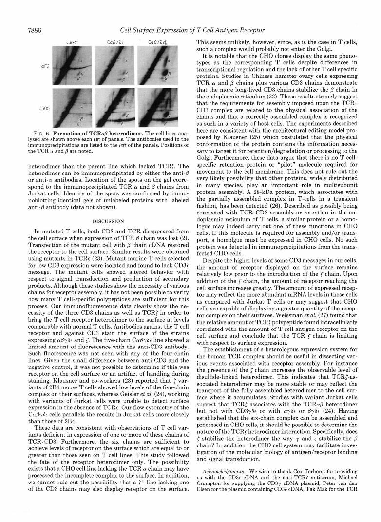
possibly receptor internalization. Immunoprecipitation with an anti-a or anti-/3 antibody of
in uiuo labeled cell proteins, followed by analysis using non- reducing-reducing two-dimensional gel electrophoresis (Fig. 6), was performed on the cells expressing the five- or six- chain complex. The TCR{ line produced more disulfide-linked
7886 Cell Surface Expression of T Cell Antigen Receptor
Jurkat CaOYGc CUBYS€[
aF2 J C 3 0 5 .
I FIG. 6. Formation of TCRaO heterodimer. The cell lines ana-
lyzed are shown above each set of panels. The antibodies used in the immunoprecipitations are listed to the left of the panels. Positions of the TCR a and are noted.
heterodimer than the parent line which lacked TCRC. The heterodimer can be immunoprecipitated by either the anti-@ or anti-a antibodies. Location of the spots on the gel corre- spond to the immunoprecipitated TCR a and p chains from Jurkat cells. Identity of the spots was confirmed by immu- noblotting identical gels of unlabeled proteins with labeled anti$ antibody (data not shown).
DISCUSSION
In mutated T cells, both CD3 and TCR disappeared from the cell surface when expression of TCR /3 chain was lost (2). Transfection of the mutant cell with /3 chain cDNA restored the receptor to the cell surface. Similar results were obtained using mutants in TCRC (23). Mutant murine T cells selected for low CD3 expression were isolated and found to lack CD3{ message. The mutant cells showed altered behavior with respect to signal transduction and production of secondary products. Although these studies show the necessity of various chains for receptor assembly, it has not been possible to verify how many T cell-specific polypeptides are sufficient for this process. Our immunofluorescence data clearly show the ne- cessity of the three CD3 chains as well as TCRC in order to bring the T cell receptor heterodimer to the surface at levels comparable with normal T cells. Antibodies against the T cell receptor and against CD3 stain the surface of the strains expressing apy6t and 5: The five-chain Capy6e line showed a limited amount of fluorescence with the anti-CD3 antibody. Such fluorescence was not seen with any of the four-chain lines. Given the small difference between anti-CD3 and the negative control, it was not possible to determine if this was receptor on the cell surface or an artifact of handling during staining. Klausner and co-workers (23) reported that { var- iants of 2B4 mouse T cells showed low levels of the five-chain complex on their surfaces, whereas Geisler et al. (24), working with variants of Jurkat cells were unable to detect surface expression in the absence of TCRC. Our flow cytometry of the Capy6c cells parallels the results in Jurkat cells more closely than those of 2B4.
These data are consistent with observations of T cell var- iants deficient in expression of one or more of these chains of TCR.CD3. Furthermore, the six chains are sufficient to achieve levels of receptor on the surface which are equal to or greater than those seen on T cell lines. This study followed the fate of the receptor heterodimer only. The possibility exists that a CHO cell line lacking the TCR a chain may have processed the incomplete complex to the surface. In addition, we cannot rule out the possibility that a {+ line lacking one of the CD3 chains may also display receptor on the surface.
This seems unlikely, however, since, as is the case in T cells, such a complex would probably not enter the Golgi.
I t is notable that the CHO clones display the same pheno- types as the corresponding T cells despite differences in transcriptional regulation and the lack of other T cell specific proteins. Studies in Chinese hamster ovary cells expressing TCR a and p chains plus various CD3 chains demonstrate that the more long-lived CD3 chains stabilize the p chain in the endoplasmic reticulum (22). These results strongly suggest that the requirements for assembly imposed upon the TCR. CD3 complex are related to the physical association of the chains and that a correctly assembled complex is recognized as such in a variety of host cells. The experiments described here are consistent with the architectural editing model pro- posed by Klausner (25) which postulated that the physical conformation of the protein contains the information neces- sary to target it for retention/degradation or processing to the Golgi. Furthermore, these data argue that there is no T cell- specific retention protein or “pilot” molecule required for movement to the cell membrane. This does not rule out the very likely possibility that other proteins, widely distributed in many species, play an important role in multisubunit protein assembly. A 28-kDa protein, which associates with the partially assembled complex in T-cells in a transient fashion, has been detected (26). Described as possibly being connected with TCR. CD3 assembly or retention in the en- doplasmic reticulum of T cells, a similar protein or a homo- logue may indeed carry out one of these functions in CHO cells. If this molecule is required for assembly and/or trans- port, a homolgue must be expressed in CHO cells. No such protein was detected in immunoprecipitations from the trans- fected CHO cells.
Despite the higher levels of some CD3 messages in our cells, the amount of receptor displayed on the surface remains relatively low prior to the introduction of the { chain. Upon addition of the { chain, the amount of receptor reaching the cell surface increases greatly. The amount of expressed recep- tor may reflect the more abundant mRNA levels in these cells as compared with Jurkat T cells or may suggest that CHO cells are capable of displaying a greater quantity of the recep- tor complex on their surfaces. Weissman et al. (27) found that the relative amount of TCRCpolypeptide found intracellularly correlated with the amount of T cell antigen receptor on the cell surface and conclude that the TCR { chain is limiting with respect to surface expression.
The establishment of a heterologous expression system for the human TCR complex should be useful in dissecting var- ious events associated with receptor assembly. For instance the presence of the r chain increases the observable level of disulfide-linked heterodimer. This indicates that TCRC-as- sociated heterodimer may be more stable or may reflect the transport of the fully assembled heterodimer to the cell sur- face where it accumulates. Studies with variant Jurkat cells suggest that TCRC associates with the TCRap heterodimer but not with CD3y6t or with ay6t or py6e (24). Having established that the six-chain complex can be assembled and processed in CHO cells, it should be possible to determine the nature of the TCRC heterodimer interaction. Specifically, does j- stabilize the heterodimer the way y and e stabilize the fi chain? In addition the CHO cell system may facilitate inves- tigation of the molecular biology of antigen/receptor binding and signal transduction.
Acknowledgments-We wish to thank Cox Terhorst for providing us with the CD3e cDNA and the anti-TCRC antiserum, Michael Crumpton for supplying the CD37 cDNA plasmid, Peter van den Elsen for the plasmid containing CD36 cDNA, Tak Mak for the TCR
Cell Surface Expression of T Cell Antigen Receptor 7887
(Y and 0 cDNAs, and Richard Klausner for the TCRCcDNA. Thanks 14. van den Elsen, P., Shepley, B.-A., Borst, J., Coligan, J. E., are due Thomas Wileman for valuable discussions of the work. Markham, A. F., Orkin, S., and Terhorst, C. (1984) Nature
REFERENCES 15. Gold, D. P., Puck, J. M., Pettey, C. L., Cho, M., Coligan, J., 1. Minami, Y., Weissman, A. M., Samelson, L. E., and Klausner, R. 16. weissman, A., H ~ ~ , D., orloff, D. G., ~ ~ d i , W. s., seuanez, H.,
Woody, J. N., and Terhorst, C. (1986) Nature 321,431-434
2. Weiss, A., and Stobo, J. D. (1984) J. Exp. Med. 160 , 1284-1299 O'Brilen, S. J., and Klausner, R. D. (1988) Proc. Natl. Acad.
3. Ohashi$ p. s.* Mak, T. w.* Van den p., Yanagiy y.y 17. Graham, F. L., and van der Erb, A. J. (1973) Virology 5 2 , 456- Sci. U. S. A. 85,9709-9713
Yoshikai, Y., Calman, A. F., Terhorst, C., Stobo, J. D., and Weiss, A. (1985) Nature 3 1 6 , 606-609
467
4. Alarcon, B., Berkhout, B., Breitmeier, E. J., and Terhorst, C. 18. Urlaub, G., and Chasin, L. A. (1980) Proc. Natl. Acad. Sci. U. S.
19. Ricciardi, R. P., Miller, J. S., and Roberts, B. E. (1979) Proc. 5. Bonifacino, J. S., Chen, C., Lippincott-Schwartz, J., Ashwell, J. Natl. Acud. Sei. U. S. A. 76,4927-4931
D., and Klausner, R. D. (1988) Proc. Natl. Acud. Sci. U. S. A. 20. Maniatis, T., Fritsch, E. F., and Sambrook, J. (1982) Molecular
312,413-418
D. (1987) Proc. Natl. Acud. Sci. U. S. A. 8 4 , 2688-2692
(1988) J. BWl. Chem. 2 6 3 , 2953-2961 A. 77,4216-4220
85,6929-6933 Cbning:A Laboratory Manual, Cold Spring Harbor Laboratory, 6. Kaufman, R. J. (1985) Proc. Natl. Acad. Sci. U. S. A. 8 2 , 689- Cold Spring Harbor, NY
7. Max, E. E., Maizel, J. V., Jr., and Leder, P. (1981) J. Biol. Chem. N. (1987) Nature 325 , 125-130
8. Mulligan, R. C., and Berg, P. (1980) Science 209, 1422-1427 Terhorst, C. (1990) J. Cell Biol. 110, 973-986 9. Subramani, s., ~ ~ l l i ~ ~ ~ , R., and B ~ ~ ~ , p., (1981) ~ ~ 1 , cell, ~ i ~ l , 23. Sussman, J. J., Bonifacino, J. S., Lippincott-Schwartz, J., Weiss-
man, A. M., Saito, T., Klausner, R. D., and Ashwell, J. D. (1988) Cell 52,85-95
693 21. Saito, T., Weiss, A., Miller, J., Norcross, M. A., and Germain, R.
256,5116-5120 22. Wileman, T., Carson, G. R., Concino, M. F., Ahmed, A., and
2,854-864 lo. Southern, p. J., and Berg, p. (1982) J. Genet. 1, 327- 24. Geisler, C,, Kuhlman, J., and Rubin, B. (1989) J , Immunol, 143,
341 11. Gritz, L., and Davies, J. (1983) Gene (Anst.) 2 5 , 179-188 12. Yanah y.9 Yoshikai, y.9 Legget6 K.3 Clark, s. p.9 Aleksander, 1.9 26. Pettey, C. L., Alarcon, B., Malin, R., Weinberg, K., and Terhorst,
25. Klausner, R. D. (1989) New Biologist 1 , 3-8
13. Krissansen, G. W., Owen, M. J., Verbi, W., and Crumpton, M. J. 27. Weissman, A. M., Frank, S. J., Orloff, D. G., Mercep, M., Ashwell,
4069-4077
and Mak, T. W. (1984) Nature 308,145-149 C. (1987) J. Biol. Chem. 2 6 2 , 4854-4859
(1986) EMBO J. 5 , 1799-1808 J. D., and Klausner, R. D. (1989) EMBO J. 8 , 3651-3656