Sinonasal Ewing Sarcoma - Advanced Radiology...
12
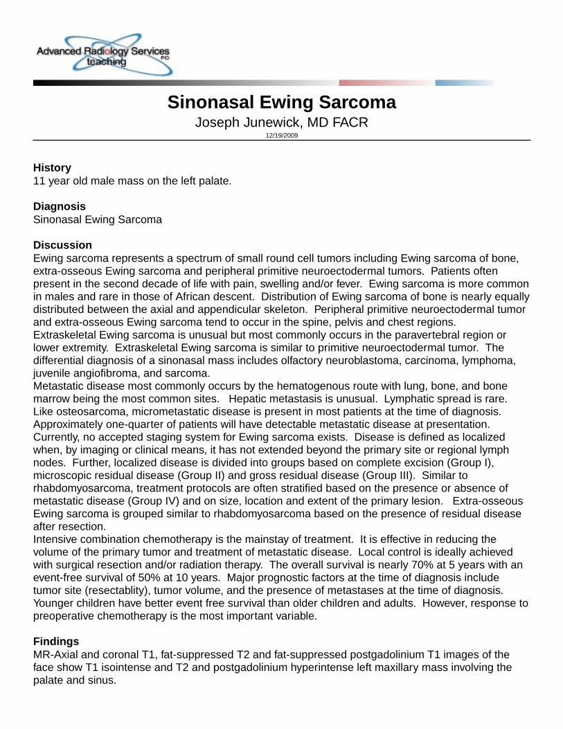
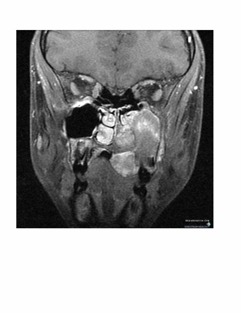
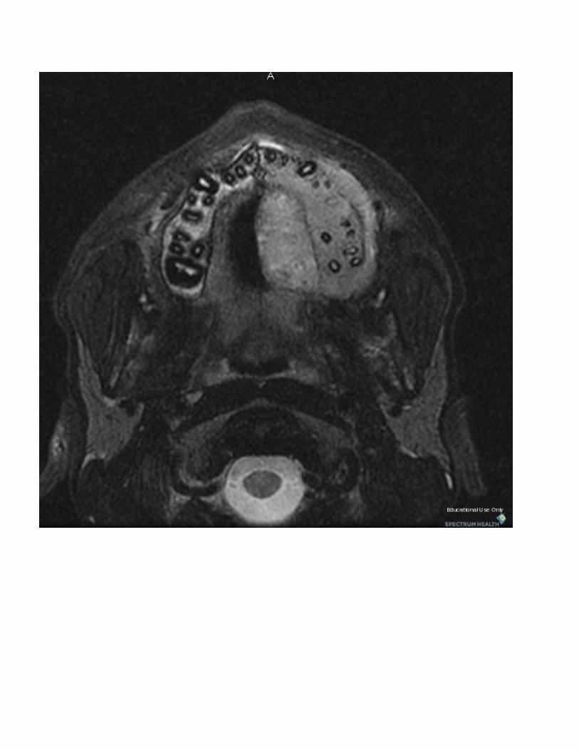
Sinonasal Ewing Sarcoma Joseph Junewick, MD FACR 12/19/2009 History 11 year old male mass on the left palate. Diagnosis Sinonasal Ewing Sarcoma Discussion Ewing sarcoma represents a spectrum of small round cell tumors including Ewing sarcoma of bone, extra-osseous Ewing sarcoma and peripheral primitive neuroectodermal tumors. Patients often present in the second decade of life with pain, swelling and/or fever. Ewing sarcoma is more common in males and rare in those of African descent. Distribution of Ewing sarcoma of bone is nearly equally distributed between the axial and appendicular skeleton. Peripheral primitive neuroectodermal tumor and extra-osseous Ewing sarcoma tend to occur in the spine, pelvis and chest regions. Extraskeletal Ewing sarcoma is unusual but most commonly occurs in the paravertebral region or lower extremity. Extraskeletal Ewing sarcoma is similar to primitive neuroectodermal tumor. The differential diagnosis of a sinonasal mass includes olfactory neuroblastoma, carcinoma, lymphoma, juvenile angiofibroma, and sarcoma. Metastatic disease most commonly occurs by the hematogenous route with lung, bone, and bone marrow being the most common sites. Hepatic metastasis is unusual. Lymphatic spread is rare. Like osteosarcoma, micrometastatic disease is present in most patients at the time of diagnosis. Approximately one-quarter of patients will have detectable metastatic disease at presentation. Currently, no accepted staging system for Ewing sarcoma exists. Disease is defined as localized when, by imaging or clinical means, it has not extended beyond the primary site or regional lymph nodes. Further, localized disease is divided into groups based on complete excision (Group I), microscopic residual disease (Group II) and gross residual disease (Group III). Similar to rhabdomyosarcoma, treatment protocols are often stratified based on the presence or absence of metastatic disease (Group IV) and on size, location and extent of the primary lesion. Extra-osseous Ewing sarcoma is grouped similar to rhabdomyosarcoma based on the presence of residual disease after resection. Intensive combination chemotherapy is the mainstay of treatment. It is effective in reducing the volume of the primary tumor and treatment of metastatic disease. Local control is ideally achieved with surgical resection and/or radiation therapy. The overall survival is nearly 70% at 5 years with an event-free survival of 50% at 10 years. Major prognostic factors at the time of diagnosis include tumor site (resectablity), tumor volume, and the presence of metastases at the time of diagnosis. Younger children have better event free survival than older children and adults. However, response to preoperative chemotherapy is the most important variable. Findings MR-Axial and coronal T1, fat-suppressed T2 and fat-suppressed postgadolinium T1 images of the face show T1 isointense and T2 and postgadolinium hyperintense left maxillary mass involving the palate and sinus.
-
Upload
phungthien -
Category
Documents
-
view
227 -
download
0
Transcript of Sinonasal Ewing Sarcoma - Advanced Radiology...
Sinonasal Ewing SarcomaJoseph Junewick, MD FACR
12/19/2009
History11 year old male mass on the left palate.
DiagnosisSinonasal Ewing Sarcoma
DiscussionEwing sarcoma represents a spectrum of small round cell tumors including Ewing sarcoma of bone,extra-osseous Ewing sarcoma and peripheral primitive neuroectodermal tumors. Patients oftenpresent in the second decade of life with pain, swelling and/or fever. Ewing sarcoma is more commonin males and rare in those of African descent. Distribution of Ewing sarcoma of bone is nearly equallydistributed between the axial and appendicular skeleton. Peripheral primitive neuroectodermal tumorand extra-osseous Ewing sarcoma tend to occur in the spine, pelvis and chest regions.Extraskeletal Ewing sarcoma is unusual but most commonly occurs in the paravertebral region orlower extremity. Extraskeletal Ewing sarcoma is similar to primitive neuroectodermal tumor. Thedifferential diagnosis of a sinonasal mass includes olfactory neuroblastoma, carcinoma, lymphoma,juvenile angiofibroma, and sarcoma.Metastatic disease most commonly occurs by the hematogenous route with lung, bone, and bonemarrow being the most common sites. Hepatic metastasis is unusual. Lymphatic spread is rare.Like osteosarcoma, micrometastatic disease is present in most patients at the time of diagnosis.Approximately one-quarter of patients will have detectable metastatic disease at presentation.Currently, no accepted staging system for Ewing sarcoma exists. Disease is defined as localizedwhen, by imaging or clinical means, it has not extended beyond the primary site or regional lymphnodes. Further, localized disease is divided into groups based on complete excision (Group I),microscopic residual disease (Group II) and gross residual disease (Group III). Similar torhabdomyosarcoma, treatment protocols are often stratified based on the presence or absence ofmetastatic disease (Group IV) and on size, location and extent of the primary lesion. Extra-osseousEwing sarcoma is grouped similar to rhabdomyosarcoma based on the presence of residual diseaseafter resection.Intensive combination chemotherapy is the mainstay of treatment. It is effective in reducing thevolume of the primary tumor and treatment of metastatic disease. Local control is ideally achievedwith surgical resection and/or radiation therapy. The overall survival is nearly 70% at 5 years with anevent-free survival of 50% at 10 years. Major prognostic factors at the time of diagnosis includetumor site (resectablity), tumor volume, and the presence of metastases at the time of diagnosis.Younger children have better event free survival than older children and adults. However, response topreoperative chemotherapy is the most important variable.
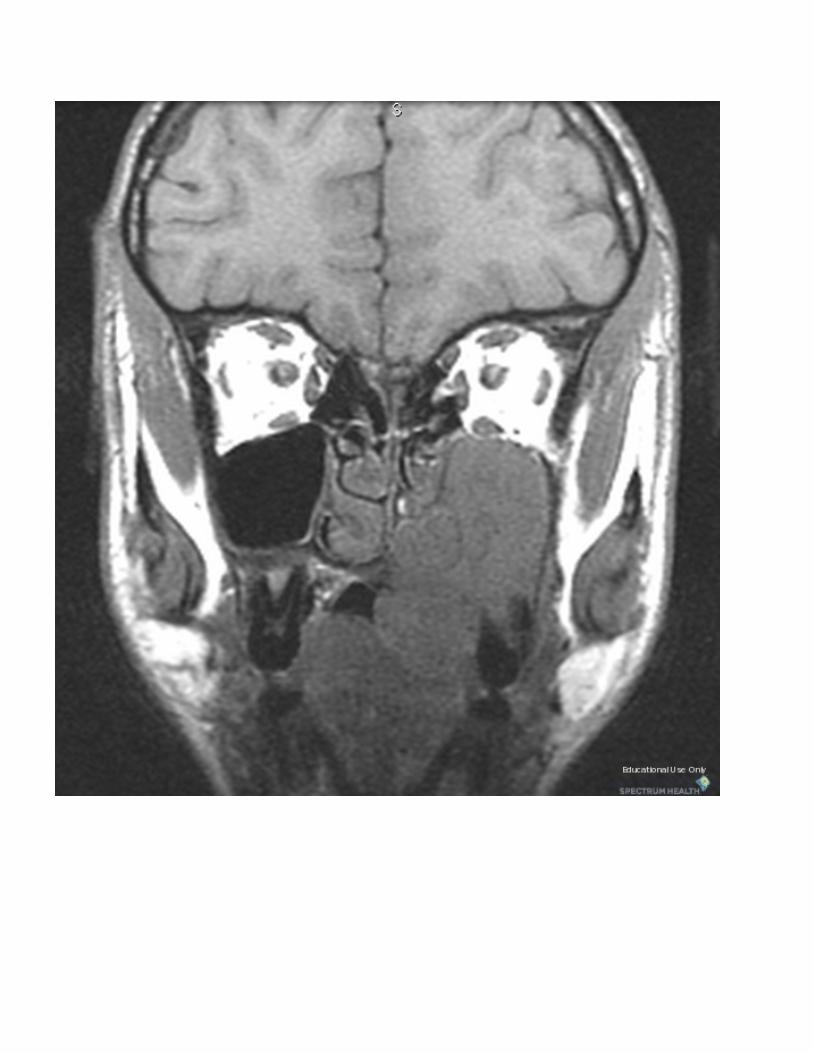
FindingsMR-Axial and coronal T1, fat-suppressed T2 and fat-suppressed postgadolinium T1 images of theface show T1 isointense and T2 and postgadolinium hyperintense left maxillary mass involving thepalate and sinus.

ReferenceReinus WR, et al. Radiology of Ewing's sarcoma: Intergroup Ewing's sarcoma (IESS). Radiographics(1984); 4:929-944.


Sponsored By
DisclaimerThis teaching site is partially funded by an educational grant from GE Healthcare and Advanced Radiology Services, PC. The material on this site isindependently controlled by Advanced Radiology Services, PC, and GE Healthcare and Spectrum Health have no influence over the content of this siteContent Download AgreementThe cases and images on this website are owned by Spectrum Health. Permission is granted (for nonprofit educational purposes) to download and printmaterials to distribute for the purpose of facilitating the education of health professionals. The authors retain all rights to the material and users arerequested to acknowledge the source of the material. Site DisclaimerThis site is developed to reach healthcare professionals and medical students. Nothing this site should be considered medical advice.Only your own doctor can help you make decisions about your medical care. If you have a specific medical question or are seeking medical care, pleasecontact your physician.The information in this website is provided for general medical education purposes only and is not meant to substitute for the independent medicaljudgment of a physician relative to diagnostic and treatment options of a specific medical condition.The viewpoints expressed in these cases are those of the authors. They do not represent an endorsement. In no event will Advanced RadiologyAssociates, PC, Spectrum Health Hospitals (Helen Devos Children's Hospital) or GE Healthcare be liable for any decision made or action taken inreliance upon the information provided through this website.