Single-scan multidimensional NMR · Single-scan multidimensional NMR Lucio Frydman Department of...
10
Single-scan multidimensional NMR Lucio Frydman Department of Chemical Physics, Weizmann Institute of Science, 76100 Rehovot, Israel Received 9 May 2005; accepted 2 June 2005 Available online 16 September 2005 Abstract Multidimensional spectroscopy plays a number of essential roles in contemporary magnetic resonance. It brings a resolution enhancement without which numerous NMR applications in organic and inorganic chemistry would be unattainable, it serves as a basic tool in the assignment and structural elucidation of complex biological structures, and it is an integral part of the image formation protocol in MRI. The present review describes a recent scheme which we have developed, enabling the acquisition of complete 2D NMR data sets within a single continuous acquisition. Provided that an analyte’s signal is sufficiently strong, the acquisition time of multidimensional NMR experiments can thus be shortened by several orders of magnitude. The new meth- odology is compatible with existing multidimensional pulse sequences (COSY, TOCSY, HSQC, MRI) and can be implemented using conventional hardware. The spatial encoding of the NMR interactions — which is the new principle underlying this ultrafast NMR protocol — is discussed, and the protocol’s performance is exemplified with a variety of homonuclear and heteronuclear 2D NMR and MRI acquisitions. To cite this article: L. Frydman, C. R. Chimie 9 (2006). © 2005 Publié par Elsevier SAS pour Académie des sciences. Résumé La spectroscopie multidimensionnelle joue des rôles essentiels dans la résonance magnétique moderne. Elle apporte une amélio- ration de la résolution, sans laquelle de nombreuses applications en chimie organique et inorganique resteraient impossibles ; elle est utilisée comme outil de base dans la détermination et l’élucidation de structures biologiques complexes et fait partie intégrante du protocole de formation de l’image en IRM. La présente revue décrit une récente approche que nous avons développée, permet- tant l’acquisition de données RMN 2D complètes en une seule acquisition continue. À condition que le signal soit suffisamment fort, le temps d’acquisition des expériences de RMN multidimensionnelle peut être raccourci de plusieurs ordres de grandeur. Cette nouvelle méthodologie est compatible avec les séquences d’impulsions multidimensionnelles (COSY,TOCSY, HSQC, IRM) et peut être mise en œuvre avec un équipement informatique conventionnel. Le codage spatial des interactions RMN, qui constitue le nouveau principe de ce protocole RMN utra-rapide, est discuté et des exemples de ses performances sont donnés, avec une variété d’acquisitions par IRM et RMN 2D homo- et hétéronucléaire. Pour citer cet article : L. Frydman, C. R. Chimie 9 (2006). © 2005 Publié par Elsevier SAS pour Académie des sciences. Keywords: Multidimensional magnetic resonance spectroscopy; Single continuous acquisition; Spatial encoding; Ultrafast NMR Mots clés : Spectroscopie multidimensionnelle de résonance magnétique ; Acquisition continue ; Codage spatial ; RMN utrarapide E-mail address: [email protected] (L. Frydman). C. R. Chimie 9 (2006) 336–345 http://france.elsevier.com/direct/CRAS2C/ 1631-0748/$ - see front matter © 2005 Publié par Elsevier SAS pour Académie des sciences. doi:10.1016/j.crci.2005.06.014
Transcript of Single-scan multidimensional NMR · Single-scan multidimensional NMR Lucio Frydman Department of...

Single-scan multidimensional NMR
Lucio Frydman
Department of Chemical Physics, Weizmann Institute of Science, 76100 Rehovot, Israel
Received 9 May 2005; accepted 2 June 2005
Available online 16 September 2005
Abstract
Multidimensional spectroscopy plays a number of essential roles in contemporary magnetic resonance. It brings a resolutionenhancement without which numerous NMR applications in organic and inorganic chemistry would be unattainable, it serves asa basic tool in the assignment and structural elucidation of complex biological structures, and it is an integral part of the imageformation protocol in MRI. The present review describes a recent scheme which we have developed, enabling the acquisition ofcomplete 2D NMR data sets within a single continuous acquisition. Provided that an analyte’s signal is sufficiently strong, theacquisition time of multidimensional NMR experiments can thus be shortened by several orders of magnitude. The new meth-odology is compatible with existing multidimensional pulse sequences (COSY, TOCSY, HSQC, MRI) and can be implementedusing conventional hardware. The spatial encoding of the NMR interactions — which is the new principle underlying thisultrafast NMR protocol — is discussed, and the protocol’s performance is exemplified with a variety of homonuclear andheteronuclear 2D NMR and MRI acquisitions. To cite this article: L. Frydman, C. R. Chimie 9 (2006).© 2005 Publié par Elsevier SAS pour Académie des sciences.
Résumé
La spectroscopie multidimensionnelle joue des rôles essentiels dans la résonance magnétique moderne. Elle apporte une amélio-ration de la résolution, sans laquelle de nombreuses applications en chimie organique et inorganique resteraient impossibles ; elleest utilisée comme outil de base dans la détermination et l’élucidation de structures biologiques complexes et fait partie intégrantedu protocole de formation de l’image en IRM. La présente revue décrit une récente approche que nous avons développée, permet-tant l’acquisition de données RMN 2D complètes en une seule acquisition continue. À condition que le signal soit suffisammentfort, le temps d’acquisition des expériences de RMN multidimensionnelle peut être raccourci de plusieurs ordres de grandeur.Cette nouvelle méthodologie est compatible avec les séquences d’impulsions multidimensionnelles (COSY, TOCSY, HSQC, IRM)et peut être mise en œuvre avec un équipement informatique conventionnel. Le codage spatial des interactions RMN, qui constituele nouveau principe de ce protocole RMN utra-rapide, est discuté et des exemples de ses performances sont donnés, avec unevariété d’acquisitions par IRM et RMN 2D homo- et hétéronucléaire. Pour citer cet article : L. Frydman, C. R. Chimie 9 (2006).© 2005 Publié par Elsevier SAS pour Académie des sciences.
Keywords: Multidimensional magnetic resonance spectroscopy; Single continuous acquisition; Spatial encoding; Ultrafast NMR
Mots clés : Spectroscopie multidimensionnelle de résonance magnétique ; Acquisition continue ; Codage spatial ; RMN utrarapide
E-mail address: [email protected] (L. Frydman).
C. R. Chimie 9 (2006) 336–345
http://france.elsevier.com/direct/CRAS2C/
1631-0748/$ - see front matter © 2005 Publié par Elsevier SAS pour Académie des sciences.doi:10.1016/j.crci.2005.06.014

1. Introduction
Few branches of spectroscopy match in eitherbreadth or depth the impact achieved by nuclear mag-netic resonance (NMR) [1]. Originated as an unex-pected by-product of the quantum mechanics revolu-tion [2], NMR remained mostly a curiosity of themolecular beam community until Bloch, Purcell andtheir coworkers demonstrated the possibility of observ-ing its manifestation in bulk [3]. This in turn triggeredthe use of nuclear spins as tiny ‘spies’ capable of char-acterizing the structure of organic molecules [4], a fea-ture that transformed NMR into one of the corner-stones of modern chemical and pharmaceuticalresearch. Over the years NMR undertook many otherunforeseen applications: it became a common researchtool in solid state chemistry and condensed matter phys-ics; it afforded one of the few methods available fordetermining the structures of proteins and nucleic acidsin their native solution state; and it expanded into medi-cine both as an imaging as well as a spectroscopy toolto study metabolism, diagnose malignancy, angio-graph non-invasively, and reveal brain function [1,5–9]. A feature that distinguishes modern NMR from thefew other techniques that might claim comparably wideimpact areas of applicability is the fact that, broadlyspeaking, one common protocol underlies most of itsvery different applications. This protocol is multidi-mensional Fourier transform NMR, nD FT NMR, ini-tially carried out along two axes [10,11] but eventuallyextended to the correlation of multiple spectral — andin the case of imaging, to the correlation of multiplespatial [9,12] — domains. nD NMR seeks to measure,spread and correlate NMR evolution frequencies. Thisis not generally carried out by exciting spectral linesbut rather by manipulations in their Fourier conjugatespace ; i.e. in the time-domain. For instance the basicscheme that started the 2D NMR revolution [10,11,13]
(1)Preparation − Evolution (t1) − Mixing − Acquisition (t2)
provides a time domain set
(2)
S(t1, t2) =
�allX2’s
dX2� �allX1’s
dX1 I(X1, X2) eiX1t1 e−t1/T2� eiX2t2 e–t2/T2
from which correlations between indirect- anddirect-domain Bohr frequencies X1, X2 can be
extracted following a discrete version of the Fourieranalysis1 :
(3)I(m1, m2) ∝ �allt2’s
dt2 � �allt1’s
dt1 S(t1, t2) e−im1t1� e−im2t2
This expression may appear as a simple extensionof 1D time-domain NMR to two dimensions, yet thissimilarity is deceiving and belies the insight involvedin the transition from one- to multi-dimensional spec-troscopy. Indeed the extraction variable against whichdata are transformed in 1D pulsed NMR is the physicalacquisition time, of which Nature has given us onlyone [13,14]. Such acquisition time becomes t2 in 2DNMR spectroscopy, leaving the problem of how tosample points in the remaining time-domain axis. Theanswer to this riddle was provided by Jeener, who pro-posed acquiring complete 2D NMR frequency correla-tion sets by relying on a parametric incrementation oft1 in N1 steps of duration Dt1(Fig. 1 A). Although inappearance a very simple solution it is actually hard toemphasize the scientific insight of this contribution,whereby a 2D correlation between evolving coher-ences is extracted from a series of statistically indepen-
1 Throughout the present work X1, X2 denote the actual values ofthe Bohr evolution frequencies experienced by the spins, while m1
and m2 represent the frequency-axis variables of a spectrum beingplotted.
Fig. 1. (A) Traditional Jeener/Ernst approach to the acquisition of2D NMR spectra, based on the acquisition of numerous independentS(t2) signals as a function of an incremented t1 time parameter (dashedarrows). (B) Mansfield’s approach to ultrafast MRI based on the conti-nuous scanning of a 2D time-domain data within a single acquisi-tion, using multiple manipulations of the interactions being correla-ted. In either case, the resulting temporal evolution obtained fromthe spins needs to be Fourier analyzed in order to extract the spectralcorrelations being sought.
337L. Frydman / C. R. Chimie 9 (2006) 336–345

dent experiments. In its stead, perhaps more indicativeand meaningful is it to note the ca. 27 000 referencesarising from a contemporary data base search on thetopic of multidimensional magnetic resonance, or thefact that in a very brief survey of its first 100 years theNobel Foundation devoted a central spot to describingthe impact of 2D NMR [15].
In spite of the unambiguous gains resulting fromupgrading NMR from one spectral dimension to two, afundamental drawback also became associated to thistransition. As each of the t1 points to be sampled alongthe indirect time-domain constitutes an independentacquisition, 2D NMR will demand tens or hundreds oftransients in order to adequately sample the t1 domain— even if the signal-to-noise ratio (S/N) is acceptablewithin a single scan. Such complication is built-in intothe scheme illustrated in eq. (1), and gets exponentiallycompounded when porting the 2D NMR principle tohigher dimensions. The slow nature of multidimen-sional spectroscopy was found particularly confiningin what eventually became the most widespread of allforms of bidimensional NMR, 2D FT MRI, an experi-ment where the spins’ positions along orthogonal ori-entations become correlated by the action of externalmagnetic field gradients [16–18]. Yet in this case theavailability of intense signals combined with the unpar-alleled flexibility with which MRI interactions couldbe manipulated enabled the eventual development ofso-called ultrafast acquisitions techniques, capable ofyielding complete 2D MRI data sets within a 0.1–1-stimescale (Fig. 1B). Principal among these methodsstands echo planar imaging, EPI [12,19], and its daugh-ter techniques [20]. This proposition was originallyjudged as ‘ahead of its time’ [21] yet eventually itbecame a cornerstone of magnetic resonance, contrib-uting with the advent of functional MRI to what hasarguably become the most exciting area in contempo-rary science.
Given the benefits that could be reaped from EPI,the question naturally arises of whether it is possible toextend its underlying principles to other, non-MRI typesof NMR experiments. And yet except for a few rarecases where the interactions to be correlated can be con-trolled independently and with nearly ideal efficiency[22–24], one is forced to conclude that the spin physicsunderlying EPI techniques could not be directly ex-ported into conventional high-resolution spectroscopy.Indeed EPI extends the principle in eq. (1) by coupling
into it the possibility of ‘walking’ through the timedomain [19,25,26]; that is, by introducing numerousperiodical switchings and/or reversals of the interac-tions being correlated so as to sample a large region ofthe 2D time-domain space within a single continuousscan (Fig. 1 B). And although of a reliability beyondarguments it clear that such ‘walk in space’approach issimply beyond the realm of the practical when relyingon stochastic mixing processes like those involved in2D Overhauser spectroscopy [13], or repeatable onlyvery few times when dealing with J-mediated coher-ence transfers like those involved in homo- or hetero-nuclear 2D correlation sequences.
Still, challenged and inspired by these MRI-derivedacquisition concepts, we sought for over a decade for aprinciple capable of reinstating the multiplexing advan-tages of 1D FT spectroscopy to multidimensional NMR— as well as potentially onto other forms of MR andnon-MR time-domain spectroscopies.Very recently onesuch principle finally crystallized [27,28], which inanalogy to the ultrafast MRI term proposed for imag-ing techniques capable of concluding their data acqui-sitions within sub-second timescales we have coined‘ultrafast NMR’. This principle provides a generalscheme for completing the acquisition of nearly arbi-trary high-resolution multidimensional experimentswithin a single scan — regardless of whether theirdimensions are 2, 3 or higher; regardless of whetherthey involve internal spin interactions, gradient-drivenones, or a combination of thereof. The goal of thepresent description is to present a brief summary of thisnew principle, and overview some of its potential appli-cations.
2. Spatial encoding and the acquisition of 2DNMR spectra within a single scan
2.1. The principle
The main aspect distinguishing ultrafast from bothtraditional 2D NMR as well as from EPI-type acquisi-tions is a departure from the homogeneous encoding ofthe spin interactions. Instead, indirect-domain evolu-tion frequencies are imparted in a heterogeneous fash-ion whereby spins positioned at different coordinates
338 L. Frydman / C. R. Chimie 9 (2006) 336–345

within the sample2 are allowed to evolve over differentextents of time prior to the mixing period. A suitableread-out of the heterogeneous evolution thus encodedcan then provide the full 2D NMR information beingsought, within a single continuous data acquisition.Given present-day NMR hardware it appears that thesimplest way to achieve this partitioning of the sampleis by activating a magnetic field gradient G = �B0 /�r,discriminating spins according to their spatial coordi-nate r. A scheme such as the one illustrated in Fig. 2can then be used to collect the indirect-domain spectralcorrelations. Such generic sequence assumes that theinitial spin evolution is triggered by a frequency-incremented RF pulse, superimposed on top of an exci-tation gradient Ge. An important demand placed on thisexcitation module is that at its conclusion, the evolu-tion phase of the excited spin packets should end upreflecting the effects of internal X1 couplings, but notthose of the ancillary Ge gradient. Numerous strategieshave already been devised for imparting such internalkind of spatial encoding [27–32], and it is likely thatfurther optimizations will arise in the near future. Inbroad terms all these spatial encoding strategies involvethe application of either discrete or continuousfrequency-shifted selective RF pulses, in combination
with alternating gradients designed so that their effectsare eventually ‘echoed away’ at the conclusion of theirradiation. The common, important point of all theseencoding alternatives is that at their conclusion, thecombined use of frequency-shifted RF pulses and ofspatially dependent resonance frequencies end upimparting on the spins an r-dependent purely internalevolution phase of the kind
(4)φe(r) = CX1(r − r0)
Here C is a spatio-temporal ratio under the experi-mentalist’s control relating a spin’s displacement r – ro
against the extent of its evolution time t1, and X1 is therotating-frame internal frequency we are attempting tomeasure. Eq. (4) amounts to a ‘winding’ of the spincoherences, a pattern analogous to the one that wouldarise if a gradient along the r^ direction were appliedfollowing a hard p/2 pulse. Analogous but not identi-cal, since unlike what happens when applying a gradi-ent, the pitch of the resulting winding will now be site-dependent. In fact, it will be governed by the indirect-domain interaction X1 conforming the observable thatwe are attempting to measure.
When inserted at the beginning of a generic 2D NMRexperiment such winding will be preserved by thehomogeneous mixing process (Fig. 2), and will conse-quently lead to no observable macroscopic signals whenconsidering the sample as a whole. If, however, the sig-nal detection is now implemented while subjecting thesample to an acquisition field gradient Ga possessingthe same geometry as Ge, this spiral of magnetizationsimparted by the X1 will eventually be unwound. Themoment at which this X1-dependent unwinding occursis easily observable, as only at its juncture will spin-packets located at different positions throughout thesample cease interfering and lead to a macroscopic over-all signal. This in turn implies that the spatial encodingof the spins interactions and this subsequent gradient-assisted readout provides a way to monitor the indirectdomain X1 evolution frequencies that affected the spins;not via the Fourier analysis of an evolving coherenceas in usual time-domain spectroscopy, but via the dis-placement observed in the position of a site-specificecho created by interfering spins located at differentpositions throughout the sample. A graphical depictionof this unusual feature — whereby the time-domain sig-nal becomes equivalent to its indirect domain fre-quency spectrum — is illustrated in Fig. 3 A, which
2 Or perhaps in the case of anisotropic media, spins oriented atinequivalent angles with respect to the magnetic field.
Fig. 2. Starting point of the single-scan 2D NMR protocol: a time-and frequency-incremented RF irradiation applied in combinationwith a suitable gradient imposes an r-dependent evolution of the X1
spin interactions to be measured (left). Following a homogeneousmixing process this spatial encoding is preserved as either a phase-or an amplitude-modulation (Center), to be subsequently decodedby the application of a second acquisition gradient that reads out theX1 frequencies as site-specific echo signals (right).
339L. Frydman / C. R. Chimie 9 (2006) 336–345
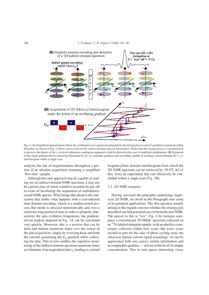
analyzes the fate of magnetizations throughout a por-tion of an ultrafast acquisition assuming a simplified‘five-slice’ sample.
Although this new approach may be capable of read-ing out an indirect-domain NMR spectrum, it may notbe a priori clear of where would its eccentricity pay offin terms of facilitating the acquisition of multidimen-sional NMR spectra. What brings this about is the real-ization that unlike what happens with a conventionaltime-domain encoding, which is a unidirectional pro-cess that needs to proceed monotonically and over arelatively long period of time in order to properly char-acterize the spin evolution frequencies, the gradient-driven readout depicted in Fig. 3A can be concludedvery quickly. Moreover, this is a process that can bedone and undone numerous times over the course ofthe data acquisition, simply by reversing back and forththe currents generating the Ga gradient while collect-ing the data. This in turn enables the repetitive moni-toring of the indirect-domain spectrum numerous timesas a function of an acquisition time t2, leading to a mixed
frequency/time-domain interferogram from which the2D NMR spectrum can be retrieved by 1D FT. All ofthis, from an experiment that can effectively be con-cluded within a single scan (Fig. 3B).
2.2. 2D NMR examples
Having surveyed the principles underlying single-scan 2D NMR, we dwell in this Paragraph onto someof its potential applications. The first question usuallyarising in this regard concerns whether the strategy justdescribed can find potential uses in biomolecular NMR.The answer to this is “yes”; Fig. 4 for instance com-pares a conventional 2D HSQC spectrum collected onan 15N-labeled ubiquitin sample, with an ultrafast coun-terpart collected within four scans (the extra scansneeded in part for the sake of phase cycling away theotherwise intense solvent signal remaining). As can beappreciated both sets convey similar information andat comparable qualities — at least at this level of sampleconcentration. This in turn opens interesting vistas
Fig. 3. (A) Simplified representation of how the combination of a spatial encoding before the mixing process and of a gradient-assisted decodingthereafter (as shown in Fig. 2) allows one to read out the indirect-domain spectral information. Notice that the mixing process is assumed hereto preserve the phases of the t1-evolved coherences; analogous arguments could be derived in the case of amplitude modulations. (B) Extensionof the single gradient-driven refocusing illustrated in (A), to a multiple gradient echo recording capable of yielding a mixed-domain S(k /m1,t2)interferogram within a single scan.
340 L. Frydman / C. R. Chimie 9 (2006) 336–345

regarding the possibility of employing real-time 2DNMR as a new tool to follow dynamic biomolecularprocesses. A second area where the short acquisitiontimes involved in ultrafast 2D NMR could find inter-esting applications is that of in vivo spectroscopy; bothbecause of the possibilities that this would open regard-ing the tracking of metabolic processes, as well as dueto the opportunities that would arise if experimentswhere 2D spectroscopy and 3D imaging scans couldbe combined while acquisition times remain withinpatient-compatible levels. Fig. 5 illustrates that also in
these areas there is a potential for ultrafast 2D NMR, atleast judging by the comparisons made on phantomand/or model systems [33].
In addition to speeding up known applications of 2Dspectroscopy, ultrafast 2D NMR could help support cer-tain new technologies that are beyond the realm of thehitherto possible. One such option involves implement-ing 2D analyses of samples subject to a continuous flowthrough the NMR sample coil — a flow which couldoriginate from either continuous sample separationrequirements as in the case of liquid chromatography,
Fig. 4. Comparison between ultrafast and conventional 2D HSQC NMR spectra recorded at 18.8 T on a 3.25 mM 15N-enriched ubiquitin sampledissolved in 93/7% H2O/D2O (courtesy of Boaz Shapira, unpublished).
Fig. 5. Comparison between ultrafast and conventional 2D TOCSY NMR spectra recorded on a 9.4-T microimaging system on a brain phantom[33]. Shown are principal cross-peaks assigned to lactate (L), N-acetyl aspartate (N), glutamate (Glu), myo-inositol (mI).
341L. Frydman / C. R. Chimie 9 (2006) 336–345

or simply as a result of the desire to impart the highestpossible rate of throughput to a chemical/physiologicalcharacterization. Under suitable sample sensitivity con-ditions the ultrafast protocol could make such a con-tinuous flow scenario compatible with real-time 2DNMR acquisitions; Fig. 6 illustrates this with a seriesof 2D TOCSY NMR spectra collected using a home-built prototype on a chemical mixture, subject to a con-tinuous chromatographic separation of its constituentcomponents [34]. Another instance where new possi-bilities could be unlocked arises when considering themerger between 2D NMR and nuclear hyperpolariza-tion methods. The latter include a number of special-ized spin physics methodologies [35–37] capable ofbuilding up nuclear magnetizations that exceed theirthermal equilibrium counterparts by several orders ofmagnitude. While the relatively long pre-polarizingperiods and less-than-perfect reproducibility character-izing these methods do not make them suitable startingpoints for conventional 2D NMR experiments, theycould constitute ideal counterparts to the single-scanacquisitions introduced in the preceding Paragraph.Fig. 7 shows a preliminary step in that direction, with asingle-scan 2D TOCSY spectrum acquired following abrief period of CIDNP polarization enhancement [38].The sub-mM concentration then required for imple-menting such study serves to exemplify the promisesheld by this new kind of NMR applications.
2.3. Spatial encoding as a new approachto ultrafast 2D MRI
The encoding principles described in the precedingparagraph could also find applications in MRI-orientedinvestigations. Here the goals sought usually go beyonda purely chemical or structural characterization, andinclude finding information about the spatial placingof the spins. There are at least two immediate ways bywhich ultrafast 2D NMR could help retrieve this infor-mation. One of these involves adapting the spatialencoding principle so as to transform it from a 2D spec-troscopy protocol into a 2D imaging one.A simple routeto achieving this is by spoiling the gradient echoingprocedure deliberately inserted along ultrafast’s indi-rect domain, imparting an evolution that instead of beingpurely spectroscopic encodes a position-related infor-mation [39]. Fig. 8 illustrates such a transition from aspectroscopic into an imaging scenario, where as usualin imaging instances we are assuming that the spin evo-lution is solely dictated by the offsets introduced bygradients, and that local interactions are negligible. Bydoing this modification one ends up with an alternativethat although based on different physical principles thanEPI, also enables the acquisition of multidimensionalMR images within a single scan. An intriguing issue iswhether this new protocol offers any competitive advan-tage over any of the existing ultrafast 2D MRI scan-ning options; at this point, this remains to be seen.
Fig. 6. Real-time identification via 2D ultrafast TOCSY 1H NMR of a mixture of three compounds being continuously separated under standardon-flow LC conditions. In the actual experiment, 2D NMR spectra were constantly acquired 32 s apart over a 15-min elution period [34]; thefigure concentrates on spectra corresponding to the drawn compounds appearing at the indicated elution times.
342 L. Frydman / C. R. Chimie 9 (2006) 336–345

In addition to the approach just described ultrafast2D NMR offers an alternative, somewhat less evidentroute to the retrieval of spatial information.As explainedearlier 2D NMR spectra arise from these experimentsby taking 2D data sets acquired as a function of k andt2, and subjecting them to FT solely along t2 — peaksalong the indirect domain appearing simply as a resultof echo phenomena upon the continued application ofGa. A question that then arises is: what would happenif one were to subject the resulting data to a second FTalong the yet unprocessed domain? Since the corre-sponding peaks stem from an acquisition that took placewhile under the action of a gradient, one could assumethat they will contain some imaging information. And
indeed under ideal conditions, the point spread func-tion characterizing each diagonal and/or cross peakalong the indirect dimension of ultrafast spectra con-tains the signature of the spatial distribution originat-ing a particular 2D NMR peak. All that is needed toretrieve such distribution is to extract this complex sig-nal and subject it to a Fourier analysis along the k/m1
domain: no additional imaging ingredient is necessary,beyond what had already been included in the originalultrafast 2D NMR pulse sequence. This in turn opens avery attractive route to spatial localization, as it meansthat the same protocol originating the 2D NMR spec-trum within a single scan can also be exploited toretrieve the spatial origin of the spins at no additional
Fig. 7. Example of the potential benefits resulting from the combination of pre-polarization and ultrafast 2D NMR methods. Both traces corres-pond to single-scan 2D TOCSY 1H NMR spectra recorded on a tyrosine-containing cyclic octapeptide dissolved in D2O at a 0.5 mM concen-tration. Shown on the left are the results observed under standard conditions; shown on the right are the results observed after pre-irradiating thesample for 0.5 s using a 480-nm light sources (2W) capable of inducing a CIDNP enhancement [38].
Fig. 8. (Left) Translating the spatial encoding idea from a purely spectroscopic setting, to one where single-scan 2D imaging of positions is thegoal [39]. In the illustrated imaging sequence, only G1 is assumed to be spatially encoded by the application of a frequency sweep; gradientevolution along the second dimension is conventional. (Right) Application of the sequence shown on the bottom left to a single-scan imagingexperiment carried out on a water phantom at 11.7 T.
343L. Frydman / C. R. Chimie 9 (2006) 336–345

expense in either acquisition time or sequence com-plexity. Fig. 9 presents an example of this procedure[40], which demonstrates the possibility of collecting
spatially-resolved information from 2D NMR spectrawhile remaining in a sub-second timescale. Retrievingsuch spatially-localized information may be quite valu-able, particularly when dealing with in vivo systemswhere the anatomical distribution associated to a par-ticular NMR peak is of essence.
3. Perspectives
Few scientists need nowadays be reminded of theimportance played by multidimensional magnetic reso-nance. Whether because of the need to characterize anew material, to elucidate a protein structure, to syn-thesize a new drug or to evaluate the possibility of dis-ease, scientists involved in a wide variety of endeavorshave already had the chance (or perhaps the forced duty)to become acquainted with those peculiar map-like plotsfirst conceived by Jeener and Ernst. The possibilitiesthat could open by speeding up the acquisition of suchmultidimensional NMR data — speeding up in someinstances by several orders of magnitude — are defi-nitely alluring. Yet instead of summarizing the variousopportunities that could thus be opened it is perhapsmore adequate to conclude the present account with aword of caution. Indeed it should be kept in mind thatin spite of all the exciting potential that ultrafast nDNMR acquisition schemes may bring, they will not per-form ‘magic’. Accelerated acquisition schemes may becapable of affording nD NMR spectra within a singlescan, but will only do so if sufficient S/N is available tomonitor the desired features within that single scan. Thisin turn highlights the importance of sensitivity asanother major goal to focus upon — even if all theseefforts are invested and then used up within a fractionof a second [41].
Acknowledgments
My thanks go to all the talented coworkers I had theluck to collaborate with over the course of this project,and in particularly to past and present members of mygroup: A. Lupulescu, B. Shapira, Y. Shrot, N. Sela, E.Morris, M. Mishkovsky, A. Tal, M. Gal, and F. Kramer.Financial support from the Ilse Katz Magnetic Reso-nance Center (Weizmann Institute), the Israel ScienceFoundation (grant 296/01), the Minerva Foundation
Fig. 9. Illustration of how the post-processing of ultrafast 2D NMRspectroscopic data can afford additional information on spatial posi-tions [40]. A single-scan 2D TOCSY 1H NMR spectrum was recor-ded on the indicated analytes, which were dissolved in separate orga-nic (CCl4, pink) and aqueous (D2O, purple) phases. The spatialprofiles indicated on the bottom resulted from extracting ca. 20 pointsin the neighborhood of the indicated 2D NMR peaks, and subjectingthe resulting data to FT against their k/m1 variable.
344 L. Frydman / C. R. Chimie 9 (2006) 336–345

(Munich), and the US National Institute of Health(GM72565) is also gratefully acknowledged.
References
[1] D.M. Grant, R.K. Harris (Eds.), Encyclopedia of NMR, JohnWiley & Sons, Chichester, UK, 1996.
[2] I.I. Rabi, J.R. Zacharias, S. Millman, P. Kusch, Phys. Rev. 53(1937) 318.
[3] J. Rigden, Rev. Mod. Phys. 58 (1988) 433.[4] Felix Bloch quoting Niels Bohr in interview to Charles
Weiner, Niels Bohr Library, American Institute of Physics,College Park, MD (1968).
[5] M.S. Cohen, Physiological NMR Spectroscopy: From Iso-lated Cells to Man, Ann. N. Y. Acad. Sci. 508, (1987).
[6] P.T. Callaghan, Principles of Nuclear Magnetic ResonanceMicroscopy, Oxford University Press, Oxford, 1991.
[7] K. Schmidt-Rohr, H.W. Spiess, Multidimensional Solid-StateNMR and Polymers, Academic Press, London, 1994.
[8] J. Cavanagh, W.J. Fairbrother, A.G. Palmer, N.J. Skelton,Protein NMR Spectroscopy: Principles and Practice, Aca-demic Press, San Diego, 1996.
[9] R.B. Buxton, An Introduction to Functional MRI: Principlesand Techniques, Cambridge University Press, Cambridge,2001.
[10] J. Jeener, Oral presentation in Ampere International SummerSchool II, Basko Polje, Yugoslavia, 1971.
[11] W.P. Aue, E. Bartholdi, R.R. Ernst, J. Chem. Phys. 64 (1976)2229.
[12] P. Mansfield, P.G. Morris, NMR Imaging in Biomedicine,Academic Press, New York, 1982.
[13] R.R. Ernst, G. Bodenhausen, A. Wokaun, Principles ofNuclear Magnetic Resonance in One and Two Dimensions,Clarendon Press, Oxford, UK, 1987.
[14] R.R. Ernst, W.A. Anderson, Rev. Sci. Instr. 37 (1966) 93.[15] Visit
www.kva.se/KVA_Root/eng/academy/publications/nobelpostersfor more information.
[16] P.C. Lauterbur, Nature 242 (1973) 190.[17] P. Mansfield, P.K. Grannel, J. Phys. C. 6 (1973) L422.[18] A. Kumar, R.R. Ernst, J. Magn. Reson. 18 (1975) 69.[19] P. Mansfield, J. Phys. C: Solid-State Phys. 10 (1977) 55.
[20] F. Schmitt, M.K. Stehling, R. Turner (Eds.), Echo PlanarImaging: Principles, Technique and Application, Springer,Berlin, 1998.
[21] P. Mansfield, A Personal View of My Involvement in theDevelopment of NMR and the Conception and Developmentof MRI, in: D.M. Grant, R.K. Harris (Eds.), Encyclopaedia ofNMR, Vol. 1, John Wiley and Sons, Chichester, UK, 1996,p. 478.
[22] A. Bax, A.F. Mehlkoff, J. Smidt, J. Magn. Reson. 40 (1980)213.
[23] L. Frydman, J. Peng, Chem. Phys. Lett. 220 (1994) 371.[24] P. Blumler, J. Jansen, B. Blumich, Solid-State Nucl. Magn.
Reson. 3 (1994) 237.[25] D.B. Twieg, Med. Phys. 10 (1983) 610.[26] D.-Q. Chen, R.B. Marr, P.C. Lauterbur, IEEE Trans. Med.
Imaging 5 (1986) 162.[27] L. Frydman, T. Scherf, A. Lupulescu, Proc. Natl Acad. Sci.
USA 99 (2002) 15858.[28] L. Frydman, T. Scherf, A. Lupulescu, J. Am. Chem. Soc. 125
(2003) 9204.[29] P. Pelupessy, J. Am. Chem. Soc. 125 (2003) 12345.[30] Y. Shrot, B. Shapira, L. Frydman, J. Magn. Reson. 171 (2004)
163.[31] B. Shapira, A. Lupulescu, Y. Shrot, L. Frydman, J. Magn.
Reson. 166 (2004) 152.[32] A. Tal, B. Shapira, L. Frydman, J. Magn. Reson. 176 (2005)
107.[33] N. Sela, H. Degani, L. Frydman, Magn. Reson. Med. 52
(2004) 893.[34] B. Shapira, A. Karton, D. Aronzon, L. Frydman, J. Am. Chem.
Soc. 126 (2004) 1262.[35] M.S. Albert, G.D. Cates, B. Driehuys, W. Happer, B. Saam,
C.S. Springer, A. Wishnia, Nature 370 (1994) 199.[36] P. Hubler, J. Bargon, Angew. Chem. Int. Ed. Engl. 39 (2000)
371.[37] J.H. Ardenkjaer-Larsen, B. Fridlund, A. Gram, G. Hansson,
L. Hansson, M.H. Lerche, R. Servin, M. Thaning, K. Golman,Proc. Natl Acad. Sci. USA 100 (2003) 10158.
[38] B. Shapira, E. Morris, A.K. Muszkat, L. Frydman, J. Am.Chem. Soc. 126 (2004) 11756.
[39] Y. Shrot, L. Frydman, J. Magn. Reson. 172 (2005) 179.[40] Y. Shrot, L. Frydman, J. Magn. Reson. 167 (2004) 42.[41] J. Wolber, F. Ellner, B. Fridlund, A. Gram, H. Johannesson,
G. Hansson, L.H. Hansson, M.H. Lerche, S. Mänsson,R. Servin, M. Thaning, K. Golman, J.H. Ardenkjaer-Larsen,Nucl. Inst. Meth. Phys. Res A 526 (2004) 173.
345L. Frydman / C. R. Chimie 9 (2006) 336–345