
Single Molecule Imaging and Tracking for High-Throughput Screening Single Molecule Imaging and...
18
Single Molecule Imaging and Tracking for High-Throughput Screening Greg Bashford Dept. of Biological Systems Engineering
-
Upload
aileen-porter -
Category
Documents
-
view
219 -
download
3
Transcript of Single Molecule Imaging and Tracking for High-Throughput Screening Single Molecule Imaging and...

Single Molecule Imaging and Tracking for High-Throughput Screening
Greg BashfordDept. of Biological Systems Engineering

Outline
Proposal overview and goalsNIH reviewRecent progress

HTS and Drug Discovery
High-throughput screening (HTS) methods have been an area of growing interest for the discovery and characterization of new drugs.
The development rate of new pharmaceutical compounds in recent years has greatly accelerated.
Thus, there is a large backlog of potential compounds needing to be screened for their therapeutic potential.
Therefore, an obvious need exists for developing new and improved HTS techniques to mitigate this backlog.

Goals and Objectives
Long-term goal: Create novel (and accelerate conventional)
rapid bioanalysis methods by capitalizing on image analysis
Objective of this application: Use computer modeling to determine the
expected effects of using single molecule imaging and tracking for applications such as HTS for pharmaceuticals
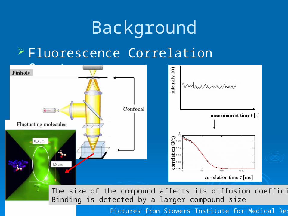
Background Fluorescence Correlation Spectroscopy
Pictures from Stowers Institute for Medical Research
The size of the compound affects its diffusion coefficientBinding is detected by a larger compound size
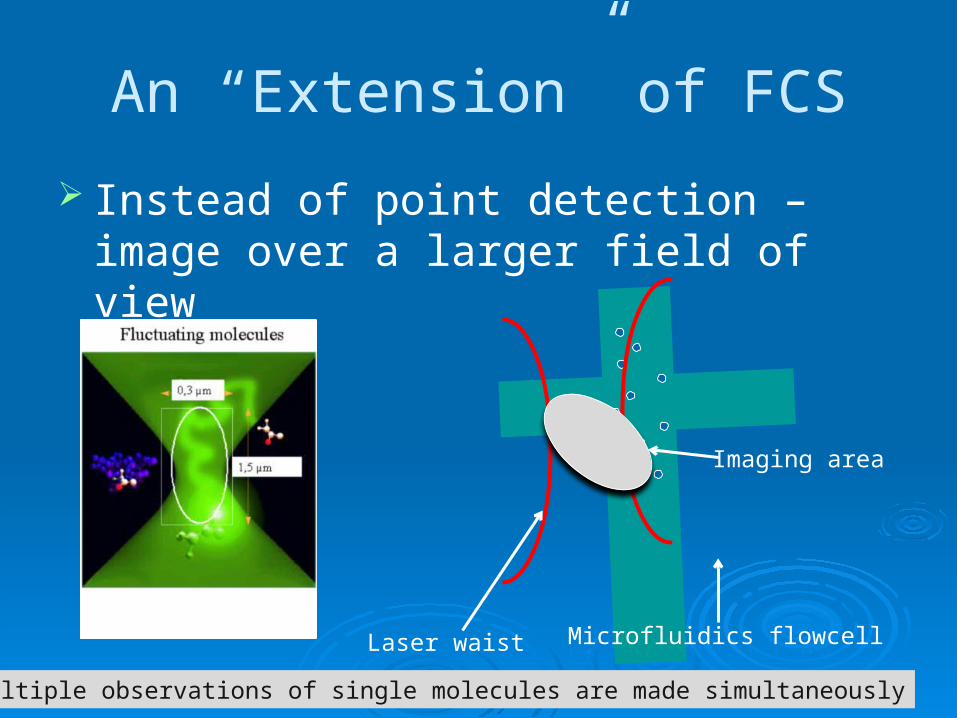
An “Extension” of FCS
Instead of point detection – image over a larger field of view
Laser waist
Imaging area
Microfluidics flowcell
Multiple observations of single molecules are made simultaneously

Single Particle Tracking (SPT)
Molecules are tracked across multiple image frames
Assumption: within each frame, any particle doesn’t move “much” (else blurring)
Frame 1 Frame 2 Frame 3

In Contrast to SPT…
Molecules are driven through the field of view by forced flow
Pressure-driven, EOF Molecules move “fast”
with respect to one image integration time
Results in blurring, or a particle “streak”
Horizontal: diffusion
Hypothesis: we can back-calculate diffusion information from the image streakF
orce
d flo
w

A Computer Simulation of SMD/SMI
Obj
CCD Detector
lem
Flow: Pressure, Eph, EOF
Through-objective TIR• Molecule transport• Flowcell interaction• Photophysics• Optics• CCD Detection• Noise
Specific Aim 1 Refine and optimize a computer model of
single fluorescent molecules imaged within a microfluidics flowcell To add:
• Molecule adsorption to flowcell wall• TIR intensity enhancement• Blinking• Readout blur• Updated objective, camera specifications
Compare with model system – DNA/SfiI complex

Specific Aim 2
Develop image-analysis based algorithms for measuring molecular interactions of ligand-receptor pairs from CCD images First, determine the “best” way to measure
diffusion

Specific Aim 2
Develop image-analysis based algorithms for measuring molecular interactions of ligand-receptor pairs from CCD images Next, determine how best to discriminate
between species of differing diffusion

Specific Aim 2
Develop image-analysis based algorithms for measuring molecular interactions of ligand-receptor pairs from CCD images Finally, determine the limits of measuring
diffusion

Specific Aim 2
Also, test the limits of feature identification How many molecules visible in this frame?

Specific Aim 3
Develop protocols in which the algorithms from Specific Aim 2 can be used to increase throughput and information content (compared to FCS) For example: Consider a sample composed of
a mixture of two different types of single molecules that have different diffusion constants - the goal of the measurement is to determine the fraction of each species

Specific Aim 3
Develop protocols in which the algorithms from Specific Aim 2 can be used to increase throughput and information content (compared to FCS) Parameters to study:
• Bulk flow• Ratio of diffusion coefficients• Concentration ratio of two species• Number of frames used in analysis

NIH Review Significance: “This project, if successful, could greatly increase the
rate of high-throughput screening and improve its efficiency and potentially its success rate … The techniques could also have broader applications for the study of the interaction of ligands with intact cells.”
Innovation: “This is a very innovative approach that will build a model for single molecule imaging that can improve screening and analysis of molecular interactions.”
Investigator: “The project investigator is highly skilled and has the resources to complete this project.”
Environment: “The environment is excellent, with a good mentoring program and the resources to perform the development. There is good complementarity to other COBRE projects.”
Section Score: Outstanding
(No changes recommended)

Current Work
Starting on Specific Aim 1 (refine model) Hosted visit from single-molecule
detection consultant (Dr. Lloyd Davis) Visited with Dr. Lyubchenko at UNMC
Goals for Spring 2009 Incorporate changes to model to allow for
diffusion measurement Submit publication with Dr. Davis