Simulation of Protein-Imprinted Polymers. 3. Imprinting Selectivity
6
Published: October 19, 2011 r2011 American Chemical Society 14469 dx.doi.org/10.1021/jp206940j | J. Phys. Chem. B 2011, 115, 14469–14474 ARTICLE pubs.acs.org/JPCB Simulation of Protein-Imprinted Polymers. 3. Imprinting Selectivity Liora Levi and Simcha Srebnik* Department of Chemical Engineering, Technion — Israel Institute of Technology, Haifa, Israel 32000 1. INTRODUCTION The formation of synthetic antibodies using molecular im- printing of biomacromolecules and proteins, 1 if successfully achieved, could potentially offer a generic and cost-effective alternative to existing biological recognition techniques such as monoclonal antibodies that are used in isolation, extraction, biosensors, and other laboratory practices. 2 6 The molecular imprinting procedure is simple and involves the interaction of a template molecule, or analyte, with functional groups via covalent or noncovalent bonds. The complex is then retained through polymerization and cross-linking of monomers to form the imprinted polymer matrix. When the template is removed, a cavity of complementary shape and functionality remains in the polymer, which can, in principle, selectively rebind the tem- plate. A number of alternative imprinting methods have been developed 7 with special interest and methods developed toward imprinting of molecules of biological relevance. Imprinting of small molecular templates, prominently drugs, has advanced significantly over the years, 7 with applications including clinical analysis, medical diagnostics, environmental monitoring, and drug delivery. 7 13 However, efforts to generate imprints of proteins have, in general, shown limited separation and show relatively high affinity toward competitor proteins. 3,5,14 The difficulties encountered with protein imprinting can be attributed to several factors, such as the use of water as solvent, the presence of multiple weak interactions on the surface of the protein, the relatively flexible conformation of the macromole- cule, and the large molecular size of the protein. 2,4,5 Various protocols have been developed to improve the performance of protein-imprinted polymers (PIPs), including surface imprinting or the epitope approach, 5,6,11 though the most extensive experimental work on PIPs has focused on three- dimensional imprinting using polyacrylamide gels (PAA) due to their biocompetability, neutrality, and inert nature that mini- mizes nonspecific interactions with proteins. Some of the im- portant experimental studies of PAA protein-imprinted gels are referred to in our previous work. 15 We recently carried out lattice Monte Carlo (MC) simulations of the imprinting process using radical polymerization of hydrogels as a simple model for such PIPs. 15,16 Apart from our work, several computational ap- proaches for molecularly imprinted polymers of small molecular templates (MIPs) have been introduced during the past decade. As recently reviewed in refs 17 19, most of these studies concentrate on prepolymerization complexation of the templates and functional monomers because a correlation between proper- ties of the prepolymerization solution and final imprinting efficiency has been shown to exist. 20 As discussed by Levi et al., 21 coarse-grained models of the imprinting process remark- ably are able to replicate and give some insight into the origins of experimentally measured trends of MIPs, including the imprint- ing effect, binding site distributions, and selectivities. Molecular simulation in this field, however, concentrates on MIPs. Our group was the first to consider coarse-grained modeling of PIPs. We introduced a lattice kinetic gelation model (KGM) 15 of a protein-imprinted polymer gel where, for simpli- city, the protein was modeled as a rigid body with randomly Received: July 20, 2011 Revised: September 22, 2011 ABSTRACT: Molecular imprinting has been extensively studied and applied as a simple technique for creating arti ficial polymer-based recognition gels for a target molecule. Although this technique is effective when targeting small molecules, attempts to extend it to larger templates, such as proteins, have, for the most part, failed to show similar success. Our group has developed a simple simulation model to study protein imprinting. In our previous studies, we investigated the structure of the protein-imprinted pore and imprinting factors of various model proteins. Here, we concentrate on imprinting conditions that affect the separation factor, or the ratio between the interaction energies of the template and a competitor protein. We study the effect of size, charge density, and surface charge distribution of the template protein and calculate the separation factor for various polymerization conditions. Our model captures the known effect of increasing imprinting factor (ratio of binding of the protein in an imprinted polymer to that of a nonimprinted polymer) with increasing surface functionality of the polymer but at the cost of reduced selectivity. Most interestingly, we observe that the surface charge distribution of the protein plays an important role in selectivity of the protein-imprinted polymer, suggesting that some proteins may be better candidates for molecularly imprinted polymers than others.
Transcript of Simulation of Protein-Imprinted Polymers. 3. Imprinting Selectivity

Published: October 19, 2011
r 2011 American Chemical Society 14469 dx.doi.org/10.1021/jp206940j | J. Phys. Chem. B 2011, 115, 14469–14474
ARTICLE
pubs.acs.org/JPCB
Simulation of Protein-Imprinted Polymers. 3. Imprinting SelectivityLiora Levi and Simcha Srebnik*
Department of Chemical Engineering, Technion — Israel Institute of Technology, Haifa, Israel 32000
1. INTRODUCTION
The formation of synthetic antibodies using molecular im-printing of biomacromolecules and proteins,1 if successfullyachieved, could potentially offer a generic and cost-effectivealternative to existing biological recognition techniques such asmonoclonal antibodies that are used in isolation, extraction,biosensors, and other laboratory practices.2�6 The molecularimprinting procedure is simple and involves the interaction of atemplate molecule, or analyte, with functional groups viacovalent or noncovalent bonds. The complex is then retainedthrough polymerization and cross-linking of monomers to formthe imprinted polymer matrix. When the template is removed, acavity of complementary shape and functionality remains in thepolymer, which can, in principle, selectively rebind the tem-plate. A number of alternative imprinting methods have beendeveloped7 with special interest andmethods developed towardimprinting of molecules of biological relevance.
Imprinting of small molecular templates, prominently drugs,has advanced significantly over the years,7 with applicationsincluding clinical analysis, medical diagnostics, environmentalmonitoring, and drug delivery.7�13 However, efforts to generateimprints of proteins have, in general, shown limited separationand show relatively high affinity toward competitor proteins.3,5,14
The difficulties encountered with protein imprinting can beattributed to several factors, such as the use of water as solvent,the presence of multiple weak interactions on the surface of theprotein, the relatively flexible conformation of the macromole-cule, and the large molecular size of the protein.2,4,5
Various protocols have been developed to improve theperformance of protein-imprinted polymers (PIPs), including
surface imprinting or the epitope approach,5,6,11 though the mostextensive experimental work on PIPs has focused on three-dimensional imprinting using polyacrylamide gels (PAA) dueto their biocompetability, neutrality, and inert nature that mini-mizes nonspecific interactions with proteins. Some of the im-portant experimental studies of PAA protein-imprinted gels arereferred to in our previous work.15 We recently carried out latticeMonte Carlo (MC) simulations of the imprinting process usingradical polymerization of hydrogels as a simple model for suchPIPs.15,16 Apart from our work, several computational ap-proaches for molecularly imprinted polymers of small moleculartemplates (MIPs) have been introduced during the past decade.As recently reviewed in refs 17�19, most of these studiesconcentrate on prepolymerization complexation of the templatesand functional monomers because a correlation between proper-ties of the prepolymerization solution and final imprintingefficiency has been shown to exist.20 As discussed by Leviet al.,21 coarse-grained models of the imprinting process remark-ably are able to replicate and give some insight into the origins ofexperimentally measured trends of MIPs, including the imprint-ing effect, binding site distributions, and selectivities.
Molecular simulation in this field, however, concentrates onMIPs. Our group was the first to consider coarse-grainedmodeling of PIPs. We introduced a lattice kinetic gelation model(KGM)15 of a protein-imprinted polymer gel where, for simpli-city, the protein was modeled as a rigid body with randomly
Received: July 20, 2011Revised: September 22, 2011
ABSTRACT:Molecular imprinting has been extensively studiedand applied as a simple technique for creating artificialpolymer-based recognition gels for a target molecule. Althoughthis technique is effective when targeting small molecules,attempts to extend it to larger templates, such as proteins,have, for the most part, failed to show similar success. Ourgroup has developed a simple simulation model to studyprotein imprinting. In our previous studies, we investigatedthe structure of the protein-imprinted pore and imprintingfactors of various model proteins. Here, we concentrate onimprinting conditions that affect the separation factor, or the ratio between the interaction energies of the template and acompetitor protein. We study the effect of size, charge density, and surface charge distribution of the template protein andcalculate the separation factor for various polymerization conditions. Our model captures the known effect of increasingimprinting factor (ratio of binding of the protein in an imprinted polymer to that of a nonimprinted polymer) with increasingsurface functionality of the polymer but at the cost of reduced selectivity. Most interestingly, we observe that the surface chargedistribution of the protein plays an important role in selectivity of the protein-imprinted polymer, suggesting that some proteinsmay be better candidates for molecularly imprinted polymers than others.

14470 dx.doi.org/10.1021/jp206940j |J. Phys. Chem. B 2011, 115, 14469–14474
The Journal of Physical Chemistry B ARTICLE
located functional sites. We carried out a systematic investigationof the structure and porosity of the imprinted gel for variouspolymerization parameters and studied the structure and func-tionality of the imprinted pore by diffusion of the model proteininside of the pore immediately following polymerization. One ofour prominent results showed that the binding energy of theprotein within the imprinted pore is strongly influenced by theoverall monomer concentration used in the prepolymerizationsolution, with minimal recognition of the protein at the percola-tion limit of protein-sized pores in the gel. PIP selectivity wasevaluated by comparing the interaction energy of the protein inthe imprinted gel with the energy of a random process (simulatedas a statistical configuration of functional monomers and cross-linkers in solution prior to equilibration and gelation). Thismeasure revealed that solution charge concentration has a con-siderable influence on the extent of recognition in the imprintedgel, achieving a maximum at relatively low charge densities, thatis, solutions containing a high concentration of functionalmonomers result in increased nonspecific interactions, as hasalso been reported in previous experimental work on proteinimprinting22 as well as simulations of model small moleculartemplates.23
We also compared binding energies and imprinting factors(IFs) of the protein within imprinted and nonimprinted poly-mers (NIPs) for various compositions and model proteins.16 Inour simulation, the IF is defined as the ratio of average bindingenergies of the template protein within the imprinted polymerand within a nonimprinted polymer. We focused in this part ontwo gel types, PIPs and the analogous templated polymers (TPs)formed by imprinting nonfunctional molecules of the same sizeand shape as the protein in the PIP. We found a significantmonotonic increase of IF with monomer concentration,Φ. HighΦ consistently showed up as the most influential factor forimprinting performance, as was seen in previous experimentalstudies,24 which, however, must be weighed against the lowdiffusion rates and protein entrapment that occur in low porosityimprinted gels.9,11,12 In contrast, TP gels showed an increase inIF with Φ only above the percolation limit, and their IF valueswere always significantly smaller than those of PIP gels, whichunderlines the essence of imprinting as it shows the significanceof combining shape and functionality in order to maximizerecognition, where the pore functionality complements thepositioning of functional groups on the template protein surface.
In this work, we focus on the separation factor, α, calculated asthe ratio of binding energies of the template protein within theimprinted pore to that of a competitive protein. Focusing on therole of protein size, protein charge density, and charge distribu-tion, we find that IF and α do not follow the same trends, so thatthe biomimetic ability of the PIP cannot always be optimized, orin other words, our results strongly suggest that certain proteinsmight be better candidates for PIPs than others.
2. SIMULATION MODEL
Our simulation presents a simple model of protein imprintingvia acrylate-based bulk free radical polymerization. Details of thesimulation are elaborated in our previous papers.15,16 Briefly, wecarry out lattice simulations, where lattice sites may be occupiedby neutral and charged (functional) monomers, cross-linkers,and the rigid cubic model protein. Empty lattice sites correspondto solvent molecules. The protein has a fraction, fp, of randomlydistributed charged residues that may interact with the charged
monomers in solution. For simplicity, a sufficiently screenedsolution is considered such that only nearest-neighbor interac-tions are considered. The charge concentration and fraction inthe polymerizing solution and the final gel are defined accordingto ϕg = Nc/L
3 and fg = Nc/N, respectively, where Nc representsthe number of charged functional monomers and N is the totalnumber ofmonomers in the simulation box of dimensionL. Notethat N is the initial number of particles (monomers and cross-linkers) in the simulation. In all simulations, we have consideredcases in which the gel fraction is close to unity, and therefore, thedifference betweenN and the actual number making up the gel isnegligible. Simulations were performed on a lattice with L = 20,which proved sufficiently large.25
The simulation model is depicted in Figure 1 in two dimensionsfor clarity. The simulation begins with random positioning of theprotein, cross-linkers, and monomers in the cubic lattice, followedby equilibration (formation of the prepolymerization complex),where noncovalent complexation takes place between the templateprotein and the functional monomers. The energy of the nearest-neighbor contact between a functional monomer and a chargedprotein residue is assigned a value of �ε, while all other nearest-neighbor contacts do not contribute to the energy. We assumescreened solution conditions, such that ionic interactions arefelt only between nearest neighbors. The dependence of thedielectric constant on the distance between charged species hasbeen indicated by recent experimental and simulation studiesof polyelectrolytes.26 Once equilibrium is reached, KGM is usedto simulate gelation,27�30 beginning with instantaneous initia-tion where radical “signatures” are randomly distributed among themonomers. Propagation of the polymerization reaction thenfollows and involves particle moves according to Metropolisacceptance rules and bond formation that takes place upon collisionof free radicals with other monomers. Gelation ends either when asingle cluster is formed, when all free radicals have terminated, orwhen the number of clusters does not change for the next 2� 105
iterations, which indicates entrapment of the remaining radicals. Atthis stage of our study, flexibility of the forming polymer chains isneglected, so that the system is quenched once gelation iscompleted and the templates are removed, providing a measureof the optimal recognition potential.20,31,32
In our previous work, we concentrated on the effect of variousparameters on the performance of the gel, focusing on the
Figure 1. Two-dimensional illustration of the lattice simulation modelfor (a) initialization and polymerization of NIP and (b) initiation,equilibration, and polymerization of PIP. The spheres represent mono-mers (white), functional monomers (blue), cross-linkers (black), neu-tral protein residues (yellow), and functional protein residues (red).

14471 dx.doi.org/10.1021/jp206940j |J. Phys. Chem. B 2011, 115, 14469–14474
The Journal of Physical Chemistry B ARTICLE
contribution of site-specific interactions through modeling gela-tion in the presence of both charged and neutral proteintemplates. The significantly higher IF seen in PIP over TP gelsindicated that our model captures the main features of theimprinting process, where the pore functionality complementsthe positioning of functional groups on the template proteinsurface. Here, we address the separation factor of PIP gels forcompetitor proteins that differ from the template in size, chargedensities, or charge distributions, as depicted in Figure 2.
3. RESULTS AND DISCUSSION
Protein versus Gel Functionality. We use the difference inbinding energy between the template protein and competitor,ΔEb = Eb
c � Ebt , as a measure of specific binding within the
imprinted pore. Figure 3 shows ΔEb as a function of the chargedresidue fraction on the protein surface (fp) and the fraction ofcharged monomers within the gel (fg). For a fixed fp, we find thatinitially increasing the amount of charged monomers in the gelincreases the probability to form specific binding, with the
difference being more pronounced at higher fg. That is, highlycharged gels more strongly associate with the template. However,energy in itself is clearly not a sufficient measure because, as iswell established,33 a high functional monomer�template ratioresults in MIPs with high nonspecific binding due to an excess ofcharges that are randomly distributed within the polymer matrix.On the other hand, ΔEb changes nonmonotonically with fp. Thedecrease in specificity is due in part to the increase in nonspecificbonds between the protein and template, but more importantly,it is due to the increasing similarity between the template and thecompetitor protein (at the extreme of fp = 1, the template andcompetitor proteins are identical). Hence, highly charged pro-teins are not expected to be good candidates for PIPs.The separation factor, α, is usually defined as the ratio between
the amount of bound template and competitor proteins. Withinthe simulation framework, one must carry out a grand canonicalsimulation that involves particle transfer from solution to gel toobtain the relative loadings of the two proteins. However, due tothe large size of the template, such transfers are extremely unlikelyand could only be carried using biased transfers to predeterminedsites of sufficient size within the gel. Because we mainly compareproteins of similar size, the dominant contribution to the partitionfunction is energetic. Hence, we calculate the average interactionenergy of our model proteins within such predetermined sites.The interaction energy of the template�monomer complex hasbeen shown to be correlated to the template�polymer bindingstrength,34,35 so that we estimate α as α = Eb
t /Ebc, where Eb
t and Ebc
are the binding energies of the template and the competitorprotein within the PIP, respectively. Figure 4 shows the effect ofΦonα for different charge fractions in the gel (fg) and on the protein(fp). While separation in general tends to improve with increasingΦ (higher separation factor), the slope ofα gradually decreases forany given charge fraction, and the decrease is more pronouncedwith high charge fractions. Moreover, for a highly charged system(circles in Figure 4), selectivity decreases with increasing geldensity, although binding of the protein consistently increaseswith increasing Φ.16 Clearly, charge multiplicity leads to non-specific binding, so that IF is not a sufficient measure in itself forpredicting imprinting efficiency. Moreover, Figure 4 clearly in-dicates that the effect of protein charge is a significantly more
Figure 2. Schematic of model proteins with (a) different charge fractions, (b) different charge distributions, and (c) different sizes. Gray representscharged residues and white neutral residues.
Figure 3. Effect of the fraction of charged monomers on the protein(black circles) and within the gel (white circles) on the difference inbinding energy between the competitor and the template protein.Φ = 0.4.

14472 dx.doi.org/10.1021/jp206940j |J. Phys. Chem. B 2011, 115, 14469–14474
The Journal of Physical Chemistry B ARTICLE
influential factor than the fraction of charged functional groups,with more pronounced selectivity for weakly charged proteins athighΦ (compare white symbols).Figure 5 shows more clearly the effect of charge on α for
different polymer volume fractions, again underlining the more
significant effect of protein charge on selectivity than functionalmonomer concentration. Furthermore, the percolation thresh-old (Φc = 0.2) distinguished the recognition behavior of the gel(Figure 5, black symbols), that is, separation is improved withincreasing charge below Φc but diminishes with increasingcharge above Φc. However, the behavior of fp at different Φ ismore complex. AboveΦc, highly charged proteins display poorerselectivity (opposite to the effect on IF16) because the presenceof a large number of charged groups on the protein surface on theone hand decreases the specificity during gelation and on theother hand improves the probability of finding similarly chargedareas on the template and on the competitor proteins. BelowΦc,however, a maximum in α is observed. For the weakly chargedproteins, adsorption is favored due to the relatively high func-tional monomer-to-template ratio. On the other extreme, thehighly charged template protein not only presents a relativelyindistinguishable surface for imprinting but also lacks a thermo-dynamic drive for the association of such a large number ofmonomers in the prepolymerization solution.We further compare the effects of fp and fg on α and IF for
Φ = 0.4 in Figure 6. Figure 6a shows that increasing charge ingeneral, both on the protein and in the gel decrease selectivity,but the effect of fp on α is clearly more significant. In contrast,at this gel density, increasing protein charge improves the IF.Hence, low concentrations of functional monomers in the gelwould lead to high imprinting efficiency and higher selectivity,albeit at low efficiencies (low binding).Imprinting simulations were performed using competitor
proteins with different charge fractions than the template pro-tein, shown in Figure 7, for example, for lysozyme (fp≈ 0.2) andcytochrome c (fp ≈ 0.6). For the same charge (but differentcharge distribution) on the competitor and the protein template,Figure 6 shows a slight decrease inαwith increasing charge of thetemplate protein (compare the outermost bars in Figure 7) dueto increased nonspecific bonding of the highly charged protein.We predict the highest selectivity for competitor proteins of lowercharge than the template protein, and in fact, when a highlycharged competitor protein is adsorbed within a gel imprintedwith a weakly charged protein,α takes on values lower than unity.That is, the best selectivity is expected for highly chargedtemplate proteins and weakly charged competitors. Reference36 observed similar results for lys (weakly charged protein)adsorption on the cytochrome c (highly charged protein)imprinted polymer gel. However, whether α takes on values lessthan unity for the highly charged cyt on a lys-imprinted gel is
Figure 5. Separation factor (α) as a function of fraction of chargedfunctional groups within the gel (black symbols, for fp = 0.5) and onthe protein surface (white symbols, for fg = 0.5) for various gel densities(Φ = 0.1 (circles), 0.2 (triangles), and 0.4 (squares);Φ = 0.2 marks thepercolation limit of protein-penetrable pores).
Figure 4. Effect of the monomer concentration (Φ) on selectivity (α)for various charge fractions of the gel (for fp = 0.5) and of the protein (forfg = 0.5).
Figure 6. Effect of gel charge (black symbols) and protein charge (white symbols) on (a) α and (b) IF for Φ = 0.4.
14473 dx.doi.org/10.1021/jp206940j |J. Phys. Chem. B 2011, 115, 14469–14474
The Journal of Physical Chemistry B ARTICLE
difficult to assess from the results of ref 36. To isolate the effect ofimprinting from that of charge, we compare the “separationfactor” between 0.2 and 0.6 charged proteins in TP, shown as thewhite columns in Figure 7. We clearly see an imprinting effect,though the effect of charge is significant. Presumably, ourpredicted results underestimate the expected results for realproteins that differ not only in charge density but also instructure, which also significantly contributes to the specificityof the imprinted pores.16
Protein Charge Distribution. Proteins differ not only in theamount of charged residues on their surface but also in thedistribution of charges. The order (or disorder) of charged groupsclearly will affect the functionality of the imprinted site and henceshould play an important role in recognition of the target protein.Five template charge distributions were considered in addition tothe random distribution studied thus far, depicted in Figure 2b,and the results are plotted in Figure 8 for the template proteinswith different charged distributions (competitor proteins have the
same charge concentration as the template but with a randomdistribution of charges). There are four features of Figure 8 thatstand out: (1) the imprinting effect is present for all proteinsconsidered (α > 1), (2) the randomly distributed templateprotein shows the poorest binding, (3) the protein with thesmallest patches shows the poorest selectivity (lowestα), and (4)there is an optimum patch size for best binding and selectivity.The even and uniform charge distribution of protein p1 leads tothe formation of uniformly functional pores, which can easily bindother charged proteins and hence shows low specificity. On theother hand, the relatively good binding and high specificity of p2suggests that proteins with a somewhat patchy charge distribution(such as cyt shown in the inset of Figure 7) may serve as bettertemplates than proteins with either a more uniform or randomcharge distribution. Very large patches, however, again lead tolower selectivity because the large surfaces of these proteins(p3�p5) present a smooth charged surface that can easily interactwith the random patches of the competitor proteins. Such charac-terization of the protein surface would be revealing and couldguide experimentation such as that by Searson and co-workers,37
who have recently considered the tuning of prepolymerizationparameters according to the functional monomer distribution onthe protein surface.Protein Size. We carried out simulations for proteins of
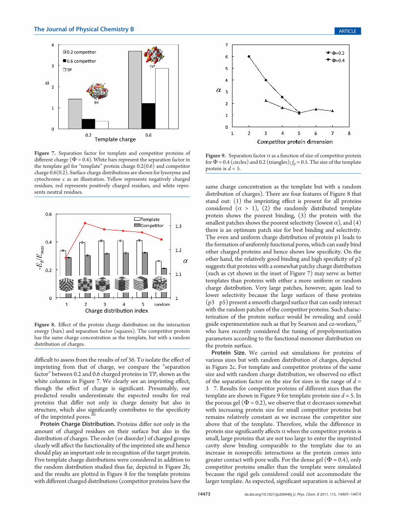
various sizes but with random distribution of charges, depictedin Figure 2c. For template and competitor proteins of the samesize and with random charge distribution, we observed no effectof the separation factor on the size for sizes in the range of d =3�7. Results for competitor proteins of different sizes than thetemplate are shown in Figure 9 for template protein size d = 5. Inthe porous gel (Φ = 0.2), we observe that α decreases somewhatwith increasing protein size for small competitor proteins butremains relatively constant as we increase the competitor sizeabove that of the template. Therefore, while the difference inprotein size significantly affects αwhen the competitor protein issmall, large proteins that are not too large to enter the imprintedcavity show binding comparable to the template due to anincrease in nonspecific interactions as the protein comes intogreater contact with pore walls. For the dense gel (Φ = 0.4), onlycompetitor proteins smaller than the template were simulatedbecause the rigid gels considered could not accommodate thelarger template. As expected, significant separation is achieved at
Figure 7. Separation factor for template and competitor proteins ofdifferent charge (Φ = 0.4). White bars represent the separation factor inthe template gel for “template” protein charge 0.2(0.6) and competitorcharge 0.6(0.2). Surface charge distributions are shown for lysozyme andcytochrome c as an illustration. Yellow represents negatively chargedresidues, red represents positively charged residues, and white repre-sents neutral residues.
Figure 8. Effect of the protein charge distribution on the interactionenergy (bars) and separation factor (squares). The competitor proteinhas the same charge concentration as the template, but with a randomdistribution of charges.
Figure 9. Separation factor α as a function of size of competitor proteinforΦ = 0.4 (circles) and 0.2 (triangles); fp = 0.5. The size of the templateprotein is d = 5.

14474 dx.doi.org/10.1021/jp206940j |J. Phys. Chem. B 2011, 115, 14469–14474
The Journal of Physical Chemistry B ARTICLE
high Φ when the size of the template protein is considerablydifferent than the competitor protein.
4. CONCLUSIONS
This work presents continuing work on the simulation ofprotein imprinting. In our previous work, we studied the effect ofdifferent simulation parameters on the gel structure, pore func-tionality, and imprinting factors (IFs) of PIP and templatedpolymer (TP) gels. In the present paper, we concentrated on theeffect of various parameters on the performance of the imprintedpolymer, focusing on the separation factor, α, which measuredthe binding capacity of the imprinted gel toward its templatecompared with a competitor protein.We studied the effect of twoproperties of the gel, the polymer density (Φ) and chargefraction within the gel (fg) on the separation factor, in additionto the size, charge density, and charge distribution of the templateprotein.
Our results show that α decreases in highly charged systemsdue to increased nonspecific binding. Hence, increasing theprotein charge impairs PIP selectivity toward the templateprotein because despite the higher IFs, it does not improve theseparation of the gel. On the other hand, there is a minimalcharge concentration that is sufficient for recognition, andtherefore, for low density gels, high charge fractions are favorable.When using competitors of various charge densities, we foundthat a highly charged competitor protein has a notable advantageover a poorly charged template. This result points to theconclusion that in cases of similar protein morphology, theimprinting process is not an efficient tool for protein separation.We conclude that proteins with a relatively high concentration ofcharged surface residues that are distributed in patches may bethe best candidates for PIPs. That is, these proteins havedistinguishing surface characteristics and can therefore be im-printed. Our current efforts, therefore, concentrate on classifyingsurface properties of proteins.
’AUTHOR INFORMATION
Corresponding Author*E-mail: [email protected]. Tel. +972-4-8293584. Fax+972-4-8295672.
’ACKNOWLEDGMENT
This research was supported, in part, by the Israel ScienceFoundation.
’REFERENCES
(1) Ge, Y.; Turner, A. P. F. Trends Biotechnol. 2008, 26, 218.(2) Bossi, A.; Bonini, F.; Turner, A. P. F.; Piletsky, S. A. Biosens.
Bioelectron. 2007, 22, 1131.(3) Hansen, D. E. Biomaterials 2007, 28, 4178.(4) Turner, N. W.; Jeans, C. W.; Brain, K. R.; Allender, C. J.; Hlady,
V.; Britt, D. W. Biotechnol. Prog. 2006, 22, 1474.(5) Zhou, X.; Li, W. Y.; He, X. W.; Chen, L. X.; Zhang, Y. K. Sep.
Purif. Rev. 2007, 36, 257.(6) Takeuchi, T.; Hishiya, T. Org. Biomol. Chem. 2008, 6, 2459.(7) Alexander, C.; Andersson, H. S.; Andersson, L. I.; Ansell, R. J.;
Kirsch, N.; Nicholls, I. A.; O’Mahony, J.; Whitcombe, M. J. J. Mol.Recognit. 2006, 19, 106.(8) Jiang, X. M.; Jiang, N.; Zhang, H. X.; Liu, M. C. Anal. Bioanal.
Chem. 2007, 389, 355.
(9) Hilt, J. Z.; Byrne, M. E. Adv. Drug Delivery Rev. 2004, 56, 1599.(10) Alvarez-Lorenzo, C.; Concheiro, A. J. Chromatogr., B 2004,
804, 231.(11) Sellergren, B.; Allender, C. J. Adv. Drug Delivery Rev. 2005,
57, 1733.(12) Duarte, A. R. C.; Casimiro, T.; Aguiar-Ricardo, A.; Simplicio,
A. L.; Duarte, C. M. M. J. Supercrit. Fluids 2006, 39, 102.(13) Zhang, H. Q.; Ye, L.; Mosbach, K. J. Mol. Recognit. 2006, 19,
248.(14) Fu, G. Q.; Yu, H.; Zhu, J. Biomaterials 2008, 29, 2138.(15) Levi, L.; Srebnik, S. J. Phys. Chem. B 2010, 114, 107.(16) Levi, L.; Srebnik, S. J. Phys. Chem. B 2010, 114, 16744.(17) Levi, L.; Raim, V.; Srebnik, S. J. Mol. Recognit. 2011In Press.(18) Nicholls, I. A.; Andersson, H. k. S.; Golker, K.; Henschel, H.;
Karlsson, B. r. C. G.; Olsson, G. D.; Rosengren, A. M.; Shoravi, S.;Suriyanarayanan, S.; Wiklander, J. G.; Wikman, S. Anal. Bioanal. Chem.2011, 400, 1771.
(19) Nicholls, I. A.; Andersson, H. S.; Charlton, C.; Henschel, H.;Karlsson, B. C. G.; Karlsson, J. G.; O’Mahony, J.; Rosengren, A. M.;Rosengren, K. J.; Wikman, S. Biosens. Bioelectron. 2009, 25, 543.
(20) Idziak, L.; Benrebouh, A.; Deschamps, F. Anal. Chim. Acta2001, 435, 137.
(21) Levi, L.; Raim, V.; Srebnik, S. J. Mol. Recognit. To Appear.(22) Janiak, D. S.; Kofinas, P. Anal. Bioanal. Chem. 2007, 389, 399.(23) Dourado, E. M. A.; Sarkisov, L. J. Chem. Phys. 2009, 130,
214701.(24) Ou, S. H.; Wu, M. C.; Chou, T. C.; Liu, C. C. Anal. Chim. Acta
2004, 504, 163.(25) Levi, L.; Srebnik, S. Macromol. Symp. 2010, 291�292, 258.(26) Yin, D. W.; Horkay, F.; Douglas, J. F.; de Pablo, J. J. J. Chem.
Phys. 2008, 129, 11.(27) Herrmann, H. J.; Landau, D. P.; Stauffer, D. Phys. Rev. Lett.
1982, 49, 412.(28) Bansil, R.; Herrmann, H. J.; Stauffer, D. Macromolecules 1984,
17, 998.(29) Ghiass, M.; Rey, A. D.; Dabir, B. Polymer 2002, 43, 989.(30) Bowman, C. N.; Peppas, N. A. Chem. Eng. Sci. 1992, 47, 1411.(31) Molinelli, A.; O’Mahony, J.; Nolan, K.; Smyth, M. R.; Jakusch,
M.; Mizaikoff, B. Anal. Chem. 2005, 77, 5196.(32) Piletsky, S. A.; Karim, K.; Piletska, E. V.; Day, C. J.; Freebairn,
K. W.; Legge, C.; Turner, A. P. F. Analyst 2001, 126, 1826.(33) Andersson, H. S.; Nicholls, I. A. Bioorg. Chem. 1997, 25, 203.(34) Wu, L. Q.; Li, Y. Z. J. Mol. Recognit. 2004, 17, 567.(35) Wu, L. Q.; Sun, B. W.; Li, Y. Z.; Chang, W. B. Analyst 2003,
128, 944.(36) Kimhi, O.; Bianco-Peled, H. Langmuir 2007, 23, 6329.(37) Zayats, M.; Kanwar, M.; Ostermeier, M.; Searson, P. C.Macro-
molecules 2011, 44, 3966.
![Fundamental Studies of Molecular Interactions in Complete …280012/FULLTEXT01.pdf · 2009. 12. 8. · imprinted polymers (MIPs) [1]. Molecular imprinting [2] has been defined as:](https://static.fdocuments.in/doc/165x107/5fdb907f6521f1278e14dd0a/fundamental-studies-of-molecular-interactions-in-complete-280012fulltext01pdf.jpg)


![High Affinity Polymers by Molecular Imprinting for Drug ...€¦ · Imprinted polymers are synthetic receptors with binding constants comparable to natural receptors [9,10], but capable](https://static.fdocuments.in/doc/165x107/60605123d701475a1426d31c/high-affinity-polymers-by-molecular-imprinting-for-drug-imprinted-polymers-are.jpg)