Simulation And Other Tools To Enhance Student Learning
13
University of Nebraska - Lincoln DigitalCommons@University of Nebraska - Lincoln Transactions of the Nebraska Academy of Sciences and Affiliated Societies Nebraska Academy of Sciences 1972 Notes on the Deciduous and Permanent Dentition of the Hyracodonts Lloyd G. Tanner University of Nebraska State Museum Larry D. Martin University of Nebraska State Museum Follow this and additional works at: hp://digitalcommons.unl.edu/tnas is Article is brought to you for free and open access by the Nebraska Academy of Sciences at DigitalCommons@University of Nebraska - Lincoln. It has been accepted for inclusion in Transactions of the Nebraska Academy of Sciences and Affiliated Societies by an authorized administrator of DigitalCommons@University of Nebraska - Lincoln. Tanner, Lloyd G. and Martin, Larry D., "Notes on the Deciduous and Permanent Dentition of the Hyracodonts" (1972). Transactions of the Nebraska Academy of Sciences and Affiliated Societies. 362. hp://digitalcommons.unl.edu/tnas/362
Transcript of Simulation And Other Tools To Enhance Student Learning

University of Nebraska - LincolnDigitalCommons@University of Nebraska - LincolnTransactions of the Nebraska Academy of Sciencesand Affiliated Societies Nebraska Academy of Sciences
1972
Notes on the Deciduous and Permanent Dentitionof the HyracodontsLloyd G. TannerUniversity of Nebraska State Museum
Larry D. MartinUniversity of Nebraska State Museum
Follow this and additional works at: http://digitalcommons.unl.edu/tnas
This Article is brought to you for free and open access by the Nebraska Academy of Sciences at DigitalCommons@University of Nebraska - Lincoln. Ithas been accepted for inclusion in Transactions of the Nebraska Academy of Sciences and Affiliated Societies by an authorized administrator ofDigitalCommons@University of Nebraska - Lincoln.
Tanner, Lloyd G. and Martin, Larry D., "Notes on the Deciduous and Permanent Dentition of the Hyracodonts" (1972). Transactionsof the Nebraska Academy of Sciences and Affiliated Societies. 362.http://digitalcommons.unl.edu/tnas/362

EAR TH SCIENCE
NOTES ON THE DECIDUOUS AND PERMANENT DENTITION OF THE HYRACODONTS
LLOYD G. TANNERI
LARRY D. MARTIN2
ABSTRACT
Study of radiographs and dissections prepared from nearly fifty Hyracodon jaws in the collections of the University of Nebraska State Museum have demonstrated the normal presence of the lower premolar one in immature jaws of this genus. This tooth is functional in the young animal but is lost with the deciduous dentition and is not replaced. In the upper dental series permanent premolar one was preceded by a deciduous premolar one, which is also characteristic of the tapirs, but is unusual in the other perissodactyla.
INTRODUCTION
The milk dentition of the Hyracodontinae is unique in several respects from the teeth of other families and subfamilies of the Order Perrisodactyla, and as a result of this, there has been some misinterpretation in the identification of premolars in the deciduous series of the Oligocene Hyracodon.
Information has been obtained through the study of a series of over one hundred hyracodont jaws in the University of Nebraska State Museum Study Collection. Fifty of these jaws were either x-rayed or dissected, in order to examine the development of the unerupted teeth.
The dental formulae for juvenile and adult hyracodonts are as follows: juvenile dentition: DI §, DC {, DP~; adult dentition: 11, C t, p!, M 1·
DESCRIPTION OF INCISORS AND CANINES
The deciduous incisors and canines are relatively small, thin teeth, about one half the size of the permanent teeth (Plate 1, Fig. b). 13 and 13 were developed fully before the MI erupted. II and ]1, and 12 and 13 followed in short succession after the permanent upper and lower molar one erupted. The canines of both the uppcr and lowcr dentition came into place after ]3 and
]3·
DISCUSSION AND DESCRIPTION OF THE LOWER DENTITION
DP 1: (Plate I, Fig. d) This is a small tooth ranging in size from 9 to 10.5 mm. antcfoposteriorly, and the transverse dimension is usually near 6 mm. It
1 Coordinator of Systematic Collections, University of Nebraska State Museum. 2 Research Assistant, Department of Geology and University of Nebraska State Museum;
field party leader, Division of Vertebrate Paleontology of the Museum.
61

EARTH SCIENCE
has a large central protoconid, a small hypoconid and an equally small paraconid. A crescent-shaped facet developed on the posterior one-half of the DP 1 [rom the occlusial wear with the protocone of Dp l (Plate I, Fig. b). DP 1
has two well-developed rulltS, a strong internal cingulum, and it 111ay or may not have an external cingulum. DP 1 was never replaced, but as the permanent premolars eru pted it was transloca ted anteriorly toward the posterior portion of the diastema where it was pushed off with relatively little reabsorption of the roots (Plate 2, Fig. a). Troxell (1921, p. 39) pOinted out the presence of
an alveolus for a lower premolar one, on the holotypic ramus of Hyracodon lciJyallus, but apparently did not realize that it was a consistant feature of hyracodont deciduous dentitions.
DP 2 (Plate I, Fig. d) The anterior portion of the tooth is narrower than the posterior dimension forming a sub·triangular shaped tooth. It has a
shorter anteroposterior dimension than DP 3' It is also characterizcd by having both internal and external cingula, and a well-developed paraconid.
DP 3: (Plate 1, Fig. d) The DP 3 is the largest of the lower deciduous premolars, it is nearly rectangular in shape, and usually has external and internal cingula. The external cingulum is normally slightly interrupted at the
mid-point of the tooth. The hypolophid and metalophid of DP 2 and DP 3 are "L" shaped rather than being rounded as arc the other teeth of the deciduous and permanent series.
DP 4: (Plate 1, Fig. d) This tooth is very molariform, smaller than DP 3'
and has an external cingulum which thins at the midpoint of the tooth. The
ectoloph is discontinuous, and the hypolophid is continuous with the internal cingulum. Since the DP 4 is very molariform it may be confused with the umvorn MI' WOod (1927, PIa te 13, Figs. 12 and 13), for example, interpreted two lower jaws to have the series "DP 2- M1 ." However, the anterior alveolus shown in his illustration is for the DP l' The corrected series would therefore be DP 1 (alv.) - DP 4' Butler (1952) also states that the corrected series for these specimens should be DP 1 - DP 4'
The ramus in young byracodonts lacks calcification relative to that of the mature jaws. The juvenile jaw in all cases observed is expanded, to make room for tbe developing teeth, at a point near the posterior portion of the jaw. There are three or four mental foramina which extend from the posterior edge of the DP 3 to the anterior of DP l' (This number is usually reduced to one or two upon maturity.) One of the youngest rami in the collection (Plate 1, Fig. a) has a depth posterior to DP3 of 16 mm., which is less than one-half
the depth the ramus would probably have had upon maturity. The sequence of tooth replacement in the hyracodonts was observed
through the usc of x-rays (Martin, 1967) and dissections. Tn the case of the deciduous teeth, no specimens of very young jaws were available for study which could be used to determine the time at which the DP 1 erupted. DP 2
and DP 3 erupted prior to the DP 4'
62

EARTH SCIENCE
PLATE 1 Pig. a. Immature Hyracodon ramus V.N.S.M. 11031 showing DP 3 - DP 4' X 1. Pig. b. The skull and lower mandible of an immature hyracodont V.N.S.M.
11036 showing the deciduous incisors, as well as Dpl - DP4, a trace of M 1 just erupting, and DP I - DP 4' X %.
Pig. c. A hyracodont M1 , and its crypt V.N.S.M. 11075, weathered out of the ramus. x 1.
Fig. d. DP 1 - MI of Hyracodon V.N.S.M.ll0n. x 1.
63

EARTH SCIENCE
...0
PLATE 2 Fig. a. An immature Hyracodon ramus U.N.S.M. 11002 with M3 about to
erupt. The fully erupted teeth are DP 1 - M2 . x %. Fig. b. A section through a Hyracodon ramus U.N.S.M. 11077 showing the
relationship of the tooth roots to the trabecular bone tissue. (not to scale).
Fig. c. A Hyracodon ramus U.N.S.M. 11059 with DP 4 - M2 erupted and P3 -P 4 erupting simultaneously. DP 4 is resting on the crown of P 4' X V2.
Fig. d. A ventral section of a Hyracodon ramus U.N.S.M. 11078 showing the trabecular bone tissue just above the mandibular canal. (not to scale).
64

EARTH SCIENCE
PLATF 3 Fig. a. A Iogitronic· print of a x-ray of an immature Hyracodon ramus
U.N.S.M. 11050. DP 4 was present on the original specimen but did not print and was later sketched in as is also true for the erupted teeth in the following figures of x-ray negatives. The crypt for P 4 is visible while that for P is not yet evident. x 1.
l'ig. b. A print or a x-ray of a Hyracodon ramus U.N.S.M. 11053 showing P 2-4
migrating upwards directly under DP 2-4' Ml _2 are erupted and M3 can be seen in its crypt in the ascending ramus. x I.
65
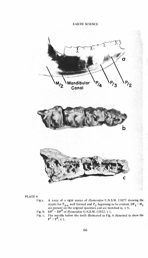
PLATE 4 Fig a.
Fig. b. Fig. c.
EARTH SCIENCE
A x-ray of a right ramus of Hyracodon D.N.S.M. 11077 showing the crypts for P 3-4 well formed and P 2 beginning to be evident. DP 1 - Ml are Fresenl on the original specimen and are sketched in. x 'h. DP - DP of Hyracodon D.N.S.M. 11022. xL The maxilla below the teeth illustrated in Fig. 6 dissected to show the pI _ p4. X 1.
66

EARTH SCIENCE
..a
PLATE 5 Fig a. DP 1-4 of Hyracodon V.N.S.M. 11026. This specimen is from the large
size group of deciduous dentitions. x 1. Fig. b. X-ray of Dendrohyrax showing positions of developing molars. x 1. Fig. c. Anterior portion of a mandible of Hyracodon with an enlarged canine.
(not to scale). Fig. d. DP 1 of Hyracodon V.N.S.M. 11076. This specimen is from the small
size group of deciduous dentitions. x 1. Fig. e. PI of Hyracodon V.N.S.M.lIOO1. x 1. Fig. f. Left DP 1-3 of Hyracodon V.N.S.M. 11002 showing pronounced
interstitial wear between DP 1 and DP 3' x 1.
67

EARTH SCIENCE
The MI , before its eruption, occupied a crypt formed by the reabsorp_ tion of the surrounding trabecular bone tissue (Plate 2, Figs. b and d), and during this time the unerupted M I is enclosed in a shell of compacted bone tissue. (Plate 1, Fig. e, shows an M I' which has been weathered out of its ramus, prior to collection, but is still surrounded by this shell of compacted bone.) The bud of the MI before translocation, is tilted only slightly in comparison to the tooth germs of the other molars. The positions of the permanent molars and premolars prior to eruption are very similar to the unerupted teeth ofhyracoids(Plate 4, Fig. a and Plate 5, Fig. b).
The M2 began to form prior to the eruption of MI and by the time roots had begun to form under the MI , the roots of the deciduous premolars had nearly reached their greatest length. Following the eruption of the MI , a small oval crypt formed, approximately posterior to the center line of the ramus, and under the DP 4 (Plate 3, Fig. a), also, at this stage, reabsorption of the DP 4 roots began. The same procedure took place under the DP 3 and at nearly the same time (Plate 4, Fig. a). Further development of these deciduous teeth followed as the ventral borders of the crypts extended gradually downward until they rested on the top of the mandibular canal. Also during these processes the crypt for the P 2 was being formed, and it too extended to the top of the mandibular canal. In this study no evidence has been found which would indicate that a crypt ever formed under the DP I' The crypts are at first oval-shaped then gradually become heart-shaped, with the apex between the roots of the deciduous premolars, reabsorbing the roots on either side (Plate 4, Fig. a).
The development of P 3 and' P 4 occurred before that of P 2 (Plate 4, Fig. a). This process of tooth formation commenced after the eruption of the MI ,
but before the roots completely formed on that molar. The last two premolars appear to have formed almost simultaneously and migrated upward together (Plate 3, Fig. b). As these teeth approached the upward limit of the dentary line they took their places nearly at the same time (Plate 2, Fig. c). In other words, by tlle time the MI and M2 have erupted and the M3 is formed (except for the roots) the roots of DP 2 through DP 4 have been almost completely reabsorbed, finally as the M3 started to erupt, the permanent premolars also commenced to extrude: The P 3 and P 4 came into place almost at this same time. According to the evidence at hand, either of these teeth may have come into place prior to the other; P 2 is the last of the premolars to be replaced in the series.
UPPER DENTITION
The premolars of the upper dentary are more molariform than are tlle permanent premolars, and the medial valleys of these teeth are open. DP! is replaced (Plate 4, Figs. band c), and the DP I is nearly one-half larger at tlle
68

EAR TH SCIENCE
anteroposterior and transverse dimensions than the permanent pI, and is triangular shaped. It has a hook-shaped metaloph which is often more elaborate in design than the metaloph of the pl. The Dp1 occludes against the DP 1 and DP 2 and the large posteriorly directed protoloph of the Dpl
wears against the posterior portion of the DP l' forming a crescent-shaped facet on the tooth. Dpl is lost with the deciduous dentition and pI mayor may not have a protoloph, if one is present, it is usually small.
In most cases Dp2 and Dp3 had a style developed on the anterior edge of the medial valleys and both had well-developed cristae. Dp4 is the largest tooth in the deciduous series and also has the medial valley open. The first three deciduous premolars all erupt at nearly the same time and DP4 is the last to take its place.
SUMMARY AND CONCLUSIONS
Very little previous work has been done on tooth replacement in the hyracodontids. Sinclair (1922, Fig. 6) does illustrate a very worn upper cheek-dentition of Hyracodon with Dpl-4. Wood, when discussing the Hyrachyidae suggests that DP + are not replaced in that family nor in Hyracodorl (Wood, 1934, p 263). Very little opposition has been offered to this view although the possibility of the replacement of the Dpl has been considered (Butler, 1952). That this situation should have been maintained for such a long period of time is strange, as we have shown in this study that the DPI and pI are very disimilar teeth (Plate 5, Figs. a and e). The presence of the pI beneath the Dpl has been demonstrated by both x-ray negatives and dissections (Plate 4, Figs. band c). The upper deciduous cheek-teeth of Hyracodol1 very clearly show that they belong to the same tooth series as the molar teeth, as they are all more molariform than their replacements. The development of the permanent premolars was very rapid as was pointed out by Sinclair (1922, p. 75) and most of it took place after the eruption of MI.
The lower dentition is exceptional in the presence of a large functional DP 1 which is not replaced, but is lost with the deciduous dentition. This tooth is absent from most Tapir and Rhinoceros dentitions, and when present is usuaIly vestigial. In Hyracodol1 the DP l' along with the larger Dp l
contributes a significant portion of the grinding surface in the mouth of the young animal. The molar teeth are formed in the ascending ramus as in the hyracoids (Plate 5, Fig. b) and moved down and then up into place behind the previously erupted teeth. The ramus apparently grows enough in length to accomodate these teeth until the eruption of M3, at which time the tooth row is translocated forward and the DP 1 is pushed off the ramus in a manner similar to that of the proboscidia. This causes a large amount of interstitial wear on the deciduous premolars (Plate 5, Fig. 0. The mandible grows in depth very rapidly before the eruption of Ml . This translocation can be
69

EARTH SCIENCE
16
2 .
7
• 5
18
-S
-7
• I I I I I I I 10 15 20 25 30 35 40
PLATE 6 Graph showing relative increase in depth of Hyracodon rami in millimeters measured posterior to DP 3 until the eruption of Ml and then the dimension is taken posterior to the P 3' The horizontal lines indicate the total range in depth, and the sample size is given below the lines. A single dot means a sample of one specimen. The measurements are taken as each tooth is beginning to erupt = ( ) and after full eruption. A decrease in depth is shown during the eruption of M3 at which time the tooth row is translocated forwardly towards the thinner portion of the ramus.
shown graphically by plotting the depth of the ramus below the posterior edge of DP 3 at the time of eruption of each of the following teeth (Plate 6). This dimension shows a decline when P 3 is used as the tooth row has moved anteriorly with eruption of M3 and the ramus tapers in that direction.
We have at the present time noted few characters which might be due to sexual dimorphism in Hyracodon. However, certain specimens which share the same cheek-tooth pattern may differ in the size of the lower canines. We
70

EARTH SCIENCE
presume that the larger canines belong to males (Plate 5, Fig. c), although this probably cannot be proved. The deciduous dentitions can be readily separated into groups of large and small size (Plate 5, Figs. a and d). These size groupS roughly correspond to the stratigraphic horizon of the specimens, with the larger being later in time. Their exact relationship to the various described species of Hyracodon have not been worked out, but they do lend additional support to the concept that more than one species is involved. This is contrary to the view presented by Scott (1941, p 841) that the several species of Hyracodon which have been erected are variants of !:!. nebraskensis (Leidy). Radinsky (1967, p. 30) concurred with Scott and lists If. /lcbraskensis as the "type and sole species." Specific differences can also be observed in the permanent dentition and in the skull leading the writers to believe that there are at least four or five separate species of Hyracodon present in the Oligocene (Tanner, 1965, p. 45).
Radinsky (1966, p. 633) includes the Hyrachyinae as a subfamily of the tapiroid family Helaletidae. The Hyrachyinae probably gave rise to the hyracodontids independently from the rhinocerotoidea and if the Hyrachyinae is included with the tapirs so should the hyracodontids which are tapir-like in the presence of Dp! , as well as in the form of their canines and incisors, be included in the Tapiroidea.
ACKNOWLEGEMENTS
We are indebted to Professors C. Bertrand Schultz and Harvey L. Gunderson of the University of Nebraska State Museum for their aid in preparation of this manuscript, also to Professor Donald T. Waggener and Carol Thompson for the preparation of x-rays used in this study. Mesdames Mary Cutler and Lorenc Bartos assisted in the final typing of the manuscript, and Mr. David Nixon prepared Plates 1 and 3, and Mr. Jerry Tanner prepared Plates 5 and 6.
REFERENCES CITED
Butler, P. M., 1952. The milk-molars of Perissodactyla, with remarks of molar occlusion: Proc. Zool. Soc.Lond., 121: 777-817.
Martin, Larry D., 1966. Tooth replacement in Hyracodon (abstract): Proe. Nebr. Acad. Sci., 76th Ann. Meeting: 15.
___ 1967. X-ray techniques for the study of fossil vertebrates: Compass of Sigma
Gamma Epsilon, 44(2): 101-103, Plates 1-3. Radinsky, Leonard B., 1966. The families of the Rhinocerotoidea. (Mammalia,
Perissodactyla): Jour. of Mamm., 47(4): 631-639. ___ 1967. A review of the Rhinocerotoid Family Hyracodontidac (Perissodactyla):
Bull. of Amer. Mus. Nat. Hist., 136(1): 1-46, Plate 1, Figs. 1-25, tables 1-6. Scott, W. B., 1941. Perissodactyla, In Scott, W. B., and G. L. Jepsen. The mammalian
fauna of the White River Oligocene: Trans. Amer. Phil. Soc., new ser., 34(3): 747-980.
Simpson, G. S., 1945. The principles of classification and a classification of mammals: Bull. Amer. Mus. Nat. Hist., 85: 1-350 and i-xvi.
Sinclair, W. J., 1922. Hyracodons from the Big Badlands of South Dakota: Proc. Amer. Phi\. Soc., 61(E): 65-79.
71

EARTH SCIENCE
Tanner, Lloyd G., 1965. The geologic history of Nebraska's Rhinoceroses: (Past Presidential Address) Proc. Nebr. Acad. Sci., 75th Ann. Meeting: 41-45.
Troxell, E. C., 1921. New species of Hyracodon: Amer. Jour. of Sci., 5th(1l): 34-40 Figs. 15 '
Wood, H. E., 1927. Some early Tertiary rhinoceroses and hyracodonts: Bull. Amer. Paleo., 13(50) 5-105, Plate 5 i-vi~ tables i-vii.
___ 1934. Revision of the Hyrachyidae: Bull. Amer. Mus. Nat. Hist., 67(5): 181-295.
1938. Casual factors in shortening tooth series with age: Jour. of Dent. --research., 17(1): 1-13.
72