Simple atrophic gastritis and gastric carcinoma
6
Gut, 1971, 12, 906-911 Simple atrophic gastritis and gastric carcinoma 1. R. WALKER,' R. G. STRICKLAND, B. UNGAR, AND I. R. MACKAY From the Clinical Research Unit, the Walter and Eliza Hall Institute of Medical Research and Royal Melbourne Hospital, Melbourne, Australia SUMMARY Gastric carcinoma was detected nine, 10, 18, and 21 years after the biopsy diagnosis of atrophic gastritis in four patients of a group of 40. The gastritis was presumed to be of the simple type. Tests of vitamin B,2 absorption in three patients gave normal results, no gastric autoantibodies were detected in the two patients tested, in all patients histological examination of the gastrectomy specimens revealed a multifocal gastritis differing from the diffuse gastritis of per- nicious anaemia and in three patients the gastritis affected the antrum, which is unusual in pernicious anaemia. The 10 % incidence of gastric carcinoma in 40 patients with simple atrophic gastritis followed for a mean period of 15 years is equivalent to that previously described in pernicious anaemia. However, in view of the relative incidence of atrophic gastritis with and without pernicious anaemia in the general adult population, it emerges that atrophic gastritis without pernicious anaemia is numerically the more important precursor of gastric carcinoma. In patients with Addisonian pernicious anaemia, in whom atrophic gastritis is invariably present, there is said to be an increased incidence of gastric carcinoma (Magnus, 1958). Moreover, gastritis with intestinal metaplasia was described in the mucosa around a gastric carcinoma or in areas remote from the tumour site in 36 of 39 stomachs removed sur- gically (Morson, 1955a and b), but it is uncertain whether the gastritis accompanying gastric malig- nancies represents an antecedent premalignant state or is a secondaryconsequential reaction to thetumour. The four case reports described here document the supervention of gastric cancer on an antecedent gastritis of long standing, and in all cases this gastritis was not of the 'autoimmune' type associated with permicious anaemia. Material and Methods PATIENTS The present four case reports were derived from long-term observations of 40 patients with atrophic gastritis, many of whom have been the subjects of previous studies from this Unit (Fairley, Turner, Mackay, and Joske, 1955; Wood, Ralston, Ungar, and Cowling, 1964). The period of observation of 'Present address: Department of Medicine, McMaster University, Hamilton, Ontario, Canada Received for publication 12 August 1971. this group of patients ranges from nine to 22 years (mean 15 years). PROCEDURES Gastric biopsies were performed with the Wood tube (Wood, Doig, Motteram, and Hughes, 1949). Gastric acid secretion was assessed over one hour by the response to submaximal (Weiden, 1949) or maximal (Kay, 1953) doses of histamine acid phosphate. LABORATORY ANALYSES Gastric acidity was measured before 1965 by titration of the gastric aspirate with 001 sodium hydroxide using Topfers reagent and phenolph- thalein as indicators, and thereafter electrometrically. Serum vitamin B12 levels were measured before 1965 by the method of Ross (1952) using Euglena gracilis Z strain and the medium of Hutner, Bach, and Ross (1956), and thereafter by the method of Anderson (1964). The normal range is 200-800 pg/ml. Vitamin Bl2 absorption was measured by the urinary ex- cretion test of Schilling (1953) as described by Ungar, Stocks, Martin, Whittingham, and Mackay (1968); the lower normal limit is taken to be 10% excretion of the administered dose. In a few in- stances absorption of vitamin Bl2 was measured by the faecal excretion method of Heinle, Welch, Scharf, Meacham, and Prusoff (1952) as described 906
-
Upload
phamkhuong -
Category
Documents
-
view
228 -
download
3
Transcript of Simple atrophic gastritis and gastric carcinoma

Gut, 1971, 12, 906-911
Simple atrophic gastritis and gastric carcinoma1. R. WALKER,' R. G. STRICKLAND, B. UNGAR, AND I. R. MACKAY
From the Clinical Research Unit, the Walter and Eliza Hall Institute ofMedical Research and Royal MelbourneHospital, Melbourne, Australia
SUMMARY Gastric carcinoma was detected nine, 10, 18, and 21 years after the biopsy diagnosisof atrophic gastritis in four patients of a group of 40. The gastritis was presumed to be of thesimple type. Tests of vitamin B,2 absorption in three patients gave normal results, no gastricautoantibodies were detected in the two patients tested, in all patients histological examination ofthe gastrectomy specimens revealed a multifocal gastritis differing from the diffuse gastritis of per-
nicious anaemia and in three patients the gastritis affected the antrum, which is unusual in perniciousanaemia.The 10% incidence of gastric carcinoma in 40 patients with simple atrophic gastritis followed for
a mean period of 15 years is equivalent to that previously described in pernicious anaemia. However,in view of the relative incidence of atrophic gastritis with and without pernicious anaemia in thegeneral adult population, it emerges that atrophic gastritis without pernicious anaemia is numericallythe more important precursor of gastric carcinoma.
In patients with Addisonian pernicious anaemia,in whom atrophic gastritis is invariably present,there is said to be an increased incidence of gastriccarcinoma (Magnus, 1958). Moreover, gastritis withintestinal metaplasia was described in the mucosaaround a gastric carcinoma or in areas remote fromthe tumour site in 36 of 39 stomachs removed sur-gically (Morson, 1955a and b), but it is uncertainwhether the gastritis accompanying gastric malig-nancies represents an antecedent premalignant stateor is a secondaryconsequential reaction tothetumour.The four case reports described here document thesupervention of gastric cancer on an antecedentgastritis of long standing, and in all cases this gastritiswas not of the 'autoimmune' type associated withpermicious anaemia.
Material and Methods
PATIENTSThe present four case reports were derived fromlong-term observations of 40 patients with atrophicgastritis, many of whom have been the subjects ofprevious studies from this Unit (Fairley, Turner,Mackay, and Joske, 1955; Wood, Ralston, Ungar,and Cowling, 1964). The period of observation of'Present address: Department of Medicine, McMaster University,Hamilton, Ontario, CanadaReceived for publication 12 August 1971.
this group of patients ranges from nine to 22 years(mean 15 years).
PROCEDURESGastric biopsies were performed with the Woodtube (Wood, Doig, Motteram, and Hughes, 1949).Gastric acid secretion was assessed over one hourby the response to submaximal (Weiden, 1949) ormaximal (Kay, 1953) doses of histamine acidphosphate.
LABORATORY ANALYSESGastric acidity was measured before 1965 bytitration of the gastric aspirate with 001 sodiumhydroxide using Topfers reagent and phenolph-thalein as indicators, and thereafter electrometrically.Serum vitamin B12 levels were measured before 1965by the method of Ross (1952) using Euglena gracilisZ strain and the medium of Hutner, Bach, and Ross(1956), and thereafter by the method of Anderson(1964). The normal range is 200-800 pg/ml. VitaminBl2 absorption was measured by the urinary ex-cretion test of Schilling (1953) as described byUngar, Stocks, Martin, Whittingham, and Mackay(1968); the lower normal limit is taken to be 10%excretion of the administered dose. In a few in-stances absorption of vitamin Bl2 was measured bythe faecal excretion method of Heinle, Welch,Scharf, Meacham, and Prusoff (1952) as described
906
Simple atrophic gastritis and gastric carcinoma
by Wood, Cowling, Ungar, and Gray (1960).Antibody to gastric intrinsic factor was detected bya modification (Ungar, 1967) of the coated charcoalmethod (Gottlieb, Lau, Wasserman, and Herbert,1965), and antibody to gastric parietal cells byindirect immunofluorescence using rat stomach assource of antigen (Whittingham and Mackay, 1969).
Case Reports
PATIENT 1G.T., a 54-year-old man, presented in 1947 withintermittent indigestion for six months. Examinationwas unremarkable, a barium meal x-ray examina-tion was normal; there was achlorhydria. Between1951 and 1961, four gastric biopsies were performed;each showed chronic atrophic gastritis with intestinalmetaplasia and achlorhydria was demonstratedrepeatedly. In 1960, vitamin B%2 absorption wasnormal and several serum vitamin B,2 estimationsgave normal results. In 1961 abdominal pain re-curred and a barium meal x-ray examinationrevealed a filling defect on the greater curvature ofthe body of the stomach. At operation an ulceratingcarcinoma was found (Fig. 1). A partial gastrectomywas performed but death occurred after several daysfrom pneumonia. At necropsy there was no evidenceof metastases.
Fig. 1 Gastrectomy specimen from patient G.T.Ulcerating carcinoma in the body ofstomach andsurrounding atrophic mucosa.
PATIENT 2
L.M., a 64-year-old woman, presented in 1946 with20 years of indigestion not relieved by alkali;examination was unremarkable. A barium meal
907
x-ray examination was normal. There was achlor-hydria. In 1951 gastroscopy showed features ofatrophic gastritis, and gastric biopsies in 1951 andon three further occasions over the following nineyears all showed chronic atrophic gastritis. Achlor-hydria was demonstrated repeatedly. In 1959-60,serum vitamin B12 levels were normal on severaloccasions. In 1960, unexplained weight loss led to abarium meal x-ray examination which showed rigidityand narrowing of the body of the stomach. A totalgastrectomy was performed but the patient died aftertwo weeks from intraperitoneal sepsis. The resectedstomach contained a non-ulcerated infiltratingcarcinoma involving the body and proximal antrum.At necropsy there was no evidence of metastases.
PATIENT 3H.M., a 40-year-old man, presented in 1950 withrecurrent biliary colic and jaundice. Cholecystectomyand exploration of the common bile duct were per-formed. Achlorhydria was then demonstrated and agastric biopsy showed atrophic gastritis with intes-tinal metaplasia. Intermittent indigestion continued.Between 1950 and 1960 a further five gastric biopsiesshowed similar changes. In 1962 the Schilling testgave a normal result and from 1959 to 1964 serumvitamin B12 estimations gave normal results. Testsfor gastric autoantibodies were negative. In 1966, abarium meal examination x-ray was done because ofpersistent indigestion. There was thought to be asmall lesser curve ulcer but this was not reviewedand the patient improved. In 1968, another bariummeal x-ray examination was performed because ofrecurrence of symptoms and revealed a polypoidcarcinoma of the body of the stomach. A Polyagastrectomy was performed. The patient hasremainedwell with no evidence of recurrence of the tumour.
PATIENT 4J.S., a 57-year-old woman, presented in 1949 witha seven-year history of indigestion. Radiologicalexaminations of the stomach and gallbladder werenormal. Gastroscopy revealed an atrophic mucosa.Several gastric biopsies revealed chronic atrophicgastritis (Fig. 2). In 1951 achlorhydria was demon-strated. From 1951 to 1965 several further gastricbiopsies showed unchanged appearances. Between1959 and 1961 the absorption of vitamin B12 wasnormal on four occasions, but in 1962 a Schillingtest result of 5.4% together with low serum vitaminB12 of 100 pg/ml, led to treatment with vitamin B12even though there were no haematological or neuro-logical signs of vitamin B12 deficiency. This wasstopped nine months later. A further Schilling test in1970 gave a normal result. In 1965 and 1970 testsfor gastric autoantibodies were negative. In 1965, a

I. R. Walker, R. G. Strickland, B. Ungar, and I. R. Mackay
Fig. 2 Gastric biopsy obtainedfrom patient J.S. in1949. Marked atrophic gastritis. (H and E, x 100.)
second barium meal x-ray examination was normal.In 1970, because of exacerbation of indigestion, a
barium meal x-ray examination and gastroscopywere done and revealed a constricting carcinoma Fig. 3 Barium meal in patient J.S. 1970. Irregular
of the body of the stomach (Fig. 3). Laparotomy constriction in body ofstomach indicating carcinoma.
revealed peritoneal and hepatic metastases. A Polyagastrectomy was performed but the patient died fourmonths later. The resected stomach showed an SUMMARY OF CASESulcerated tumour infiltrating the body and proximal The details of the four cases are summarized in theantrum. Table. Gastric carcinoma was detected nine, 10, 18,
Patient Barium Gastro- Gastric Acid Vitamin B,1 Serum B,2 Parietal Intrinsic Site and Histological Featuresfrom(sex and Meal scopy Biopsies Secretion Absorption (pg/ml) Cell Factor Type of Gastrectomy Specimenage) (no. and (no. oftests) (%) Antibody Antibody Carcinoma
date) Body Antral IntestinalMucosa Mucosa Metaplasla,
G.T. N, 1947 6(1951-60) 5(1947-61) 57.31,1960 520,1957 NT NT Body, ul- Multifocal Diffuse Focal(M 54) Ca, 1961 Ca, 1961 AG Achlorhydria 2301 cerating atrophic atrophic
260 .1960 anaplastic gastritis gastritis333 J adeno-
carcinoma
L.M. N, 1946 6(1951-60) 5(1946-60) NT 328 NT NT Body, linitis Multi- Normal Absent(F 64) N, 1951 AG, 1951 AG Achlorhydria 290 >1959 plastica, focal
Ca, 1960 303J adeno- atrophic250, 1960 carcinoma gastritis
H.M. ?GU 1966 - 6(1950-60) 2(1950-60) 15', 1962 303,1959 Negative, Negative, Body, Multifocal Diffuse Focal(M 40) Ca, 1968 AG Achlorhydria 290,1960 1964 1964 polypoid atrophic atrophic
220,1962 mucinous gastritis gastritis550, 1964 Negative, Negative, adeno-
1969 1969 carcinomaJ.S. N, 1949 AG, 1949 16(1949-70) 2(1951-59) 491 300,1957 Negative, Negative, Body, Multifocal Diffuse Focal(F 57) N, 1965 AG Achlorhydria 83 15 001959 1965 1965 ulcerating atrophic atrophic (absent
Ca, 1970 Ca, 1970 14,' 1961 107f Negative, Negative, anaplastic gastritis gastritis close to5,'1962 1280,1970 1970 1970 adeno- carcinoma)
10-6,' 1970 carcinoma
Table Clinical and pathological details offour patients with atrophic gastritis developing gastric carcinomaNTcnot tested;N= normal; Ca= carcinoma;GU =gastric ulcer;AG = atrophic gastritis'Faecal excretion (normal 30-90 V. absorption)'Schilling test (normal > 10% excretion)
908
Simple atrophic gastritis and gastric carcinoma
and 21 years after the diagnosis by gastric biopsy ofatrophic gastritis. Three of the four patients hadbarium meal x-ray examinations at presentationand two of these also had a gastroscopy, and gastriccarcinoma was not evident; in the other patient 18years elapsed before carcinoma was detected. Thesefour patients all presented with indigestion. Two ofthe patients were not tested for circulating gastricautoantibodies, both having died before such testswere available; the other two gave negative resultsto tests for both parietal cell and intrinsic factorantibodies. Serum vitamin B12 levels were estimatedin all patients and were normal on several occasionsin three; they were decreased in the fourth patientwho, however, had only a transiently abnormalvitamin B12 absorption. Three of the patients wereblood group A and the fourth (G.T.) was group B.
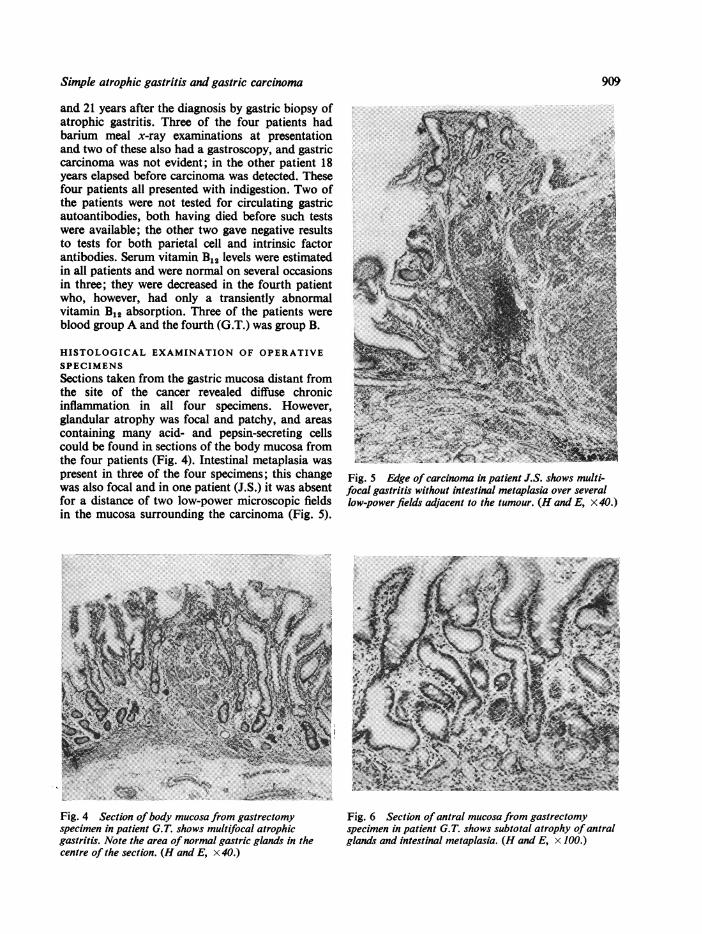
HISTOLOGICAL EXAMINATION OF OPERATIVESPECIMENSSections taken from the gastric mucosa distant fromthe site of the cancer revealed diffuse chronicinflammation in all four specimens. However,glandular atrophy was focal and patchy, and areascontaining many acid- and pepsin-secreting cellscould be found in sections of the body mucosa fromthe four patients (Fig. 4). Intestinal metaplasia waspresent in three of the four specimens; this changewas also focal and in one patient (J.S.) it was absentfor a distance of two low-power microscopic fieldsin the mucosa surrounding the carcinoma (Fig. 5).
Fig. 5 Edge of carcinoma in patient J.S. shows multi-focal gastritis without intestinal metaplasia over severallow-power fields adjacent to the tumour. (H and E, x 40.)
Fig. 4 Section ofbody mucosa from gastrectomy Fig. 6 Section of antral mucosa from gastrectomyspecimen in patient G.T. shows multifocal atrophic specimen in patient G.T. shows subtotal atrophy of antralgastritis. Note the area ofnormal gastric glands in the glands and intestinal metaplasia. (H and E, x 100.)centre of the section. (H and E, x 40.)
909

I. R. Walker, R. G. Strickland, B. Ungar, and L R. Mackay
The mucosa in patient L.M. showed no intestinalmetaplasia either overlying the carcinoma or insections of mucosa proximally or distally. In threeof the four specimens (from patients J.S., G.T., andH.M.) sections of the antral mucosa showed diffuseatrophy with intestinal metaplasia (Fig. 6).
Discussion
The four cases described are remarkable in relationto the long period during which atrophic gastritiswas demonstrable before the detection of carcinoma.Long-term surveys of cases of atrophic gastritisproven at biopsy are few. Fairley et al (1955) fromthis Unit found no gastric carcinomas in 32patients over a five-year survey. Siurala, Varis, andWiljasalo (1966), in a survey covering 10-15 years,found that nine gastric carcinomas developed in agroup of 116 patients with atrophic gastritis, whereasno carcinomas developed in groups with normalgastric mucosa or with superficial gastritis. Theoccurrence of carcinoma of the stomach in four ofour40 patients with atrophic gastritis followed for up to22 years is comparable, and indicates that patientswith this condition incur a substantial risk ofdeveloping malignancy.The four patients presented here were considered to
have 'simple' atrophic gastritis for the followingreasons. First, vitamin B12 absorption was normalin the three patients tested, and vitamin B12 deficiencydid not develop over a 14-year period of observationin the fourth patient. Second, in the two patients sotested, gastric autoantibodies were not detected.The patients already mentioned described bySiurala et al (1966) were not screened for the pre-sence of gastric autoantibodies and it is possible thattheir series included patients with latent perniciousanaemia, in that 21 of 103 patients tested had de-creased vitamin B12 absorption. Third, the pattern ofthe gastritis was of the multifocal type in all fourpatients; the mucosal changes in simple atrophicgastritis not associated with gastric autoantibodiesare usually multifocal in distribution (te Velde,Hoedemaeker, Anders, and Nieweg, 1966). Fourth,the antral mucosa was found to be severely involvedby gastritis in three of the patients, whereas inpatients with pernicious anaemia the antral mucosais spared and only the body and fundic mucosa showdiffuse atrophy (Magnus and Ungley, 1938).Kaplan and Rigler (1945) reported a 12% in-
cidence of gastric carcinoma in fully developedpernicious anaemia. Shearman, Finlayson, Wilson,and Samson (1966), however, suggested that the ex-tent of this association had been underestimated. Ifpernicious anaemia significantly contributed to theoverall prevalence of carcinoma of the stomach
then a higher than normal incidence of parietal celland intrinsic factor antibodies could be expected incarcinoma of the stomach. However, the findings ofUngar, Strickland, and Francis (1971) indicate thatthis is not the case. In fact Kravetz, van Noorden,and Spiro (1967) found that only 8% of 38 patientswith gastric carcinoma showed parietal cell antibodyeven though 80% of these patients had advancedgastritis. These observations indicate that thepredisposition to carcinoma of the stomach is basedon chronic gastritis per se and not particularly ongastritis with autoimmune features. Indeed, ifsimple atrophic gastritis, were shown to be morelikely to predispose to gastric carcinoma than auto-immune gastritis, then the presence of parietal cellantibody would take on a new significance.
It has been suggested that carcinoma of thestomach arising in patients with pernicious anaemiamay originate in areas of intestinal metaplasia(Siurala, Eramaa, and Tapiovaara, 1959). Theevidence from the present case studies indicates thatthis may not be true for carcinoma arising fromsimple atrophic gastritis, as in one patient intestinalmetaplasia was not observed in multiple sectionsfromall areas of the resected stomach, and in a furtherpatient the mucosa immediately surrounding thecarcinoma was free from this change.The suggested linking of atrophic gastritis with
gastric carcinoma must take into account the fre-quency of gastritis in the normal population. Biopsystudy of a normal population has been performedin a Finnish commune (Siurala, Isokoski, Varis,and Kekki, 1968), with the result that 28% of 142randomly selected subjects aged from 15 to 65 wereshown to have atrophic gastritis. However, whenonly instances of moderate to severe atrophicgastritis are considered the incidence was 10 %,and one-third ofthese subjects had circulating parietalcell antibody (Isokoski, Krohn, Varis, and Siurala,1969). Comparable figures for an unselected sampleof the Australian population are not available.The 28% incidence of severe atrophic gastritis in1,000 consecutive gastric biopsies reported fromthis Unit by Joske, Finckh, and Wood (1955) givessome indication of its prevalence in Australia,although most of these patients had gastrointestinaldiseases.
If it be accepted, that-for an adult population-the prevalence of atrophic gastritis lies between 10%and 28% (Siurala et al, 1968), that the prevalenceof pernicious anaemia as judged from a hospitalpopulation over the age of 50 years is 1 % (Ungar,1968), and that there is an incidence of gastriccarcinoma of 10% in both, then for every case ofgastric carcinoma supervening on pernicious anaemiathere will be some 10-28 cases supervening in
910

Simple atrophic gastritis and gastric carcinoma 911
atrophic gastritis without pernicious anaemia. It isunknown what determines the actual occurrence ofgastric carcinoma in certain subjects with eitherform of atrophic gastritis. What can be said is thatatrophic gastritis without pernicious anaemia is anumerically more important precursor of gastriccarcinoma than is the atrophic gastritis which isassociated with pernicious anaemia.
The authors wish to thank Drs I. J. Wood and D. C.Cowling for their early documentation of thepatients described in this study and Dr P. S. Bhathalfor reviewing the pathological specimens andproviding the photomicrographs.
Requests for reprints should be addressed toDr R. G. Strickland, Clinical Research Unit,Walter and Eliza Hall Institute of Medical Research,P.O., Royal Melbourne Hospital, Victoria, 3050,Australia.
This study was supported by grants from theNational Health and Medical Research Council ofAustralia (R.G.S., I.R.M.).
References
Anderson, B. B. (1964). Investigations into the Euglena method forthe assay of the vitamin B1, in serum. J. clin. Path., 17, 14-26.
Fairley K. F., Turner, C. N., Mackay, M. A., and Joske, R. A. (1955).Atrophic gastritis: a five-year survey of thirty-two cases provenby gastric biopsy. Med. J. Aust., 2, 1085-1088.
Gottlieb, C., Lau, K.-S., Wasserman, L. R., and Herbert, V. (1965)Rapid charcoal assay for intrinsic factor (IF), gastric juice un-saturated B1, binding capacity, antibody to IF and serumunsaturated B,2 binding capacity. Blood, 25, 875-884.
Heinle, R. W., Welch, A. D., Scharf, V., Meacham, G. C., andPrusoff, W. H. (1952). Studies of excretion and absorption ofCo'°-abeled vitamin B,2 in pernicious anaemia. Trans. Ass.Amer. Phycns, 65, 214-222.
Hutner, S. H., Bach, M. K., and Ross, G. I. M. (1956). A sugarcontaining basal rredium for vitamin B,, assay with Euglena:application to body fluids. J. Protozool., 3, 101-112.
Imai, T., Kubo, T., and Watanabe, H. (1971). Chronic gastritis inJapanese with reference to high incidence of gastric carcinoma.J. nat. Cancer Inst., 47, 179-195.
Isokoski, M., Krohn, K., Varis, K., and Siurala, M. (1969). Parietalcell and intrinsic factor antibodies in a Finnish rural populationsample. Scand. J. Gastroent., 4, 521-527.
Joske, R. A., Finckh, E. S., and Wood, I. J. (1955). Gastric biopsy-A study of 1000 consecutive successful gastric biopsies. Quart.J. Med., 24, 269-294.
Kaplan, H. S., and Rigler, L. G. (1945). Pernicious anaemia and car-cinoma of the stomach. Amer. J. med. Sci., 209, 339-348.
Kay, A. W. (1953). Effect of large doses of histamine on gastricsecretion of HCI: an augmented histamine test. Brit. med. J.,2,77-80.
Kravetz, R. E., Van Noorden, S., and Spiro, H. M. (1967). Parietal-cell antibodies in patients with duodenal ulcer and gastriccancer. Lancet, 1, 235-237.
Magnus, H. A. (1958). A re-assessment of the gastric lesion in per-nicious anaemia. J. clin. Path., 11, 289-295.
Magnus, H. A., and Ungley, C. C. (1938). The gastric lesion in per-nicious anaemia. Lancet, 1, 420-421.
Morson, B. C. (1955a). Carcinoma arising from areas of intestinalmetaplasia in the gastric mucosa. Brit. J. Cancer, 9, 377-385.
Morson, B. C. (1955b). Intestinal metaplasia of the gastric mucosa.Brit. J. Cancer, 9, 365-376.
Ross, G. I. M. (1952). Vitamin B,, assay in body fluids using Eugknagrailis. J. clin. Path., 5, 250-256.
Schilling, R. F. (1953). Intrinsic factor studies II: The effect of gastricjuice on the urinary excretion of radioactivity after the oraladministration of radioactive vitamin B,,. J. Lab. clin. Med.,42,860866.
Shearman, D. J. C., Finlayson, N. D. C., Wilson, R., and Samson,R. R. (1966). Carcinoma of the stomach and early perniciousanaemia. Lancet, 2, 403-405.
Siurala, M.,Eramaa, E., and Tapiovaara, J. (1959). Pernicious anemiaand gastric carcinoma. Acta med. scand., 164, 431-436.
Siurala, M., Isokoski, M., Varis, K., and Kekki, M. (1968). Pre-valence of gastritis in a rural population. Scand. J. Gastroent.,3,211-223.
Siurala, M., Varis, K., and Wiljasalo, M. (1966). Studies of patientswith atrophic gastritis: A ten to fifteen year follow-up. Scand.J. Gastroent., 1, 40-48.
Te Velde, K., Hoedemaeker, P. J., Anders, G. J. P. A., Arends, A.,and Nieweg, H. 0. (1966). A comparative morphological andfunctional study of gastritis with and without autoantibodies.Gastroenterology, 51, 138-148.
Ungar, B. (1967). Detection and titration of antibody to intrinsicfactor by a modified charcoal method. Aust. J. exp. Biol. med.Sci., 45, 317-321.
Ungar, B. (1968). Antibody to gastric intrinsic factor in blood donorsand hospital patients. Aust. Ann. Med., 17, 107-109.
Ungar, B., Stocks, A. E., Martin, F. I. R., Whittingham, S., andMackay, I. R. (1968). Intrinsic-factor antibody, parietal-cellantibody and latent pernicious anaemia in diabetes mellitus.Lancet, 2, 415-418.
Ungar, B., Strickland, R. G., and Francis, C. M. (1971). The preval-ence and significance of circulating antibodies to gastric in-trinsic factor and parietal cells in gastric carcinoma. Gut, 12,903-905.
Weiden, S. (1949). Some observations on the secretion of the stomachwith special reference to mucus. Med. J. Aust., 2, 81-83.
Whittingham, S., and Mackay, I. R. (1969). Laboratory methods fordiagnosis of autoimmune disease. Med. J. Aust., 1, 1200-1205.
Wood, 1. J., Cowling, D. C., Ungar, B., and Gray, A. (1960). Serumvitamin B1, levels in chronic atrophic gastritis. Aust. Ann. Med.,9,309-317.
Wood, I. J., Doig, R. K., Motteram, R., and Hughes, A. (1949).Report on 55 biopsies using a new flexible gastric biopsy tube.Lancet, 1, 18-21.
Wood, I. J., Ralston, M., Ungar, B., and Cowling, D. C. (1964).Vitamin B,, deficiency in chronic gastritis. Gut, 5, 27-37.
AddendumIn a recent report (Imai, Kubo, and Watanabe,1971), in which chronic gastritis with antralinvolvement and gastric cancer in Americansand Japanese were compared, it appeared thatchronic gastritis could determine the much higherprevalence of gastric cancer in Japanese.