Signalling and trafficking of the cysteinyl leukotriene receptors in...
83
Signalling and trafficking of the cysteinyl leukotriene receptors in intestinal epithelial cells Parhamifar, Ladan Published: 2009-01-01 Link to publication Citation for published version (APA): Parhamifar, L. (2009). Signalling and trafficking of the cysteinyl leukotriene receptors in intestinal epithelial cells Department of Laboratory Medicine, Lund University General rights Copyright and moral rights for the publications made accessible in the public portal are retained by the authors and/or other copyright owners and it is a condition of accessing publications that users recognise and abide by the legal requirements associated with these rights. • Users may download and print one copy of any publication from the public portal for the purpose of private study or research. • You may not further distribute the material or use it for any profit-making activity or commercial gain • You may freely distribute the URL identifying the publication in the public portal
Transcript of Signalling and trafficking of the cysteinyl leukotriene receptors in...
LUND UNIVERSITY
PO Box 117221 00 Lund+46 46-222 00 00
Signalling and trafficking of the cysteinyl leukotriene receptors in intestinal epithelialcells
Parhamifar, Ladan
Published: 2009-01-01
Link to publication
Citation for published version (APA):Parhamifar, L. (2009). Signalling and trafficking of the cysteinyl leukotriene receptors in intestinal epithelial cellsDepartment of Laboratory Medicine, Lund University
General rightsCopyright and moral rights for the publications made accessible in the public portal are retained by the authorsand/or other copyright owners and it is a condition of accessing publications that users recognise and abide by thelegal requirements associated with these rights.
• Users may download and print one copy of any publication from the public portal for the purpose of privatestudy or research. • You may not further distribute the material or use it for any profit-making activity or commercial gain • You may freely distribute the URL identifying the publication in the public portal

From the Department of Laboratory Medicine,
Division of Cell Pathology,
Lund University, Malmö, Sweden
Signalling and trafficking of the cysteinyl leukotriene receptors
in intestinal epithelial cells
Ladan Parhamifar
Academic dissertation By due permission of the Faculty of Medicine, Lund University, Sweden
To be defended at the main lecture hall, Pathology building, Malmö University Hospital, Malmö on Friday January 23rd, 2009 at 09.15 for
the degree of Doctor of Philosophy, Faculty of Medicine.
Faculty Opponent: Professor Catherine Godson, College of Life Sciences, Conway Institute Belfield, Dublin 4, Ireland
139

3
For my parents N oth in g s ho ck s me . I'm a s c i en t i s t .
Harri s on Ford (1942 -), a s Ind ian a Jone s The h i ghe s t r e su l t o f e du cat ion i s t o l eran ce . He len Ke l le r (1880 - 1968), 'Opt im i sm,' 1903

4
TABLE OF CONTENTS LIST OF PAPERS 5 LIST OF ABBREVIATIONS 6 INTRODUCTION 7 BACKGROUND 8 1. The intestinal epithelium 8 2. Inflammatory Bowel Disease (IBD) 11 3. Colon cancer 13 4. Colon cancer and Inflammatory Bowel Disease 14 Highlighted signalling molecules 17
5. Phospholipase A2 (PLA2) 17 6. Eicosanoids 22 Cyclooxygenase (COX) and Prostaglandins (PG) 23 Leukotrienes 247. Cysteinly Leukotriene receptors (CysLTRs) 278. Mitogen activated protein kinases (MAPKs) 329. Nuclear factor kappa B (NFκB) 3310. Protein Kinase C (PKC) 3511. G-protein coupled receptors (GPCRs) 36 12. GPCR dimerization and other GPCR interacting proteins 3913. GPCR desensitization 4314. Internalisation of GPCRs 44- Internalisation of GPCRs and caveolin 44 - GPCR internalisation and clathrin 45- GPCR internalisation and β-arrestin 46- GPCR internalisation and Rab proteins 4815. Nuclear GPCR 50THE PRESENT INVESTIGATION 52RESULTS AND DISCUSSION Papers I, II and III 53SUMMARY 59 POPULÄRVETENSKAPLIG SAMMANFATTNING 60 ACKNOWLEDGEMENTS 62REFERENCES 65

5
LIST OF PAPERS
This thesis is based on the following papers, which are referred to in the text by their Roman numerals:
I. Parhamifar L, Jeppsson B, Sjölander A.
Activation of cPLA2 is required for leukotriene D4-induced proliferation in CRC
cells. Carcinogenesis. 2005 Nov; 26(11): 1988-98.
II. Parhamifar, L, Yudina, Y, Vilhardt, F and Sjölander, A
Nuclear trafficking and signalling of the G-protein coupled receptor, CysLT1 in intestinal epithelial cells (Manuscript)
III. Parhamifar L, Vilhardt F, Mögerlin M and Sjölander A Co-dependent localization and ligand specific trafficking of Cysteinyl Leukotriene receptors (Manuscript)
Reprints were made with permission from publisher: ©2005 by the Oxford University Press
5

6
LIST OF ABBREVIATIONS AA arachidonic acid ADP adenosine 5′-diphosphate APC adenomatous polyposis coli ATP adenosine 5′-triphosphate BSA bovine serum albumin cAMP Cyclic adenosine mono phosphate CD Crohn’s Disease COX Cyclooxygenase CRC Colorectal cancer CREB cAMP response element binding CysLT Cysteinyl leukotriene CysLT1R Cysteinyl Leukotriene 1 receptor CysLT2R Cysteinyl Leukotriene 2 receptor ER Endoplasmatic reticulum FAP Familial adenomatous polyposis FLAP Five Lipoxygenase Activating Protein GDP Guanosine diphosphate GI Gastrointestinal GPCR G-protein coupled receptor HNPCC Hereditary non-polypus colorectal cancer IBD Inflammatory bowel disease LO Lipoxygenase LT Leukotriene MAPK Mitogen-activated protein kinase NFκB Nuclear Factor κB NLS Nuclear localization signal PBS-T Phosphate buffer saline – Tween PG Prostaglandin PI Phosphoinositol PK Protein kinase PL Phospholipase PLA2 Phospholipase A2 PPAR-γ Peroxisome Proliferator Activated Receptor- γ PTX Pertussis toxin RAMP Receptor activity modifying protein RGS Regulator of G-protein signalling TCF T cell factor TNF-α Tumour necrosis factor α TRAF TNF receptor associated factor UC Ulcerative colitis
6
7
INTRODUCTION Inflammatory responses are designated to protect the host from injuries and / or foreign invasion. The colon houses 300-500 species of bacteria, responsible for producing vitamins and other important products from food transversing the digestive system. The bacteria are prevented from entering the rest of the body by a single layer of intestinal epithelial cells, this barrier functions as the first line of defence against foreign invaders. Due to its large number of bacteria the colon maintains a low level of inflammation. An imbalance of this inflammatory state has been suggested as a possible cause of inflammatory bowel disease (IBD). Prolonged inflammatory conditions arise when the inflammatory response fails to resolve the injury, leading to continuous recruitment of inflammatory cells and inflammatory mediators to the site of injury. This continuous inflammatory response leads to tissue damage, such as the disruption of the epithelial barrier. These responses are thought to be the pre disposition to colorectal cancer. Leukotrienes are lipid mediators produced by inflammatory cells and implicated in various inflammatory responses. High levels of leukotrienes are found in patients suffering from IBD. In intestinal epithelial cells Leukotriene D4 (LTD4) can induce cell growth, increase cell survival and induce cell migration by regulating various enzymes and proteins shown to be involved in cancer progression and development. LTD4 elicits these effects through the Cysteinyl leukotriene receptor 1 (CysLT1R). However, leukotrienes can bind to four different G-protein coupled receptors (GPCRs), the work in this thesis fouses on two of the best characterized CysLTRs; the CysLT1R and the CysLT2R. The balance between the two first receptors has been suggested to play a role in the prognosis of colorectal cancer patients. Increased CysLT1 expression correlates with a poor survival rate, whereas increased CysLT2 correlates with a better prognosis. With this in mind, the aim of the work in this thesis has been to investigate the cysteinyl leukotriene receptors CysLT1 and 2 signalling and their regulation in particular the trafficking.
7

8
BACKGROUND 1. The intestinal epithelium The small and large intestine are the last part of the gastrointestinal (GI) tract.
In the small intestine food components such as fats, proteins and
carbohydrates are broken down. The nutrient components are then transferred
to the bloodstream. The large intestine absorbs the remaining water and
removes the waste products (1,2).
The intestinal epithelium consists of a single layer of epithelial cells(3).
They provide a barrier for the indigestible components and the gut microflora,
keeping these within the lumen of the intestine.
Figure1. The large intestine.
In the lining of the mammalian intestine different classes of epithelial cells
termed goblet, endocrine and paneth cells can be found. Paneth cells are only
found in the small intestine and constitute one of the differences between the
small and large intestine. Whilst the goblet cells secrete protective mucins, the
8

9
endocrine cells secrete various gut hormones and the paneth cells secrete anti-
bacterial proteins (4-6). The majority of cells are the absorptive cells are
termed enterocytes (small intestine) or colonocytes (large intestine), and are
positioned on a basement membrane, where the intra epithelial lymphocytes
can be found migrating in and out of the intestinal epithelium.
The small intestine consists of villi and crypts whilst the large intestine
consists only of crypts (3,5) (Fig.2). The villi in the small intestine mainly
consist of enterocytes. At the bottom of the crypts in the small intestine reside
the stem cells and the paneth cells. The paneth cells occupy the three first
positions of the crypt whilst the stem cells are positioned fourth. New cells that
are made at the bottom of the crypts by the stem cells, migrate up to the villi
while dividing and differentiating into specialised intestinal epithelial cells
(Fig.2) (4,5).
Figure2. Cells of the small intestine (Crosnier, Nature Publishing group, 2006)
Cells in transit divide three to four times before they differentiate into
mature cells. Once the cells have reached the top of the villi they undergo
apoptosis and sloth off in to the lumen (6). In the colon, proliferative cells
9

10
occupy two thirds of the crypt and the top third consists of the differentiated
cells.
Renewal of the epithelium in the small intestine takes place every 4-6 days
whilst turnover of the colonic epithelium can take 3-8 days. This high
regenerative potential is what makes the intestine so fascinating but it is also
its achilles heal as dys-regulation of this system has dangerous outcomes (4,5).
Colonisation of the gastrointestinal tract begins after birth and the intestinal
tract ends up with between 300-500 different bacterial species. The stomach
and the small intestine only have a few species of bacteria but the in the colon
the microflora outnumber host eukaryotic cells by 10 fold (7). The
colonisation of the colon is affected by the environment and the food that the
baby first eats. The first bacteria to colonise the colon can decide the gene
expression patterns in the epithelial cells and create a more favourable
environment for themselves and inhibit the growth of other bacteria. The
normal microflora consists of mainly bacteria a few viruses and fungi (8). This
normal intestinal flora is essential for the function of the intestine, however
some of these bacteria are potentially pathogenic and can cause infections if
the epithelial barrier has been disrupted (9). Therefore, due to its commensal
bacteria the colon always produces a low level of inflammation (immune
surveillance) to be able to distinguish between pathogens and “normal”
commensal bacteria. This is accomplished by the existence of intra epithelial
immune cells such as macrophages and dendritic cells as well as the
Gastrointestinal Associated Lympoid Tissue (GALT) are lymphoid like
sections, important in facilitating immune surveillance along the GI tract,
which consist immune cells, such as dendritic cells, T and B lymphocytes. In
the small intestine the GALT is known as the Peyer's patches. M cells are
specialised epithelial cells that are located on top of the Peyer’s Patches. The
M cells have loose intercellular binding, allowing the intestinal microflora
easy access to the underlying lymphoid tissue. Dendritic cells present in the
Payers Patches process the microbes they come into contact with and present
10
11
them to the T lymphocytes. In other GALT along the GI tract, the dendritic
cells can sample antigens directly from the intestinal lumen, or receive
microbes which have transversed the intestinal epithelial cells (10,11).
Specialised membrane and intracellular receptors, the Toll-like receptors
(TLRs) and the NOD or NOD-like receptors (NLRs), respectively, are
responsible for recognition of the microbial antigens also known as pathogen
associated molecular patterns (PAMPS). These PAMPs induce tolerance
towards the normal commensal bacteria through stimulating signalling
pathways through the TLRs, NODs and NLRs. However dysregulation of
the tolerogenic signalling pathways, for example through receptor mutation
can lead to the development of prolonged or chronic inflammation (12-14).
Inflammation is a response to injury or pathogen invasion. Under normal
conditions the initiation of inflammation also includes signals for the
termination and resolution of inflammation (15-18). Prolonged inflammatory
conditions arise when the stimulus for the acute state of inflammation is not
terminated (8,19) leading to the development of inflammatory bowel disease
(IBD) (20).
2. Inflammatory bowel disease
IBD is an umbrella term for several conditions (collagenous colitis,
Lymphocytic colitis, Ischemic colitis, Behcet’s colitis, infective colitis,
intermediate colitis, diversion colitis) however there are two major conditions,
ulcerative colitis (UC) and Crohn’s disease (CD) (21). Even though it is
difficult to compare studies geographically due to methodological differences
it is suggested that the incidence of IBD is higher in developed countries(22).
Patients suffering from IBD often more vulnerable to other chronic
inflammatory conditions. Currently three dispositions are hypothesised to be
required for the development of IBD: 1) genetic factors, for example
mutations to proteins involved in immune surveillance; 2) luminal factors such
11

12
as the microflora and food antigens, for example a low fibre diet is linked to
the development of IBD; and 3) environmental factors for example smoking
increases the risk for the development of CD (21).
The two major conditions of IBD have similarities but also major
differences. CD can affect any part of the GI tract, displaying typical
discontinuous transmural inflammation and is associated with a T helper cell -
1 response, over-production of IL-12/IL-23 and IFN-γ. Research has also
identified mutations in the NOD2 protein as being a cause for CD in a sub-
population of patients (23,24). UC on the other hand primarily affects the
colon and extending rectum with the inflammation continuous and confined to
the mucosa and sub mucosa, is mainly a T helper cell -2 response, mediated by
an over-production of IL-13. Interestingly smoking increases the risk for CD
whilst providing a protective factor against UC (7,14).
Despite the differences in the two different conditions, UC and CD also
share many common features. In both cases inflammation causes mucosal
destruction, which leads to loss of mucosal barrier and absorptive function.
These two events have similar symptoms, such as abdominal pain and bloody
diarrhoea. Furthermore both UC and CD have been associated with alterations
in STAT-3 and NKX2-3. Various mouse models demonstrate an important
role for the commensal bacteria in IBD (9,14,23,25).
Current non-invasive treatments for IBD mainly centre around the
inhibition of the dysregulated inflammatory process. Commonly 5-
aminosalicylates are used to dampen the inflammation (26) either alone or in
combination with corticosteroids, which are more immunosuppressive drugs
(27). However, tolerance can develop, therefore in the case of Crohn’s disease
several chimeric antibodies have been developed which bind to tumour
necrosis factor-α (TNF-α), a major cytokine and driving factor behind IBD.
Patients receiving this treatment can also unfortunately relapse (28). Thus,
12
13
ultimately, the only cure to date, (which is only possible in ulcerative colitis) is
surgical resection (29).
3. Colon cancer Colorectal cancer (CRC) is the third most common form of cancer in the
western world (World health organization 2007). most common malignancy
of the GI, with the overall survival rate lower that 50% (30). CRC is the
second leading cause of cancer related deaths in the United States. In Sweden
5000 new cases of CRC are diagnosed every year (Swedish National board of
Health and Disease, 1998). Most, if not all, malignant colorectal tumours
(carcinomas) arise from pre-existing benign tumours (adenomas)
(Sugarbaker et al., 1985). The rate of deaths caused by CRC can be and has
been reduced by early detection with colonoscopy and testing of stool
samples (31). Various factors have been suggested to increase the risk for
developing CRC such as; cancer elsewhere in the body, colorectal polyps,
CD, family history of CRC and UC (32). Low fibre, high fat diets and
smoking have also been suggested to be risk factors for developing CRC.
Genetic predisposition increases the chance of developing CRC, however the
majority of cases occur sporadically (33). The loss of tumour suppressor genes
which drive the sporadic adenoma carcinoma have been proposed to be a
contributing factor (33). The three major carcinogenic pathways that lead to
sporadic CRC and colitis associated CRC are; chromosomal instability,
microsatelite instability and hyper methylation. Hyper methylation of
promoter regions of cell cycle(32), DNA repair and cell adhesion genes, leads
to silencing of these genes and thereby promotes CRC. In sporadic CRC the
dysplastic precursor is a discrete focus of neoplasia, the adenomatous polyp
that can be removed. The majority of spontaneous colorectal cancers (about
85%) are the results of chromosomal instability, the rest are due to
microsatelite instability (MSI) or failure of the mismatch repair system
(34,35). Both of these cases lead to the accumulation of mutations which
13

14
eventually could become carcinogenic. Chromosomal instability leads to
abnormal chromosomal segregation and abnormal DNA content and
eventually loss of chromosomal material and loss of function of tumour
suppressor genes such as adenomatous polyposis coli (APC) and p53 (a key
regulator of the cell cycle)(36). This results in activation of proto-oncogenes,
such as c-myc and cyclin D1. In microsatelite instability, the loss of DNA
miss match repair genes leads to loss of colonic homeostasis and neoplastic
growth. Mutations in the RAS/RAF pathway, the p53 pathway, and several
other genes/pathways drive tumour progression towards malignancy and
metastasis (32).
Genetic predisposition to colorectal cancer depends on the presence or
absence of polyps. Patients with hereditary non-polyposis colorectal cancer
have an inherited mutation in an allele of a DNA mismatch repair gene(37).
Patients with familial adenomatous polyposis (FAP) have a mutation in the
APC gene. This pathway however is altered in approximately 95% of CRCs.
FAP patients are characterised by the early onset of adenomatous colorectal
polyps in the large intestine (38).
4. Colon cancer and Inflammatory Bowel Disease The link between cancer and inflammation, has long been suggested and dates
back to Virchow in 1863. IBD is one of three high risk factors for developing
CRC, as patients with UC develop colorectal cancer 10 years earlier than
patients with sporadic colorectal cancer (39). The other two high risk factors
are having a genetic predisposition namely familial adenomatous polyposis
(FAP) and hereditary non-polypus colorectal cancer (HNPCC) (32,39,40).
Dysplasia in IBD can be flat, multifocal, localised or diffuse, with the
duration and surface affected by colitis increasing the risk for developing CRC
(32). In sporadic CRC the cancer progresses from adenoma to carcinoma,
whilst in IBD induced CRC, the progression is from inflammation to dysplasia
14

15
and then carcinoma (41). Colitis associated CRC affects individuals at a young
age and has a more proximal distribution in the colon.
There is a lot of evidence to support the link between IBD and CRC.
Tumours have been observed to behave very similarly to wounds that fail to
heal, since they surround themselves with a milieu similar to that of a chronic
inflammatory state, which for example gives rise to the production of matrix
metalloproteinases, tissue degradation, leukocyte infiltration and angiogenesis.
Many proteins have been observed to be highly up-regulated in both IBD
tissue and CRC. Possibly one of the most important proteins to be highlighted
are the cyclo-oxygenase (COX) enzymes, in particular COX-2. This inducible
enzyme is over expressed in 90% of CRC (42). The induction of expression is
seen early in acute inflammation, and high expression levels have been shown
to be present in UC and CD. COX-2 produces a prostaglandin (PG) precursor
(a member of the eicosanoid family of lipid mediators), with the main PG in
the intestine being PGE2. Accordingly, the membrane receptor for PGE2 along
with this PG are also over-expressed in IBD and CRC (43).
A further example linking CRC and IBD include, the observation that p53
mutations which are very common in CRC have been found in the inflamed
mucosa from UC patients who did not have cancer. Also, methylation is a way
of controlling gene transcription, with many proto-oncogenes being
methylated in CRC. Several genes that precede dysplasia have been found
methylated in UC (32,41).
Reactive oxygen species (ROS) and nitrogen oxygen species (NOS) are
highly reactive molecules that are increased in the inflamed mucosa and
remain elevated in colonic neoplasms (Fig. 3). ROS and NOS have also been
15
16
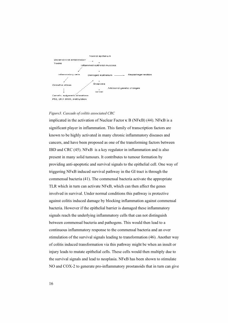
Figure3. Cascade of colitis associated CRC
implicated in the activation of Nuclear Factor κ B (NFκB) (44). NFκB is a
significant player in inflammation. This family of transcription factors are
known to be highly activated in many chronic inflammatory diseases and
cancers, and have been proposed as one of the transforming factors between
IBD and CRC (45). NFκB is a key regulator in inflammation and is also
present in many solid tumours. It contributes to tumour formation by
providing anti-apoptotic and survival signals to the epithelial cell. One way of
triggering NFκB induced survival pathway in the GI tract is through the
commensal bacteria (41). The commensal bacteria activate the appropriate
TLR which in turn can activate NFκB, which can then affect the genes
involved in survival. Under normal conditions this pathway is protective
against colitis induced damage by blocking inflammation against commensal
bacteria. However if the epithelial barrier is damaged these inflammatory
signals reach the underlying inflammatory cells that can not distinguish
between commensal bacteria and pathogens. This would then lead to a
continuous inflammatory response to the commensal bacteria and an over
stimulation of the survival signals leading to transformation (46). Another way
of colitis induced transformation via this pathway might be when an insult or
injury leads to mutate epithelial cells. These cells would then multiply due to
the survival signals and lead to neoplasia. NFκB has been shown to stimulate
NO and COX-2 to generate pro-inflammatory prostanoids that in turn can give
16
17
rise to carcinogenic effects. Active NFκB has also been found in inflamed
mucosa in IBD. A known upstream effector of NFκB is the pro-inflammatory
Tumour Necrosis Factor α (TNF-α). Antibodies against TNF-α are currently in
the treatment of CD and UC. On the other hand PPAR-γ inhibits NFκB and is
effective in reducing inflammation in CD (7,9). Impaired expression of PPAR-
γ has been demonstrated in colonic epithelial cells in CD.
As previously mentioned the eicosanoids and the enzymes that produce
them have been implicated in inflammation and CRC (39,47). Another
enzyme 5-lipoxygenase (5-LO) produces other eicosanoids, the products, the
cysteinyl leukotrienes (CysLTs) are the basis for the investigation presented in
this thesis.
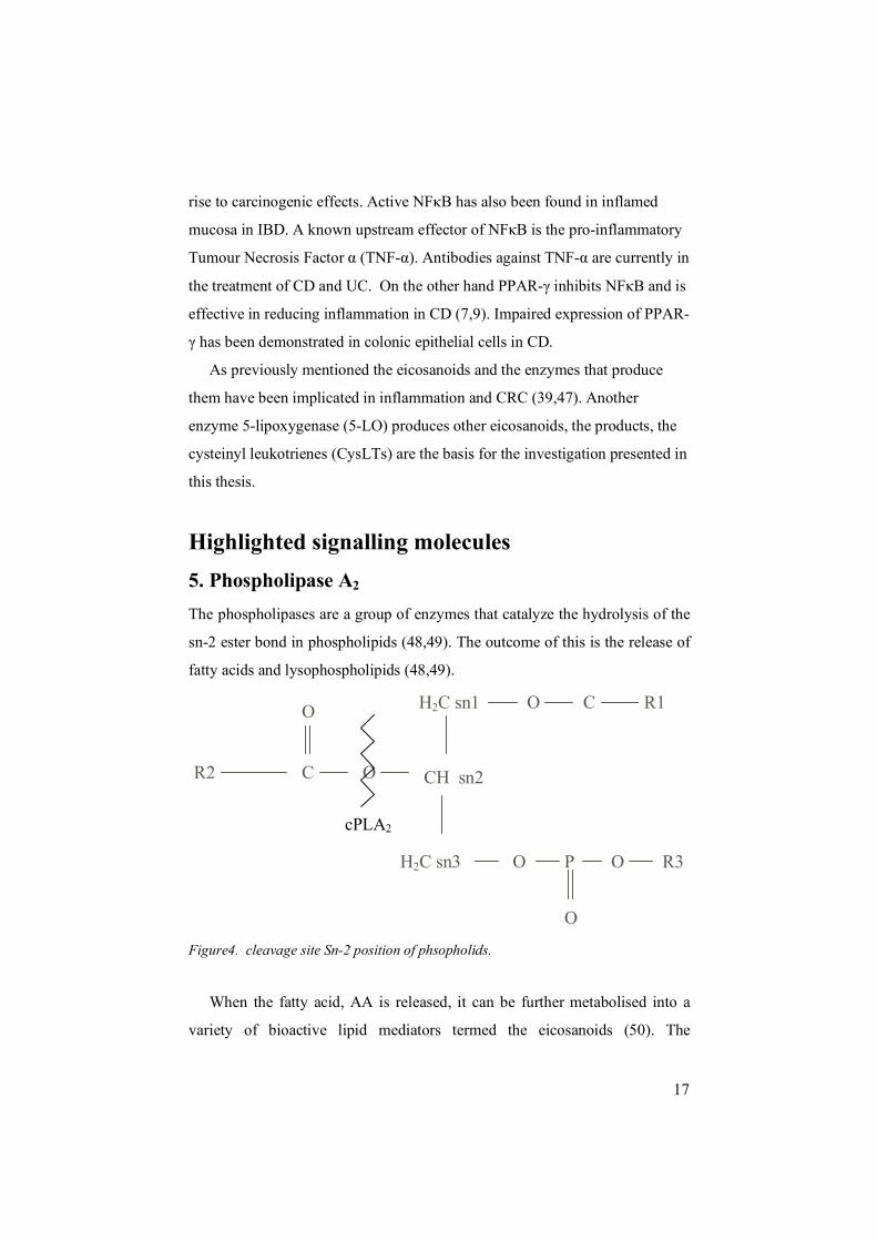
Highlighted signalling molecules 5. Phospholipase A2 The phospholipases are a group of enzymes that catalyze the hydrolysis of the
sn-2 ester bond in phospholipids (48,49). The outcome of this is the release of
fatty acids and lysophospholipids (48,49).
Figure4. cleavage site Sn-2 position of phsopholids.
When the fatty acid, AA is released, it can be further metabolised into a
variety of bioactive lipid mediators termed the eicosanoids (50). The
R2 C
O
O CH sn2
H2C sn3 O P O R3
O
H2C sn1 O C R1
cPLA2
17

18
eicosanoids (discussed below) are a family of potent mediators involved in
many different conditions, such as inflammation and cancer (47,51,52). The
lysophospholipids can in turn be converted into lysophosphatidic acid platelet
activating factor (PAF) which also is a potent signalling molecule implicated
in for example inflammation (53,54).There are currently 15 known groups and
subgroups of Phospholipase A2s (PLA2) (55-57). These groups are separated
by characteristics of the PLA2s such as their; structure, requirement for
calcium and size. This divides the PLA2s in to five major groups; the sPLA2s
(secreted), the cPLA2s (cytosolic), the iPLA2s (calcium independent), the PAF
acetyl hydrolases and the lysosomal PLA2s. The sPLA2 group have a low
molecular weight (14-19kDa) and are calcium dependent (55,56). The
different members of the sPLA2 family have been implicated in the digestion
of phospholipids in the stomach (58,59), rheumatoid arthritis (60), hydrolyzing
membranes of gram negative bacteria (anti-microbal agents) (61,62),
atherosclerosis and IBD (63-65). It has also been demonstrated to decrease the
size and development of tumours in mice. Increased expression in mice, has
been correlated with a better survival, however this data contradicts the results
detected in humans, where the enzyme is up-regulated and may be
contributing to the progression of CRC (66). The exact role of sPLA2 in
eicosanoid production remains unexplained, but might depend on cell type and
stimuli.
The iPLA2 group have molecular weights ranging from 28-146 kDa.
Whilst they do not have fatty acid chain specificity, they are thought of as
house keeping genes but also been shown to be involved in apoptosis by
delaying or inhibiting cell death induced by death receptors (55).
The PAF acetylhydrolase group (also shown to be anti-inflammatory) do
not require calcium for their activity, they range from 26-45 kDa, and some
are secreted and some are intracellular but they all hydrolyse the acetyl group
from the sn-2 position of PAF (67-69). The PAF acetylhydrolase IIA is
secreted and can also hydrolyse short fatty acids from the sn-2 position, it can
18
19
bind to both the high density lipoprotein and low density lipoprotein
cholesterol molecules (69,70) and not surprisingly has been implicated in
cardiovascular disease (71). Lysosomal PLA2 is the most recently discovered
being found in bovine brain and does not require calcium for its activity (72).
The cPLA2s are serine esterases, they have a molecular weight of 61-114
kDa, and all but cPLA2-γ/IVC require calcium. The different cPLA2s have
different specificities for fatty acids in the sn-2 position with cPLA2-α/IVA
being specific for AA containing phospholipids whilst cPLA2-β and γ have
very little specificity for AA (73,74). A major role of cPLA-α is in the
production of eicosanoids. cPLA2-α is an 85kDa enzyme, isolated from
neutrophils and platelets (75,76). It is composed of a C2 calcium binding
domain, that is required for its membrane translocation, α and β -hydrolase
domains with the catalytic site, and the serine 727 and 505 that are
phosphorylated by the Mitogen Activated Protein Kinases (MAPK) and
MAPK activated proteins. The phosphorylation of the cPLA2s is important for
the lipid enzyme interaction. (55,77). Even though cPLA2-α requires calcium
for its translocation and membrane binding, phosphoinositol diphosphate
(PIP2) has been shown to activate the protein in a calcium independent
manner. The PIP2 binding domain is in the catalytic site but has been shown to
require the C2 domain (78). The importance of this protein in inflammation
has been demonstrated in knock-out mice, which have post-ischemic injury,
acute lung injury and significantly decreased allergic responses. Furthermore,
the first study in this thesis demonstrates that the pro-inflammatory mediator
leukotriene D4 (LTD4) can activate and increase expression of cPLA2-α (79).
19
20
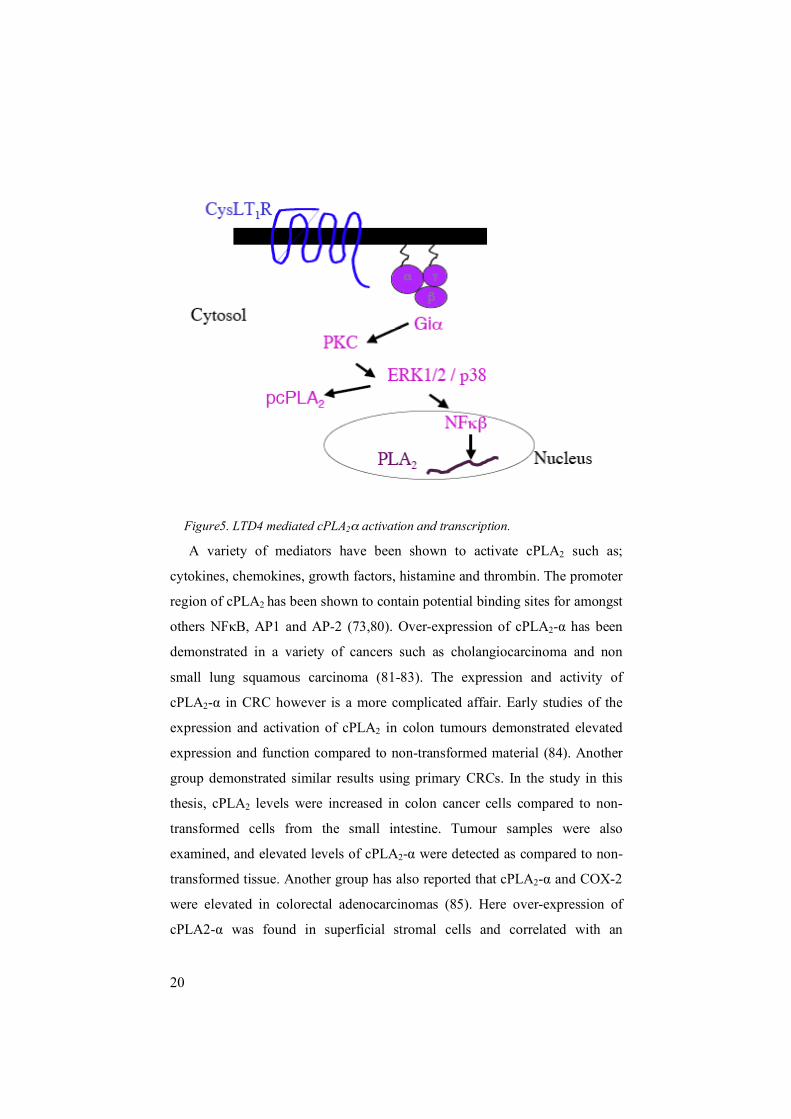
Figure5. LTD4 mediated cPLA2α activation and transcription.
A variety of mediators have been shown to activate cPLA2 such as;
cytokines, chemokines, growth factors, histamine and thrombin. The promoter
region of cPLA2 has been shown to contain potential binding sites for amongst
others NFκB, AP1 and AP-2 (73,80). Over-expression of cPLA2-α has been
demonstrated in a variety of cancers such as cholangiocarcinoma and non
small lung squamous carcinoma (81-83). The expression and activity of
cPLA2-α in CRC however is a more complicated affair. Early studies of the
expression and activation of cPLA2 in colon tumours demonstrated elevated
expression and function compared to non-transformed material (84). Another
group demonstrated similar results using primary CRCs. In the study in this
thesis, cPLA2 levels were increased in colon cancer cells compared to non-
transformed cells from the small intestine. Tumour samples were also
examined, and elevated levels of cPLA2-α were detected as compared to non-
transformed tissue. Another group has also reported that cPLA2-α and COX-2
were elevated in colorectal adenocarcinomas (85). Here over-expression of
cPLA2-α was found in superficial stromal cells and correlated with an
20

21
increased COX-2 expression and high micro vessel density (85), suggesting a
regulatory role for cPLA2-α in COX-2 induced angiogenesis. This group
analysed another set of CRC specimens and found elevated levels of cPLA2-α,
but could not correlate them to any specific stage, histological pattern or
microsatelite instability (86). They could however, yet again show a
correlation between cPLA2-α and COX-2 in half of the samples (86). A mouse
model using APC min mice produced conflicting results concerning cPLA2-α
in colon cancer (87). This group found a decrease in the expression of cPLA2-
α in five human colorectal cancers. The same study also reported that despite
low levels of cPLA2-α, COX-2 expression was elevated. These studies were
further explored (88) and 13/27 samples showed elevation of COX-2 and in 11
of these 13 samples cPLA2-α was reported absent. The authors suggest that the
low levels of cPLA2-α and high levels of COX-2, probably disrupts the levels
of AA (high levels of AA induces apoptosis) leading to escape from apoptosis.
In another study by the same group, AOM induced colon cancer, was
investigated in cPLA2-α knockout mice, which showed increased tumour size
and number in the colon (89). The authors also observed enhanced
tumorogenesis despite low levels of COX-2 in their model (89). Further APC
min mouse studies have shown that in the small intestine, cPLA2 is the
dominant source of AA for COX-2 (90) and that deletion of cPLA2 reduces
tumour size and/or number even in the colon (90-92). The conflicting results
can also be seen in the regulation of cPLA2-α by AA metabolites. In a study
using prostate cancer cells (PC-3), AA was added directly to the cells. This
lead to a transcriptional activation of cPLA2-α and COX-2 in a dose dependent
manner, and an increase of PGE2 production. Interestingly the authors show
that a COX-2 inhibitor blocked the up-regulation of cPLA2-α and COX-2,
suggesting that PGE2 can up-regulate cPLA2-α (100). Again there is a
contradictory study preformed in mouse lung fibroblasts, where the addition of
PGs could only up- regulate COX-2 but not cPLA2-α (101). These data
suggest that the regulation of cPLA2-α to some extent might be tissue specific.
21

22
Surprisingly, a very recent study reported the findings of cPLA2-α deficiency
in a 45 year old white American male of Italian decent. The study showed that
this man suffered from multiple ulcers in the small intestine and concluded
that cPLA2-α plays an important role in intestinal homeostasis and integrity.
Furthermore he had platelet dysfunction and globally decreased eicosanoid
production. The findings suggest that the production of eicosanoids in platelet
and leukocytes is almost entirely dependent on cPLA2-α. However, the study
also showed that the outcome of several colonoscopies preformed, were
normal despite the absence of cPLA2-α. Moreover, the authors reported that
their results indicate that cPLA2-α provides AA for virtually all biosynthesis of
eicosanoids by platelets, leukocytes and the cells from which the CysLTs are
derived (93).
6. Eicosanoid The eicosanoids are biologically active fatty acid metabolites with a twenty
carbon chain backbone (94). Their name originates from the Greek word
eicosa, which means twenty. Eicosanoid is the umbrella term for metabolites
of AA. AA is found in membrane phospholipid bilayers. Once released by
PLA2, AA can be further metabolized by the COXs, LOs, cytochrome p450 or
non-enzymatically. When oxygenated by COX, AA gives rise to PGs,
prostacyclin or thromboxanes. Oxygenation by LOs gives rise to LTs,
lipoxins, hepoxillins or monohydroxy fatty acids (49,95-98). Cytochrome
p450 metabolism of AA forms epoxy fatty acids or dihydroxy fatty acids.
Non-enzymatic metabolism of AA produces isoprotnes or isoleaukotrienes(99-
101).
22

23
Figure6. Eicosanoid production.
Cyclooxygenases and Prostaglandins The COX enzymes initiate the synthesis of PGs, and can be divided into two
established isoforms; COX-1 and COX-2. The enzymes share a 60%
homology (95,102,103) and consist of three domains; an EGF domain, a
membrane binding domain and a catalytic domain that controls both its COX
and peroxidase activity (104). COX-1 is a house keeping enzyme and
produces basal levels of PGs. COX-1 is ubiquitously expressed and needed for
homeostasis, for example in maintaining the epithelial barrier (105,106). It
should however be mentioned that COX-1 has been shown to play a role in
ovarian cancer (107). COX-2 is an inducible enzyme and its expression can be
mediated by many inflammatory mediators (108). It has also been shown to be
over-expressed in IBD, CRC (90%) and many cancers. Inhibitors of the COXs
have been used for over a 100 years (109-111). Non-steroidal anti-
inflammatory drugs (NSAIDs; aspirin, indometacin, and ibuprofen) block both
COX-1 and COX-2. Specific COX-2 inhibitors have been shown to reduce
tumour size and colonic polyps (42). These positive effects are however
23
24
clouded by the cardiovascular side effects which were seen with a particular
COX-2 specific inhibitor in a subset of patients being treated for Rheumatoid
arthritis, leading to it being pulled from the market (105). However, other
COX-2 specific inhibitors are still used in treating inflammatory conditions
although with greater caution (112,113). With this in mind, there is also data
demonstrating that COX-2 also is involved in anti-inflammatory and pro-
resolution pathways, and perhaps COX-2 inhibitors are not optimal in this
regard either (104,114). AA is metabolized by COXs to the PG precursor
PGH2. PGH2 is then further metabolized to PGI2, PGE2, PGD2 or PGF2α via
specific PG synthases (42). The PGs were discovered in 1935 and have since
then been implicated in various signalling pathways (115). Similarly to COX-
2, PGE2 has been implicated in IBD and CRC. PGE2 has been shown to
increase cell survival, proliferation and migration and be directly involved in
angiogenesis (116-118). The effects of PGs are exerted through G-protein
coupled receptors termed EP1-4. These receptors have been shown to be
expressed at the plasma membrane as well as the nuclear membrane (118-
120).
Leukotrienes The leukotrienes (LTs) are named after their structure and the cells that they
were discovered in. Leuko means from white blood cells and trienes stands for
the three double bonds in their structure (121). They are inflammatory
mediators derived from AA through the action of 5-LO (121). AA is a 20-
carbon polyunsaturated fatty acid derived from food or from the conversion of
the essential fatty acid linoleic acid (94). It is usually found esterified in the
membrane phospholipids and is released by PLA2 (94). Glucorticosteroids,
common treatments for inflammatory conditions, act by inhibiting the
transcription of PLA2 (122). 5-LO which belongs to a family of LOs
comprising of 5-, 8-, 12- and 15-LO (123,124) is responsible for initial step in
the production of the LTs converting AA to LTA4.
24

25
The unstable LTA4 produced by 5-LO is rapidly converted to LTB4 or the
CysLTs; LTC4, LTD4 and LTE4 (52,125,126). All LTs exert their effects
through G-protein Coupled Receptors (GPCRs). The CysLT receptors are
termed CysLT1, CysLT2, CysLTE4 and GPR17 (127,128) and have been
shown to be involved in various inflammatory conditions such as asthma,
IBD, cancer, and arthrosclerosis (129). The CysLTs are produced upon
immune and inflammatory stimuli (50,130).
LTD4 has been shown to induce cell proliferation, survival (131,132) and
cell migration (133), as well as up-regulating several anti-apoptotic and
survival proteins such as BCL-2, β-catenin(134) and COX-2, in intestinal
epithelial cells.(135). LTD4 induces proliferation in epithelial cells via two
distinct pathways. One pathway is mediated by Protein Kinase C (PKC) -ε,
MEK1/2 and ERK1/2 leading to p90RSK activation; the other pathway is
mediated through PKC-α and CREB (131,132). Furthermore LTD4 is this
thesis has been shown to induce cell proliferation through cPLA2-α (79). The
same study also shows that the activation of cPLA2-α is mediated through
CysLT1R, a pertussis toxin (PTX) sensitive G-protein, PKC, p38, ERK1/2 and
calcium. Correspondingly, in renal messengial cells, LTD4 induced
proliferation requires activation of ERK1/2, p38, phosphatidyl inositol 3-
kinase (PI3-K) and PKC (136) and in mast cells, LTD4 also induces
proliferation (137). In addition to cPLA2-α activation, LTD4 is shown to the
induce expression of cPLA2-α in a similar fashion to the proliferation, which
suggested a role for NFκB. In another study LTD4 induced CysLT1R
signalling in HEK293 cells stably expressing CysLT1 lead to IL-8 expression
in a NFκB and AP1 dependent manner (138). Recent data from our group also
confirmed the short term activation of NFκB by LTD4 (unpublished data),
although long term stimulation with LTD4 does not seem to activate NFκB
(139). Interestingly, in contrast to LTD4 induction of proliferation, LTC4 has
been demonstrated to promote differentiation in CRC cells (140) and have a
chemotactic effect (141).
25
26
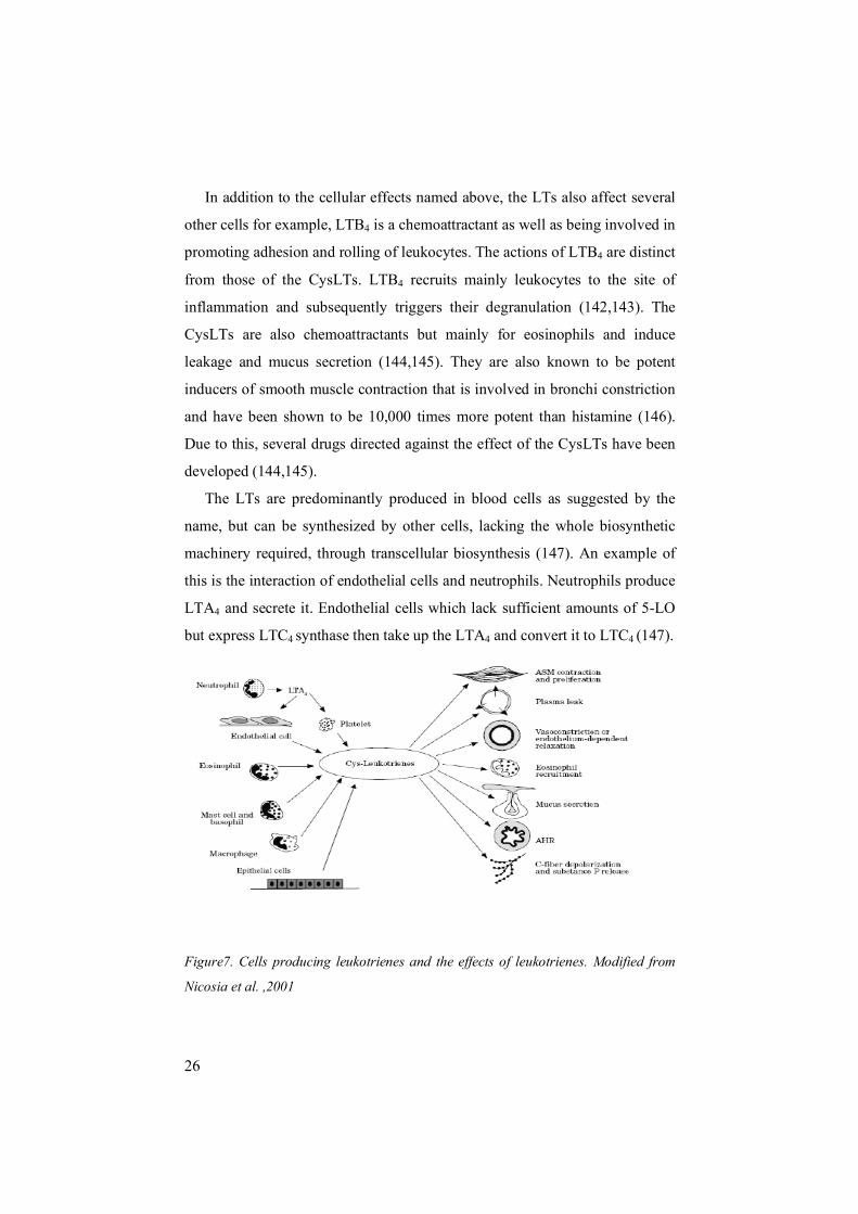
In addition to the cellular effects named above, the LTs also affect several
other cells for example, LTB4 is a chemoattractant as well as being involved in
promoting adhesion and rolling of leukocytes. The actions of LTB4 are distinct
from those of the CysLTs. LTB4 recruits mainly leukocytes to the site of
inflammation and subsequently triggers their degranulation (142,143). The
CysLTs are also chemoattractants but mainly for eosinophils and induce
leakage and mucus secretion (144,145). They are also known to be potent
inducers of smooth muscle contraction that is involved in bronchi constriction
and have been shown to be 10,000 times more potent than histamine (146).
Due to this, several drugs directed against the effect of the CysLTs have been
developed (144,145).
The LTs are predominantly produced in blood cells as suggested by the
name, but can be synthesized by other cells, lacking the whole biosynthetic
machinery required, through transcellular biosynthesis (147). An example of
this is the interaction of endothelial cells and neutrophils. Neutrophils produce
LTA4 and secrete it. Endothelial cells which lack sufficient amounts of 5-LO
but express LTC4 synthase then take up the LTA4 and convert it to LTC4 (147).
Figure7. Cells producing leukotrienes and the effects of leukotrienes. Modified from
Nicosia et al. ,2001
26
27
In epithelial cells, LTD4 can induce the production of CysLTs in an autocrine
fashion (148) as wells as inducing production of PGs via COX-2 induction
(149). The cell death induced by COX-2 inhibitors in intestinal epithelial cells
could be escaped by the cells through LTD4 stimulation (150). LTD4 has also
been shown to induce a rapid calcium response in intestinal epithelial cells as
well as CRC cells (151,152). This calcium response has been demonstrated to
be mediated through a PTX sensitive G-protein, leading to cAMP release and
PKA activation (153,154). In resting cells 5-LO is located in the cytoplasm but
when activated it translocates to the nuclear membrane, and with the help of
Five Lipoxygenase Activating Protein (FLAP) converts the released AA to
LTA4 (155).
7. Cysteinyl leukotriene receptors The LT receptors are GPCRs that were cloned and characterised late in the
1990’s (156-160). The first receptor to be cloned was the first LTB4 receptor,
BLT1R (161) and only three years later a second LTB4 receptor; the BLT2R
was cloned (162). BLT1 is the high affinity receptor for LTB4 and BLT2 is the
low affinity receptor. The CysLTs have so far been shown to exert their effects
mainly through two receptors; the CysLT1R and CysLT2R (156-160).
However, recent discoveries reveal the existence of two new receptors; the
orphan receptor GPR17 (163) which has higher affinity for LTC4 than LTD4
and a potential CysLTE4R (127) with a high affinity for LTE4. GPR17 has also
been shown to bind uracil nucleotides (124). The CysLTs bind CysLT1R with
the following different affinities; LTD4<LTC4<LTE4. LTD4 binds CysLT1R
with the highest affinity (EC50 = 0.4nM) whilst LTC4 binds CysLT1R with a
350 times lower affinity (EC50 = 21nM). LTE4 is a less potent agonist and has
been shown to sometimes act as a partial agonist (130,159,160,164). LTD4 and
LTC4 have equal affinity for CysLT2R and LTE4 has an even lower affinity
(158).
27

28
Figure8. The Cysteinyl leukotriene receptor 1.
The CysLT1R is encoded on the X chromosomes while the CysLT2R is
encoded by chromosome 13 and the two receptors share a mere 38%
homology. Both receptors were cloned almost at the same time in 1999.
CysLT1R has a molecular mass of 38 kDa in its monomeric form (159,160)
yet is often detected oligomerized (165). The CysLT1R has four potential N-
glycosylation sites and many potential PKC and PKA phosphorylation sites in
its third intracellular loop and C-terminus (160). It is known to be involved in
various signalling pathways and cellular functions such as; MAPK activation,
calcium increases, actin reorganization, proliferation, survival, differentiation,
and migration (79,166,167). In the last 20 years, an array of various inhibitors
targetting the CysLT1R, have been developed. Montelukast, Zafirlukast,
Pranlukast (IC50 = 1.8–4.9nM) are some of the examples of inhibitors for the
CysLT1R (159).
The CysLT2R is a protein of 346 amino acids and migrates at 49-58 kDa
(168). It also contains 4 N-glycolysation sites mainly in its N-terminus and
PKC and PKA phosphorylation sites in its third internal loop and C terminal
tail (158). Binding studies in Cos-7 cells with this receptor reveal the presence
of two binding sites, one low and one high for LTD4. In another study the
affinity for LTC4 for CysLT2R was detected in the nano molar range (156-
158) whilst all of the CysLT1R selective antagonists were demonstrated to be
28
29
inactive in a competition assay. Bay u9773 which has been suggested as a
CysLT2R antagonist but was later shown to also be a partial agonist of both
receptors (156,158). HEK-293 cells transfected with the CysLT2R also
demonstrated that LTC4 and LTD4 are equipotent agonists but that LTE4 is a
partial agonist (156-158).
The distribution of CysLT1R and CysLT2R receptors is mostly overlapping
but high expression of CysLT2R has been detected in heart, brain and the
adrenal glands (128,156-158). Intestinal epithelial and CRC cells express both
receptors (140,169,170) and the expression of each receptor in CRC cells
varies, although in general CysLT2R seems to be down regulated in CRC
patient material whilst CysLT1R seems to be up-regulated in comparison to the
surrounding non-transformed tissue (140,169,171).
The recently discovered GPR17 has been shown to have a higher affinity
for LTC4 than LTD4 (163). The idea that additional receptors may exist came
from a few unexplainable data such as: LTC4 or LTE4 failing to activate
CysLT1R or CysLT2R and also from the fact that the response from LTC4 was
more potent than that of LTD4, which does not fit with the affinity studies
preformed on the CysLT1R and CysLT2R (172-175). In 2001 a report
described that CysLT1R and another yet unidentified receptor, although not the
CysLT2R, both responded to uracil nucleotides and CysLTs. Later another
study investigated the orphan receptor GPR17 and found that it responds to
both CysLTs and extracellular nucleotides (163). Interestingly two CysLT1R
inhibitors (pranlukast and montelukast) could block the LTD4 induced
response of GPR17 and as could two antagonists of the P2YR family (163).
GPR17 binds the CysLTs in the nano molar range whilst the potency range for
the extracellular nucleotides is in the micro molar range (UDP-galactose =
UDP > UDP-glucose). All the responses from GPR17 have been demonstrated
to be PTX sensitive and both types of agonists induce a calcium response and
inhibition of forskolin induced cAMP formation. GPR17 is highly expressed
in tissue undergoing ischemic injury such as heart, brain and kidney
29
30
suggesting its involvement in these conditions. Infact knock-down or
inhibition of GPR17 by either CysLT1R or P2YR antagonists protects against
brain damage (163). A recent study preformed in CysLT1R / CysLT2R
knockout mice showed a LTE4 induced vascular permeability response. This
led the authors to hypothesize that yet another CysLT receptor existed with
high affinity for LTE4. The authors continued by showing that in mice lacking
CysLT1R and CysLT2R, LTC4 and LTD4 could induce vascular leak but to a
lesser extent than LTE4. The effect of LTE4 was 64-fold greater in deficient
mice than the wild type mice. The response from all three ligands in double
deficient mice could be blocked by pranlukast, a CysLT1R inhibitor. The
authors concluded from this data the existence of a new CysLT receptor,
termed CysLTE4R (127).
CysLT1R internalisation has been studied in Cos-7 cells transfected with
the receptor and stimulated with LTD4. In this study CysLT1R was internalised
in a PKC dependent manner, where PKC phosphorylated the CysLT1R in the
C-terminus, and that lack of this phosphorylation impaired receptor
internalisation. Furthermore, the same study identified that the internalisation
of CysLT1R is β-arrestin independent (176). In contrast to this, a separate
study preformed in U937 human macrophage like cells, also transfected with
CysLT1R provided evidence that the LTD4 induced desensitization of
CysLT1R is not PKC dependent but GRK2 dependent. However, the PKC
phosphorylation of CysLT1R was shown to be important in the extracellular
nucleotide induced CysLT1R desensitisation, although this does not lead to its
internalisation, rather it shows a rapid recovery of the receptor (165). Data
from the second project in this thesis demonstrates a clathrin, EPS15, β-
arrestin-2, Rab5 mediated internalisation of CysLT1R upon LTD4 stimulation.
Various signalling pathways have been established for CysLT1R in a
variety of cells and tissues. The signalling of CysLT2R has been less clarified
due to the lack of specific inhibitors. Contrasting reports regarding the
expression and signalling of CysLT1 and CysLT2 in HUVEC cells have been
30
31
reported. Some reports have identified the expression of CysLT1R in these
cells coupled with a calcium response from both LTD4 and LTC4 in these cells
that can be blocked by Pobilukast. Other groups have stated that these cells
mainly express the CysLT2R and have shown that this expression is highly up
regulated by IL-4 and also that CysLT2R is responsible for the calcium signals
exerted by LTD4, LTC4 and Bay u9773 (177-179). Additionally, LTD4
induced CysLT2R activation in HUVEC cells has been shown to up-regulate
37 early inducible genes, which included for example early growth response
(EGR) and COX-2 (180). In a study with mast cells the authors use MK 571, a
selective CysLT1R inhibitor, and show that the IL-8 production by the IL-4
primed and CysLT or UDP stimulated cells was not inhibited, but that Bay
u9773 was able to evoke IL-8 production. Interestingly, they show that
inhibition of p38 blocked the IL-8 production, suggesting a role for p38
downstream of CysLT2R (181). Studies in human coronary artery stimulated
with LTC4 show a calcium response which is unable to be blocked by
CysLT1R antagonists (141). In oxygen/glucose deprived PC12 cells, cell death
was shown to may be mediated by CysLT2R since Bay u9773 inhibited this
effect and CysLT1R seemed to reduce cell death (182).
A novel mechanism for receptor regulation was described for CysLT2R,
which has been suggested to negatively regulate the plasma membrane
expression and signalling of CysLT1R in mast cells. Moreover the authors
gave evidence for dimerization of CysLT1R and CysLT2R (183). Studies
preformed by J.A Boyce and co-workers recently reported by Rovati et. al,
suggest CysLT1R/ CysLT2R dimers at the nuclear membrane of mast cells
(128).
Our group have published data indicating the existence of both receptors at
the nuclear envelope in intestinal epithelial cells and CRC cells (140,169).
Furthermore, CysLT1R can localise to the outer nuclear membrane and
translocate from the plasma membrane to the nuclear membrane upon
stimulation with LTD4 (169). The COX-2 gene up-regulation mediated by
31

32
LTD4 requires the internalisation of the receptor and we hypothesise that this
signal is mediated through the translocation of CysLT1R to the nuclear
membrane. In contrast to COX-2, Cyclin D1 mRNA up-regulation and the
LTD4 induced ERK1/2 phosphorylation are increased when receptor
internalisation is blocked. This leads us to hypothesise that the plasma
membrane CysLT1R can continue to signal and respond to LTD4 when its
internalisation is blocked and can therefore increase the signals mediated from
the plasma membrane.
8. Mitogen activated protein kinases Mitogen activated protein kinases (MAPKs) are serine threonine kinases
involved in a variety of signalling cascades mediating cellular responses such
as proliferation, apoptosis, differentiation and survival. MAPKs need to be
phosphorylated for full activation and the pathways they regulate are
sometimes cross linked or dependent on each other (184). In contrast, MAPKs
are negatively regulated by de-phosphorylation. There are seven families of
MAPKs and they can be dived in the classical MAPKs (ERK1/2, p38, JNK
and ERK5) and the atypical MAPKs (ERK3, 4, 7 and NLK) (185,186).
Phosphorylation of MAPKs occurs in their catalytic loop and these
phosphorylations are regulated by upstream MEKS/MAPKK. MEKs in turn
are regulated by MAPKKK, such as Raf. In summary different stimuli activate
MAPKKK that in turn activate MAPKK and MAPKs (187). Raf can be
activated by a family of small monomeric GTPases such as Ras. Ras has been
shown to be mutated in 30% of all cancers and B-Raf is mutated in 60% of all
malignant melanomas (188).A classical pathway is that of mitogens or growth
factors activating PKC which in turn can lead to the activation of Ras and Raf.
Raf then activates MEK1,2 which phosphorylate and activate ERK1/2.
ERK1/2 activation leads to the activation of various transcription factors such
as NFκB, inducing cell survival and proliferation (186,189-192). The p38
MAPK family consists of four isoforms; α, β, γ and ε (189). They are activated
32
33
by stress and inflammatory cytokines. Most stimuli that activate p38 can also
activate JNK. MEK6 can activate all p38 isoforms whilst MEK3 activates α
and β (193). p38 has been shown to be critical for normal immune and
inflammatory responses (194). Mitogens or growth factors can also activate
(via MEK1-4 or MEK3, 6) the p38 MAPK which has been shown to be
involved in differentiation, apoptosis but also cell growth and survival.
Downstream targets of p38 include p53 and NFκB (45,195,196). Both p38 and
ERK1/2 have been implicated in IBD. ERK1/2 has been demonstrated to be
over expressed and also overly active in IBD (186). Many cytokines can
activate ERK1/2 such as IL-21 and IL-1(186,189). p38 and JNK families have
however been the centre of investigation concerning MAPKs and IBD
(197,198). Inhibition of p38 and JNK have been shown to reduce cytokine
production (199).
9. NF-κB
The NF-κB family are transcription factors involved in many inflammatory
pathways. They have been suggested to play a major role in IBD and CRC
(41,45). Epithelial cells isolated from IBD patients show increased expression
of NFκB. Furthermore, constitutively active NFκB has been found in human
cancer cell lines as well as in tumours from patients suffering from CRC,
breast cancer, leukemia and prostate cancer. Suppression of NFκB in these
tumour samples inhibits proliferation, causes cell cycle arrest and leads to
apoptosis (200). Many cytokines and growth factors such as TNF-α or EGF
mediate their proliferative effects through activation of NFκB. It has been
suggested that NFκB is one of the major links between inflammation and
cancer (200). This family consists of five members, p50, p52, p65 (RelA), c-
33

34
Rel and RelB, which share an N-terminal Rel homology domain (RHD)
responsible for DNA binding, nuclear targeting and homo and
heterodimerisation. NF-κB dimers bind to various pro-inflammatory, anti-
apoptotic and cell cycle regulating genes. It is however only the p65, c-Rel and
RelB that are able to directly activate transcription of target genes. P50 and
p52 need to dimerize with p65, c-Rel and RelB, for gene activation whilst
homo-dimerization of p50 and p52 can suppress transcription. NFκB was first
discovered in inflammatory cells but has since been shown to be expressed in
various cell types (45,200,201). When activated NFκB translocates to the
nucleus, whilst in its inactive state NF-κB dimers are bound to specific
inhibitory proteins IkBs, in the cytoplasm. The IкBs can be divided in to IKB-
α, β and γ. They keep NF-κB in its inactive state by masking its nuclear
localization sequence. IкB-α has also been shown to translocate to the nucleus,
inhibit NFкB from binding to its target genes and export NFкB out of the
nucleus. However upon stimuli, the IкBs are phosphorylated, ubiquitinated
and sent for degradation. This then release NFкB to translocate to the nucleus
and bind to its target genes (201). The activation of NFкB can be divided in to
the classical and the alternative pathways. The classical pathway can be
activated by for example pro-inflammatory cytokines such as TNF-α or IL-1,
bacterial LPS and viruses. These signals lead to activation of IKK (IKB kinase
complex) which is composed of IKK-α and β as well as the regulatory NEMO.
The catalytic subunits (IKK α and β) phosphorylate serine residues in the IKB
proteins. In the classical pathway this phosphorylation is mediated mainly
through the IKK-β and results in IкB being sent for degradation and NFкB
translocating to the nucleus. In the alternative pathway, IKK-α is predominant
in the activation process. In this pathway p100, the precursor of p52, is cleaved
into p52, which then translocates to the nucleus (202-204). NEMO is not
absolutely required for the alternative pathway. Some of the inducers of the
classical pathway can also induce the alternative pathway, such as TNF-
receptor family member CD40. Other NFкB activators are the Nod proteins (1
34

35
and 2) which are cytoplasmic receptors for microbial ligands, or the TLR that
via TRAF6 and MEK1/3 can activate the IKK complex. MEK 1,3 can activate
and regulate both ERK1/2 and p38, both of which have been shown to be
involved in activation of cPLA2-α and proliferation of intestinal epithelial
cells (201).
Figure9. Classical and atypical activating pathways of NFκB.
http://www.biomedcentral.com/nspprimers/nfkb/full
10. Protein kinase C The protein kinase C (PKC) family consists of 10 mammalian isoforms
divided into three groups; the classical, novel and the atypical. The classical
PKCs include the α, βI, βII, and γ PKCs. These isoforms need calcium,
diacylglycerol (DAG) and phosphatidyl serine (PS) for activation. The novel
PKCs consist of PKCδ, PKCε, PKCη, PKCµ, PKCθ, and do not require
calcium but do require DAG and PS for activation. The atypical PKCs (PKCζ,
PKCι/λ) only require PS for activation (187,205). PKCs are activated by
phosphorylation and binding of PKCs to the cytoplasmic side of the plasma
membrane. The membrane targeting domains are the C1 domain which binds
DAG and C2 which binds acidic lipids. PKCs are serine threonine kinases and
35

36
consist of a regulatory N-terminal that binds calcium, co-factors and lipids (C1
and C2). When co-factors bind these domains they release a pseudo substrate
that otherwise blocks the activation site of PKCs. They also have a catalytic C-
terminal that is an ATP substrate binding domain (206). PKCs are involved in
many different pathways and signalling cascades by phosphorylating many
different proteins. Three types of amino acids are known to be phosphorylated;
serines, threonines and tyrosines. When PKC phosphorylates other proteins in
their serine or threonine site, it changes the state of these downstream proteins
and activates or allows them to bind to other proteins. GPCRs, tyrosine kinase
receptors, ion channels, and transcription factors and enzymes such as PLA2s
are just some of the types of proteins that are phosphorylated by PKCs (207).
In the case of GPCRs PKC has been shown to phosphorylate them once they
have been activated by their ligand. This phosphorylation then can desensitize
and/or help the GPCR associate with the endocytic machinery (207). Two
ways of inhibiting PKCs include the use of calphostin C which competes with
DAG/PS and inhibits the membrane translocation of PKC. The other way is
used by GFX and Gö inhibitors which inhibit the catalytic activity of PKC by
competing with ATP (208,209). We have previously shown several PKC
members to be important in LTD4 mediated signalling. PKCδ was shown to be
important for stress fibre production induced by LTD4 (210), whilst
interestingly two PKCs namely PKCα and ε, are involved in cell proliferation
(132).
11. G-protein coupled receptor structure and signalling G-protein coupled receptors (GPCRs) are a family of seven transmembrane
spanning receptors that sense molecules outside of the cells and activate
intracellular signal transduction pathways and cellular responses by coupling
to heterotrimeric G-proteins (211).
GPCRs are one of the largest known families in the genome. There are
approximately 865 potential GPCRs which equals about 3.5% of the human
36
37
genome set (212,213). Approximately 30%-45% of all patented drugs in
current clinic use are GPCRs (213,214). GPCRs contain 7 hydrophobic
stretches of 22-25 residues which are long enough to cross a membrane when
folded (7 transmembrane domains) creating three external and three internal
loops with an external N-terminal and a cytoplasmic C-terminus. The trans-
membrane domains of GPCRs are highly conserved. The bovine rhodopsin
GPCR has been crystallized and its structure has served as a template for other
GPCRs (215) (216). The GPCRs have a variety of different ligands including;
hormones, neurotransmitters, chemokines, calcium ions, sensory receptors for
various odorants, tastes and photons of light. There are two broadly defined
functional classes of GPCRs; The sensory receptor homologs and the non-
sensory receptor homologs. In 2004 the Nobel prize went to Linda Buck and
Richard Axel for demonstrating that odour precipitation results from several
different GPCRs on different neurons (217). The GPCRs can further be
classified into three families based on their sequence. The first family
(Family1/A) has the closest structure to Rhodopsin and is the largest family.
They have a short N and C terminus and include most of the olfactory
receptors but also 200 of the non-olfactory receptors belong to this group
(218). The second family (2/B) only has 25 members and they are
characterised by their long N-terminus and the fact that they mainly activate
cAMP through the Gs G-protein. This family includes the parathyroid
hormone receptor, secretin receptor, adrenomedulin receptor and glucagon
receptor (218). The third family (3/C) have long N and C terminals, mostly
bind their ligands in the N-terminus and include the GABAbR, calcium
sensing receptors, some taste receptors and the orphan receptors, where the
natural ligands are unknown (218).
GPCRs have been shown to exist in a conformation equilibrium between
inactive and active state in the absence of ligand (219). The binding of ligands
shifts the equilibrium either towards the active or inactive state. Three types of
ligands exist; agonists which shift the equilibrium to active state, inverse
37

38
agonists which shift the state to inactive and also prefer to bind receptors in the
inactive equilibrium state. The inverse agonists have been demonstrated to
have the opposite effect of agonists. There are also neutral antagonists that
bind the GPCR and lock it in an inactive state (220). As mentioned above this
activation state can also be induced spontaneously without ligand as well as in
receptors containing mutations that lead to constitutive activity which has been
demonstrated in various pathological conditions (221). There are four different
mutations known in the Rhodopsin receptor that causes it to be constitutively
active that can lead to blindness (222). Mutations creating a constitutively α-1-
β -adregenic receptor have been demonstrated to have mitogenic and
tumorogenic effects (223).
Malignant cells or their surrounding stromal cells often abuse the functions of
GPCRs to be able to proliferate, induce angiogenesis (growth, migration and
blood supply) and survive apoptosis. This is often achieved by either
expressing constitutively active GPCRs or more commonly by over-
expressing particular GPCRs (224). Furthermore over-expression of
constitutively active G-proteins have also been implicated in cancer, the most
potent ones being G-α12/13 (225,226).
Heteromeric G-proteins are composed of the α, β and γ subunits. In their
inactive state the α subunit of the G-protein is bound to guanine dinucleotide
phosphate (GDP). Upon activation the GDP is exchanged for a guanine
trinucleotide phosphate (GTP). This leads to the disassociation of the α subunit
from the β and γ subunits. These subunits can stimulate several downstream
pathways. The α subunit has an intrinsic ability to hydrolyse the GTP back to
GDP. The G-proteins are classified according to their α subunit. The G-αs
stimulates cAMP production by stimulating adenylylcyclase whilst the G-αi
inhibits cAMP production. The G-αq/11 activate PLC which cleaves PIP2 to
IP3 and DAG. G-α12/13 activate monomeric Rho GTPases. There are several
regulating proteins that can catalyze the GDP-GTP and GTP-GDP exchange,
but there are also two toxins used that can inhibit these activities. The first one
38

39
is Pertussis toxin which is a bacterial toxin that keeps the α subunit of the Gi
family of G-proteins in GDP/inactive state by catalysing the ADP-ribosylation
thus blocking its interaction with GPCRs. There is also the use of cholera
toxin, another bacterial toxin. This toxin inhibits the GTPase activity of the α
subunit of the Gs family of G-proteins keeping it in a constitutively active
state by ADP ribosylating it (227,228). GPCRs are a family of intrinsic
hydrophobic proteins only found in higher eurokaryotes, located both at the
plasma membrane and nuclear envelope of cells (169).
The β and γ G-protein subunits, are also able to play a signalling role. For
example our group has previously shown that LTD4 stimulation of calcium
intracellular release, requires the release of inositoltriphosphate from
diacylglycerol. This is achieved through the action of phospholipase C –γ,
which required “docking” with the β and γ subunits to become functional
(229).
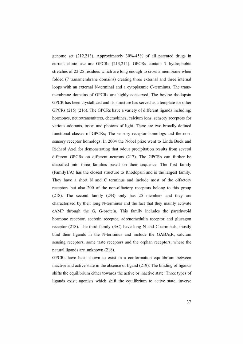
12. GPCR dimerization and other GPCR interacting
proteins GPCRs can signal as hetero- and/or homo-dimers or oligomers. Moreover,
dimerization has been shown to be needed for proper expression, stronger
ligand binding, phosphorylation and internalization.
39
40
Figure9. GPCR dimerization. Modified from Carrillo, J. J. et al. J. Biol. Chem.
2003;278:42578-42587
Dimerized GPCRs have been shown to have signalling properties distinct form
those of the monomeric receptors (230,231). The GABAB receptors are a part
of the family C GPCRs and they brought attention to the functional relevance
of receptor dimerization. It was demonstrated that when the GABABR1 was
expressed alone it did not have the ability to be efficiently trafficked to the cell
surface (232). Six years after the cloning of this receptor the GABABR2 was
cloned and studies on this receptor showed that it could not bind ligands when
expressed alone. However, when the two receptors were expressed together
they were efficiently expressed at the plasma membrane and also able to
respond to the GABA ligands (233). It has been suggested that GABAB1 and 2
receptors can dimerize via the transmembrane or N-terminal regions (234).
However, the GABABR1 is found in many regions of the brain where
expression of the GABAR2 is not detected. (235). Surprisingly it was found
that non of the mGluR (closest related family) or 30 other GPCRs could
40
41
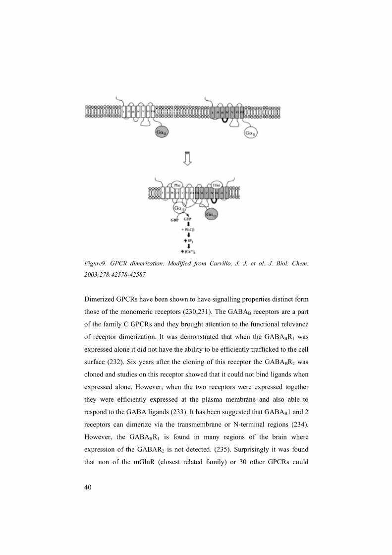
effectively target the GABAB1R to the cell surface (236). Several other
receptors have been shown to dimerize for different functions. Table 1.
demonstrates various receptors, their partners and the effect of the
dimerization (237).
Table1. GPCR heterodimerisation. Modified from Prinster et. al. Pharmacological
Reviews.
41

42
The importance of homo-dimerization has also been demonstrated as in the
case of β2ARs where interruption of the homo-dimerization decreases β2AR
induced cAMP production. (238). Targeting of GPCRs to the correct site has
been shown to play an important role in homeostasis as miss localization has
been demonstrated in various pathological conditions such as retinitis
pigmentosa. This condition arises from intra cellular accumulation of mutant
forms of the Rhodopsin receptor. Similar pathological accumulations have
been demonstrated for the vasopressin and gonadotropin-releasing hormone
receptors (239).
There are various proteins known to interact with GPCRs and deliver them
to the cell surface, link them to their downstream effectors and ensure
specificity and activation of diverse pathways. Proteins involved in regulating
GPCR ER export and GPCR folding include the ER chaperons; calnexins,
which help the folding of newly synthesized proteins and send improperly
folded proteins to degradation. Accessory proteins cyclophilins are also
involved in folding, and RAMPs are involved in ER export as well as proper
folding, signalling, internalization, recycling and degradation (240). A-kinase
anchoring proteins have been shown to link GPCRs to PKA or G-proteins
(241,242). Some AKAPS have been shown to increase receptor
phosphorylation and downstream ERK1/2 signalling whilst others have been
shown to induce desensitization (242). Other proteins involved in bringing
together GPCRs with their downstream effectors for rapid and efficient
signalling are the Homer proteins and the InaD proteins (243). The InaD
proteins contain PDZ domains, a domain known to interact with the C-termini
of proteins including GPCRs (244). Scaffolding/chaperon proteins bind and
recognize specific motifs on GPCRs (245). Furthermore, several motifs and
domains of GPCRs have been demonstrated to be required for surface
expression, nuclear localization and ER-retention (245).
42
43
13. GPCR desensitization There are many regulatory mechanisms involved in GPCR signalling. One of
which is receptor desensitization (246). This mechanism involves GPCR
phosphorylation which is mediated by PKC, PKA or G-protein receptor
kinases (GRKs) (247). The first two mentioned kinases directly uncouple
GPCRs from G-proteins. PKC and PKA can mediate both homo and
heterolouges desensitization. These two terminologies refer to whether the
receptor desensitization is initiated by the ligand of the receptor or by another
GPCR-ligand interaction. An example of this is the heterologous
desensitization of the CysLT1R by UDP or ATP (165). This desensitization is
mediated through PKC and leads to a more rapid receptor recovery than the
homologous desensitization (165). In addition, PKA is capable of
phosphorylating the GPCR in such a manner that it switches its binding from
Gs to Gi. This has been demonstrated for the β2 adregenic receptor (β2AR)
and the prostacyclin receptor. This switch favours downstream activation of
MAPKs such as ERK1/2 (248-250). GRKs only phosphorylate ligand
occupied/ligand activated GPCRs. It is often associated with the classical β -
arrestin dependent desensitization because GRKs are suggested to promote β -
arrestin binding to the GPCR which inhibits the GPCR from further binding
G-proteins (251). β-arrestins are often involved in receptor internalization
(discussed below). There are four arrestins, two of which are expressed in the
retina called visual arrestin and cone arrestin. The other two arrestins, β-
arrestin-1/arrestin-2 and β-arrestin-2/arrestin-3 are expressed in most tissues.
The importance of GRKs and β-arrestin are demonstrated in knock-out mice
where various GPCR desensitization, signalling and regulation are impaired
(218). Receptor desensitization can be regulated on G-protein level as well by
the RGS family. These proteins accelerate the hydrolysis of GTP to GDP
speeding up deactivation of the signal induced (252).
43

44
14. Internalization of GPCRs GPCR internalization is a complex yet extensively studied mechanism
involving various scaffolding and regulatory proteins. GPCRs can internalize
via three known pathways; clathrin coated pits, uncoated vesicles and caveolae
(253). Internalization of GPCRs is initiated by ligand binding, which leads to a
conformational change initiating receptor signalling. This subsequently leads
to phosphorylation of the receptor by PKA, PKC or GRKs (as discussed
above). From here the GPCRs are targeted to vesicles transporting them inside
the cell to translocate, recycle or be degraded. Some receptors have actually
been shown to have the ability of continuing to signal or even initiate new
signalling pathways from the endosomes (254). Though internalization is a
series of sequentially regulated events, studies have demonstrated that various
steps have the ability to determine or play a part in receptor fate. The ligand
binding has the ability to determine the fate of the receptor. This has been
demonstrated for example for the β -2 adregenic receptor. Under normal
conditions when the receptor is activated by its ligand it predominantly
undergoes recycling, however upon prolonged or repeated exposure to its
ligand, the receptor is degraded (255). Different ligands have also been
implicated in inducing different endocytic sorting and trafficking of the same
GPCR (256).
Internalization of GPCRs and caveolin Caveolae are 50-100nm flasked shaped membrane invaginations that are rich
in cholesterol as well as the caveolin proteins (as indicated by the name) (257).
There are three caveolins; caveolin1, 2 and 3. Caveolins-1 and 2 are strictly co
localised and they have been implicated to require each other for proper
function (258-260). It is not fully understood how receptors are targeted or
internalized via caveolae or uncoated vesicles. But it is speculated that the
transmembrane regions can interact with cholesterol found in caveolae or
44

45
uncoated vesicles and it has been shown that cholesterol can modulate the
affinity of some GPCRs (261,262). Caveolae are localised both near and on
the plasma membranes well as intracellularly. Interestingly many proteins
such as the EGFR, heteromeric G-proteins, GPCRs and their interacting
proteins are positioned in caveolae (263). This is thought to be a mechanism
that makes signalling and trafficking of GPCRs efficient (263). However,
disruption of caveolae has been shown to prevent internalization of the
endothelin receptor ETB and VIP receptors (263). Caveolin-1 in vivo and in
vitro animal experiments have shown a suppressive effect of caveolin-1 in
transformation and breast tumorogenesis (264,265). Contradictory to this,
studies on human breast and prostate cancer show a positive correlation of
caveolin-1 expression and tumorogenesis. Another study demonstrated
elevated levels of caveolin-1 in adenocarcinoma of the colon whilst the
adenomas and normal colonic epithelium showed little or no staining and
Caveolin-2 absent (266). Conversely in another study CRC cell lines and
human tumour epithelial mucosa were shown to express low or undetectable
levels of caveolin-1 (267).
GPCR internalization and clathrin
Clathrin mediated endocytosis is a much more extensively studied mechanism.
Clathrin coated pits are made up of clathrin heavy and light chain as well as a
dozen regulatory proteins (268).
Clathrin mediated endocytosis of GPCRs usually includes the β-arrestin
dependent internalization, although β-arrestin independent clathrin mediated
internalization will also be addressed in this section.
When a GPCR is activated by its ligand and subsequently phosphorylated,
this phosphorylation recruits β -arrestin. Phosphorylation of the GPCR has
also been demonstrated to be able to determine the sorting of the receptor.
This is exemplified in the case of the CXCR chemokine receptor where
mutation of its phosphorylation sites inhibits it from being ubiquitinated and
targeted to the lysosomes (269). Ubiquitin is known for its ability to sort
45

46
proteins to the proteosome (270), this mechanism includes GPCRs as well, but
only in the biosynthetic pathway (271). In endocytic sorting, ubiquitin
molecules added on to the C-terminus of the GPCRs targets them for
lysosomal degradation (272) or effects the rate of internalization (273,274).
The family of ESCRT proteins are highly conserved and have been
demonstrated to be involved in the lysosomal sorting of GPCRs. This
lysosomal sorting can be through the ubiquitin pathway but GPCRs can be
sorted to lysosomes by ESCRT in the absence of ubiquitination as well. (256).
This suggests that phosphorylation can be an initial step to the post
translational modifications made on a GPCR upon trafficking.
Furthermore, phosphorylated GPCRs recruit β-arrestin that functions as
a scaffolding protein and links the GPCR to the heavy chain of clathrin and
to β2 subunit of AP-2 by directly binding to these proteins. AP-2 is a
clathrin adaptor protein involved in early events of vesicle formation. AP-2
targets receptors containing tyrosine or di-leucine based motifs and binds
them as well as binding clathrin coats and Eps-15 (another clathrin adaptor
protein first discovered in the EGFR endocytic pathway). Inhibition of
clathrin pit formation is often achieved by using dominant negative Eps-15
constructs. Eps15 has been shown to be both phosphorylated as well as
ubiquitinated. The phosphorylation of Eps-15 has been connected to its role
in internalization, although the role of the ubiquitination is still poorly
understood (275,276). The clathrin coated pit complex recruits the large
GTPase Dynamin via β -arrestin. Dynamin is involved in the pinching off
the coated pit from the plasma membrane (277).
GPCR internalization and β -arrestin β -arrestin mediated binding targets the GPCR to clathrin coated pits at the cell
surface (268). Furthermore, ubiquitination of β-arrestin has been shown to
promote internalization of GPCRs. This is suggested to occur through
46

47
enhancing the affinity of β -arrestin to the plasma membrane (278).
Furthermore, β-arrestin also plays a role in receptor sorting. Transient binding
of GPCRs to arrestin that is terminated shortly after internalization mediates a
rapid recycling whilst a more prominent binding mediates a slow recycling
(251,253). Once inside the cell, GPCRs can be de-phosphorylated and
returned to the plasma membrane, but this requires that β-arrestin is
disassociated from the GPCR. ERK 1/2 activation have been implicated in β-
arrestin mediated signalling. Dominant negative mutants of arrestins have
been shown to attenuate GPCR induced ERK1/2 phosphorylation (279,280).
However β -arrestin has also been shown to mediate ERK1/2 signalling in
parallel to effecting G-protein mediated signalling. Over-expression of
arrestins attenuated G-protein mediated signalling of the angiogenesis 1
receptor (AT1R) but increased ERK1/2 phosphorylation through the same
receptor (281,282). β-arrestin has the ability of effecting the duration and
magnitude of ERK1/2 signalling as well as its localization. This has been
demonstrated by the fact that weak interactions between β-arrestin and GPCRs
mediated ERK1/2 translocation to the nucleus, activation of transcription
factors (Elk and c-Myc) and induction of proliferative/mitogenic responses. In
a similar manner, stable interactions between β -arrestin and GPCRs can
mediate ERK1/2 signalling with the difference that the ERK1/2 signal retained
in the cytosol preventing agonist induced cell proliferation (282,283). With all
this in mind some GPCR internalization has been suggested to be arrestin-
independent. The LTB4 receptor BLT1R has been demonstrated to internalize
in an arrestin independent but GRK dependent manner. In this study dominant
negative constructs of GRK2 could inhibit internalization of the BLT1R but
dominant negative constructs of β –arrestins were ineffective. Furthermore, the
authors demonstrated a lack of co-immunoprecipitation and co-localization of
β -arrestins with the BLT1R (284). This does not rule out the possibility that
the interaction of β-arrestin and the BLT1R might be weak and transient and
therefore not detected, therefore β -arrestin double (β-arrestin-1 and-2) knock-
47

48
out mice models should be used (285,286). This has been preformed in studies
with the protease activated receptor 1 where internalization is not effect and
suggested to be arrestin-independent (287). Similar studies to both models
mentioned above have been preformed for the CysLT1 receptor, where over-
expression of β -arrestin increased CysLT1 internalization and dominant
negative constructs of β -arrestin demonstrate a non-significant decrease of
receptor internalization. However, MEF cells from β -arrestin double knockout
mice demonstrated a loss of CysLT1 from the surface similar to that of the
wild type mice (176). We preformed β -arrestin-2 siRNA studies in intestinal
epithelial and CRC cells and demonstrate an almost abolished internalization
of CysLT1R.
GPCR internalization and Rab proteins The Rab GTPases are small monomeric G-proteins and constitute the largest
family of monomeric GTPases with over 60 different members (288). They
are involved in various cellular processes such as trafficking, endocytosis,
exocytosis cell growth and differentiation (289). They are post-transitionally
modified which allows them to associate with various membranes such as the
Golgi-, nucleus, mitochondria-, ER-, plasma- membranes, endosomes and
lysosomes (290). They act to coordinate the trafficking between these
membrane bodies.
48
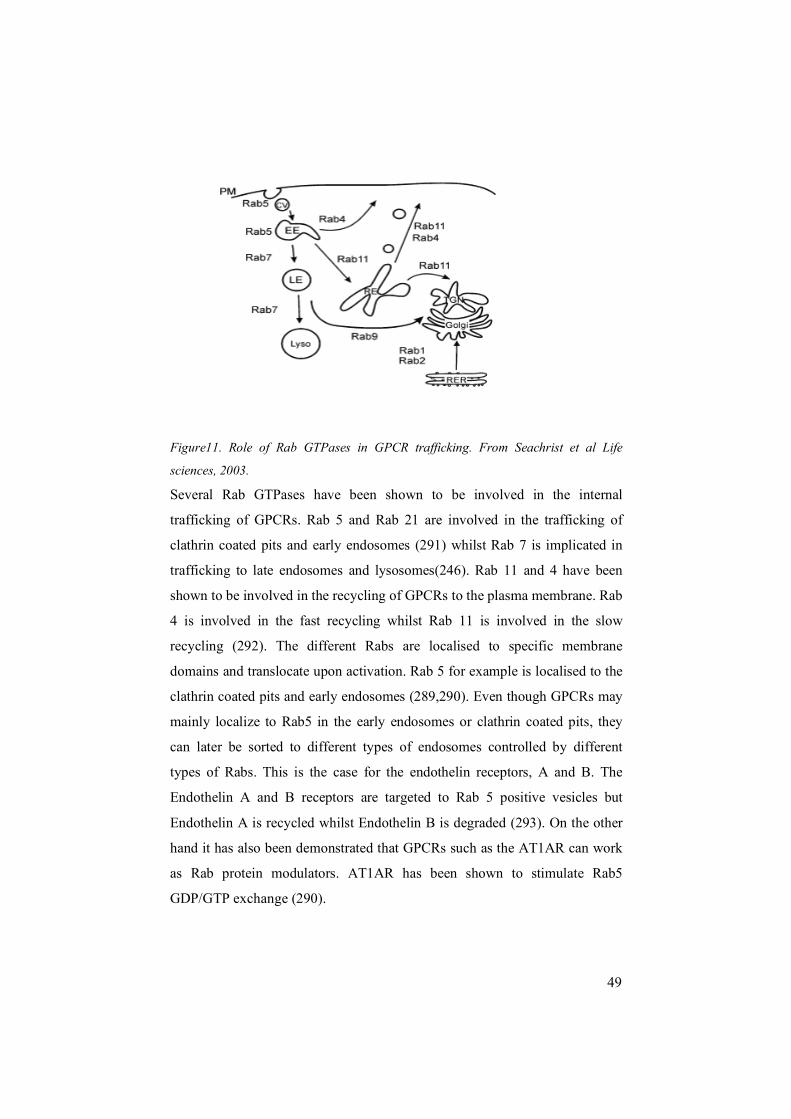
49
Figure11. Role of Rab GTPases in GPCR trafficking. From Seachrist et al Life
sciences, 2003.
Several Rab GTPases have been shown to be involved in the internal
trafficking of GPCRs. Rab 5 and Rab 21 are involved in the trafficking of
clathrin coated pits and early endosomes (291) whilst Rab 7 is implicated in
trafficking to late endosomes and lysosomes(246). Rab 11 and 4 have been
shown to be involved in the recycling of GPCRs to the plasma membrane. Rab
4 is involved in the fast recycling whilst Rab 11 is involved in the slow
recycling (292). The different Rabs are localised to specific membrane
domains and translocate upon activation. Rab 5 for example is localised to the
clathrin coated pits and early endosomes (289,290). Even though GPCRs may
mainly localize to Rab5 in the early endosomes or clathrin coated pits, they
can later be sorted to different types of endosomes controlled by different
types of Rabs. This is the case for the endothelin receptors, A and B. The
Endothelin A and B receptors are targeted to Rab 5 positive vesicles but
Endothelin A is recycled whilst Endothelin B is degraded (293). On the other
hand it has also been demonstrated that GPCRs such as the AT1AR can work
as Rab protein modulators. AT1AR has been shown to stimulate Rab5
GDP/GTP exchange (290).
49
50
15. Nuclear GPCR Over time a greater number of GPCRs have been found at the nuclear
membrane and even inside the nucleus (such as the PAFR) (294). They have
either been shown to be there under basal conditions such as the prostaglandin
receptors (EP1-4) (295) and be further increased upon stimulation, such as the
CysLT1R (169) or translocate there upon ligand dependent or independent
activation (296). The GPCRs have been shown to be inserted into the nuclear
membrane and exert signalling pathways or gene transcriptions that are
diverse from those of the plasma membrane (297). Interestingly, no difference
in the size and sequence has been detected in any of the GPCRs (297). There
are several examples of GPCRs signalling at the nucleus. The first examples
are GPCRs at the plasma membrane that initiate signalling pathways at the
nuclear membrane. The GPCR for LPA (lisopphosphatidic acid) activates a Gi
dependent pathway that results in activation of phospholipase D1 (PLD1) that
in turn leads to conversion of phosphatidylcholine to DAG and phosphatidic
acid (PA). The chemokine receptor CXCR4 has been shown to induce
ERK1/2 nuclear transclocation and induction of transcription and proliferation
in a Gi dependent manner. Both of these pathways are mediated through the
plasma membrane GPCR eliciting a nuclear response from the plasma
membrane (296). As discussed above β-arrestin can retain ERK1/2 in the
cytoplasm and this retention can be mediated by GPCRs. The GPCR
Neurokinin-1 for example has been shown to induce β-arrestin dependent
retention of ERK1/2 in the cytoplasm while the β2 adrenergic receptor induces
β-arrestin dependent ERK1/2 translocation (298-300). An example of different
signalling pathways induced by the same receptor at the nuclear membrane
compared to the plasma membrane is that of the EP3Rs. The receptors signal
for cAMP or IP3 generation from the plasma membrane whereas the nuclear
membrane EP3R induces calcium signals and induces gene-transcription
(119,301). Moreover, stimulation of the nuclei with PAF and LPA induces a
calcium dependent ERK1/2 pathway and NFκB binding to DNA. The calcium
50

51
induction induced by lipid ligands of these three nuclear localised GPCRs lead
to specific gene-up regulations (119,297,301,302). PAF or LPA have been
demonstrated to induce gene expression of eNOS, COX-2 and iNOS. These
signalling cascades were mediated through MAPK and NFκB (297).
Considering the cascade of signalling molecules that reside near or in the
nuclear membrane such as; cPLA2, PKC, ERK1/2, β-arrestin, 5-LO and COX-
2 (which give rise to the production of lipid ligands) and calcium channels, it
is not so farfetched to believe that signalling pathways can be orchestrated
here. Some GPCRs contain nuclear localization signals (NLS) or even ER-
retention sequences (PAFR and CysLT1R) (169) yet deletion or mutation of
these sequences do not change the nuclear localization of the GPCRs. In line
with this there are GPCRs that do not contain any of these types of sequences,
such as the LPA1R (297). The LPA1R has been shown in both clathrin and
caveolae domains in the plasma membrane but co localizes only with caveolin
in nuclear fractions. Considering the role caveolae play in bringing signalling
molecules together this is rather interesting. Moreover, not all GPCRs that are
found to reside at the nuclear membrane in certain tissue or cells are found to
do so in other types of cell or tissue (297).
51
52
THE PRESENT INVESTIGATION
AIM
The general aim of this work was to further investigate signalling and
trafficking of the cysteinyl leukotriene receptors and their role in CRC.
Specific aims of the present work
1. To explore the role of cPLA2 in LTD4 induced cell proliferation
2. To explore the regulatory mechanism of trafficking of the CysLT1R in
non-transformed and CRC cells as increased expression of this receptor
has been correlated with a poorer prognosis in CRC.
3. To validate the trafficking of the CysLT1R and the CysLT2R in relation
to each other as increased expression of the CysLT2R has been
correlated with a good prognosis and the balance and signalling of
these receptors may play a role in CRC.
52

53
RESULTS AND DISCUSSION Paper I One of the characteristics of chronic inflammation is a continuous production
of inflammatory mediators (32). LTs are inflammatory mediators derived from
AA. AA is released by PLA2s, particularly cPLA2-α from the phospholipid
bilayer of cellular membranes and can further be metabolized by 5-LO to
produce LTs or COX-2 to produce PGs (94). Indeed previous studies from our
group have demonstrated that LTD4 is able to induce COX-2 expression as
well as up-regulate LTC4 synthase and 5-LO in intestinal epithelial cells (303).
We have shown that these cells have the ability to produce CysLTs and that
this production can mediate proliferation and survival in intestinal epithelial
cells (148,303). Additionally, we have published data that LTD4 can up-
regulate 5-LO and LTC4 synthase in CRC cells (303). In the first paper of this
thesis LTD4 was observed to be able to activate cPLA2-α after 10 minutes of
stimulation, and that this activation consists of phosphorylation of cPLA2-α
and its translocation from the cytosol to an intact nuclear fraction. The same
study also revealed that LTD4 in a similar fashion could induce production of
cPLA2-α after 90 minutes. Both these pathways are mediated through the
CysLT1R, a PTX sensitive G-protein, PKC, ERK1/2, p38 and require a
calcium signal. The LTD4 induced expression of cPLA2-α is suggested to be
mediated through NFκB as an inhibitor of this protein blocked the cPLA2-α
up-regulation. This is further supported by data from our group that LTD4 can
induce NFκB expression at both mRNA and protein level at 60 minutes of
stimulation (unpublished data). The results from Paper I provide evidence for
the theory that LTD4 can induce its own production in intestinal epithelial and
CRC cells. We also demonstrate that LTD4 can induce cell proliferation,
which has been previously shown to go through two separate pathways; one
involving a PTX sensitive G-protein and one Ras dependent but PTX
independent pathway. This latter pathway activated PKC-ε, Raf-1 and ERK1/2
(131). In PaperI the LTD4 mediated proliferation reveals it to be cPLA2-α
53
54
dependent. Interestingly the cPLA2-α inhibitors even decreased the basal level
of proliferation. Whether this cPLA2-α proliferation involves a third pathway
or a crosstalk with the other pathways was not clear. It is however interesting
in the concept of inflammation induced cancer, that LTD4 induces a rapid
activation of cPLA2-α and that a longer stimulation results in the production of
cPLA2-α and eventually proliferation of the cells. We also observed an
increase of cPLA2-α in CRC cells and tissue compared to non-transformed
cells and tissue. This increased expression was similarly seen for COX-2 and
correlates well with the theory that cPLA2-α and COX-2 play a role in
different malignancies including CRC, strengthening the idea that cPLA2-α is
the predominant source for eicosanoid production (81-83,85,86,90-92). There
are however studies describing decreased cPLA2-α levels in CRC. The study
in this case suggests that cPLA2-α is involved in apoptosis and the decreased
expression of it, leads to cell survival (87,89).
Other studies including our own with colorectal patient material, contradict
this data, as increased cPLA2-α expression was evident. A recent study
investigating a patient with cPLA2-α deficiency indicated an important global
role for cPLA2-α in eicosanoid production and shows no malignancy in the
colon (93).
PaperII
A key regulation in GPCR signalling is internalisation. Our group has
previously identified two very interesting points about the CysLT1R; its
nuclear localisation and translocation (169) as well as its role in CRC
prognosis (171). High levels of the CysLT1R have been correlated with a
poorer survival rate in material from CRC patients. In this context, we wanted
to investigate a regulatory mechanism of the receptor in non-transformed
intestinal epithelial cells compared to CRC cells. Our results showed a rapid
internalisation of the receptor from the plasma membrane already at 5 minutes
and recycling of the receptor back to the plasma membrane within 20 minutes
after agonist removal in non-transformed cells. However, continuous
54

55
stimulation with LTD4 up to 60 minutes increased of the expression of the
receptor in a nuclear fraction in both non-transformed as well as CRC cells.
We have previously published that the CysLT1R is also localised to the outer
nuclear membrane (169). This internalisation and increase of the CysLT1R at
the nuclear membrane is a clathrin dependent mechanism, as inhibition of
clathrin blocks internalisation and CysLT1R accumulation at the nuclear
membrane. Furthermore, the CysLT1R internalisation is a β-arrestin-2
dependent process as siRNA against β-arrestin-2 blocks loss of the CysLT1R
from the cells surface. A previous study investigating internalisation of the
CysLT1R demonstrated a PKC dependent but β-arrestin independent
internalisation of the receptor (176). Another study showed that the LTD4
induced internalisation of the CysLT1R is not PKC dependent but GRK2
dependent (165). These data suggest that the internalisation of the CysLT1R
may vary in different systems. Since our study is preformed on cells
endogenously expressing the receptor we believe that the internalisation route
characterised in PaperII is representative for intestinal epithelial cells and CRC
cells. Moreover, we present data that the internalised CysLT1R is targeted to
Rab5 positive endosomes. The inhibition of clathrin led to blockade of the
CysLT1R internalisation in both non-transformed as well as CRC cells.
However, in the CRC cells an increase of the CysLT1R at the plasma
membrane was detected. Indeed CysLT1R internalisation is delayed compared
to non-transformed cells and also difficult to detect endogenously using
confocal microscopy (169) and western blot analysis. Intriguingly an
accumulation of the CysLT1R is detected rapidly after LTD4 stimulation and
this accumulation is clathrin sensitive. These results suggest a rapid turnover
of the CysLT1R at the plasma membrane of CRC cells explaining the increase
at the plasma membrane detected upon inhibition of clathrin coated pit
formation and LTD4 stimulation. FACS analysis only detects internalised
CysLT1R after 20min to 1h in various CRC cells yet the accumulation of
CysLT1R at the nuclear membrane is detected already 10-15 min of LTD4
55

56
stimulation. This could be explained by a low amount of receptor being
internalised from the plasma membrane at earlier time points that are difficult
to detect. It does however not exclude the possibility of internal pools of the
receptor translocating to both the plasma membrane as well as the nuclear
membrane of CRC cells upon LTD4 stimulation. This would also support the
increased amount of CysLT1R detected at the plasma membrane fo CRC cells.
As mentioned above inhibition of clathrin pit formation and stimulation with
LTD4 blocks the internalisation of CysLT1R from the plasma membrane and
its up regulation at the nuclear membrane in non-transformed cells. This study
also demonstrates ERK1/2 phsophorylation and cyclinD1 mRNA up
regulation induced by LTD4 in non transformed intestinal epithelial cells.
Interestingly both these signalling events further increase upon inhibition of
clathrin coated pit formation and LTD4 stimulation (1h), suggesting that the
signalling is mediated through the CysLT1R at the plasma membrane since this
treatment renders the receptor to stay at the plasma membrane and continue
signalling. In contrast to this LTD4 induced COX-2 is inhibited by upon
inhibition of clathrin coated pit formation suggesting that this signal is
dependent on upon inhibition of CysLT1R internalisation. Furthermore, we
speculate that the COX-2 signal is mediated through the CysLT1R at the
nuclear membrane since CysLT1R seems to translocate to nuclear membrane
upon LTD4 stimulation. However, we can not exclude the possibility of
CysLT1R signalling from endosomes.
PaperIII
Previous publications from our group have demonstrated different roles for
LTD4 and LTC4. Whilst LTD4 has been shown to induce proliferation,
migration and cell survival (131-133), LTC4 has been shown to promote cell
differentiation (140). Furthermore, upregulation of the CysLT1R has been
correlated with a poorer survival prognosis whilst increased CysLT2R
correlates with a better prognosis (140,171). In this study we wanted to
investigate the trafficking of the two cysteinyl leukotriene receptors; CysLT1R
56
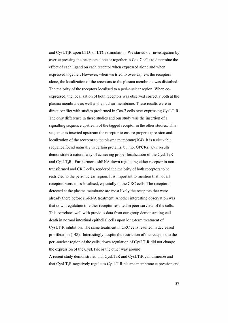
57
and CysLT2R upon LTD4 or LTC4 stimulation. We started our investigation by
over-expressing the receptors alone or together in Cos-7 cells to determine the
effect of each ligand on each receptor when expressed alone and when
expressed together. However, when we tried to over-express the receptors
alone, the localization of the receptors to the plasma membrane was disturbed.
The majority of the receptors localised to a peri-nuclear region. When co-
expressed, the localization of both receptors was observed correctly both at the
plasma membrane as well as the nuclear membrane. These results were in
direct conflict with studies preformed in Cos-7 cells over expressing CysLT1R.
The only difference in these studies and our study was the insertion of a
signalling sequence upstream of the tagged receptor in the other studies. This
sequence is inserted upstream the receptor to ensure proper expression and
localization of the receptor to the plasma membrane(304). It is a cleavable
sequence found naturally in certain proteins, but not GPCRs. Our results
demonstrate a natural way of achieving proper localization of the CysLT1R
and CysLT2R. Furthermore, shRNA down regulating either receptor in non-
transformed and CRC cells, rendered the majority of both receptors to be
restricted to the peri-nuclear region. It is important to mention that not all
receptors were miss-localised, especially in the CRC cells. The receptors
detected at the plasma membrane are most likely the receptors that were
already there before sh-RNA treatment. Another interesting observation was
that down regulation of either receptor resulted in poor survival of the cells.
This correlates well with previous data from our group demonstrating cell
death in normal intestinal epithelial cells upon long-term treatment of
CysLT1R inhibition. The same treatment in CRC cells resulted in decreased
proliferation (148). Interestingly despite the restriction of the receptors to the
peri-nuclear region of the cells, down regulation of CysLT1R did not change
the expression of the CysLT2R or the other way around.
A recent study demonstrated that CysLT1R and CysLT2R can dimerize and
that CysLT2R negatively regulates CysLT1R plasma membrane expression and
57

58
signalling in mast cells (183). These conflicting results may be due to tissue
specificity of receptor regulation. Conflicting results by different groups have
been demonstrated in the internalisation of the CysLT1R. Furthermore, we
hypothesize that a balance in the expression of CysLT1R and CysLT2R is most
likely responsible for proper expression of the two receptors as low levels of
CysLT2R has been detected in some cancer cells where the expression of the
CysLT1R at the plasma membrane is not altered. Most likely a low level of
CysLT2R is required for proper localization of the CysLT1R and vice versa.
Furthermore, we investigated the effect of LTC4 on both the CysLT1R and the
CysLT2 R. Interestingly both receptors were internalised from the plasma
membrane upon LTC4 stimulation. Moreover, a dimerization assay
demonstrated that the CysLT1R and CysLT2R were dimerized both at basal
levels as well as upon LTC4 stimulation. No dynamics of either were detected
at the nuclear membrane upon LTC4 stimulation. Stimulation with LTD4
resulted mainly in CysLT1R internalisation. Furthermore, stimulation with
LTD4 decreases the CysLT1R and CysLT2 R dimerization. This correlates well
with our previous results that the effect of LTD4 is mediated through the
CysLT1R.
58
60
SUMMARY
In summary, we have shown that:
1. LTD4 via the CysLT1R can produce and activate cPLA2-α, which
is involved in LTD4 mediated intestinal epithelial cell
proliferation.
2. CysLT1R is internalised and increased at the nuclear membrane in
a clathrin, β -arrestin, Rab 5 dependent manner, and that the LTD4
induced COX-2 requires CysLT1R internalisation.
3. CysLT1 and CysLT2 may need each other for proper plasma
membrane localization.
4. LTC4 internalizes both CysLT1 and CysLT2 receptors, whilst
LTD4 mainly internalizes the CysLT1R.
5. CysLT1 and CysLT2 receptors can hetero-dimerize, this
dimerization is decreased upon LTD4 stimulation
59

61
POPULÄRVETENSKAPLIG
SAMMANFATTNING Inflammation är en mekanism som kroppen använder för att skydda oss mot
patogener och fysiska skador i olika vävnader. Detta karaktäriseras av feber,
svullnad och rodnader. Ett nätverk av specialiserade signalvägar och
mediatorer initierar, reglerar och avslutar den inflammatoriska processen.
Rubbning i dessa signalvägar kan leda till att inflammationsprocessen inte
avslutas eller fungerar som den ska. Detta kan t.ex. hända när kroppen börjar
reagera på sina egna antikroppar och detta tillstånd kallas för autoimmun
sjukdom. En stor del av vårt immunförsvar sitter i mag-tarmkanalen, främst i
tjocktarmen som innehåller ett stort antal bakterier. Dessa bakterier börjar leva
i tarmen från det att vi föds och fortsätter föröka sig i arter allt eftersom vi
utsätts för miljön och maten omkring oss. Balansen av dessa bakterier är viktig
för en välfungerande tarm och ett effektivt immunsystem. Specialiserade celler
i tarmen omsluter dessa bakterier och håller de skilda från immunceller och
resten av kroppen. Dessa specialiserade celler består till största del av olika
epitelceller som kan känna av bakterie produkter genom speciella receptorer.
Dessa celler kan skilja på vilka bakterier som är egna och vilka som inte är
egna. Detta medför att en låg nivå av inflammation alltid existerar i tarmen.
Om epitelcellerna av olika anledningar skulle ta skada och tarmbakterierna
skulle komma i kontakt med de underliggande cellerna uppstår en
inflammation som karaktäriseras som akut. Men om kroppen inte kan reparera
denna skada övergår den akuta inflammationen till ett tillstånd som kallas
kronisk inflammation (Inflammatory bowel disease, IBD). Detta innebär att en
konstant nivå av inflammation hela tiden påverkar cellerna i tarmen.
Epitelcellerna kan då ta skada av det här genom muteringar i sitt DNA. Dessa
muteringar leder oftast till ökad cell delning vilket i sin tur kan leda till fler
muterade celler. Om tillräckligt många muteringar och muterade celler
ansamlas och fortsätter delar sig kan detta leda till uppkomsten av en tumör
som sedan utnyttjar de inflammatoriska cellerna för att sprida sig. Denna
60
62
fortskridning från en inflammatorisk respons till cancer är en teori som
föreslogs redan 1863 av en forskare vid namn Virchow. Den är också grunden
för de studier som presenteras i denna avhandling. Den modell som används är
normala epitelceller och koloncancer celler och de inflammatoriska
mediatorerna som undersöks är cysteinyl leukotrienerna (LTC4, LTD4 och
LTE4). Dessa inflammatoriska mediatorer bildas från arakidonsyra som frisätts
från cellmembran främst av ett enzym kallat cPLA2α. Tidigare studier från vår
grupp har visat att LTD4 kan inducera celldelning, cellöverlevnad samt
cellmigration i normala tarmepitelceller, tre mekanismer som används av
cancer celler för överlevnad. Dessa LTD4 inducerade mekanismer har visats
vara genom hög affinitets receptorn för LTD4, nämligen CysLT1R. LTC4 har
föreslagits ha en roll i celldifferentiering. Leukotrienerna har 4 olika
receptorer; CysLT1, CysLT2, GPR17 och CysLTE4R. Ett ökat uttryck av
CysLT1R har påvisats i material från koloncancer patienter. Detta ökade
uttryck har visats ge en dålig överlevnadsprognos medan ökat uttryck av
CysLT2R ger en bättre prognos. Den första studien i denna avhandlig visar att
LTD4 via CysLT1R kan aktivera cPLA2-alpha och att denna aktivering är ett
av stegen i en signalväg som lever till LTD4 inducerad celldelning.
Signalvägen som aktiverar cPLA2α via LTD4 har också karaktäriserats i större
detalj denna studie. Vi visar involveringen av protein kinase C (PKC), MAP
kinaserna; ERK1/2 och p38 samt transkriptionsfaktorn NFкB. I den andra
studien har internaliseringen av CysLT1R undersökts. Internalisering av
receptorer är ett nyckelsteg som definierar varaktigheten av en signalväg. Vi
visar här att CysLT1R internaliseras via clathrin vesiklar, Rab5 positiva
endosomer och β -arrestin-2. Utöver detta visar vi att CysLT1R ökar i kärnan
och föreslår att den där kan inducera ökad COX-2 mRNA. I den tredje studien
visar vi att CysLT1R och CysLT2R kan dimerisera. Vi visar även att LTC4 kan
inducera internalisering av båda receptorena, men att LTD4 främst
internaliserar CysLT1R. Utöver detta visar vi att LTD4 minskar dimeriseringen
mellan CysLT1 och CysLT2R.
61
64
ACKNOWLEDGEMENTS The work presented in this thesis has been preformed at Cell and Experimental Pathology, Malmö University Hospital. It has been supported by grants from the Royal Physiographic Society in Lund. First and foremost, I want to express my sincere gratitude to my supervisor Anita Sjölander. I have thoroughly enjoyed being a part of your lab, learning from your experience, and being influenced by your never-ending enthusiasm for science. Your criticisms and our long discussions about projects and science have been of great help to me. I learned a lot more about science than just lab-work, grant applications and scientific writing from you. Your honesty and easy going attitude has been of great help during my PhD. Prof. Tommy Andersson, during most of my PhD time, you were the head of the department and I would like to thank you for your contribution to our working environment. Maria Juhas, thank you for maintaining the lab so well and for being so patient with us. It has been so much fun working with you and your advice and excellent lab-skills have been invaluable throughout the years. We have had a lot of laughs about food, fashion and all that other stuff that exists in a land far far away from the lab. You are the definition of a true lady. Next time we are in San Francisco together we will find a good sushi bar. Lena Axelsson, for always lending me things (trolley, keys mm.) from your lab, and for the much appreciated attempt at getting calcium signals from cells in a passage where they just don’t want to respond. Anki, Monica and Ulla; you are like the three little angels from Sleeping beauty. You work your magic and make sure all the paper work, trips and salaries are fixed. Thank you for that. We have shared many laughs. See you in Australia. Alva, you were the first one to help me with all these things, I miss your stories. Frederik Vilhardt, thank you for all the work with the shRNA, the antibodies and the discussions about trafficking and microscope pictures. Mattias Mögerlin, thank you for all the help with the electron microscope pictures, despite microscope and memory sticks breaking down. The past members of Cell and Experimental pathology: Christian Kamp-Nielsen and Joan Campel-Tofte thank you for the initial help on the receptor
62
65
projects. Yulyana Yudina, my co-author in the first manuscript of this thesis. Thank you for sharing the frustration of working with GPCR internalization. Sailaja for sharing my enthusiasm for Disney and for always being so kind and friendly. Your secret dance for good western blots is safe with me. Karim, for making me laugh so much, I think Ireland is the perfect place for you. Marina, thank you for being such a great person to share my office with and for all the Ukrainian celebrations, massages, dinners and drinks outside of the lab. Tarras, you have a great voice and a wonderful personality, it has been a pleasure getting to know you. Christian H when I finally started understanding what you say in your Danish accent, I have found you to be a really funny and kind person. I can even over-look your love for football. Shamsa you are the best older sister ever! You are like Anthony Hopkins I could listen to your voice for hours. Thank you for being such a compassionate and kind person. Karin L, thank you for all the molecular biology help and your wonderful personality. Catharina, for being such a cool person and for the chats and medical advise. Annette, Simone, Veronika and Maite I enjoyed working with you in the lab, thank you for making me feel welcome in the “PhD-room” when I first started. Totte, you are a breath of fresh air with your insane sport interests and your enthusiasm. The present member of Cell and Experimental Pathology I would like to thank all my collegues in Cell Pathology for a great working enviorment (Jian, Yuan, Hala, Astrid, Cecilia). Jian you are so funny, Yuan thank you for teaching me how to make dumplings and for discussing weird food customs, Hala for speaking to me in French all the time even though I have no idea what you are saying I know you are a lovely person. Thank you for the tea. Astrid for introducing me to all the boards on the medical faculty. My two brilliant students Emily and Linda, I could not have asked for more enthusiastic and smart students. Thank you for your contribution to my work. Ramins group: Ramin, it was nice to come to a lab that had such a good impression of Persian people, thank you for that and for hiring Gina. Gina, my other room mate, thank you for being so happy all the time. I look forward to laughing and talking to you every day in the office. Thank you for all the wonderful food recipes. You have become a very good friend. Gang, you have a great sense of humour (and wonderful hair). Raji, Thank you for teaching me about Indian culture. Exp-pathology: Jill, thank you for so many things; the advice, the help in and out of the lab, the discussions but most of all for being a true friend and being
63

66
on my side and standing up for me when I really needed it the most. Richard, thank you for being such a good friend and for bringing me Maryan Keyes books, even though you don’t like buying them. I think you are the funniest person I know. Caroline, thank you for all the fun times and the walks in the mornings. Elin thank you for all the chats, laughs and for Endnote. Vicky, for the short time I have known you I have really grown to like you a lot. Thank you for taking me home when I had hick-ups. Anders, thank you for all the coffee times, I admire your somewhat hidden sarcasm. I would also like to thank all the other present and past members of cell and Experimental pathology for creating a creative atmosphere. Friends and Family: I would like to thank my parents Parviz and Mitra to whom this thesis is dedicated and my brother Amir. Without your support, endless encouragement and unconditional love I could not have accomplished any of this. Thank you for always guiding me. I would also like to thank my mother and father in laws Gillian and Ernest for the wonderful times in England and for your love and support. My aunt Roya who has been like my second mother in Malmö. Thank you for always listening and understanding, for all the good food and cosy movie nights. My cousins Aida, Sanaz, Sara and Lilly for all the good times growing up together and for still being like sisters. All my family in Stockholm The Hassanis: Manijeh, Farhad, Yohannes and Abtin, thank you for being there and being such a wonderful part of my life. The Welin-Berger family: Katty, Johan, Philip, Nadia and Simon thank you for all the lovely weeks, ski-trips, shopping and movie nights. My family in Australia; Mohsen, Mandana, Ranna, Ramtin and Moshtaba for showing me such a great time in Australia. My closest friends; Azade and Mariam (and your families) for showing me the other side of life and for always being there for me. You are the best. Kaveh for your sense of humour, your wonderful personality and just so much more. I am lucky to have you as a friend. Alex & Richard all the laughs and great time together. Sussie for filling me in on all the latest celebrity happenings and for introducing me to painting classes, where we can bitch about science. Karin for helping sort out all the mess I make from time to time. I big thank you to all my other friends and family. Saving the best for last; I would like to thank my wonderful husband Oliver. You are my inspiration and the joy in my life. I don’t know what I would do without you. There are no words to express how I feel about you.
64

68
REFERENCES
1. Llopis, M., Antolin, M., Guarner, F., Salas, A., and Malagelada, J. R. (2005) Gut 54, 955-959
2. Flint, H. J., Duncan, S. H., Scott, K. P., and Louis, P. (2007) Environ Microbiol 9, 1101-1111
3. Sancho, E., Batlle, E., and Clevers, H. (2004) Annu Rev Cell Dev Biol 20, 695-723
4. Crosnier, C., Stamataki, D., and Lewis, J. (2006) Nature reviews 7, 349-359
5. Fre, S., Vignjevic, D., Schoumacher, M., Duffy, S. L., Janssen, K. P., Robine, S., and Louvard, D. (2008) Advances in cancer research 100, 85-111
6. Radtke, F., and Clevers, H. (2005) Science 307, 1904-1909 7. McKay, D. M. (2005) Memorias do Instituto Oswaldo Cruz
100 Suppl 1, 205-210 8. Tlaskalova-Hogenova, H., Stepankova, R., Hudcovic, T.,
Tuckova, L., Cukrowska, B., Lodinova-Zadnikova, R., Kozakova, H., Rossmann, P., Bartova, J., Sokol, D., Funda, D. P., Borovska, D., Rehakova, Z., Sinkora, J., Hofman, J., Drastich, P., and Kokesova, A. (2004) Immunology letters 93, 97-108
9. Guarner, F., and Malagelada, J. R. (2003) Lancet 361, 512-519
10. Sanders, D. S. (2005) J Clin Pathol 58, 568-572 11. Hanaway, P. (2006) Altern Ther Health Med 12, 52-60; quiz
61-52 12. Fukata, M., and Abreu, M. T. (2007) Biochemical Society
transactions 35, 1473-1478 13. Fukata, M., Chen, A., Vamadevan, A. S., Cohen, J.,
Breglio, K., Krishnareddy, S., Hsu, D., Xu, R., Harpaz, N., Dannenberg, A. J., Subbaramaiah, K., Cooper, H. S., Itzkowitz, S. H., and Abreu, M. T. (2007) Gastroenterology 133, 1869-1881
14. Strober, W., Fuss, I., and Mannon, P. (2007) J Clin Invest 117, 514-521
15. Serhan, C. N. (2008) J Periodontol 79, 1520-1526 16. Serhan, C. N., and Chiang, N. (2008) Br J Pharmacol 153
Suppl 1, S200-215
65
69
17. Serhan, C. N., Chiang, N., and Van Dyke, T. E. (2008) Nat Rev Immunol 8, 349-361
18. Serhan, C. N., Yacoubian, S., and Yang, R. (2008) Annu Rev Pathol 3, 279-312
19. Verdu, E. F., and Collins, S. M. (2004) Best Pract Res Clin Gastroenterol 18, 315-321
20. Rieder, F., Brenmoehl, J., Leeb, S., Scholmerich, J., and Rogler, G. (2007) Gut 56, 130-139
21. McFarland, L. V. (2008) World J Gastroenterol 14, 2625-2629
22. Gismera, C. S., and Aladren, B. S. (2008) World J Gastroenterol 14, 5491-5498
23. Cho, J. H. (2008) Nat Rev Immunol 8, 458-466 24. Shi, D., Das, J., and Das, G. (2006) Cell Res 16, 70-74 25. Guarner, F. (2006) Digestion 73 Suppl 1, 5-12 26. van Bodegraven, A. A., and Mulder, C. J. (2006) World J
Gastroenterol 12, 6115-6123 27. Pierik, M., Rutgeerts, P., Vlietinck, R., and Vermeire, S.
(2006) World J Gastroenterol 12, 3657-3667 28. Shepela, C. (2008) Minn Med 91, 42-45 29. Hwang, J. M., and Varma, M. G. (2008) World J
Gastroenterol 14, 2678-2690 30. Boyle, P., and Leon, M. E. (2002) Br Med Bull 64, 1-25 31. Lieberman, D. (2008) Gastrointest Endosc Clin N Am 18,
595-605, xi 32. Itzkowitz, S. H., and Yio, X. (2004) Am J Physiol
Gastrointest Liver Physiol 287, G7-17 33. Fearon, E. R., and Vogelstein, B. (1990) Cell 61, 759-767 34. Grady, W. M., and Carethers, J. M. (2008)
Gastroenterology 135, 1079-1099 35. Alrawi, S. J., Schiff, M., Carroll, R. E., Dayton, M., Gibbs,
J. F., Kulavlat, M., Tan, D., Berman, K., Stoler, D. L., and Anderson, G. R. (2006) Anticancer Res 26, 107-119
36. Watson, A. J. (2006) Crit Rev Oncol Hematol 57, 107-121 37. Su, L. K., Kinzler, K. W., Vogelstein, B., Preisinger, A. C.,
Moser, A. R., Luongo, C., Gould, K. A., and Dove, W. F. (1992) Science 256, 668-670
38. Senda, T., Iizuka-Kogo, A., Onouchi, T., and Shimomura, A. (2007) Med Mol Morphol 40, 68-81
39. Balkwill, F., and Mantovani, A. (2001) Lancet 357, 539-545 40. Coussens, L. M., and Werb, Z. (2002) Nature 420, 860-867
66
70
41. Clevers, H. (2004) Cell 118, 671-674 42. Wang, D., and Dubois, R. N. (2006) Gut 55, 115-122 43. Cha, Y. I., and DuBois, R. N. (2007) Annu Rev Med 58, 239-
252 44. Zhang, Y., and Chen, F. (2004) Cancer Res 64, 1902-1905 45. Atreya, I., Atreya, R., and Neurath, M. F. (2008) J Intern
Med 263, 591-596 46. Burstein, E., and Fearon, E. R. (2008) J Clin Invest 118,
464-467 47. Stenson, W. F. (1990) Scand J Gastroenterol Suppl. 172, 13-
18 48. Balsinde, J., Balboa, M. A., Li, W. H., Llopis, J., and
Dennis, E. A. (2000) J Immunol 164, 5398-5402 49. Balsinde, J., Winstead, M. V., and Dennis, E. A. (2002)
FEBS Lett 531, 2-6 50. Funk, C. D. (2001) Science 294, 1871-1875 51. Schumert, R., Towner, J., and Zipser, R. D. (1988) Dig Dis
Sci. 33, 58S-64S 52. Soberman, R. J., and Christmas, P. (2003) J Clin Invest.
111, 1107-1113 53. Moolenaar, W. H., van Meeteren, L. A., and Giepmans, B.
N. (2004) Bioessays 26, 870-881 54. Prescott, S. M., Zimmerman, G. A., Stafforini, D. M., and
McIntyre, T. M. (2000) Annu Rev Biochem 69, 419-445 55. Schaloske, R. H., and Dennis, E. A. (2006) Biochim Biophys
Acta 1761, 1246-1259 56. Six, D. A., and Dennis, E. A. (2000) Biochim Biophys Acta
1488, 1-19 57. Dennis, E. A. (1994) J Biol Chem 269, 13057-13060 58. Eerola, L. I., Surrel, F., Nevalainen, T. J., Gelb, M. H.,
Lambeau, G., and Laine, V. J. (2006) Biochim Biophys Acta 1761, 745-756
59. Richmond, B. L., Boileau, A. C., Zheng, S., Huggins, K. W., Granholm, N. A., Tso, P., and Hui, D. Y. (2001) Gastroenterology 120, 1193-1202
60. Seilhamer, J. J., Pruzanski, W., Vadas, P., Plant, S., Miller, J. A., Kloss, J., and Johnson, L. K. (1989) J Biol Chem 264, 5335-5338
61. Buckland, A. G., and Wilton, D. C. (2000) Biochim Biophys Acta 1488, 71-82
67

71
62. Buckland, A. G., Heeley, E. L., and Wilton, D. C. (2000) Biochim Biophys Acta 1484, 195-206
63. Nevalainen, T. J., Haapamaki, M. M., and Gronroos, J. M. (2000) Biochim Biophys Acta 1488, 83-90
64. Pruzanski, W., Lambeau, L., Lazdunsky, M., Cho, W., Kopilov, J., and Kuksis, A. (2005) Biochim Biophys Acta 1736, 38-50
65. Murakami, M., and Kudo, I. (2003) Curr Opin Lipidol 14, 431-436
66. Kennedy, B. P., Soravia, C., Moffat, J., Xia, L., Hiruki, T., Collins, S., Gallinger, S., and Bapat, B. (1998) Cancer Res 58, 500-503
67. Tjoelker, L. W., Eberhardt, C., Unger, J., Trong, H. L., Zimmerman, G. A., McIntyre, T. M., Stafforini, D. M., Prescott, S. M., and Gray, P. W. (1995) J Biol Chem 270, 25481-25487
68. Tjoelker, L. W., Wilder, C., Eberhardt, C., Stafforini, D. M., Dietsch, G., Schimpf, B., Hooper, S., Le Trong, H., Cousens, L. S., Zimmerman, G. A., and et al. (1995) Nature 374, 549-553
69. Min, J. H., Jain, M. K., Wilder, C., Paul, L., Apitz-Castro, R., Aspleaf, D. C., and Gelb, M. H. (1999) Biochemistry 38, 12935-12942
70. Gardner, A. A., Reichert, E. C., Topham, M. K., and Stafforini, D. M. (2008) J Biol Chem 283, 17099-17106
71. Koenig, W., and Khuseyinova, N. (2008) Cardiovasc Drugs Ther
72. Hiraoka, M., Abe, A., and Shayman, J. A. (2002) J Biol Chem 277, 10090-10099
73. Clark, J. D., Lin, L. L., Kriz, R. W., Ramesha, C. S., Sultzman, L. A., Lin, A. Y., Milona, N., and Knopf, J. L. (1991) Cell 65, 1043-1051
74. Ghosh, M., Tucker, D. E., Burchett, S. A., and Leslie, C. C. (2006) Prog Lipid Res 45, 487-510
75. Kramer, R. M., Checani, G. C., Deykin, A., Pritzker, C. R., and Deykin, D. (1986) Biochim Biophys Acta 878, 394-403
76. Alonso, F., Henson, P. M., and Leslie, C. C. (1986) Biochim Biophys Acta 878, 273-280
77. Kita, Y., Ohto, T., Uozumi, N., and Shimizu, T. (2006) Biochim Biophys Acta 1761, 1317-1322
68

72
78. Six, D. A., and Dennis, E. A. (2003) J Biol Chem 278, 23842-23850
79. Parhamifar, L., Jeppsson, B., and Sjolander, A. (2005) Carcinogenesis 26, 1988-1998
80. Kramer, R. M., and Sharp, J. D. (1997) FEBS Lett 410, 49-53
81. Han, C., Demetris, A. J., Michalopoulos, G., Shelhamer, J. H., and Wu, T. (2002) Am J Physiol Gastrointest Liver Physiol 282, G586-597
82. Heasley, L. E., Thaler, S., Nicks, M., Price, B., Skorecki, K., and Nemenoff, R. A. (1997) J Biol Chem 272, 14501-14504
83. Van Putten, V., Refaat, Z., Dessev, C., Blaine, S., Wick, M., Butterfield, L., Han, S. Y., Heasley, L. E., and Nemenoff, R. A. (2001) J Biol Chem 276, 1226-1232
84. Soydan, A. S., Tavares, I. A., Weech, P. K., Temblay, N. M., and Bennett, A. (1996) Eur J Cancer 32A, 1781-1787
85. Wendum, D., Comperat, E., Boelle, P. Y., Parc, R., Masliah, J., Trugnan, G., and Flejou, J. F. (2005) Mod Pathol 18, 212-220
86. Panel, V., Boelle, P. Y., Ayala-Sanmartin, J., Jouniaux, A. M., Hamelin, R., Masliah, J., Trugnan, G., Flejou, J. F., and Wendum, D. (2006) Cancer Lett 243, 255-263
87. Dong, M., Guda, K., Nambiar, P. R., Rezaie, A., Belinsky, G. S., Lambeau, G., Giardina, C., and Rosenberg, D. W. (2003) Carcinogenesis 24, 307-315
88. Dong, M., Johnson, M., Rezaie, A., Ilsley, J. N., Nakanishi, M., Sanders, M. M., Forouhar, F., Levine, J., Montrose, D. C., Giardina, C., and Rosenberg, D. W. (2005) Clin Cancer Res 11, 2265-2271
89. Ilsley, J. N., Nakanishi, M., Flynn, C., Belinsky, G. S., De Guise, S., Adib, J. N., Dobrowsky, R. T., Bonventre, J. V., and Rosenberg, D. W. (2005) Cancer Res 65, 2636-2643
90. Hong, K. H., Bonventre, J. C., O'Leary, E., Bonventre, J. V., and Lander, E. S. (2001) Proc Natl Acad Sci U S A 98, 3935-3939
91. Takaku, K., Sonoshita, M., Sasaki, N., Uozumi, N., Doi, Y., Shimizu, T., and Taketo, M. M. (2000) J Biol Chem 275, 34013-34016
69

73
92. Oshima, M., Dinchuk, J. E., Kargman, S. L., Oshima, H., Hancock, B., Kwong, E., Trzaskos, J. M., Evans, J. F., and Taketo, M. M. (1996) Cell 87, 803-809
93. Adler, D. H., Cogan, J. D., Phillips, J. A., 3rd, Schnetz-Boutaud, N., Milne, G. L., Iverson, T., Stein, J. A., Brenner, D. A., Morrow, J. D., Boutaud, O., and Oates, J. A. (2008) J Clin Invest 118, 2121-2131
94. Dennis, E. A. ( 2000) Am J Respir Crit Care Med. 161, S32-35
95. Smith, W. L., DeWitt, D. L., and Garavito, R. M. (2000) Annu Rev Biochem 69, 145-182
96. Smith, W. L., Marnett, L. J., and DeWitt, D. L. (1991) Pharmacol Ther 49, 153-179
97. Murakami, M., Das, S., Kim, Y. J., Cho, W., and Kudo, I. (2003) FEBS Lett 546, 251-256
98. Murakami, M., and Kudo, I. (2002) J Biochem (Tokyo) 131, 285-292
99. Harrison, K. A., and Murphy, R. C. (1995) J Biol Chem. 270, 17273-17278
100. Morrow, J. D., Awad, J. A., Boss, H. J., Blair, I. A., and Roberts, L. J. (1992) Proc Natl Acad Sci U S A. 89, 10721-10725
101. Rzigalinski, B. A., Willoughby, K. A., Hoffman, S. W., Falck, J. R., and Ellis, E. F. (1999) J Biol Chem. 274, 175-182
102. Wendum, D., Masliah, J., Trugnan, G., and Flejou, J. F. (2004) Virchows Arch 445, 327-333
103. Warner, T. D., and Mitchell, J. A. (2002) Proc Natl Acad Sci U S A 99, 13371-13373
104. Marnett, L. J., and DuBois, R. N. (2002) Annu Rev Pharmacol Toxicol 42, 55-80
105. Ferrandez, A., Prescott, S., and Burt, R. W. (2003) Curr Pharm Des 9, 2229-2251
106. Sakamoto, C. (1998) J Gastroenterol 33, 618-624 107. Li, S., Miner, K., Fannin, R., Carl Barrett, J., and Davis, B.
J. (2004) Gynecol Oncol 92, 622-627 108. Wu, K. K., Liou, J. Y., and Cieslik, K. (2005)
Arteriosclerosis, thrombosis, and vascular biology 25, 679-685
109. Cao, Y., and Prescott, S. M. (2002) J Cell Physiol 190, 279-286
70

74
110. Singer, II, Kawka, D. W., Schloemann, S., Tessner, T., Riehl, T., and Stenson, W. F. (1998) Gastroenterology 115, 297-306
111. Williams, C. S., Shattuck-Brandt, R. L., and DuBois, R. N. (1999) Expert Opin Investig Drugs 8, 1-12
112. Singh, P., and Mittal, A. (2008) Mini Rev Med Chem 8, 73-90
113. Sooriakumaran, P. (2006) Postgrad Med J 82, 242-245 114. Rossi, A., Kapahi, P., Natoli, G., Takahashi, T., Chen, Y.,
Karin, M., and Santoro, M. G. (2000) Nature 403, 103-108 115. Simmons, D. L., Botting, R. M., and Hla, T. (2004)
Pharmacol Rev 56, 387-437 116. Wang, M. T., Honn, K. V., and Nie, D. (2007) Cancer
Metastasis Rev 26, 525-534 117. Stasinopoulos, I., O'Brien, D. R., Wildes, F., Glunde, K.,
and Bhujwalla, Z. M. (2007) Mol Cancer Res 5, 435-442 118. Konturek, P. C., Kania, J., Burnat, G., Hahn, E. G., and
Konturek, S. J. (2005) J Physiol Pharmacol 56 Suppl 5, 57-73
119. Bhattacharya, M., Peri, K., Ribeiro-da-Silva, A., Almazan, G., Shichi, H., Hou, X., Varma, D. R., and Chemtob, S. (1999) J Biol Chem 274, 15719-15724
120. Breyer, M. D., and Breyer, R. M. (2000) Am J Physiol Renal Physiol 279, F12-23
121. Samuelsson, B. (2000) Am J Respir Crit Care Med. 161, S2-6 122. Bailey, J. M. (1991) Biofactors 3, 97-102 123. Levick, S. P., Loch, D. C., Taylor, S. M., and Janicki, J. S.
(2007) J Immunol 178, 641-646 124. Flamand, N., Mancuso, P., Serezani, C. H., and Brock, T.
G. (2007) Cell Mol Life Sci 64, 2657-2670 125. Murphy, R. C., and Gijon, M. A. (2007) Biochem J 405,
379-395 126. Leier, I., Jedlitschky, G., Buchholz, U., and Keppler, D.
(1994) Eur J Biochem 220, 599-606 127. Maekawa, A., Kanaoka, Y., Xing, W., and Austen, K. F.
(2008) Proc Natl Acad Sci U S A 105, 16695-16700 128. Rovati, G. E., and Capra, V. (2007) ScientificWorldJournal
7, 1375-1392 129. Samuelsson, B. (1983) Science 220, 568-575 130. Capra, V., Thompson, M. D., Sala, A., Cole, D. E., Folco,
G., and Rovati, G. E. (2007) Med Res Rev 27, 469-527
71
75
131. Paruchuri, S., Hallberg, B., Juhas, M., Larsson, C., and Sjolander, A. (2002) J Cell Sci. 115, 1883-1893
132. Paruchuri, S., and Sjölander, A. (2003) J Biol Chem 278, 45577-45585
133. Paruchuri, S., Broom, O., Dib, K., and Sjolander, A. (2005) J Biol Chem 280, 13538-13544
134. Mezhybovska, M., Wikstrom, K., Ohd, J. F., and Sjolander, A. (2006) J Biol Chem 281, 6776-6784
135. Massoumi, R., and Sjolander, A. (2007) ScientificWorldJournal 7, 1413-1421
136. McMahon, B., Stenson, C., McPhillips, F., Fanning, A., Brady, H. R., and Godson, C. (2000) J Biol Chem 275, 27566-27575
137. Jiang, Y., Kanaoka, Y., Feng, C., Nocka, K., Rao, S., and Boyce, J. A. (2006) J Immunol 177, 2755-2759
138. Thompson, C., Cloutier, A., Bosse, Y., Thivierge, M., Gouill, C. L., Larivee, P., McDonald, P. P., Stankova, J., and Rola-Pleszczynski, M. (2006) Am J Respir Cell Mol Biol 35, 697-704
139. Bengtsson, A. M., Massoumi, R., and Sjolander, A. (2008) Prostaglandins Other Lipid Mediat 85, 100-106
140. Magnusson, C., Ehrnstrom, R., Olsen, J., and Sjolander, A. (2007) Cancer Res 67, 9190-9198
141. Kamohara, M., Takasaki, J., Matsumoto, M., Matsumoto, S., Saito, T., Soga, T., Matsushime, H., and Furuichi, K. (2001) Biochem Biophys Res Commun 287, 1088-1092
142. Dahlen, S. E., Bjork, J., Hedqvist, P., Arfors, K. E., Hammarstrom, S., Lindgren, J. A., and Samuelsson, B. (1981) Proc Natl Acad Sci U S A. 78, 3887-3891
143. Ford-Hutchinson, A. W., Bray, M. A., Doig, M. V., Shipley, M. E., and Smith, M. J. (1980) Nature 286, 264-265
144. Hasday, J. D., Meltzer, S. S., Moore, W. C., Wisniewski, P., Hebel, J. R., Lanni, C., Dube, L. M., and Bleecker, E. R. (2000) Am J Respir Crit Care Med 161, 1229-1236
145. Tohda, Y., Fujimura, M., Taniguchi, H., Takagi, K., Igarashi, T., Yasuhara, H., Takahashi, K., and Nakajima, S. (2002) Clin Exp Allergy 32, 1180-1186
146. Sala, A., Zarini, S., and Bolla, M. (1998) Biochemistry 63, 84-92
147. Folco, G., and Murphy, R. C. (2006) Pharmacol Rev 58, 375-388
72
76
148. Paruchuri, S., Mezhybovska, M., Juhas, M., and Sjolander, A. (2006) Oncogene 25, 6660-6665
149. Mezhybovska, M., Wikstrom, K., Ohd, J. F., and Sjolander, A. (2005) Biochemical Society transactions 33, 698-700
150. Öhd, J. F., Wikström, K., and Sjölander, A. (2000) Gastroenterology 119, 1007-1018
151. Sjolander, A., Gronroos, E., Hammarstrom, S., and Andersson, T. (1990) J Biol Chem 265, 20976-20981
152. Massoumi, R., Nielsen, C. K., Azemovic, D., and Sjölander, A. (2003) Exp Cell Res 289, 342-351
153. Grönroos, E., Thodeti, C. K., and Sjölander, A. (1998) Cell Calcium 24, 9-16.
154. Sjölander, A., Grönroos, E., Hammarström, S., and Andersson, T. (1990) J Biol Chem 265, 20976-20981
155. Peters-Golden, M., and Brock, T. G. (2003) Prostaglandins Leukot Essent Fatty Acids 69, 99-109
156. Nothacker, H. P., Wang, Z., Zhu, Y., Reinscheid, R. K., Lin, S. H., and Civelli, O. (2000) Mol Pharmacol 58, 1601-1608
157. Takasaki, J., Kamohara, M., Matsumoto, M., Saito, T., Sugimoto, T., Ohishi, T., Ishii, H., Ota, T., Nishikawa, T., Kawai, Y., Masuho, Y., Isogai, T., Suzuki, Y., Sugano, S., and Furuichi, K. (2000) Biochem Biophys Res Commun. 274, 316-322
158. Heise, C. E., O'Dowd, B. F., Figueroa, D. J., Sawyer, N., Nguyen, T., Im, D. S., Stocco, R., Bellefeuille, J. N., Abramovitz, M., Cheng, R., Williams, D. L., Jr., Zeng, Z., Liu, Q., Ma, L., Clements, M. K., Coulombe, N., Liu, Y., Austin, C. P., George, S. R., O'Neill, G. P., Metters, K. M., Lynch, K. R., and Evans, J. F. (2000) J Biol Chem 275, 30531-30536.
159. Sarau, H. M., Ames, R. S., Chambers, J., Ellis, C., Elshourbagy, N., Foley, J. J., Schmidt, D. B., Muccitelli, R. M., Jenkins, O., Murdock, P. R., and al., e. (1999) Mol. Pharmacol. 56, 657-663
160. Lynch, K. R., O'Neill, G. P., Liu, Q., Im, D. S., Sawyer, N., Metters, K. M., Coulombe, N., Abramovitz, M., Figueroa, D. J., Zeng, Z., Connolly, B. M., Bai, C., Austin, C. P., Chateauneuf, A., Stocco, R., Greig, G. M., Kargman, S., Hooks, S. B., Hosfield, E., Williams, D. L., Jr., Ford-
73

77
Hutchinson, A. W., Caskey, C. T., and Evans, J. F. (1999) Nature 399, 789-793.
161. Yokomizo, T., Izumi, T., Chang, K., Takuwa, Y., and Shimizu, T. (1997) Nature 387, 620-624
162. Yokomizo, T., Masuda, K., Kato, K., Toda, A., Izumi, T., and Shimizu, T. (2000) Am J Respir Crit Care Med 161, S51-55.
163. Ciana, P., Fumagalli, M., Trincavelli, M. L., Verderio, C., Rosa, P., Lecca, D., Ferrario, S., Parravicini, C., Capra, V., Gelosa, P., Guerrini, U., Belcredito, S., Cimino, M., Sironi, L., Tremoli, E., Rovati, G. E., Martini, C., and Abbracchio, M. P. (2006) EMBO J 25, 4615-4627
164. Bandeira-Melo, C., and Weller, P. F. (2003) Prostaglandins Leukot Essent Fatty Acids 69, 135-143
165. Capra, V., Ravasi, S., Accomazzo, M. R., Citro, S., Grimoldi, M., Abbracchio, M. P., and Rovati, G. E. (2005) J Cell Sci 118, 5625-5636
166. Pollock, K., and Creba, J. (1990) Cell Signal 2, 563-568 167. Winkler, J. D., Sarau, H. M., Foley, J. J., and Crooke, S. T.
(1988) Biochem Biophys Res Commun 157, 521-529 168. Gauvreau, G. M., Plitt, J. R., Baatjes, A., and MacGlashan,
D. W. (2005) J Allergy Clin Immunol 116, 80-87 169. Nielsen Kamp, C., Campbell, J., Öhd, J. F., Mörgelin, M.,
Riesbeck, K., Landberg, G., and Sjölander, A. (2005) Cancer Res. 65
170. Nielsen, C. K., Massoumi, R., Sonnerlind, M., and Sjolander, A. (2005) Exp Cell Res 302, 31-39
171. Öhd, J. F., Nielsen, C. K., Campbell, J., Landberg, G., Löfberg, H., and Sjölander, A. (2003) Gastroenterology 124, 57-70
172. Pedersen, K. E., Bochner, B. S., and Undem, B. J. (1997) The Journal of pharmacology and experimental therapeutics 281, 655-662
173. Bandeira-Melo, C., Woods, L. J., Phoofolo, M., and Weller, P. F. (2002) J Exp Med 196, 841-850
174. Back, M., Norel, X., Walch, L., Gascard, J., de Montpreville, V., Dahlen, S., and Brink, C. (2000) Eur J Pharmacol 401, 389-395
175. Walch, L., Norel, X., Back, M., Gascard, J. P., Dahlen, S. E., and Brink, C. (2002) Br J Pharmacol 137, 1339-1345
74

78
176. Naik, S., Billington, C. K., Pascual, R. M., Deshpande, D. A., Stefano, F. P., Kohout, T. A., Eckman, D. M., Benovic, J. L., and Penn, R. B. (2005) J Biol Chem 280, 8722-8732
177. Sjostrom, M., Jakobsson, P. J., Heimburger, M., Palmblad, J., and Haeggstrom, J. Z. (2001) Eur J Biochem 268, 2578-2586
178. Heimburger, M., and Palmblad, J. E. (1996) Clin Exp Immunol 103, 454-460
179. Sjostrom, M., Johansson, A. S., Schroder, O., Qiu, H., Palmblad, J., and Haeggstrom, J. Z. (2003) Arteriosclerosis, thrombosis, and vascular biology 23, e37-41
180. Uzonyi, B., Lotzer, K., Jahn, S., Kramer, C., Hildner, M., Bretschneider, E., Radke, D., Beer, M., Vollandt, R., Evans, J. F., Funk, C. D., and Habenicht, A. J. (2006) Proc Natl Acad Sci U S A 103, 6326-6331
181. Mellor, E. A., Frank, N., Soler, D., Hodge, M. R., Lora, J. M., Austen, K. F., and Boyce, J. A. (2003) Proc Natl Acad Sci U S A 100, 11589-11593
182. Qian, X. D., Wei, E. Q., Zhang, L., Sheng, W. W., Wang, M. L., Zhang, W. P., and Chen, Z. (2006) Eur J Pharmacol 549, 35-40
183. Jiang, Y., Borrelli, L. A., Kanaoka, Y., Bacskai, B. J., and Boyce, J. A. (2007) Blood 110, 3263-3270
184. Schindler, J. F., Monahan, J. B., and Smith, W. G. (2007) J Dent Res 86, 800-811
185. Coulombe, P., and Meloche, S. (2007) Biochim Biophys Acta 1773, 1376-1387
186. Chen, Z., Gibson, T. B., Robinson, F., Silvestro, L., Pearson, G., Xu, B., Wright, A., Vanderbilt, C., and Cobb, M. H. (2001) Chem Rev 101, 2449-2476
187. Ron, D., and Kazanietz, M. G. (1999) FASEB J 13, 1658-1676
188. Lawrence, M. C., Jivan, A., Shao, C., Duan, L., Goad, D., Zaganjor, E., Osborne, J., McGlynn, K., Stippec, S., Earnest, S., Chen, W., and Cobb, M. H. (2008) Cell Res 18, 436-442
189. Roux, P. P., and Blenis, J. (2004) Microbiol Mol Biol Rev 68, 320-344
190. Ralph, J. A., and Morand, E. F. (2008) Expert Opin Ther Targets 12, 795-808
75

79
191. Delaney, J., Chiarello, R., Villar, D., Kandalam, U., Castejon, A. M., and Clark, M. A. (2008) Neurochem Res 33, 545-550
192. Guha, M., O'Connell, M. A., Pawlinski, R., Hollis, A., McGovern, P., Yan, S. F., Stern, D., and Mackman, N. (2001) Blood 98, 1429-1439
193. Zhang, R., He, X., Liu, W., Lu, M., Hsieh, J. T., and Min, W. (2003) J Clin Invest 111, 1933-1943
194. Dong, W., Liu, Y., Peng, J., Chen, L., Zou, T., Xiao, H., Liu, Z., Li, W., Bu, Y., and Qi, Y. (2006) J Biol Chem 281, 26029-26040
195. Hollenbach, E., Neumann, M., Vieth, M., Roessner, A., Malfertheiner, P., and Naumann, M. (2004) FASEB J 18, 1550-1552
196. Subbaramaiah, K., Marmo, T. P., Dixon, D. A., and Dannenberg, A. J. (2003) J Biol Chem 278, 37637-37647
197. Waetzig, G. H., Seegert, D., Rosenstiel, P., Nikolaus, S., and Schreiber, S. (2002) J Immunol 168, 5342-5351
198. Dahan, S., Roda, G., Pinn, D., Roth-Walter, F., Kamalu, O., Martin, A. P., and Mayer, L. (2008) Gastroenterology 134, 192-203
199. Lee, J. C., Laydon, J. T., McDonnell, P. C., Gallagher, T. F., Kumar, S., Green, D., McNulty, D., Blumenthal, M. J., Heys, J. R., Landvatter, S. W., and et al. (1994) Nature 372, 739-746
200. Aggarwal, B. B. (2004) Cancer Cell 6, 203-208 201. Hayden, M. S., and Ghosh, S. (2004) Genes Dev 18, 2195-
2224 202. Karin, M., and Ben-Neriah, Y. (2000) Annu Rev Immunol
18, 621-663 203. Karin, M., and Delhase, M. (2000) Semin Immunol 12, 85-
98 204. Gilmore, T. D. (2006) Oncogene 25, 6680-6684 205. Mellor, H., and Parker, P. J. (1998) Biochem J. 332, 281-
292 206. Newton, A. C., and Johnson, J. E. (1998) Biochim Biophys
Acta 1376, 155-172 207. Liu, J. P. (1996) Mol Cell Endocrinol 116, 1-29 208. Bradshaw, D., Hill, C. H., Nixon, J. S., and Wilkinson, S. E.
(1993) Agents Actions. 38, 135-147
76

80
209. Davies, S. P., Reddy, H., Caivano, M., and Cohen, P. (2000) Biochem J. 351, 95-105
210. Massoumi, R., Larsson, C., and Sjölander, A. (2002) J Cell Sci 115, 3509-3515
211. Schoneberg, T., Schulz, A., and Gudermann, T. (2002) Rev Physiol Biochem Pharmacol 144, 143-227
212. Fredriksson, R., Lagerstrom, M. C., Lundin, L. G., and Schioth, H. B. (2003) Mol Pharmacol 63, 1256-1272
213. Drews, J. (2000) Science 287, 1960-1964 214. Hopkins, A. L., and Groom, C. R. (2002) Nat Rev Drug
Discov 1, 727-730 215. Palczewski, K., Kumasaka, T., Hori, T., Behnke, C. A.,
Motoshima, H., Fox, B. A., Le Trong, I., Teller, D. C., Okada, T., Stenkamp, R. E., Yamamoto, M., and Miyano, M. (2000) Science 289, 739-745
216. Van Rhee, A. M., Fischer, B., Van Galen, P. J., and Jacobson, K. A. (1995) Drug Des Discov 13, 133-154
217. Buck, L., and Axel, R. (1991) Cell 65, 175-187 218. Pierce, K. L., Premont, R. T., and Lefkowitz, R. J. (2002)
Nat Rev Mol Cell Biol 3, 639-650 219. Kenakin, T. (2004) Trends in pharmacological sciences 25,
186-192 220. Dupre, D. J., Rola-Pleszczynski, M., and Stankova, J.
(2004) Biochem Cell Biol 82, 676-680 221. Lefkowitz, R. J., Cotecchia, S., Samama, P., and Costa, T.
(1993) Trends in pharmacological sciences 14, 303-307 222. Parnot, C., Miserey-Lenkei, S., Bardin, S., Corvol, P., and
Clauser, E. (2002) Trends Endocrinol Metab 13, 336-343 223. Allen, L. F., Lefkowitz, R. J., Caron, M. G., and Cotecchia,
S. (1991) Proc Natl Acad Sci U S A 88, 11354-11358 224. Dorsam, R. T., and Gutkind, J. S. (2007) Nat Rev Cancer 7,
79-94 225. Dhanasekaran, N., and Dermott, J. M. (1996) Cell Signal 8,
235-245 226. Radhika, V., and Dhanasekaran, N. (2001) Oncogene 20,
1607-1614 227. Spiegelberg, B. D., and Hamm, H. E. (2007) Curr Opin
Genet Dev 17, 40-44 228. Medkova, M., Preininger, A. M., Yu, N. J., Hubbell, W. L.,
and Hamm, H. E. (2002) Biochemistry 41, 9962-9972
77
81
229. Thodeti, C. K., Adolfsson, J., Juhas, M., and Sjölander, A. (2000) J. Biol. Chem. 275, 9849-9853
230. Milligan, G. (2004) Mol Pharmacol 66, 1-7 231. Bai, M. (2004) Cell Signal 16, 175-186 232. Couve, A., Filippov, A. K., Connolly, C. N., Bettler, B.,
Brown, D. A., and Moss, S. J. (1998) J Biol Chem 273, 26361-26367
233. Marshall, F. H., White, J., Main, M., Green, A., and Wise, A. (1999) Biochemical Society transactions 27, 530-535
234. Pagano, A., Rovelli, G., Mosbacher, J., Lohmann, T., Duthey, B., Stauffer, D., Ristig, D., Schuler, V., Meigel, I., Lampert, C., Stein, T., Prezeau, L., Blahos, J., Pin, J., Froestl, W., Kuhn, R., Heid, J., Kaupmann, K., and Bettler, B. (2001) J Neurosci 21, 1189-1202
235. Charara, A., Galvan, A., Kuwajima, M., Hall, R. A., and Smith, Y. (2004) J Comp Neurol 476, 65-79
236. Balasubramanian, S., Teissere, J. A., Raju, D. V., and Hall, R. A. (2004) J Biol Chem 279, 18840-18850
237. Prinster, S. C., Hague, C., and Hall, R. A. (2005) Pharmacol Rev 57, 289-298
238. Hebert, T. E., Moffett, S., Morello, J. P., Loisel, T. P., Bichet, D. G., Barret, C., and Bouvier, M. (1996) J Biol Chem 271, 16384-16392
239. Tan, C. M., Brady, A. E., Nickols, H. H., Wang, Q., and Limbird, L. E. (2004) Annu Rev Pharmacol Toxicol 44, 559-609
240. Brady, A. E., and Limbird, L. E. (2002) Cell Signal 14, 297-309
241. Dodge, K., and Scott, J. D. (2000) FEBS Lett 476, 58-61 242. Fraser, I. D., Cong, M., Kim, J., Rollins, E. N., Daaka, Y.,
Lefkowitz, R. J., and Scott, J. D. (2000) Curr Biol 10, 409-412
243. Tsunoda, S., Sierralta, J., Sun, Y., Bodner, R., Suzuki, E., Becker, A., Socolich, M., and Zuker, C. S. (1997) Nature 388, 243-249
244. Hall, R. A., and Lefkowitz, R. J. (2002) Circ Res 91, 672-680
245. Dong, C., Filipeanu, C. M., Duvernay, M. T., and Wu, G. (2007) Biochim Biophys Acta 1768, 853-870
246. Ferguson, S. S. (2001) Pharmacol Rev 53, 1-24
78
82
247. Pitcher, J. A., Freedman, N. J., and Lefkowitz, R. J. (1998) Annu Rev Biochem 67, 653-692
248. Daaka, Y., Luttrell, L. M., and Lefkowitz, R. J. (1997) Nature 390, 88-91
249. Zamah, A. M., Delahunty, M., Luttrell, L. M., and Lefkowitz, R. J. (2002) J Biol Chem 277, 31249-31256
250. Lawler, O. A., Miggin, S. M., and Kinsella, B. T. (2001) J Biol Chem 276, 33596-33607
251. Krupnick, J. G., and Benovic, J. L. (1998) Annu Rev Pharmacol Toxicol 38, 289-319
252. De Vries, L., Zheng, B., Fischer, T., Elenko, E., and Farquhar, M. G. (2000) Annu Rev Pharmacol Toxicol 40, 235-271
253. Claing, A., Laporte, S. A., Caron, M. G., and Lefkowitz, R. J. (2002) Prog Neurobiol 66, 61-79
254. Sorkin, A., and Von Zastrow, M. (2002) Nat Rev Mol Cell Biol 3, 600-614
255. Tsao, P., Cao, T., and von Zastrow, M. (2001) Trends in pharmacological sciences 22, 91-96
256. Hanyaloglu, A. C., and von Zastrow, M. (2008) Annu Rev Pharmacol Toxicol 48, 537-568
257. Anderson, R. G. (1998) Annu Rev Biochem 67, 199-225 258. Okamoto, T., Schlegel, A., Scherer, P. E., and Lisanti, M.
P. (1998) J Biol Chem 273, 5419-5422 259. Engelman, J. A., Zhang, X. L., and Lisanti, M. P. (1998)
FEBS Lett 436, 403-410 260. Scherer, P. E., Lewis, R. Y., Volonte, D., Engelman, J. A.,
Galbiati, F., Couet, J., Kohtz, D. S., van Donselaar, E., Peters, P., and Lisanti, M. P. (1997) J Biol Chem 272, 29337-29346
261. Gimpl, G., Burger, K., Politowska, E., Ciarkowski, J., and Fahrenholz, F. (2000) Exp Physiol 85 Spec No, 41S-49S
262. Gimpl, G., and Fahrenholz, F. (2000) Eur J Biochem 267, 2483-2497
263. Smart, E. J., Graf, G. A., McNiven, M. A., Sessa, W. C., Engelman, J. A., Scherer, P. E., Okamoto, T., and Lisanti, M. P. (1999) Molecular and cellular biology 19, 7289-7304
264. Engelman, J. A., Lee, R. J., Karnezis, A., Bearss, D. J., Webster, M., Siegel, P., Muller, W. J., Windle, J. J., Pestell, R. G., and Lisanti, M. P. (1998) J Biol Chem 273, 20448-20455
79

83
265. Yang, G., Truong, L. D., Timme, T. L., Ren, C., Wheeler, T. M., Park, S. H., Nasu, Y., Bangma, C. H., Kattan, M. W., Scardino, P. T., and Thompson, T. C. (1998) Clin Cancer Res 4, 1873-1880
266. Fine, S. W., Lisanti, M. P., Galbiati, F., and Li, M. (2001) Am J Clin Pathol 115, 719-724
267. Bender, F. C., Reymond, M. A., Bron, C., and Quest, A. F. (2000) Cancer Res 60, 5870-5878
268. Wolfe, B. L., and Trejo, J. (2007) Traffic 8, 462-470 269. Marchese, A., and Benovic, J. L. (2001) J Biol Chem 276,
45509-45512 270. Glickman, M. H., and Ciechanover, A. (2002) Physiol Rev
82, 373-428 271. Petaja-Repo, U. E., Hogue, M., Bhalla, S., Laperriere, A.,
Morello, J. P., and Bouvier, M. (2002) EMBO J 21, 1628-1637
272. Shenoy, S. K., McDonald, P. H., Kohout, T. A., and Lefkowitz, R. J. (2001) Science 294, 1307-1313
273. Wolfe, B. L., Marchese, A., and Trejo, J. (2007) J Cell Biol 177, 905-916
274. Hanyaloglu, A. C., McCullagh, E., and von Zastrow, M. (2005) EMBO J 24, 2265-2283
275. Benmerah, A., Poupon, V., Cerf-Bensussan, N., and Dautry-Varsat, A. (2000) J Biol Chem 275, 3288-3295
276. Klapisz, E., Sorokina, I., Lemeer, S., Pijnenburg, M., Verkleij, A. J., and van Bergen en Henegouwen, P. M. (2002) J Biol Chem 277, 30746-30753
277. Zhang, J., Ferguson, S. S., Barak, L. S., Menard, L., and Caron, M. G. (1996) J Biol Chem 271, 18302-18305
278. Shenoy, S. K., and Lefkowitz, R. J. (2003) J Biol Chem 278, 14498-14506
279. DeFea, K. A., Zalevsky, J., Thoma, M. S., Dery, O., Mullins, R. D., and Bunnett, N. W. (2000) J Cell Biol 148, 1267-1281
280. Charest, P. G., and Bouvier, M. (2003) J Biol Chem 278, 41541-41551
281. Wei, H., Ahn, S., Barnes, W. G., and Lefkowitz, R. J. (2004) J Biol Chem 279, 48255-48261
282. Tohgo, A., Pierce, K. L., Choy, E. W., Lefkowitz, R. J., and Luttrell, L. M. (2002) J Biol Chem 277, 9429-9436
80
84
283. Tohgo, A., Choy, E. W., Gesty-Palmer, D., Pierce, K. L., Laporte, S., Oakley, R. H., Caron, M. G., Lefkowitz, R. J., and Luttrell, L. M. (2003) J Biol Chem 278, 6258-6267
284. Chen, Z., Gaudreau, R., Le Gouill, C., Rola-Pleszczynski, M., and Stankova, J. (2004) Mol Pharmacol 66, 377-386
285. Kohout, T. A., Lin, F. S., Perry, S. J., Conner, D. A., and Lefkowitz, R. J. (2001) Proc Natl Acad Sci U S A 98, 1601-1606
286. van Koppen, C. J., and Jakobs, K. H. (2004) Mol Pharmacol 66, 365-367
287. Vines, C. M., Revankar, C. M., Maestas, D. C., LaRusch, L. L., Cimino, D. F., Kohout, T. A., Lefkowitz, R. J., and Prossnitz, E. R. (2003) J Biol Chem 278, 41581-41584
288. Colicelli, J. (2004) Sci STKE 2004, RE13 289. Schwartz, S. L., Cao, C., Pylypenko, O., Rak, A., and
Wandinger-Ness, A. (2007) J Cell Sci 120, 3905-3910 290. Seachrist, J. L., and Ferguson, S. S. (2003) Life Sci 74, 225-
235 291. Bucci, C., Parton, R. G., Mather, I. H., Stunnenberg, H.,
Simons, K., Hoflack, B., and Zerial, M. (1992) Cell 70, 715-728
292. Sonnichsen, B., De Renzis, S., Nielsen, E., Rietdorf, J., and Zerial, M. (2000) J Cell Biol 149, 901-914
293. Bremnes, T., Paasche, J. D., Mehlum, A., Sandberg, C., Bremnes, B., and Attramadal, H. (2000) J Biol Chem 275, 17596-17604
294. Schievella, A. R., Regier, M. K., Smith, W. L., and Lin, L. L. (1995) J Biol Chem 270, 30749-30754
295. Gobeil, F., Jr, D. I., M., M. A., Vazquez-Tello, A., Bernier, S. G., Abran, D., Hou, X., Beauchamp, M. H., Quiniou, C., Bouayad, A., Choufani, S., Bhattacharya, M., Molotchnikoff, S., Ribeiro-Da-Silva, A., Varma, D. R., Bkaily, G., and Chemtob, S. (2002) Circ Res 90, 682-689
296. Goetzl, E. J. (2007) FASEB J 21, 638-642 297. Zhu, T., Gobeil, F., Vazquez-Tello, A., Leduc, M.,
Rihakova, L., Bossolasco, M., Bkaily, G., Peri, K., Varma, D. R., Orvoine, R., and Chemtob, S. (2006) Can J Physiol Pharmacol 84, 377-391
298. Kobayashi, H., Narita, Y., Nishida, M., and Kurose, H. (2005) Cell Signal 17, 1248-1253
81
85
299. Gripentrog, J. M., and Miettinen, H. M. (2005) Cell Signal 17, 1300-1311
300. Jafri, F., El-Shewy, H. M., Lee, M. H., Kelly, M., Luttrell, D. K., and Luttrell, L. M. (2006) J Biol Chem 281, 19346-19357
301. Bhattacharya, M., Peri, K. G., Almazan, G., Ribeiro-da-Silva, A., Shichi, H., Durocher, Y., Abramovitz, M., Hou, X., Varma, D. R., and Chemtob, S. (1998) Proc Natl Acad Sci U S A 95, 15792-15797
302. Bazan, N. G., Squinto, S. P., Braquet, P., Panetta, T., and Marcheselli, V. L. (1991) Lipids 26, 1236-1242
303. Yudina, Y., Parhamifar, L., Bengtsson, A. M., Juhas, M., and Sjolander, A. (2008) Prostaglandins Leukot Essent Fatty Acids 79, 223-231
304. Guan, X. M., Kobilka, T. S., and Kobilka, B. K. (1992) J Biol Chem 267, 21995-21998
82