Side-effects to Antibiotics in Cystic Fibrosis: Dental...
8
Arch. Dis. Childh., 1967, 42, 311. Side-effects to Antibiotics in Cystic Fibrosis: Dental Changes in Relation to Antibiotic Administration J. N. SWALLOW*, JACQUELINE DE HALLER, and WINIFRED F. YOUNG From the Department of Child Dental Health, London Hospital Dental Institute, and Queen Elizabeth Hospital for Children, London A discoloration of the primary and permanent dentitions associated with the administration of tetracycline antibiotics to children suffering from cystic fibrosis has been widely reported. The present investigation was carried out at Queen Elizabeth Hospital for Children, London, in an attempt to correlate certain dental findings with the type of tetracycline administered and the severity of the underlying disease. In recent years at this hospital (Young, 1964) long courses of tetracycline, or its derivatives, in full dosage were given as the drug of choice to children with cystic fibrosis who had, or were suspected of having, a Staphylococcus pyogenes infection during and following episodes of lower respiratory infection. It was valued as a broad-spectrum antibiotic with minimal side-effects, and was used, except in those with evidence of infection resistant to it, in all cases judged to need antibiotic therapy. Latterly, other suitable antibiotics have become available, and they have often been chosen as drugs of choice in place of the tetracyclines. The prevalence of dental side- effects to the tetracyclines presented here has strengthened the preference for these alternatives. Material and Methods The study comprised 80 children suffering from cystic fibrosis who were attending the hospital regularly for supervision of their treatment. 63 children were selected for dental examination, mainly on the grounds of availability. The teeth were examined under artificial light for the presence of caries and periodontal disease. No probe was used for this examination. The upper and lower incisors were compared with 30 artificial teeth ranging in colour from very light yellow to a dark grey- brown. The artificial tooth which most closely resembl- ed the colour of the natural incisors was considered as representing the shade of the individual's teeth and was so recorded. The room was then darkened and the Received September 14, 1966. * Present address: The Welsh National School of Medicine, Dental School, Heath, Cardiff. teeth illuminated with ultra-violet light from a standard Wood's lamp. The incisor teeth were examined for the presence of a yellow fluorescence. Standards Severity index. The 30 artificial teeth were arranged in order from the lightest to the darkest. Three experienced dental surgeons then divided the 30 teeth into 6 groups, A being the most severely discoloured to F representing those teeth not appreciably discoloured. Because of the small numbers in each group, it was decided to pool A and B, C and D, and E and F, in order to facilitate analysis. These pooled groups contained: (1) severely discoloured; (2) moderately discoloured; and (3) minimally discoloured teeth. It was realized that the third group included children without any discoloration, but this was felt desirable as the range of normality of tooth coloration has not been accurately determined. Calculation of tetracycline dosages Total dosage. In the majority of patients the total amount of tetracycline prescribed was well documented, as was the type of tetracycline administered. These results are given in Table I. Dosage during tooth formation. Tetracyclines are deposited in the enamel and dentine during calcification, and the tetracyclines in the dentine are largely respons- ible for the observed discoloration (Owen, 1963). It was decided to calculate the dosage during the calcification of the tooth crown, using widely accepted standards (Massler and Schour, 1944). For the primary incisors it was the amount of tetracycline administered between birth and 1 year. The total amount of tetracycline prescribed during the period of 6 months to 5 years was regarded as being the amount of tetracycline received dur- ing calcification of the permanent incisors. The dates of crown completion were slightly modified by the findings of Frankel and Hawes (1964). In view of their work, it was also decided to calculate dosage during an altemative period for the permanent teeth, and this was the total amount of tetracycline prescribed during the period of 6 months to 2 years. Radiological severity of disease. Cases are grouped according to the radiological severity of their 311 on 30 May 2018 by guest. Protected by copyright. http://adc.bmj.com/ Arch Dis Child: first published as 10.1136/adc.42.223.311 on 1 June 1967. Downloaded from
Transcript of Side-effects to Antibiotics in Cystic Fibrosis: Dental...
Arch. Dis. Childh., 1967, 42, 311.
Side-effects to Antibiotics in Cystic Fibrosis: DentalChanges in Relation to Antibiotic Administration
J. N. SWALLOW*, JACQUELINE DE HALLER, and WINIFRED F. YOUNGFrom the Department of Child Dental Health, London Hospital Dental Institute, and Queen Elizabeth Hospital
for Children, London
A discoloration of the primary and permanentdentitions associated with the administration oftetracycline antibiotics to children suffering fromcystic fibrosis has been widely reported. Thepresent investigation was carried out at QueenElizabeth Hospital for Children, London, in anattempt to correlate certain dental findings with thetype of tetracycline administered and the severity ofthe underlying disease.
In recent years at this hospital (Young, 1964) longcourses of tetracycline, or its derivatives, in fulldosage were given as the drug of choice to childrenwith cystic fibrosis who had, or were suspected ofhaving, a Staphylococcus pyogenes infection duringand following episodes oflower respiratory infection.It was valued as a broad-spectrum antibiotic withminimal side-effects, and was used, except in thosewith evidence of infection resistant to it, in all casesjudged to need antibiotic therapy. Latterly, othersuitable antibiotics have become available, and theyhave often been chosen as drugs of choice in place ofthe tetracyclines. The prevalence of dental side-effects to the tetracyclines presented here hasstrengthened the preference for these alternatives.
Material and MethodsThe study comprised 80 children suffering from
cystic fibrosis who were attending the hospital regularlyfor supervision of their treatment. 63 children wereselected for dental examination, mainly on the groundsof availability. The teeth were examined under artificiallight for the presence of caries and periodontal disease.No probe was used for this examination. The upperand lower incisors were compared with 30 artificial teethranging in colour from very light yellow to a dark grey-brown. The artificial tooth which most closely resembl-ed the colour of the natural incisors was considered asrepresenting the shade of the individual's teeth and wasso recorded. The room was then darkened and the
Received September 14, 1966.* Present address: The Welsh National School of Medicine,
Dental School, Heath, Cardiff.
teeth illuminated with ultra-violet light from a standardWood's lamp. The incisor teeth were examined for thepresence of a yellow fluorescence.
StandardsSeverity index. The 30 artificial teeth were arranged
in order from the lightest to the darkest. Threeexperienced dental surgeons then divided the 30 teethinto 6 groups, A being the most severely discoloured to Frepresenting those teeth not appreciably discoloured.Because of the small numbers in each group, it wasdecided to pool A and B, C and D, and E and F, in orderto facilitate analysis. These pooled groups contained:(1) severely discoloured; (2) moderately discoloured; and(3) minimally discoloured teeth. It was realized that thethird group included children without any discoloration,but this was felt desirable as the range of normality oftooth coloration has not been accurately determined.
Calculation of tetracycline dosagesTotal dosage. In the majority of patients the total
amount of tetracycline prescribed was well documented,as was the type of tetracycline administered. Theseresults are given in Table I.
Dosage during tooth formation. Tetracyclines aredeposited in the enamel and dentine during calcification,and the tetracyclines in the dentine are largely respons-ible for the observed discoloration (Owen, 1963). It wasdecided to calculate the dosage during the calcification ofthe tooth crown, using widely accepted standards(Massler and Schour, 1944). For the primary incisorsit was the amount of tetracycline administered betweenbirth and 1 year. The total amount of tetracyclineprescribed during the period of 6 months to 5 years wasregarded as being the amount oftetracycline received dur-ing calcification of the permanent incisors. The dates ofcrown completion were slightly modified by the findingsof Frankel and Hawes (1964). In view of their work, itwas also decided to calculate dosage during an altemativeperiod for the permanent teeth, and this was the totalamount of tetracycline prescribed during the period of6 months to 2 years.
Radiological severity of disease. Cases aregrouped according to the radiological severity of their
311
on 30 May 2018 by guest. P
rotected by copyright.http://adc.bm
j.com/
Arch D
is Child: first published as 10.1136/adc.42.223.311 on 1 June 1967. D
ownloaded from

312 Swallow, Haller, and Young
TABLE I
Dental Findings in Children With Cystic Fibrosis of the Pancreas
Drug and Dosage During Severity Index*Age at Type of Total Tooth Formation (g.)Exami- Tetra- Dosage Fluores- DMF** dmf** Groupttnation cycline* (g.) Prim: 0-1 yr. Perm: 6 mth.- Perma- Primary cence§
Perm: 6 mth.- 2 yr. nent Incisors5 yr. Incisors
Findings in Boys
2 O/T 2 *5/2 *5 T 2*5 - C 0 - 0 I2 T 30 T 15 - B V - 0 I2 Nil - - - D 0 - 0 I2 T/O 3/12 T 3/0 6 - E 0 - 0 I2 T 27 T 27 - E v - 0 I3 O/C 54/7 0 24/T 7 - E 0 - 0 I3 T 290 T 140 - F v - 2 III3 0 590 Nil - F 0 - 2 III3 ? 0 ? ? F 0 _ 2 III3 T 31 T 20 _ E 0 _ 0 I4 0 70 Nil - E 0 - 20 I4 T 2 T 2 _ E v 0 I4 T 200 T 15 - C V - 0 I4 T ? - C 0 - 0 I5 T 7 Nil - D 0 - 7 I6 0 12 0 5 - E 0 0 0 I6 T 40 T 5 - E 0 0 0 I6 T 392 T 2 - F 0 0 7 I6 ? ? Nil - E 0 0 0 ?7 0 44 0 41 0 41 E - 0 0 0 I7t 0 500 0 51/484 0 451 A A V 0 0 I10 O/C 548/110 0 323/C 110 0 15/C 100 B - 0 0 0 II10 T 324 T 254 T 196 B - N/ 0 0 I10 O/C 342/40 0 100/C 40 0 50/C 40 E - 0 0 4 I11 O/C 1236/21 0 340/C 21 0 32/C 19 E - V 0 0 III11 0 1099 0 548 0 343 A - 0 0 0 III11 C 175 C 175 C 95 C - 0 0 0 II13 O/C 2286/95 0 1000 C 95/0 80 A - 0 0 2 II14 ? ? ? ? F - 0 0 0 I14 C 750 C 75 Nil A - 0 3 1 III15 O/C 494/229 0 5/C 229 C 38-5 A - 0 6 0 II
Findings in Girls
1 Nil - - - F 0 - 0 ?2 Nil --- F 0 - 0 I2 Nil --- F 0 - 2 I3 T 36 T 3 - E V - 0 I3 T 7 T 2 |A V - 0 13 0 245 Nil - F 0 - 1 I4 Nil - - - E 0 - 3 ?4 T 404 T 15 - E 0 - 0 I4 0 163 ? - C 0 - 0 1I6 T 241 T 78 - E 0 0 1 I6 ? ? E 0 0 0 ?6 T 525 T 395 Nil F _ 0 4 8 II6 0 150 Nil - F 0 0 2 I6 T 302 T 12 - F 0 0 0 II7t ? ? Nil Nil F F 0 0 8 ?7 ? ? T 10 T 10 E - 0 0 0 III7 ? ? T300 T150 E - V 0 0 ?7 Nil - - Nil E - 0 0 0 III7 0 68 ? ? E - 0 0 0 1I7 0 41 ? ? A - 0 0 0 1I7 T/O 28/9 ? ? F - 0 0 0 II8 T 426 T ? T 20 A - 0 2 7 II8 T 562 T 175 Nil E - 0 0 0 I8 0 845 ? ? E - 0 2 0 II10 T 100 T ? ? F - 0 0 0 II10 C/T 325/503 C ? ? C - 0 0 1 I11 ? ? T? ? A - v 0 0 III11 ? ? C ? ? A - 0 0 0 II11 C/0 48/575 C 100 C43 B - 0 4 3 I12 0 355 Nil Nil E - 0 4 0 1I12 T/C 398/317 T 185/C 185 C 103/0 32 A - 0 4 0 II14 T/C 904/5 T 35 ? F - 0 8 0 1I
* Type of tetracycline: T-tetracycline; 0-oxytetracycline; C-chlortetracycline.t Child who has both primary and permanent incisors present.t Severity index: A, B-severely discoloured; C, D-moderately discoloured; E, F-minimally discoloured.S Fluorescence: /-characteristic bright yellow under ultra-violet light; 0-no fluorescence.** DMF-number of decayed, missing, and filled permanent teeth; dmf-number of decayed, missing and filled primary teeth.tt Group-radiological severity of the disease: I-no evidence of permanent lung damage; II-minimal localized areas of damage; and
III-multiple areas of shadowing which persist in serial films.
on 30 May 2018 by guest. P
rotected by copyright.http://adc.bm
j.com/
Arch D
is Child: first published as 10.1136/adc.42.223.311 on 1 June 1967. D
ownloaded from

Side-effects to Antibiotics in Cystic FibrosisTABLE II
Number of Children With Discoloured Teeth
313
Severely discoloured ..Moderately discoloured ..Minimally discoloured ..
Total
TABLE IIIANumber of Children With Incisors Exhibiting Characteristic Fluorescence Under Ultra-violet Light
TABLE IIIBNumber of Children Who Received Various Types of Tetracycline and Whose Incisors Exhibited Characteristic
Fluorescence Under Ultra-violet Light
Oxytetracycline Tetracycline Chlortetracycline Mixed No Tetracyclines
With fluorescence . 1 (9%) 10 (40%) 0 (0%) 1 (13%) 0 (0%)Without fluorescence 10 (91%) 15 (60%) 5 (100%) 7 (87%) 6 (100%)
Total .. 11(100%) 25 (100%) 5 (100%) 8 (100%) 6 (100%)
disease. Those in Group I have no evidence of perma-nent lung damage; those in Group II have minimallocalized areas of damage; and those in Group III havemultiple areas of shadowing which persist in serial films(Keith, de Haller, and Young, 1966).
ResultsThe findings on the children examined are given
in Table I. It should be noted that two childrenhad both primary and permanent incisors present,and this explains the apparent disparity in totals inthe subsequent tables.
Prevalence of discoloured teeth. Fifteen(24%) ofthe children had incisors that were classifiedas being severely discoloured, 8 (13%) had incisorsthat were moderately discoloured, and 40 (63%) hadincisors that were minimally discoloured. Table IIshows the number of children with discolouredprimary and permanent teeth. It will be seen thatproportionately more permanent teeth were gradedseverely discoloured than primary teeth (X2 = 8 * 372 d. off.; p < 0'01 > 0 001).
Presence of fluorescence. Twelve (19%)children had incisors which fluoresced bright yellow
7
under the ultra-violet light. Of these 12 children,5 had teeth graded severely discoloured, 1 moderate-ly discoloured, and 6 minimally discoloured.Information was available on the type of drug takenat any time in 55 of the 63 children. It will be notedthat proportionately more children who hadreceived tetracycline had incisors which fluorescedin the characteristic manner than the children whohad received oxytetracycline, chlortetracycline, or amixture of these drugs (Table III).
Prevalence of dental caries. The dmf (totalnumber of decayed, missing, and filled primaryteeth) and the DMF (total number of decayed,missing, and filled permanent teeth) for each childare given in Table I and plotted in the Figure. Thenumber of children examined was too small to drawany firm conclusions as to whether their cariesexperience was high, average, or low compared withother children. However, in the Figure the averagefigures per year of age for either sex are plottedagainst average figures for 1500 physically handi-capped children, attending day schools, who wereexamined at the same time using similar methods.It will be seen that there is a tendency for the cariesexperience of the children with cystic fibrosis to belower than that of the physically handicapped
on 30 May 2018 by guest. P
rotected by copyright.http://adc.bm
j.com/
Arch D
is Child: first published as 10.1136/adc.42.223.311 on 1 June 1967. D
ownloaded from

Swallow, Hailer, and Young
Average dmf
per Child5
30 0
1 o 0 o
2 4 6 8 10 12 14 1610 Age in Years
8Average DMF
per Child 6 j/4
2~~~~~~~~~0OJS.
---- Physically handicapped girls at day schoolsPhysically boys
o Girls with cystic fibrosis* Boys
FIG.-Dental caries prevalence in children with cysticfibrosis.
children, though no definite conclusions can bedrawn from this.Only 1 child of the 63 seen had any form of
hypoplasia of the teeth. This was a 12-year-oldgirl who had linear striations on the upper and lowerpermanent incisors.
Gingivitis was also very rare. Only 2 childrenexamined were felt to have any form of gingivitis:one had a mild marginal gingivitis, the other a moresevere gingival infection. 6 of the children examinedhad supragingival calculus, which fluoresced a brightorange colour under ultraviolet light.
Type of tetracycline and severity of toothdiscoloration. Table IV shows the number ofpatients who received, at any time, the various typesof tetracycline and indicates the severity of toothdiscoloration. This information was available on
55 children. The relation between the type oftetracycline and the severity of the staining wassimilar in both the primary and permanent denti-tions. Children who received chlortetracycline andoxytetracycline had proportionately more severelydiscoloured teeth than children who receivedtetracycline or a mixture of the tetracyclines.However, if one compares the number of childrenwith severely and moderately discoloured teeth, whotook oxytetracycline, with those who took tetra-cycline, there is no association between the types ofdrug taken and the severity of staining (X2 = 1 971 d. off.; p > 0O05).
Six children had no tetracycline therapy. Onlyone of these was thought to have moderately dis-coloured teeth, the others having minimally dis-coloured teeth.
Relation between type, and dosage of tetra-cycline, and severity of tooth discoloration.
Total dosage. The total dosage and type oftetracycline were known for 53 children. Table Vgives the mean total dosages and range, together with
TABLE IVNumber of Children Who Received the Types of Tetracycline and the Severity of their Tooth Discoloration
Severely Discoloured Moderately Discoloured Minimally Discoloured
Primary IncisorsOxytetracycline 1 1 2Tetracycline .2 4 10Chlortetracycline .0 0 0Mixed .0 0 2No tetracyclines 0 1 4
Permanent IncisorsOxytetracycline 4 0 4Tetracycline .3 0 6Chlortetracycline .3 2 0Mixed .3 0 3No tetracyclines 0 0 1
Both Dentitions No. of Children Severely Discoloured Moderately Discoloured Minimally Discoloured
Oxytetracycline .11 (100%) 4 (41-7%) 1 (8-3%) 6 (50%)Tetracycline 25 (100%) 5 (20%) 4( 16%) 16 (64%)Chlortetracycline .5 (100%) 3 (60%) 2 (40%) 0 (0%)Mixed 8 (100%) 3 (37*5%) 0 (0%) 5 (62*5%)No tetracyclines .6 (100%) 0 (0%) 1 (16-7%) 5 (83-3%)
314
on 30 May 2018 by guest. P
rotected by copyright.http://adc.bm
j.com/
Arch D
is Child: first published as 10.1136/adc.42.223.311 on 1 June 1967. D
ownloaded from
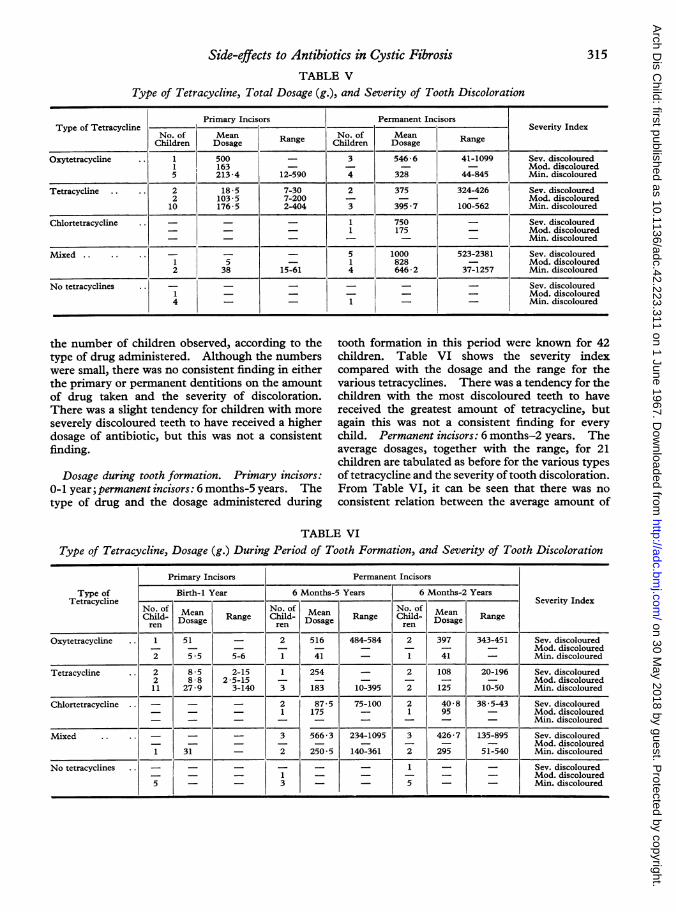
Side-effects to Antibiotics in Cystic FibrosisTABLE V
315
Type of Tetracycline, Total Dosage (g.), and Severity of Tooth Discoloration
Primary Incisors Permanent IncisorsType of Tetracycline .______________-SeverityIndex
No. of MeanRange
No. of MeanRangeChildren Dosaage Range Children Doaage Range
Oxytetracycline .. 1 500 _ 3 546-6 41-1099 Sev. discoloured1 163 - - - - Mod. discoloured5 213-4 12-590 4 328 44-845 Min. discoloured
Tetracycline .. .. 2 18-5 7-30 2 375 324-426 Sev. discoloured2 103 5 7-200 - - - Mod. discoloured10 176 5 2-404 3 395*7 100-562 Min. discoloured
Chlortetracycline .. - - - 1 750 - Sev. discoloured- _ - 1 175 - Mod. discoloured-_- - - - - Min. discoloured
Mixed.- - . 5 1000 523-2381 Sev. discoloured1 5 - 1 828 - Mod. discoloured2 38 15-61 4 646 2 37-1257 Min. discoloured
No tetracyclines - - - - - - Sev. discoloured1 - - - - - Mod. discoloured4 - 1 - Min. discoloured
the number of children observed, according to thetype of drug administered. Although the numberswere small, there was no consistent finding in eitherthe primary or permanent dentitions on the amountof drug taken and the severity of discoloration.There was a slight tendency for children with moreseverely discoloured teeth to have received a higherdosage of antibiotic, but this was not a consistentfinding.
Dosage during tooth formation. Primary incisors:0-lyear;permanentincisors:6months-5years. Thetype of drug and the dosage administered during
tooth formation in this period were known for 42children. Table VI shows the severity indexcompared with the dosage and the range for thevarious tetracyclines. There was a tendency for thechildren with the most discoloured teeth to havereceived the greatest amount of tetracycline, butagain this was not a consistent finding for every
child. Permanent incisors: 6 months-2 years. Theaverage dosages, together with the range, for 21children are tabulated as before for the various typesof tetracycline and the severity oftooth discoloration.From Table VI, it can be seen that there was no
consistent relation between the average amount of
LE VIType of Tetracycline, Dosage (g.) During Period of Tooth Formation, and Severity of Tooth Discoloration
Primary Incisors Permanent Incisors
Type of Birth-1 Year 6 Months-5 Years 6 Months-2 YearsTetracycline
of _No. .Severity Index
Child- Mean Range Child- Dosa Range Child- Doea Rangeren Dosage ren Doage ren aoage Rag
Oxytetracycline .. 1 51 _ 2 516 484-584 2 397 343-451 Sev. discoloured- - - - - - - - - Mod. discoloured2 5-5 5-6 1 41 _ 1 41 - Min. discoloured
Tetracycline .. 2 8 5 2-15 1 254 = 2 108 20-196 Sev. discoloured2 8 8 2-5-15 - - - - - - Mod. diacoloured11 27-9 3-140 3 183 10-395 2 125 10-50 Min. discoloured
Chlortetracycline . - - - 2 87 5 75-100 2 40*8 38*5-43 Sev. discoloured- _ _ 1 175 - 1 95 - Mod. discoloured_ - - - - - - - - Min. discoloured
Mixed .. .. - - - 3 566 3 234-1095 3 426-7 135-895 Sev. discoloured_ - - - - - - - - Mod. discoloured1 31 - 2 250-5 140-361 2 295 51-540 Min. discoloured
No tetracyclines .. - - - - - - 1 - - Sev. discoloured- _ - 1 - _ - - _ Mod. discoloured5 3 5 Min. discoloured
on 30 May 2018 by guest. P
rotected by copyright.http://adc.bm
j.com/
Arch D
is Child: first published as 10.1136/adc.42.223.311 on 1 June 1967. D
ownloaded from

Swallow, Haller, and YoungTABLE VII
Radiological Severity of Disease, Severity of Tooth Discoloration, and Number of Children Affected(Primary and Permanent Dentitions Combined)
Group I Group II
Severely discoloured 6 (16-7%) 6 (50%)Moderately discoloured . . 7 (19-4%) 1 (8 3%)Minimally discoloured 23 (63 9%) 5 (41-7%)Total .. 36 (100%) 12 (100%)
drug taken and the degree of tooth discolora-tion.
Radiological severity of disease and severityof tooth discoloration. Table VII summarizesthe findings on the children who were groupedaccording to the radiological severity of their diseaseand the severity of their tooth discoloration.Although there was some tendency for the childrenin Groups II and III to have proportionately moreseverely discoloured teeth than the children inGroup I, when the children with severely andmoderately discoloured teeth are pooled and com-pared with those with minimally discoloured teeth,there is no significant association (X2 = 2 06 2 d. off.; p > 0 05). It should be noted that the data inthis Table relate to both the primary and thepermanent dentitions.
Discussion
Shwachman, Fekete, Kulczycki, and Foley(1958-59) described the presence of pigmentedteeth in children with cystic fibrosis, who hadreceived tetracyclines as part of their treatment.The presence of discoloured teeth in children withthis disease has also been described by Zegarelli,Denning, Kutscher, Tuoti, and di Sant'Agnese(1960,1961), Zegarelli, Kutscher, Denning, Saporito,Slaughter, and Fahn (1962), Zegarelli, Denning,Kutscher, Fahn, Kirschner, and Slaughter (1963b),and by Sullivan (1963). It is now clear that thediscoloration of teeth found in children with cysticfibrosis is not a sign of the disease, but is the resultof the intake of tetracyclines during the formation ofthe teeth. Incriminated are four main types oftetracyclines: tetracycline, oxytetracycline, chlor-tetracycline, and demethylchlortetracycline. Theliterature on the various effects of these drugs hasbeen reviewed thoroughly by Johnson (1964), andtheir effect on the teeth has been discussed in aleading article (Lancet, 1966). Initially, it was felt(Wallman and Hilton, 1962a) that a bright yellowfluorescence was diagnostic of the presence of
tetracycline in the teeth, but it is now generallybelieved that this phenomenon is eliminated orconsiderably reduced by exposure to light (Storey,1963). In the work on the effects of tetracycline onthe teeth there are three main areas which haveproduced controversy. These are: (1) the degree ofdiscoloration related to the type of tetracyclineadministered; (2) the association of hypoplasticdefects with tetracycline therapy; and (3) the effectof tetracycline therapy on the caries prevalence.
Type of tetracycline and degree ofdiscolora-tion. Wallman and Hilton (1962a) believed thattetracycline produced a more severe discolorationthan oxytetracycline, a view supported by Weyman(1965). The latter felt that there was a remarkablecorrespondence with the type of tetracycline and thecolour of the teeth. Those who had receivedtetracycline had yellow teeth, those who had receivedchlortetracycline had grey-brown teeth. She con-cluded that oxytetracycline probably produced lessstaining than the other types of drugs. Ibsen,Urist, Sognnaes, and Keen (1965), working withNew Zealand rabbits, concluded that tetracyclineand demethylchlortetracycline discoloured teeth to agreater extent than chlortetracycline and oxytetra-cycline, chlortetracycline producing very littlestaining either before or after exposure to light.Owen (1963) similarly found in dogs that chlor-tetracycline did not stain teeth. The children withcystic fibrosis, reported by Shwachman et al.(1958-59), had received large doses of chlortetra-cycline, though some had also received oxytetra-cycline and tetracycline to a lesser degree.The reason for these different findings cannot be
easily explained. It may well be that the stainingproduced by tetracyclines in the experimentalanimal is not comparable to the staining producedin man. The present work does not clarify thematter either. No great differences were observedin children who had received any of the forms oftetracycline, but it should be pointed out thatmatching the colour of teeth with a shade guide islargely subjective-what appears a dark green-
316
on 30 May 2018 by guest. P
rotected by copyright.http://adc.bm
j.com/
Arch D
is Child: first published as 10.1136/adc.42.223.311 on 1 June 1967. D
ownloaded from

Side-effects to Antibiotics in Cystic Fibrosisbrown to one observer may well seem orange toanother.The dosage level has been described as being
critical. Bevelander and Nakahara (1966) citePindborg in believing that it is not so much theduration of the treatment that is important as thetotal dosage of the drug administered, and theysupported this with experiments with the white rat.The present survey does not seem to support theview that the total dosage is the important factor.However, it does give further weight to the belief ofZegarelli, Denning, Kutscher, Fahn, and Kirschner(1963a) that there is no striking relation between thecoloration of the patients' teeth and the severity oftheir illness.Few children receive as much tetracycline as have
those in this series, the majority of whom have hadextensive courses of the drug over periods of monthsand sometimes years. Whereas in animals andadults, receiving a short course, it might be possibleto measure quantitatively the amount of stainingproduced and relate it to the drug dosage, in child-ren with cystic fibrosis this is hardly a practicableproposition. It is also possible that the diseaseitself might modify the uptake of the tetracycline, aview advanced by Zegarelli et al. (1963a, b).
Association of hypoplastic defects withtetracycline therapy. Wallman and Hilton(1962a, b) believed that the observed enamelhypoplasia was caused by the tetracycline drugsrather than by the disease being treated, a viewsupported by Witkop and Wolf (1963). Harcourt,Johnson, and Storey (1962) also found that someteeth from children who had received large doses oftetracycline had hypoplastic enamel, but they feltthat this was due to the metabolic upsets.
In the present study macroscopic defects werenoted in only one patient. However, this does notmean that a microscopical examination of the teethwould not have revealed the presence of a disturb-ance of mineralization either in the dentine or in theenamel.
Effect of tetracycline therapy on cariesprevalence. Because of the interest in thebacteriological aspects of dental caries and thepossibilities of caries prevention by the use ofantibiotics, there have been several reports on therelation between caries prevalence and tetracyclinetherapy. Bevelander and Nakahara (1966) inter-preted Waliman and Hilton (1962a) as believing thatthe teeth that demonstrated tetracycline discolora-tion were highly susceptible to caries. On theother hand, Weyman and Porteous (1963) claimed
that the caries incidence was normal in a group ofchildren who had received tetracyclines. Sur-prisingly little reference has been made to the cariesprevalence in children with cystic fibrosis. H. M.Kopel (personal communication, 1966) believes,from a clinical observation, that the teeth of thesechildren are less susceptible to caries, and thepresent material seems to lend weight to this belief.Although the numbers were small, the trend was forthe children to have a lower caries prevalence. Itmight be argued that the discoloration produced bythe tetracyclines masks any colour changes producedby caries and thus makes identification of thisdisease difficult. However, in children of a wideage range, such as in the present survey, it would bereasonable to suppose that any masking effect wouldbe lost, since there would be a visible breakdown ofthe tooth substance as the caries process advanced.
Summary and ConclusionsDental examinations were carried out on 63
children suffering from cystic fibrosis. 36.5% ofthe children had appreciably discoloured teeth. Nofirm relation could be established between the typeof tetracycline administered and the degree ofdiscoloration, the radiographic severity of the cysticfibrosis, or the severity of respiratory infection.Neither could a relation be demonstrated betweenthe total dosage and the severity of tooth discolora-tion, nor the dosage estimated as being taken duringtooth formation. The incisor teeth of children whohad received tetracycline showed considerably morecharacteristic fluorescence under ultraviolet lightthan those of children who had received other formsof the drug.The present report substantiates the findings of
other workers, that children with cystic fibrosis, whoare receiving tetracycline therapy, are liable to havediscoloured teeth. However, the relatively smallnumber of children examined means that manyquestions remain unanswered, particularly the effectof the various tetracyclines on the type of discolora-tion produced and at what stage of tooth develop-ment the tetracyclines produce discoloration. Moreobjective assessments of the effect of tetracyclinetherapy on the teeth will have to be made beforethese questions can be answered. It is apparentthat children receiving large doses of tetracycline areliable to have disfiguring staining of the teeth and, ifalternative antibiotics are available which do notbelong to the tetracycline family, these should beemployed instead.We wish to express our gratitude to the consultant
medical staff of the hospital for referring the cases to us,and to Dr. M. Mearns and Miss V. Ambridge for their
317
on 30 May 2018 by guest. P
rotected by copyright.http://adc.bm
j.com/
Arch D
is Child: first published as 10.1136/adc.42.223.311 on 1 June 1967. D
ownloaded from

318 Swallow, Hailer, and Younghelp in collecting the data, and to Mrs. P. Charlton forsecretarial assistance.
RERNCESBevelander, G., and Nakahara, H. J. (1966). The effect of diverse
amounts of tetracycline on fluorescence and coloration ofteeth. J7. Pediat., 68, 114.
Frankel, M. A., and Hawes, R. R. (1964). Tetracycline antibioticsand tooth discoloration. _7. oral Ther., 1, 147.
Harcourt, J. K., Johnson, N. W., and Storey, E. (1962). In vivoincorporation of tetracycline in the teeth of man. Arch. oralBiol., 7, 431.
Ibsen, K. H., Urist, M. R., Sognnaes, R. F., and Keen, J. (1965).Differences among tetracyclines with respect to the staining ofteeth. J7. Pediat., 67, 459.
Johnson, R. H. (1964). The tetracyclines: a review ofthe literature-1948 through 1963. .7. oral Ther., 1, 190.
Keith, C. G., de Haller, J., and Young, W. F. (1966). Side-effectsto antibiotics in cystic fibrosis. I. Ocular changes in relationto antibiotic administration and to severity of pulmonaryinvolvement. Arch. Dis. Childh., 41, 262.
Lancet (1966). Tetracyclines and the teeth. 1, 917.Massler, M., and Schour, I. (1944). Atlas ofthe Mouth and Adjacent
Parts in Health and Disease. Bureau of Public Relations of theAmerican Dental Association, Chicago.
Owen, L. N. (1963). The effects of administering tetracyclines toyoung dogs with particular references to localization of the drugsin the teeth. Arch. oral Biol., 8, 715.
Shwachman, H., Fekete, E., Kulczycki, L. L., and Foley, G. E.(1958-59). The effect of long term antibiotic therapy inpatients with cystic fibrosis of the pancreas. Antibiot. Ann.,692.
Storey, E. (1963). Experimental tetracycline administration. J.dent. Res., 42, 5.
Sullivan, R. E. (1963). Discoloration of the teeth in patients withcystic fibrosis. Dental thesis, Graduate College, University ofNebraska.
Wallman, I. S., and Hilton, H. B. (1962a). Teeth pigmented bytetracycline. Lancet, 1, 827.
-, and- (1962b). Prematurity, tetracycline and oxytetra-cycline in tooth development. ibid., 2, 720.
Weyman, J. (1965). The clinical appearances of tetracycline stain-ing of the teeth. Brit. dent.j., 118, 289.
-, and Porteous, J. R. (1963). Tetracycline staining of teeth: areport on clinical material. J. dent. Res., 42, 1111.
Witkop, C. J., and Wolf, R. 0. (1963). Hypoplasia and intrinsicstaining of enamel following tetracycline therapy. J. Amer.med. Ass., 185, 1008.
Young, W. F. (1964). Management. In Cystic Fibrosis, a Symposi-um, p. 67. Chest and Heart Association, London.
Zegarelli, E. V., Denning, C. R., Kutscher, A. H., Fahn, B.,and Kirschner, G. (1963a). Discoloration of teeth in patientswith cystic fibrosis of the pancreas: relation to severity of disease.N. Y. St. dent. J., 29, 75.
,,-,-, -, and Slaughter, T. W. (1963b). Dis-coloration of teethin patients with cysticfibrosis of the pancreas:role of tetracycline therapy. Clin. Pediat. (Philad.), 2, 329.-, -, -, Tuoti, F., and di Sant'Agnese, P. A. (1960).Tooth discoloration in cystic fibrosis. Pediatrics, 26, 1050.
-,-, -,- , and- (1961). Discoloration of the teethin patients with cystic fibrosis of the pancreas: a preliminaryreport. N. Y. St. dent. J., 27, 237.
-, Kutscher, A. H., Denning, C. R., Saporito, R., Slaughter,T. W., and Fahn, B. (1962). Coloration of teeth in patientswith cystic fibrosis of the pancreas. Part II. Oral Surg., 15,929.
on 30 May 2018 by guest. P
rotected by copyright.http://adc.bm
j.com/
Arch D
is Child: first published as 10.1136/adc.42.223.311 on 1 June 1967. D
ownloaded from