Shock Symposium Presentation - heart.org
39
Ultrasound in Shock Todd W. Sarge, MD Assistant Professor in Anaesthesia Harvard School of Medicine Beth Israel Deaconess Medical Center Boston, MA 2017 Shock Symposium - The Latest in Resuscitation and Critical Care May 5th, 2017
Transcript of Shock Symposium Presentation - heart.org

U l t r a s o u n d i n S h o c kTodd W. Sarge, MD
Assistant Professor in AnaesthesiaHarvard School of Medicine
Beth Israel Deaconess Medical CenterBoston, MA
2017 Shock Symposium - The Latest in Resuscitation and Critical CareMay 5th, 2017

Objectives
✤ Define “Focused Ultrasound” and rationale for use in practice
✤ Review FATE / FAST / FEEL exams
✤ Review a diagnostic algorithm for Ultrasound in shock
✤ Present real clinical case examples

Focused Ultrasound - Levels
Price et al. Cardiovascular Ultrasound 2008
Appropriate level of competence for
most clinicians

The name game…
✤ FATE (Jensen MB, Eur J Anaesthesiol 2004)✤ FEEL (Breitkreutz R, Crit Care Med 2007)✤ BLEEP (Pershad J, Pediatrics 2004)✤ Goal-directed limited Echo (Manasia AR, J Cardiothorac Vasc Anesth 2005)✤ Goal-oriented hand-held Echo (Vignon P, Intensive Care Med 2007)✤ CLUE (Kimura B, Am J Cardiol 2007)✤ RACE (McLean A, Crit Care Resusc 2007)✤ FOCUS (Beaulieu Y, Crit Care Med 2007)✤ BEAT (Gunst M, J Trauma 2008)
Everyone

FATE - What is it?
✤ F ocused
✤ A ssessment
✤ T ransthoracic
✤ E chocardiography
Ultrasound protocol utilizing basic TTE skills to
exclude obvious cardio-pulmonary pathology and
evaluate basic cardiac dimensions/function and
causes for shock.
http://www.fate-protocol.com/#https://itunes.apple.com/us/app/fate-card/id413628612?mt=8

FATE - Rationale
✤ Basic cardiac ultrasound can augment other clinical skills
✤ Invasive monitors (e.g. Swanz-Ganz catheters) have not been shown to improve outcome
✤ Ultrasound echo machines are more feasible and portable for use by clinicians at the bedside
✤ Indicated in any patient with hemodynamic instability or shock.

FATE - Windows
✤ 1. Subcostal
✤ 2. Apical
✤ 3. Para-sternal (Long and Short Axis)
✤ 4. Pleural (Left and Right)
Four Standard Windows:

FATE - Views
✤ 1. Subcostal 4-ch
✤ 2. Apical 4-ch
✤ 3. Para-sternal (Long and Short Axis-mid pap)
✤ 4. Pleural (Left and Right)
BASIC Views

FATE - Views
✤ 1. Subcostal - IVC
✤ 2. Apical 2 and 3-ch
✤ 3. Para-sternal (Short Axis-mitral and aortic)
EXTENDED Views

FATE - Goals

FATE - Validation
✤ FATE protocol provided useful images that contributed positively in 227 out of 233 echo studies (97%)✤ ~37% of echo’s revealed NEW information and were DECISIVE in ~24%

Clinical Case #1 - History
✤ 87 y.o. F with no PMHx presents with chest pain and found to have contained rupture of large thoraco-abdominal aneurysm
✤ Underwent emergent endograft of aneurysm
✤ POD # 3 – develops hypotension and SOB on the floor necessitating tx to the ICU
✤ Patient intubated and stabilized on pressors and undergoes chest CT revealing left hemothorax
✤ Thoracic surgery is consulted and plans for a VAT’s washout via right chest
✤ Bedside TTE is performed...

Clinical Case #1 - Bedside TTE

Clinical Case #1 - Bedside TTE

Clinical Case #1 - Bedside TTE

Clinical Case #1 - Bedside TTE

Clinical Case #1 - Diagnosis

FAST - Exam

FAST - What is it?
✤ F ocused
✤ A ssessment with
✤ S onagraphy in
✤ T rauma
Ultrasound protocol to rapidly identify blood in
the pericardial space, intra-peritoneal cavity and
pleural space and based on the premise that blood will pool to dependent areas.
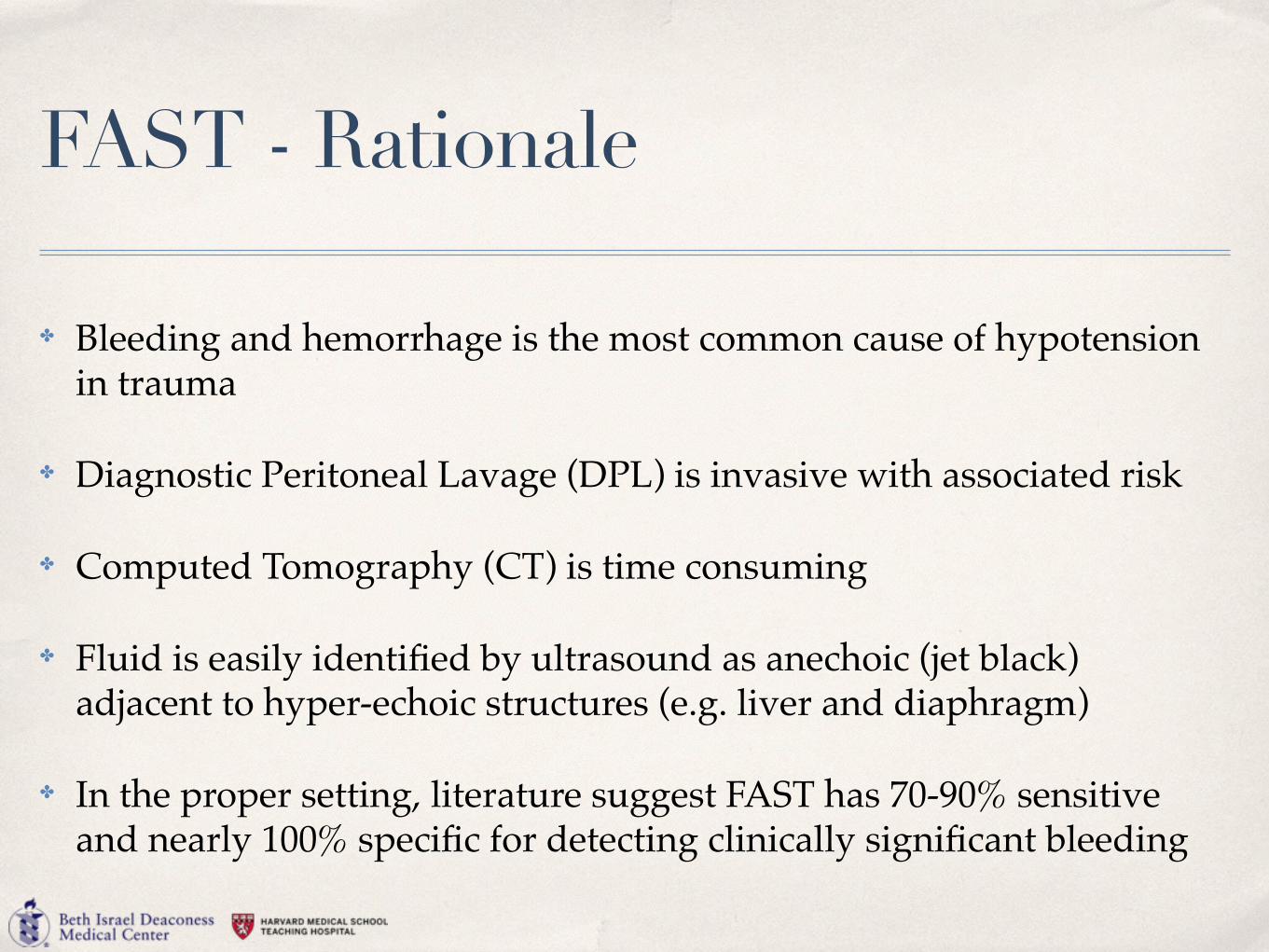
FAST - Rationale
✤ Bleeding and hemorrhage is the most common cause of hypotension in trauma
✤ Diagnostic Peritoneal Lavage (DPL) is invasive with associated risk
✤ Computed Tomography (CT) is time consuming
✤ Fluid is easily identified by ultrasound as anechoic (jet black) adjacent to hyper-echoic structures (e.g. liver and diaphragm)
✤ In the proper setting, literature suggest FAST has 70-90% sensitive and nearly 100% specific for detecting clinically significant bleeding

FAST - Advantages
✤ Ultrasound is portable, repeatable and non-invasive
✤ Can be performed rapidly at the bedside
✤ Can be done simultaneously with resuscitation efforts
✤ Does not require use of contrast or radiation exposure (safe in pregnancy and pediatrics)

FAST Goals
✤ Rapid detection of:
✤ Hemoperitoneum
✤ Hemopericardium
✤ Hemothorax
✤ Pneumothorax (eFAST)

FAST Indications / Contraindications
✤ INDICATIONS:✤ Blunt or penetrating thoraco-abdominal trauma✤ Trauma / Abdominal Pain in pregnancy✤ Unexplained Hypotension in ANY patient
✤ CONTRAINDICATIONS:✤ Any immediate indication for OR (e.g. evisceration or
ruptured diaphragm on outside hospital imaging, etc.)

FAST - Views
✤ Cardiac
✤ Peri-Hepatic (Right Upper Quadrant)
✤ Peri-Splenic (Left Upper Quandrant)
✤ Pelvic
Four FAST Views:

FAST - Where to Begin?
RUQ LUQ

Peri-Splenic Pelvic
Subcostal / Cardiac Peri-Hepatic

Clinical Case #2
✤ 71 y.o. female POD #9 from L3-L5 lami awaiting d/c to rehab
✤ Pt found unresponsive, agonal breathing --> Code Blue called
✤ Pt appeared pale with a distended abdomen. Hypotensive (SBP 80’s)
✤ Volume initiated and tx to ICU. Poor trans-thoracic windows prompting TEE
✤ PMHx: Fibrolipoma of spinal cord; DJD Lumbar spine; Chronic Pain syndrome; GERD; Anxiety; Depression

Clinical Case #2

FEEL - Exam

FEEL - What is it?
✤ F ocused
✤ E chocardiographic
✤ E valuation in
✤ L ife Support
Ultrasound protocol to rapidly identify mechanical causes of cardiac arrest in
PEA.


Whentoapply
Breitkreutz et al. Crit Care Med 2007;35[supp]:S150-S161

FEEL - Rationale for use
Identify (or rule out) 4 mechanical causes of PEA:
1. Tamponade
2. Hypovolemia
3. Pulmonary Embolism
4. Severe LV Dysfunction (MI)

FEEL - Rationale for avoiding


Clinical Case #3
✤ 67 y.o. male POD #3 from toe amputation
✤ Pt found unresponsive without palpable pulse - CPR initiated
✤ Ultrasound performed during CPR
✤ PMHx: Peripheral Vascular Disease; HTN; GERD

Clinical Case #3

Ultrasound Shock Algorithm
TTE Pleural Ultrasound
Abdominal Ultrasound (FATE / FAST / FEEL)
Pericardial effusion
Severely hypokinetic LV
Severely enlarged akinetic RV
Small hyperdynamic LV, collapsed IVC
Hypercontractile LV
Tamponade Pump failure Pulmonary embolism
Hypovolemia Sepsis
No sliding sign Stratosphere
sign
Pneumothorax
Courtesy of Sajid Shahul, MD

THANK YOU!