Shock and hemodynamic monitorization -...
23
Shock and hemodynamic monitorization Nilüfer Yalındağ Öztürk Marmara University Pendik Research and Training Hospital
Transcript of Shock and hemodynamic monitorization -...

Shock and hemodynamic monitorization
Nilüfer Yalındağ Öztürk Marmara University
Pendik Research and Training Hospital


Shock
• Leading cause of morbidity and mortality
• Worldwide: dehydration and hypovolemic shock 6-20 M deaths
• Adults vs pediatrics: Less mortality in pediatric sepsis
Mortality
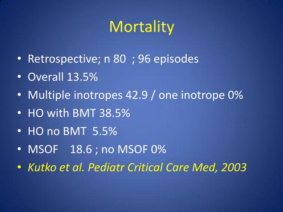
• Retrospective; n 80 ; 96 episodes
• Overall 13.5%
• Multiple inotropes 42.9 / one inotrope 0%
• HO with BMT 38.5%
• HO no BMT 5.5%
• MSOF 18.6 ; no MSOF 0%
• Kutko et al. Pediatr Critical Care Med, 2003
Mortality
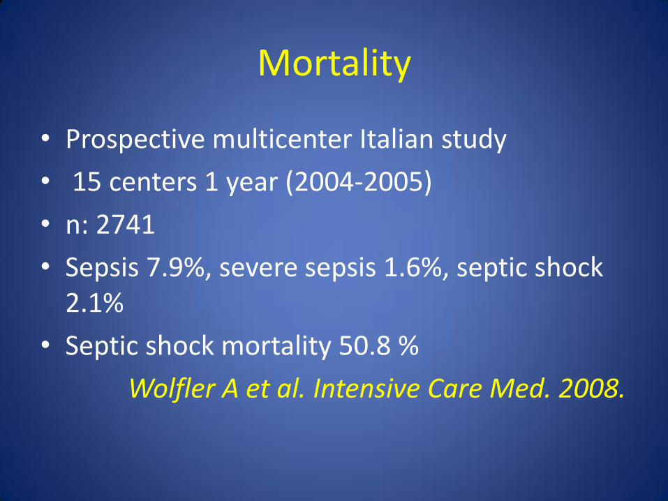
• Prospective multicenter Italian study
• 15 centers 1 year (2004-2005)
• n: 2741
• Sepsis 7.9%, severe sepsis 1.6%, septic shock 2.1%
• Septic shock mortality 50.8 %
Wolfler A et al. Intensive Care Med. 2008.
Mortality
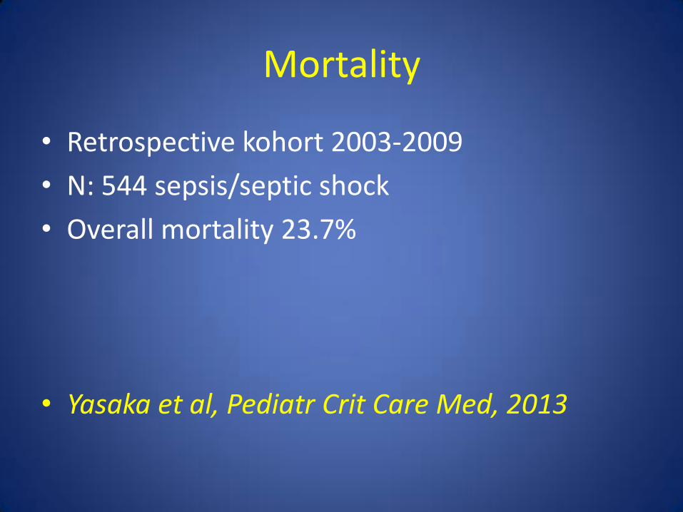
• Retrospective kohort 2003-2009
• N: 544 sepsis/septic shock
• Overall mortality 23.7%
• Yasaka et al, Pediatr Crit Care Med, 2013

Clinical findings
• Tachycardia
• Cold and clammy extremities
• Skin mottling
• Oliguria
• Mental status changes
• Tachypnea
• Hypotension

Hemodynamic monitorization
• Focuses on the adequacy of the circulation
• Limited by existing heart-lung interactions

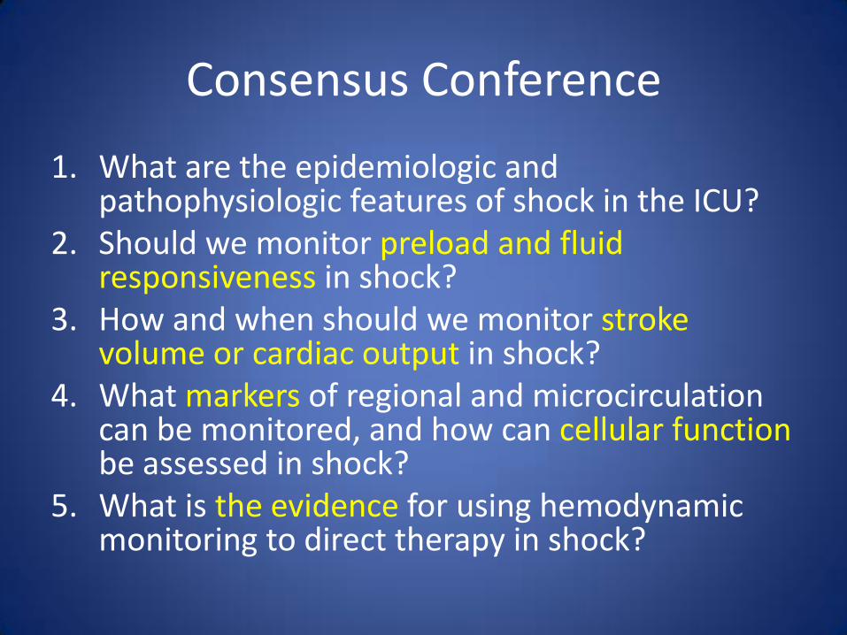
Consensus Conference
1. What are the epidemiologic and pathophysiologic features of shock in the ICU?
2. Should we monitor preload and fluid responsiveness in shock?
3. How and when should we monitor stroke volume or cardiac output in shock?
4. What markers of regional and microcirculation can be monitored, and how can cellular function be assessed in shock?
5. What is the evidence for using hemodynamic monitoring to direct therapy in shock?
1.What are the epidemiologic and pathophysiologic features of shock in the ICU?
• 1. A life threatening , generalized maldistribution of blood flow resulting in failure to deliver and/ or utilize adequate amounts of oxygen, leading to tissue dysoxia.
Level 1; QoE moderate B

1. What are the epidemiologic and pathophysiologic features of shock in the ICU?
• 2. Hypotension
SBP <90
or 40 mmHg decrease from baseline,
or MAP<65
while commonly present, should not be required to define shock.
Shock requires evidence of inadequate tissue perfusion on PE.
Level 1; QoE moderate B

1. What are the epidemiologic and pathophysiologic features of shock in the ICU?
• 3. In absence of hypotension, when shock is suggested by H+P, recommend/ that a marker of inadequate tissue perfusion be measured
• ( decreased Scv O2, SvO2, increased blood lactate, base deficit, perf related low pH)
• Level 1; QoE moderate B

4. Apart from lactate and base deficit, current evidence does not support the routine use of biomarkers for diagnosis or staging of shock.
Level 1; QoE high A

• 5. Target BP initial shock resussitation
• For uncontrolled hemorrhage : MAP 40 until bleeding surgically controlled.
• Level 1; QoE moderate B
• For TBI without systemic hemorrhage MAP 90 Level 1; QoE low C
• For other shock states MAP >65
• Level 1; QoE moderate B

2: Should we monitor preload and fluid responsiveness in shock?
• Preload measurement alone not to be used to
predict fluid responsiveness
Level 1; QoE mod B
In shock low values of commonly used static measures of preload (CVP, RAP, PAOP- eg <4 mmHg) and ventricular volumes, should lead to fluid resussitation with careful HD monitoring.
Level 1; QoE low C

2: Should we monitor preload and fluid responsiveness in shock?
• Fluid challenge to predict responsiveness.
– FC ( 250 cc crystalloid or colloid equivalent in 10-15 min)
or
– straight leg raise aiming CVP rise at least of 2.
• Positive response – measures of improved cardiac fx and tissue perfusion.
• Level 1; QoE low C
• Do not recommend routine use of dynamic measures of fluid responsiveness
– ( including but not limited to pulse pressure variation, aortic flow changes, systolic pressure variation, respiratory systolic variation test, collapse of vena cava)
– Level 1; QoE high A
• There may be some advantage to these in highly selective patients
– Level 1; QoE moderate B

3.How and when should we monitor stroke volume or cardiac output in shock?
• 1. Routine measurement of CO in patients
with shock not recommended
• ( Level 1; QoE moderate B)
• 2. We suggest considering echo or measurement of CO in patients with clinical evidence of ventricular failure and persistant shock despite initial fluid resuscitation.
• ( Level 2; QoE moderate B)

What is the evidence for using hemodynamic monitoring to direct therapy in shock?
• 1.We recommend frequent measurement of
blood pressure and physical examination variables ( including signs of hypoperf , urine output, mental status) in patient with history and clinical findings suggestive of shock.
• 2.We recommend invasive BP measurement in refractory shock .
• Level 1; QoE very low D

• We do not recommend routine use of PAC for patients with shock
• Level 1; QoE high A • -We recommend initiating goal directed therapy
without delay, in patients presenting with septic shock ( within 6 hrs or less) particulary where ScvO2 is below 70%.
• Level 1; QoE mod B • -We do not recommend targeting supranormal oxygen
delivery in patients with shock • Level 1; QoE high A
Summary
• No monitor is associated with improved outcome unless coupled with appropriate therapy
• Early recognition, monitorization and therapy may change outcome.
• Less invasive functional HDM may be the future of goal directed therapy

Thank you for your attention


![SHOCK[1] - Hypovolemic Shock](https://static.fdocuments.in/doc/165x107/58edc1bc1a28abae538b4711/shock1-hypovolemic-shock.jpg)