Shock: A Review of Pathophysiology and Management. Part II · PDF fileShock: A Review of...
19
Shock: A Review of Pathophysiology and Management. Part II L. I. G. WORTHLEY Department of Critical Care Medicine, Flinders Medical Centre, Adelaide, SOUTH AUSTRALIA ABSTRACT Objective: To review pathophysiology and management of hypovolaemic, cardiogenic and septic shock in a two part presentation. Data sources: Articles and published peer-review abstracts and a review of studies reported from 1994 to 1998 and identified through a MEDLINE search of the English language literature on septic shock, cardiogenic shock and hypovolaemic shock. Summary of review: The pathophysiological effects of cardiogenic and hypovolaemic shock are related predominantly to a reduction in preload and myocardial contractility, respectively, whereas the pathophysiological effects of septic shock result largely from the overwhelming production of inflammatory mediators. The excessive inflammatory response results in haemodynamic compromise and widespread tissue injury. While the understanding of the acute inflammatory reaction has improved, therapies to modulate the chemical mediators responsible for the organ dysfunction associated with this reaction have not altered mortality, and in some instances may have increased it. Treatment of septic shock is still largely supportive, using intravenous fluids and inotropic agents to provide adequate tissue perfusion while the infective lesion is managed with antibiotic therapy and surgical drainage of septic focus. Conclusions: Septic shock is provoked by an excessive acute inflammatory response to an infection. Management of the shock is supportive using fluids and inotropic agents, while antibiotic therapy and surgical drainage of the septic focus take effect. Immunomodulation of the acute inflammatory response causing septic shock has not improved mortality. (Critical Care and Resuscitation 2000; 2: 66-84) Key Words: Shock, distributive shock, septic shock, sepsis, systemic inflammatory response syndrome ious (e.g. bacteria, fungi, viruses) and noninfectious (e.g. pancreatitis, multiple trauma, burns, infarction, biliary peritonitis, anaphylaxis) processes. Distributive shock is a name given to shock caused by the systemic inflammatory response syndrome, or shock provoked by the inhibition, or absence, of sympa- thetic tone (e.g. neurogenic shock). The definition of clinical syndromes due to infection include: 1-5 The systemic inflammatory response syndrome (SIRS) is a clinical syndrome characterised by, but not limited to, two or more of the following: • sepsis (i.e. SIRS caused by infection), • severe sepsis (i.e. sepsis associated with organ dysfunction, hypoperfusion - including that which may be reflected by lactic acidosis, oliguria, altered mental status - or hypotension) and, 1. a body temperature of > 38°C or < 36°C 2. a heart rate of > 90 beats/min 3. respiratory rate > 20 breaths/min or PaCO 2 of < 32 mmHg • septic shock (i.e. sepsis which is associated with hypotension and perfusion abnormalities despite adequate fluid resuscitation). 4. a WBC count of > 12,000 /mm 3 or < 4000 /mm 3 or the presence of > 10% immature neutrophils and is caused by widespread inflammation due to infect- The differential diagnosis of septic shock, includes Correspondence to: Dr. L. I. G. Worthley, Department of Critical Care Medicine, Flinders Medical Centre, Bedford Park, South Australia 5042 66
-
Upload
duongduong -
Category
Documents
-
view
218 -
download
3
Transcript of Shock: A Review of Pathophysiology and Management. Part II · PDF fileShock: A Review of...

Shock: A Review of Pathophysiology and Management. Part II
L. I. G. WORTHLEY Department of Critical Care Medicine, Flinders Medical Centre, Adelaide, SOUTH AUSTRALIA
ABSTRACT Objective: To review pathophysiology and management of hypovolaemic, cardiogenic and septic shock in a two part presentation. Data sources: Articles and published peer-review abstracts and a review of studies reported from 1994 to 1998 and identified through a MEDLINE search of the English language literature on septic shock, cardiogenic shock and hypovolaemic shock. Summary of review: The pathophysiological effects of cardiogenic and hypovolaemic shock are related predominantly to a reduction in preload and myocardial contractility, respectively, whereas the pathophysiological effects of septic shock result largely from the overwhelming production of inflammatory mediators. The excessive inflammatory response results in haemodynamic compromise and widespread tissue injury. While the understanding of the acute inflammatory reaction has improved, therapies to modulate the chemical mediators responsible for the organ dysfunction associated with this reaction have not altered mortality, and in some instances may have increased it. Treatment of septic shock is still largely supportive, using intravenous fluids and inotropic agents to provide adequate tissue perfusion while the infective lesion is managed with antibiotic therapy and surgical drainage of septic focus. Conclusions: Septic shock is provoked by an excessive acute inflammatory response to an infection. Management of the shock is supportive using fluids and inotropic agents, while antibiotic therapy and surgical drainage of the septic focus take effect. Immunomodulation of the acute inflammatory response causing septic shock has not improved mortality. (Critical Care and Resuscitation 2000; 2: 66-84)
Key Words: Shock, distributive shock, septic shock, sepsis, systemic inflammatory response syndrome
ious (e.g. bacteria, fungi, viruses) and noninfectious (e.g. pancreatitis, multiple trauma, burns, infarction, biliary peritonitis, anaphylaxis) processes.
Distributive shock is a name given to shock caused by the systemic inflammatory response syndrome, or shock provoked by the inhibition, or absence, of sympa-thetic tone (e.g. neurogenic shock). The definition of clinical syndromes due to infection
include:1-5 The systemic inflammatory response syndrome (SIRS) is a clinical syndrome characterised by, but not limited to, two or more of the following:
• sepsis (i.e. SIRS caused by infection),
• severe sepsis (i.e. sepsis associated with organ dysfunction, hypoperfusion - including that which may be reflected by lactic acidosis, oliguria, altered mental status - or hypotension) and,
1. a body temperature of > 38°C or < 36°C 2. a heart rate of > 90 beats/min 3. respiratory rate > 20 breaths/min or PaCO2 of < 32
mmHg • septic shock (i.e. sepsis which is associated with hypotension and perfusion abnormalities despite adequate fluid resuscitation).
4. a WBC count of > 12,000 /mm3 or < 4000 /mm3 or the presence of > 10% immature neutrophils
and is caused by widespread inflammation due to infect- The differential diagnosis of septic shock, includes Correspondence to: Dr. L. I. G. Worthley, Department of Critical Care Medicine, Flinders Medical Centre, Bedford Park, South Australia 5042
66
Critical Care and Resuscitation 2000; 2: 66-84 L. I. G. WORTHLEY
the cell protoplasm, largely as teichoic acids. The cell wall of Gram-positive bacteria is non toxic. Endotoxin is the lipopolysaccharide outer coat of Gram-negative organisms and consists of three main parts:6
adrenal crisis, thyrotoxic crisis, delirium tremens, salicylate overdose, and malignant hyperpyrexia. In this section the pathophysiology and management of septic shock will be presented. - an outer branched chain polysaccharide portion (i.e.
the O antigen), CAUSES Septic shock is usually provoked by exogenous agents (e.g. endotoxin, exotoxin, superantigens) leading to the excess production of endogenous inflammatory mediators. The mediators of septic shock are listed in Table 1.4,5
- a mid portion R antigen polysaccharide core, and - an inner toxic lipid A portion that is normally
attached to the cell membrane of the bacterium (i.e. is concealed and therefore has low antigenicity).
The inner Lipid A portion is similar for many pathogenic Gram-negative bacteria and accounts for the majority of the toxicity of endotoxin. If the patient develops Gram-negative bacteraemia and possesses the appropriate IgM or IgG antibodies to the many possible O or R antigens of the outer and mid portion of the lipopolysaccharide coat, the opsonisation and phago-cytosis of invading organisms will be sufficient to prevent the bacterium releasing lipid A. Endotoxaemia only occurs if there is disruption of the bacterium by complement or large doses of bactericidal antibiotics.7 Levels of antibody to lipid A are normally very low, and removal of Lipid A occurs as a slow inactivation by α-1-lipoprotein esterase, and reticuloendothelial system (RES) removal of platelet-bound and high density lipoprotein bound lipid A.8 The limulus test has been used to assay endotoxin. However, its lack of sensitivity and specificity (i.e. numerous false positive and false negative results), has rendered the test of little clinical value.9
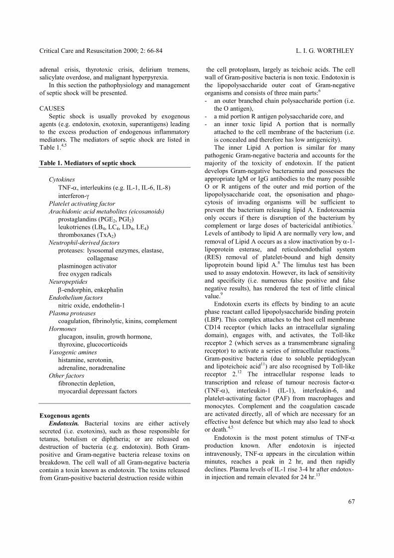
Table 1. Mediators of septic shock Cytokines TNF-α, interleukins (e.g. IL-1, IL-6, IL-8) interferon-γ Platelet activating factor Arachidonic acid metabolites (eicosanoids) prostaglandins (PGE2, PGI2) leukotrienes (LB4, LC4, LD4, LE4) thromboxanes (TxA2) Neutrophil-derived factors proteases: lysosomal enzymes, elastase, collagenase plasminogen activator free oxygen radicals Neuropeptides β-endorphin, enkephalin Endothelium factors
Endotoxin exerts its effects by binding to an acute phase reactant called lipopolysaccharide binding protein (LBP). This complex attaches to the host cell membrane CD14 receptor (which lacks an intracellular signaling domain), engages with, and activates, the Toll-like receptor 2 (which serves as a transmembrane signaling receptor) to activate a series of intracellular reactions.10 Gram-positive bacteria (due to soluble peptidoglycan and lipoteichoic acid11) are also recognised by Toll-like receptor 2.12 The intracellular response leads to transcription and release of tumour necrosis factor-α (TNF-α), interleukin-1 (IL-1), interleukin-6, and platelet-activating factor (PAF) from macrophages and monocytes. Complement and the coagulation cascade are activated directly, all of which are necessary for an effective host defence but which may also lead to shock or death.4,5
nitric oxide, endothelin-1 Plasma proteases coagulation, fibrinolytic, kinins, complement Hormones glucagon, insulin, growth hormone, thyroxine, glucocorticoids Vasogenic amines histamine, serotonin, adrenaline, noradrenaline Other factors fibronectin depletion, myocardial depressant factors Exogenous agents Endotoxin. Bacterial toxins are either actively secreted (i.e. exotoxins), such as those responsible for tetanus, botulism or diphtheria; or are released on destruction of bacteria (e.g. endotoxin). Both Gram-positive and Gram-negative bacteria release toxins on breakdown. The cell wall of all Gram-negative bacteria contain a toxin known as endotoxin. The toxins released from Gram-positive bacterial destruction reside within
Endotoxin is the most potent stimulus of TNF-α production known. After endotoxin is injected intravenously, TNF-α appears in the circulation within minutes, reaches a peak in 2 hr, and then rapidly declines. Plasma levels of IL-1 rise 3-4 hr after endotox- in injection and remain elevated for 24 hr.13
67

L. I. G. WORTHLEY Critical Care and Resuscitation 2000; 2: 66-84
Chemical mediators - integrins (e.g. lymphocyte-function-associated-antigen-1 or LFA-1) and immunoglobulin super-family (e.g. intracelular adhesion molecule 1 or ICAM-1, vascular cell adhesion molecule or VCAM) a family of glycoprotein cell surface adhesion receptors important in the neutrophil/ endothelial adhesion phase of ‘firm adherence’ leading to enhanced adherence of neutrophils to the endothelium, and
Cytokines. At low concentrations, cytokines (e.g. TNF-α, IL-1) are important ‘communication proteins’ which are essential for cell-to-cell signalling, transmiting information by binding to specific transmembrane receptors to regulate immunologic and physiologic events.14 IL-1 and TNF-α mediate local phagocytic cell emigration and activation and release of lipid derived mediators (e.g. PGE2, thromboxane, PAF). IL-1 induces interleukin-8 (IL-8) synthesis, which in turn is a potent neutrophil and monocyte chemotactic factor and stimulates the release of enzymes from neutrophils. While the acute changes in hepatic protein synthesis (i.e. the acute phase response) can be induced by IL-1 and TNF-α, it is thought to be caused largely by interleukin-6 (IL-6).15 TNF-α, IL-1, and IL-6 stimulate their own secretion; both TNF-α and IL-1 stimulate the secretion of IL-6, whereas IL-6 inhibits both IL-1and TNF-α secretion.16
- platelet endothelial-cell-adhesion molecule-1 or PECAM-1 (important in neutrophil transmigrat-ion),
- enhances polymorphonuclear leucocyte activity by stimulating phagocytic activity (it has only a weak effect on T cells),
- is directly toxic to endothelial cells, increasing capillary permeability, activating kinin and comple-ment cascades, causing disseminated intravascular coagulation (DIC) and haemorrhagic necrosis, which in turn leads to gastrointestinal haemorrhage, acute renal failure, acute respiratory distress syndrome (ARDS) as well as cardiac failure (i.e. TNF-α has a negative inotropic action21) hepatic abnormalities, and pulmonary dysfunction,22
At higher concentrations the proinflammatory cytokines (e.g. TNF-α, IL-1, IL-6, IL-8, interferon-γ) can exert potentially harmful biologic effects, ranging from tissue and organ dysfunction to a life threatening systemic reaction. Low concentrations of the counter-regulatory cytokines, for example interleukin-4 (IL-4), interleukin-10 (IL-10), interleukin-11 (IL-11) and interleukin-13 (IL-13) may also be detrimental.
- induces fever through a direct effect on hypothalamic neurones and through IL-1 product-ion, and,
- inhibits lipoprotein lipase activity reducing plasma clearance of lipids.
Tumour necrosis factor-α (TNF-α): TNF-α (or cachectin)17 is a macrophage polypeptide hormone (that can also be produced by glial cells, Kupffer cells, mast cells, and natural killer T and B lymphocytes18) which binds with high affinity to muscle cells and adipocytes. Serum levels of TNF-α are usually undetectable during health and are increased during sepsis and critical illness by the many endogenous and exogenous stimulating factors produced by bacteria, viruses, tumours and cell damage.
TNF-α is the first cytokine to appear in the circulation after experimental and clinical endotoxaemia (detected after 45 min, peaking after 90 min)23 and, in man, has a half life of 20 min,18,24,25 which increases in the absence of corticosteroids (e.g. up to 4 - 8 h in adrenalectomized animals18). Recent studies indicate that the overstimulation of TNF-α biosynthesis in septicaemia is a critical step in triggering SIRS and causing septic shock and multiple organ dysfunction syndrome in septic patients, as mice, rabbits and baboons, immunised against TNF-α or treated with polyclonal antiserum directed against TNF-α are protected against the lethal effect of endotoxin.22 Corticosteroids given before (but not after) an endotoxin infusion will inhibit TNF-α biosynthesis.
TNF-α binds to at least two distinct membrane-associated receptors (i.e. TNF-R1 and TNF-R2) and TNF-binding proteins (i.e. soluble TNF receptors) and, - induces release of IL-1, IL-6, IL-8 (which attracts
and activates neutrophils), PAF, and the eicosanoids (i.e. leukotrienes, thromboxane A2, prostaglandins) by neutrophil and endothelial cells, and may even promote its own release,
Interleukin-1 (IL-1). The IL-1 family consists of three structurally related polypeptides, two agonists (interleukin-1α and interleukin-1β) and an antagonist (interleukin-1-receptor antagonist).26 While interleukin-1α and interleukin-1β have different amino acid sequences, they are structurally related, and act through the same cell-surface receptors (i.e. share biologic activities).26 Both interleukin-1α and interleukin-1β are produced by monocytic phagocytes (e.g. monocytes,
- increases expression of the endothelial surface glycoproteins,19,20 i.e; - selectins (a group of structurally related glyco-
proteins that utilize protein-carbohydrate interaction to mediate cell adhesion and are important in the early transient neutrophil/ endothelial adhesion phase, or ‘rolling’ phase, during inflammation),
68

Critical Care and Resuscitation 2000; 2: 66-84 L. I. G. WORTHLEY
tissue macrophages, phagocytic lining cells of the liver and spleen). Nearly all infections, immunologic reactions and inflammatory processes stimulate mono-cytic phagocytes to synthesise and liberate IL-1 into the circulation,27 which, - induces release of TNF-α, IL-6, IL-8, PAF, and the
eicosanoids (i.e. leukotrienes, thromboxane A2, prostaglandins) by neutrophil and endothelial cells, and may even promote its own release,
- activates T lymphocytes, is chemotactic for T cells, and stimulates lymphocyte B-cell proliferation and antibody production,
- acts with TNF-α to promote adhesion of endothelial cells, polymorphonuclear cells, eosinophils, baso-phils, and monocytes,
- induces fever, by stimulating synthesis of prostaglandin E2 in the anterior hypothalamus to elevate the hypothalamic temperature set level,
- increases the release of circulating neutrophils from the bone marrow and is chemotactic for neutrophils,
- increases hepatic production of acute phase reactants (e.g. complement, haptoglobin, ceruloplasmin, fibrinogen, plasminogen, C-reactive protein) and reduces albumin, prealbumin and transferrin production,
- increases skeletal muscle catabolism (by increasing prostaglandin E2 production) and liberates amino acids for hepatic protein and inflammatory tissue production,
- induces slow-wave sleep, and - reduces serum Fe and Zn. The action of IL-1 is regulated through a cell membrane receptor, which has a natural inhibitor known as IL-1ra (interleukin-1 receptor antagonist) which inhibits the hypotension and leucocytosis induced by IL-1, and increases survival of animals injected with endotoxin.13 Corticosteroids reduce the production of IL-1. IL-1 and TNF-α are different compounds with similar biological actions (although IL-1 does not cause DIC or neutropenia, c.f. TNF-α). During the inflammatory response they always appear together and always act synergistically. Arachidonic acid metabolites (eicosanoids). The eicosanoids are a class of endogenous mediators derived from 20-carbon unsaturated fatty acid precursors, primarily eicosatetraenoic acid (arachidonic acid). Following the release of arachidonic acid from tissue stores (due to the action of phospholipase A2 on phospholipid in cell membranes), it is transformed by the action of either cyclooxygenase (present in all cell
walls) into unstable endoperoxidases (PGG2 PGH2) and then into a variety of vasoactive substances including prostaglandins (PGD2 PGE2 PGF2 PGI2) and thromboxane (TXA2); or 5-lipooxygenase (found only in myeloid cells, i.e. monocytes, eosinophils, basophils, alveolar macrophages, and mast cells) in the presence of a nuclear membrane cofactor (5-lipoxygenase-activating protein or FLAP), to generate an unstable intermediate, 5-hydroperoxyl-eicosatetraenoic acid (5-HPETE). The latter converts to the unstable leukotriene A4 (LTA4) which is rapidly converted to either LTB4 or LTC4, the latter of which is transported extracellularly to be converted to LTD4 and finally to LTE4 (Figure 1). These compounds can increase vascular permeability, increase mucus secretion and can cause bronchospasm.28 Elevated levels of prostacyclin (PGI2) occur 1-6 hr after the onset of septic shock and causes vasodilation, inhibits platelet activation, and disperses platelet aggregates. Prostaglandin-E2 (PGE2) dilates bronchi and blood vessels while prostaglandin-F2α (PGF2α) constricts them. Thromboxane (TXA2) is a potent vasocon-strictor.29 Platelet activating factor (PAF). Platelet activating factor is a potent phospholipid mediator that leads to amplification of cytokine release. It causes vasocon-striction and bronchoconstriction, but in very low doses induces vasodilation and increased venular permeability with a potency 100-10 000 times greater than histamine. It also causes increased leukocyte adhesion to the endothelium (by enhancing integrin binding), chemotaxis, degranulation and oxidative ‘bursts’ (i.e. all of the cardinal features of acute inflammation).30 The chemical structure of PAF is acetyl-glyceryl-ether-phosphorylcholine. It mediates its effects by a single G protein-coupled receptor, and its effects are regulated by a family of inactivating PAF acetylhydrolases. Neutrophil derived factors. The neutrophil derived factors released during shock consist of: Lysosomal enzymes and neutrophil proteases: these are proteolytic enzymes released from monocytes and polymorphonuclear leucocytes. In the presence of hypoxia and acidosis these enzymes destroy structural proteins, and activate coagulation, complement and kinase systems and can cause myocardial depression and coronary vasoconstriction. Free oxygen radicals: superoxide and other free oxygen radicals released by aggregated leucocytes have been implicated in damaging endothelium and producing increase in capillary permeability and capillary disruption.
69

L. I. G. WORTHLEY Critical Care and Resuscitation 2000; 2: 66-84
Figure 1. Arachidonic acid metabolites and their role in inflammation.
Endothelin-1. Endothelin-1 is a 21 amino acid peptide which is synthesised de novo by the endothelium, from preproendothelin-1, which undergoes an initial processing to form the 38 amino acid peptide, proendothelin-1, which is in turn cleaved by an endothelin converting enzyme (ECE), forming endothelin-1.38 Endothelin-1 acts on surface receptors of vascular smooth muscle (endothelin A receptor) activating phospholipase C and producing the secondary messengers inositol 1,4,5-triphosphate (which releases Ca2+ from the sarcoplasmic reticulum) and diacylglycerol (which activates protein kinase C), causing contraction of the smooth muscle cell.38 In addition, endothelin-1 potentiates the effects of other vasoconstrictor hormones (e.g. noradrenaline, serotonin) and stimulates proliferation of smooth muscle cells. It is usually produced in response to hypoxia, ischaemia and shear stress.39
Neuropeptides (e.g. endorphins). β-endorphin and adrenocorticotropic hormone (ACTH) are derived from the common precursor pro-opicortin and are released in equimolar amounts from the anterior pituitary in response to stress or endotoxin. It is thought that pituitary endorphins enter the central nervous system to react with specific opiate receptors, to enhance vagal-cholinergic tone, reducing cardiac output and mean arterial blood pressure.31 It has been suggested, however, that at least part of the effect involves alteration of the efferent sympathetic nervous system.32
Endothelial factors Nitric oxide. Nitric oxide is thought to be the mediator largely responsible for the sustained vasodilation in septic shock.33,34 It is also thought to be the mediator responsible largely for reduced myocardial contractility,35 hepatic damage, microvascular hyperpermeability and intestinal barrier dysfunction in septic shock.36 Endotoxin and other proinflammatory agents induce a release of PAF, TNF-α, IL-1, and interferon-gamma, and enhance the synthesis of nitric oxide by endothelial constitutive nitric oxide synthase (in the early phase of septic shock) and vascular smooth muscle cell inducible nitric oxide synthase (in the later phase of septic shock).37
Plasma proteases (e.g. kinin, coagulation and complement activation). The activation of pre-kallikrein forms proteolytic enzymes of bradykinin, causing peripheral vasodilation, myocardial depression, DIC, complement activation and increased capillary permeability.
70
Critical Care and Resuscitation 2000; 2: 66-84 L. I. G. WORTHLEY
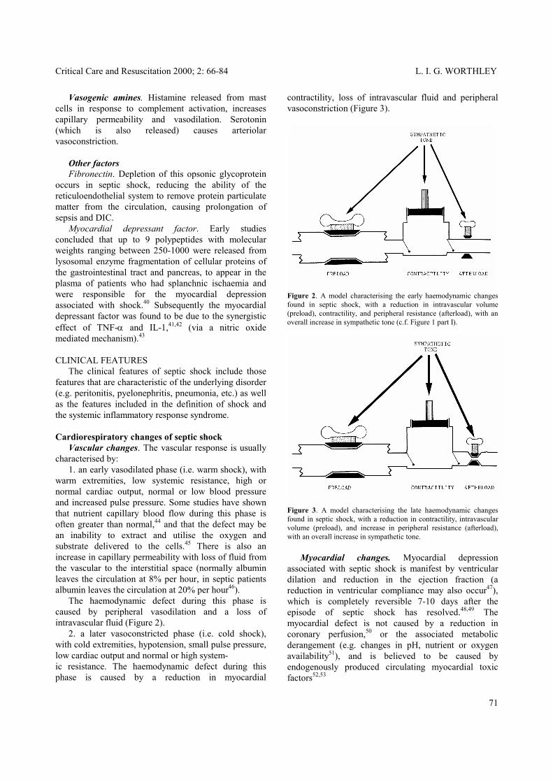
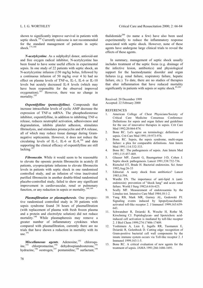
Vasogenic amines. Histamine released from mast cells in response to complement activation, increases capillary permeability and vasodilation. Serotonin (which is also released) causes arteriolar vasoconstriction. Other factors Fibronectin. Depletion of this opsonic glycoprotein occurs in septic shock, reducing the ability of the reticuloendothelial system to remove protein particulate matter from the circulation, causing prolongation of sepsis and DIC. Myocardial depressant factor. Early studies concluded that up to 9 polypeptides with molecular weights ranging between 250-1000 were released from lysosomal enzyme fragmentation of cellular proteins of the gastrointestinal tract and pancreas, to appear in the plasma of patients who had splanchnic ischaemia and were responsible for the myocardial depression associated with shock.40 Subsequently the myocardial depressant factor was found to be due to the synergistic effect of TNF-α and IL-1,41,42 (via a nitric oxide mediated mechanism).43 CLINICAL FEATURES The clinical features of septic shock include those features that are characteristic of the underlying disorder (e.g. peritonitis, pyelonephritis, pneumonia, etc.) as well as the features included in the definition of shock and the systemic inflammatory response syndrome. Cardiorespiratory changes of septic shock Vascular changes. The vascular response is usually characterised by: 1. an early vasodilated phase (i.e. warm shock), with warm extremities, low systemic resistance, high or normal cardiac output, normal or low blood pressure and increased pulse pressure. Some studies have shown that nutrient capillary blood flow during this phase is often greater than normal,44 and that the defect may be an inability to extract and utilise the oxygen and substrate delivered to the cells.45 There is also an increase in capillary permeability with loss of fluid from the vascular to the interstitial space (normally albumin leaves the circulation at 8% per hour, in septic patients albumin leaves the circulation at 20% per hour46). The haemodynamic defect during this phase is caused by peripheral vasodilation and a loss of intravascular fluid (Figure 2). 2. a later vasoconstricted phase (i.e. cold shock), with cold extremities, hypotension, small pulse pressure, low cardiac output and normal or high system- ic resistance. The haemodynamic defect during this phase is caused by a reduction in myocardial
contractility, loss of intravascular fluid and peripheral vasoconstriction (Figure 3).
Figure 2. A model characterising the early haemodynamic changes found in septic shock, with a reduction in intravascular volume (preload), contractility, and peripheral resistance (afterload), with an overall increase in sympathetic tone (c.f. Figure 1 part I).
Figure 3. A model characterising the late haemodynamic changes found in septic shock, with a reduction in contractility, intravascular volume (preload), and increase in peripheral resistance (afterload), with an overall increase in sympathetic tone. Myocardial changes. Myocardial depression associated with septic shock is manifest by ventricular dilation and reduction in the ejection fraction (a reduction in ventricular compliance may also occur47), which is completely reversible 7-10 days after the episode of septic shock has resolved.48,49 The myocardial defect is not caused by a reduction in coronary perfusion,50 or the associated metabolic derangement (e.g. changes in pH, nutrient or oxygen availability51), and is believed to be caused by endogenously produced circulating myocardial toxic factors52,53
71

L. I. G. WORTHLEY Critical Care and Resuscitation 2000; 2: 66-84
INVESTIGATIONS TNF-α depresses myocardial function and may play a key role in directly producing the myocardial depression in septic shock.54 In isolation, IL-1 does not depress myocardial function whereas IL-2 and endotoxin do.55 In one study, intravenous endotoxin in normal subjects caused an increase in plasma TNF-α levels after 1 hr, which then returned to normal before a progressive depression of myocardial contractility occurred, indicating that either TNF-α had a delayed effect on cardiac function or other cardiovascular depressant mediators were produced following the endotoxaemia.55 The vasodilation and reduction in cardiac contractility caused by TNF-α may be caused by an associated increase in intracellular cGMP56 (caused by TNF-α activation of inducible nitric oxide synthase, increasing nitric oxide production which in turn increases intracellular cGMP57). Downregulation or dysfunction of β-adrenergic receptors may also be responsible for some of the haemodynamic effects associated with sepsis.58-60
Apart from culturing various fluids (i.e. pus, blood sputum, urine, etc), investigations of patients with septic shock are largely centred on diagnosing the cause, (e.g. pneumonia, endocarditis, peritonitis, pyelonephritis, cholangitis, etc). Elevation of acute phase reactants (e.g. C-reactive protein), proinflammatory cytokines (e.g. TNF-α, IL-1, IL-6), nitric oxide production markers (e.g. plasma methemoglobin, nitrite/nitrate concentrations69) and non-specific markers of septic shock (e.g. pro-calcitonin),70 have been used to diagnose the presence and monitor the treatment of sepsis. However, they are of little help in diagnosing its cause. Moreover, many cytokines are released sporadically and have a very short plasma half-life (i.e. low sensitivity for the diagnosis of sepsis) with only IL-6 and IL-8 having any utility in the estimation of presence, severity and outcome of sepsis.71 TREATMENT Management of septic shock usually includes treatment of the septic focus (e.g. drainage of abscess, antibiotics), management of the haemodynamic disorder and organ failures (e.g. renal failure, respiratory failure, hepatic failure, etc), with immunotherapy in experimental studies revealing the possibility of new treatments (Table 2).
Currently it is believed that the myocardial depression is predominantly due to the synergistic effect of TNF-α and IL-1, via a nitric oxide mediated mechanism.41-43 Oxygen utilisation in septic shock A reduction in peripheral oxygen extraction has been reported in septic shock where an otherwise more than adequate oxygen supply exists.61 Some studies have also demonstrated that an increase in oxygen delivery is associated with an improvement in tissue oxygen extraction, indicating that oxygen consumption may be delivery-dependent in patients with septic shock.62,63 The oxygen extraction defect has been suggested to be due to either an elevation in the anaerobic threshold of oxygen delivery or peripheral AV shunting.61
Haemodynamic therapy This commonly focuses upon methods to improve tissue perfusion, which may be achieved by optimising preload, contractility and afterload, although the correct circulatory distribution to each organ is the desired goal. In the animal model, appropriate antibiotics and cardiovascular support have a synergistic effect in reducing the mortality associated with septic shock, although the effect of cardiovascular support is due largely to the intravenous fluid administered rather than the inotropic agent.72
Other studies, however, have failed to demonstrate the cardiac output dependent oxygen extraction in sepsis and believe that the increase in oxygen extraction with increase in oxygen delivery may be due to an increase in myocardial oxygen extraction with increase in cardiac output64 or is artifactual (e.g. due to mathematical coupling).65,66 At comparable increases of cardiac index and oxygen delivery, there is no significant difference in the increase in oxygen consumption between hypovolaemic shock and septic shock patients,67 supporting the experimental findings that the mitochondrial oxygen utilisation in septic shock is probably unaltered68 and that the observation of a delivery dependent oxygen consumption in septic shock is probably artifactual.
Preload. In patients with septic shock there is an increased pulmonary capillary permeability and an increased risk of non cardiogenic pulmonary oedema if the PAoP is increased above 10 mmHg.73,74 Thus, intravenous fluids (e.g. blood, or 0.9% saline solutions with or without colloid) are usually administered to achieve PAoP values up to 10 mmHg, and values greater than this are attempted with care. Contractility. In clinical practice, following the replacement of an intravascular volume deficit, if the
72

Critical Care and Resuscitation 2000; 2: 66-84 L. I. G. WORTHLEY
Afterload. When inotropic agents are used, if the haemodynamic variables reveal a low systemic vascular resistance (due to increased synthesis of nitric oxide, activation of the vascular ATP-sensitive K+ channel or vasopressin deficiency76), then inotropic agents with a peripheral vasoconstricting effect (e.g. adrenaline,77 noradrenaline78) are often chosen. Likewise, if the peripheral vascular resistance is high, inotropic agents with vasodilating effects (e.g. isoprenaline) may be used.79
Table 2. Haemodynamic and immunotherapy used in the management of septic shock Haemodynamic therapy Preload Blood, colloids, crystalloids Contractility Catecholamines (adrenaline, noradrenaline, dobutamine, dopamine) Digoxin
While therapy aims to improve peripheral perfusion (to improve cerebral, cardiac, renal, hepatic, and gastrointestinal function), as the predominant defect in septic shock is a vasomotor abnormality, treatment to alter this defect (e.g. nitric oxide synthase inhibitors, vasopressin, bradykinin antagonists, endothelin-1 antagonists, ATP-MgCl2) has been proposed.
Afterload Pressor sympathomimetics (adrenaline, noradrenaline, dopamine, metaraminol, aramine) Dilator sympathomimetics (isoprenaline) NO synthase inhibitors (L-NMMA, L-NAME, L-NMA) Other agents:
vasopressin Nitric oxide synthase inhibitors: As sepsis increases the activity of inducible nitric oxide synthase, the formation of the vasodilator nitric oxide from L-arginine is increased. The synthesis of nitric oxide can be inhibited by nitric oxide synthase inhibitors, for example NG-monomethyl-L-arginine (L-NMMA) or NG-nitro-L- arginine methyl ester (L-NAME), and both have been reported to reverse the peripheral vasodilation in patients with septic shock.80,81 The use of these agents in septic shock, however, is still experimental as an increase in hepatic damage in endotoxin treated mice during L-NMMA adminis-tration has been reported,82,83 and administration of nitric oxide synthase inhibitors in the experimental model reduces blood flow to most organs including brain, heart and kidney.37 In one clinical report of two patients with unresponsive septic shock, prolonged infusions of L-NMMA (e.g. 27 - 72 hours) were associated with sudden death due to acute left ventricular failure.84 A recent large randomised, placebo controlled, multicentred trial of the non-selective nitric oxide synthase inhibitor L- NG-methylarginine (NMA) hydrochloride for the treatment of patients with septic shock was terminated as it was associated with a significant increase in mortality.85,86 Methylene blue (a potent guanylate cyclase inhibitor) has also been used to inhibit nitric oxide activity during sepsis,87 causing a transient increase in arterial pressure and reduction in reduction in plasma lactate, although there was no measurable increase in cellular oxygen availability.88
endothelin-1 inhibitors bradykinin antagonists ATP-MgCl2 Immunomotherapy Anti-lipid A, endotoxin neutralising protein Lipid A analogues, CD14 antibody Cytokine inhibition or stimulation (e.g. TNF-α, IL-1, IL-4, IL-6, IL-8, IL-10) Platelet activating factor inhibition Anti-adhesion molecules G-CSF, inteferon-γ, immunoglobulin Arachidonic acid metabolite inhibitors Coagulation factors and coagulation factor inhibitors Hormones (e.g. glucocorticoids, glucagon, insulin, growth hormone, thyroxine, TRH) Other therapy Naloxone, oxpentifylline, N-acetylcysteine, fibronectin, plasmafiltration, plasmapheresis, adenosine, chloroquine, chlorpromazine, surfactant, dehydroepiandrosterone, hydrazine, oestrogen, pentamidine, thalidomide patient is still hypotensive and the cardiac output and peripheral perfusion are judged insufficient, inotropic agents are often used. As coronary perfusion is not primarily reduced in septic shock50 and down-regulation of the adrenergic receptors may be present,58 any of the catecholamines (e.g. intravenous adrenaline, isoprenal-ine or noradrenaline from 2 - 20 µg/min or dobutamine or dopamine from 2 - 20 µg/kg/min) may be used to advantage. Digoxin (0.75 - 1.0 mg/70kg i.v) may also be used,75 particularly in the presence of cardiac dilation and atrial fibrillation.
Currently, nitric oxide synthase inhibitors cannot be recommended for the management of septic shock. However, nitric oxide scavangers (e.g. vitamin B12) to reduce the effect of nitric oxide production may be more useful. In the experimental model hydroxo-cobalamin
73

L. I. G. WORTHLEY Critical Care and Resuscitation 2000; 2: 66-84
has been shown to reduce mortality associated with hypotensive endotoxemia.89 Vasopressin: Vasopressin causes vasoconstriction by stimulating the vascular V1 receptors and deficiency of vasopressin has been described in patients in septic shock.90 In these patients, an infusion of vasopressin at 0.04 u per minute increased the systolic blood pressure from 92 to 146 mmHg and at 0.01 u per minute increased the systolic blood pressure from 83 to 115 mmHg.90 In another study of patients with septic shock, vasopressin at 0.04 u per minute increased the systolic blood pressure from 98 + 5 mmHg to 125 + 8 mmHg.91 However, there are no prospective randomised trials to show a beneficial effect on mortality with vasopressin therapy in patients with septic shock. Bradykinin antagonists: Bradykinin is a vasoactive peptide which acts on specific receptors (e.g. BK1 and BK2 receptors) to cause an increase in vascular permeability, vasodilation, pain and neurotransmitter release. The BK2 receptor is present on most tissues throughout the body and modulates most of the actions of the kallikrein-kinin system. The BK2 bradykinin antagonist, CP-0127, significantly increases survival in animal models of endotoxic shock.92 However, one randomised double-blind placebo controlled trial of CP-0127 in patients with the systemic inflammatory response syndrome with either hypotension or dysfunction of two organ systems, revealed no significant effect on survival at 28 days, although, there was an improvement in survival in a subset of patients with Gram-negative infections.93 Endothelin-1 antagonists: Selective and nonselective endothelin receptor inhibitors, monoclonal antibodies to endothelin and endothelin converting enzyme inhibit-ors, have been developed as possible therapeutic agents for vasospastic diseases. However, there have been no studies that have shown benefit in using these agents in septic shock.94 ATP-MgCl2: Adenosine triphosphate complexed with magnesium chloride (ATP-MgCl2) has been used as a vasodilator and an energy source to improve survival in experimental shock,95 although currently there are no studies which have demonstrated a clear benefit with the use this agent in patients with shock. Therapy to increase oxygen delivery. Haemo-dymamic management of shock is usually directed to achieve a MAP between 60 - 80 mmHg and cardiac index > 2.5 L/min/m2. However, some authors believe that supernormal haemodynamic values are required to
reduce mortality in the critically ill patient (e.g. D O& 2 > 600 mL/min/m2, in association with a cardiac index > 4.5 L/min/m2 and a OV& 2 > 170 mL/min/m2),96,97 although, recent large prospective randomised, controlled clinical studies have demonstrated improved,98 unchanged,99 and decreased100 survivals when volume expansion and inotropic agents were used in an attempt to achieve these therapeutic goals. In the largest controlled study of critically ill patients, intravascular volume expansion, inotropic agents, and vasodilator agents were used to increase the cardiac index (in one group) to greater than 4.5 L/min/m2, or the mixed venous oxygen saturation to 70% or greater (in another group). Both therapeutic interventions were not associated with a reduction in mortality.99 Currently, supranormal haemodynamic goals are not recommended for the management of a patient with septic shock. Immunotherapy Anti-lipid A. Anti-lipid A is an antitoxin and not an opsonin and will not aid in the resolution of the Gram-negative infection. Infusions of either human antiserum (i.e. polyclonal antibody) or E5 murine monoclonal antiendotoxin IgM antibody (2 mg/kg once daily for two days), both of which cross react with lipid A and lipopolysaccharide structures from a broad range of pathogenic Gram-negative organisms, have been reported to reduce mortality ranging from 23% - 40% to 12% - 30% and increase the rate of reversal of multiple organ failure (e.g. DIC, acute renal failure and ARDS) in patients with Gram-negative endotoxaemia without shock.101-104 However, these initial results from subset analysis have not been confirmed, as a large prospective randomised controlled trial, showed that E5 had no effect on mortality in non-shocked patients with Gram-negative sepsis.105 In another study, the mortality in patients with Gram-negative bacteraemia and shock decreased from 57 to 33% by the use of a single 100 mg dose of HA-1A human monoclonal antiendotoxin IgM antibody.106 However, no benefit was demonstrated in patients with sepsis who did not prove to have Gram-negative bacteraemia,106 and an interim analysis of a double blind, placebo controlled trial of HA-1A has revealed that it may even increase the mortality in this group of patients.107 In a large multicentered randomised, double-blind, placebo-controlled trial of HA-1A in patients with septic shock, HA-1A was not effective in reducing the 14 day mortality in patients with Gram-negative bacteraemia and septic shock,108 and in a controlled trial of HA-1A in a canine model of Gram-negative septic shock, mortality was increased.109
74

Critical Care and Resuscitation 2000; 2: 66-84 L. I. G. WORTHLEY
The place of anti- lipid A therapy in patients who have Gram-negative sepsis, is at best not yet clear; therefore these agents should only be used in patients who are involved in clinical trials.110-112 Endotoxin neutralizing protein. A number of endogenous neutrophil proteins (e.g. bactericidal permeability-increasing protein) have a higher affinity for endotoxin than LBP and therefore compete with LBP for binding to endotoxin, neutralizing many of its adverse biological effects.113 In a preliminary clinical study in children with severe meningococcaemia, the use of a recombinant amino-terminal fragment of human bactericidal permeability-increasing protein appeared to reduce mortality.114 Polymyxin B binds to lipopolysaccharide and has endotoxin neutralizing capacity113,115 and extracorporeal removal of endotoxin from plasma by absorption to polymyxin B has been tried with some success in animal models,116 and in clinical practice.117 Lipid A analogues. In animal models, mono-phosphoryl lipid A (MPL) if administered before Gram-negative sepsis, blocks the effects of endotoxin on macrophages, neutrophils and endothelial cells.113 Clinical efficacy of MPL in patients with septic shock has not yet been demonstrated. CD14 antibody. Inhibition of the endotoxin/LBP complex binding to cells using monoclonal antibodies to CD14 has been used experimentally to reduce macrophage and neutrophil responses to endotoxin. Soluble CD14 and CD14 antibodies have not yet undergone clinical trials. Proinflammatory cytokine inhibition. While cytokine inhibition may be achieved by antibodies or inhibitors to the cytokine or cytokine receptor,14 cytokines (e.g. TNF-α and IL-1) possess both patho-genic and protective roles and and their inhibition may not benefit patients with septic shock.118 Specific blockade of the proinflammatory cytokines IL-1 or TNF-α, by using antibodies to TNF-α or IL-1 receptors, have reduced the morbidity and mortality associated with experimental septic shock,13,14,119-121 although, depending on the dose of the IL-1 receptor antagonist used, mortality associated with experimental infection may be either reduced or increased.122 In two recent randomized double-blind, placebo-controlled, multicenter trials of patients with severe sepsis (defined in both studies as ‘sepsis syndrome’), statistically significant increases in survival in patients with or without shock, were not demonstrated in patients treated with either TNF-α antibody,123 or with
recombinant IL-1 receptor antagonist.124 In a randomised, double-blind, placebo-controlled, multi-center study of 141 patients with septic shock, an infusion of TNF-R2 receptor linked with the Fc portion of human IgG1 (neutralizing circulating TNF-α) did not reduce mortality, and in high doses may have increased mortality.125 In a similar trial infusing TNF-R1 receptor linked with Fc portion of human IgG1 in patients with severe sepsis or septic shock, there was no significant reduction in mortality (although there was a trend towards a reduction in mortality).126 In another large randomised, multicentred, double blind, placebo-controlled trial using a single infusion of TNF-α murine monoclonal antibody in patients with septic shock found no reduction in shock or 28 day mortality.127 As IL-6 inhibits TNF-α and IL-1 production, recombinant IL-6 (or agents that stimulate IL-6 secretion, for example β2 adrenergic agonists and α2 adrenergic antagonists) may be useful in the management of patients with septic shock.128 There have been no clinical studies using IL-6 that have shown a reduction in mortality in patients with sepsis or septic shock; also there have been no clinical studies using IL-8 or anti-IL-8 agents in patients with sepsis.129 If adrenaline (due to both β1 and β2 adrenergic receptor effects) or aminophylline is administered three hours before an injection of endotoxin, TNF-α production is reduced, an effect which may be mediated by an increase in IL-10 production.130 The heat shock response (initiating a group of intracellular chaperone proteins) inhibits the pro-inflammatory gene expression involved in the patho-physiology of sepsis131 and protects cells against the cytotoxicity of TNFα.132 However, there have been no studies utilising this effect in septic patients. Anti-inflammatory cytokine stimulation. IL-4, IL-10 and IL-13 inhibit the production of pro-inflammatory cytokines such as IL-1, IL-6 and TNF-α, following activation of monocytes by endotoxin.92 IL-10 also enhances production of IL-1ra. While preliminary data showed that administration of IL-10 prevented death in mice following endotoxin-induced toxic shock,133 there have been no clinical studies showing the benefits of these agents in septic shock. In one study of hospitalised febrile patients, a high ratio of plasma IL-10/TNF-α was associated with a high mortality, leading to the cautioning against a wide-spread use of proinflammatory cytokine inhibition (or perhaps anti-inflammatory stimulation) in patients with sepsis.134 Platelet-activating factor inhibition. In one study of 262 patients with severe sepsis, platelet-activating factor receptor antagonist decreased the mortality by 42% in a
75

L. I. G. WORTHLEY Critical Care and Resuscitation 2000; 2: 66-84
subset of patients with documented Gram-negative sepsis,135 with an adjusted reduction in mortality of 39%.136 However, a beneficial effect was not observed in patients with Gram-positive sepsis.136 Anti-adhesion molecules. In animal studies, administration of monoclonal antibodies to ICAM-1 has reduced tissue injury caused by endotoxaemia.92 In a study of nine patients with septic shock, an intravenous bolus of murine monoclonal antibody to E-selectin was associated with resolution of shock in all patients and reversal of organ failure in eight patients.137 However, prospective, randomised and controlled clinical studies with these agents have not yet been performed. Granulocyte colony-stimulating factor (G-CSF). G-CSF is a glycoprotein that stimulates activation, proloferation and differentiation of neutrophil prog-enator cells.138 In experimental studies, pretreatement with G-CSF has been associated with a reduction in TNF-α and a reduction in mortality caused by endotoxin administration.139 In normal human subjects, G-CSF (300 µg, subcutaneously) increased granulocyte and monocyte counts, plasma TNF-α, soluble TNF receptors and IL-1 receptor antagonist levels.140 When 300 µg of G-CSF was given 12 hours before endotoxin, then TNF-α, soluble TNF receptors and IL-1 receptor antagonist levels were increased compared with controls.140 Prospective, randomised and controlled clinical studies with G-CSF in septic or shocked patients have not yet been performed. Inteferon-γ. Inteferon-γ has immuno-stimulatory properties and has been used in critically ill trauma and burns patients in an attempt to reduce the mortality associated with infection and sepsis. Trials to date have revealed no reduction in infection rate or decrease in mortality when interferon-γ has been used in either of these groups.141,142 Immunoglobulin. In sepsis and septic shock, immunoglobulin has been administered in an attempt to improve serum bactericidal activity due to neutralizing and opsonizing immunoglobulin (IgG- and IgM-antibodies), to stimulate phagocytosis and neutralisation of bacterial endo- and exotoxins, and to modify and suppress proinflammatory cytokine release from endotoxin- and superantigen-activated blood cells. While one study documented a reduction in septic complications (e.g pneumonia and non-catheter-related infections) in trauma patients given intravenous immunoglobulin,143 another study did not.144 Although there have been no multicentre prospective randomised controlled studies that have shown a reduction in
mortality by the use of intravenous immune globulin in septic patients,145 a recent meta-analysis, which included 23 trials concluded that polyclonal human immuno-globulin significantly reduces mortality when used as an adjuvant treatment for sepsis and septic shock.146 Arachidonic acid metabolite inhibitors. Despite the impressive experimental evidence supporting the use of cyclooxygenase inhibitors in septic shock,147 clinical evidence of efficacy is lacking, and adverse effects (e.g. renal failure, bronchospasm) are well documented.58 In one randomised, double-blind, placebo-controlled trial of patients with sepsis, ibuprofen did not prevent the development of shock or ARDS and did not improve survival.148 Coagulation factors and coagulation factor inhibitors. While coagulation factors and platelets are often administered in patients with sepsis and DIC who have low levels of these factors and are bleeding, they have not been found to reduce mortality when administered specifically to manage septic shock. Also inhibition of thrombin generation with heparin, has not been associated with improved survival.149 Antithrombin III infusions have been reported to be of use in patients with severe DIC,150,151 although no significant reduction in mortality has been observed.152 While one study of ATIII infusions in critically ill patients without severe DIC but who had acquired low levels of ATIII, appeared to be without benefit,153 another double blind placebo controlled study of patients requiring respiratory and/or hemodynamic support because of severe sepsis and/or post-surgery complications found that antithrombin III infusions to normalize plasma antithrombin activity had a net beneficial effect on the 30-day survival.154 Protein C infusions (100 IU/kg 8-hourly for 24 hr and thereafter according to plasma protein C levels) have also been used to treat patients with sepsis-induced DIC155 (particularly when associated with meningo-coccal disease156). Thrombomodulin infusions have been shown to have benificial effects in the experimental model of DIC, although currently no studies on thrombomodulin treatment in humans with DIC have been reported.152 Infusions of recombinant tissue factor pathway inhibitor (TFPI) have also been shown to have benificial effects in the experimental model of DIC, although no studies on TFPI treatment in humans with DIC have been reported.152 Secondary fibrinolysis associated with DIC should not be inhibited. Despite experimental evidence supporting the efficacy of plasma coagulation factor inhibitors (e.g. ATIII, aprotinin) in septic shock, clinical data
76

Critical Care and Resuscitation 2000; 2: 66-84 L. I. G. WORTHLEY
supporting the efficacy of these agents are still lacking.157 Currently, several large clinical trials reviewing the effects of coagulation inhibitors (e.g. protein C, ATIII, TFPI and thrombomodulin) in patients with sepsis and septic shock are underway. Corticosteroids. The adrenal glands normally respond to stress by increasing cortisol excretion by up to 300 mg/day. Thus, hydrocortisone 300 mg/day is all that is required to correct adrenal insufficiency associat-ed with shock (e.g. Addisonian crisis). The use of a massive 24 hr dose of a glucocorticoid (e.g. 1 - 2 g/70 kg of methylprednisolone, or 100-200 mg/70 kg of dexamethasone), for patients in early septic shock, to stabilise lysosomal membranes, inhibit complement-induced polymorphonuclear leucocyte aggregation, and inhibit endothelial cell cytotoxic effects of arachidonic acid derivatives, molecular oxygen and lysosomal enzymes, remains controversial.158 These inflammatory effects are also required by the host to rid itself of tissue infection, and their suppression by glucocorticoids reduces the ability of the neutrophil to kill bacteria.159 In septic patients, who are vasodilated and hypotensive, pharmacoogical doses of glucocorticoids have been shown to enhance the vasoconstrictor actions of noradrenaline160 and angiotensin II, increase trans-cription of the β2-adrenoreceptor gene, reduce desens-itisation and downregulation of the β2-adrenoreceptor161 and appear to have beneficial haemodymanic effects.162 In two prospective, randomised, and controlled clinical studies in patients with septic shock, hydrocortisone (100 mg 8-hourly for 5 days163 or 100 mg bolus followed by 0.18 mg/kg/hr until shock reversed then 0.08 mg/kg/hr for 6 days164) improved the haemodynamic status compared with the control group. However they did so without significantly altering the mortality rate. The reduction in mortality associated with corticosteroid administration has only been consistently demonstrated in animals if given before, with, or immediately after the injection of endotoxin.165 If corticosteroids are given a few hours later, the effect is lost. Furthermore, in the experimental animal, cortico-steroids administered without antibiotics are associated with 100% mortality, which may in clinical practice be analogous to giving corticosteroids to a patient who has a resistant infection.159 In an early double-blind prospective trial, Schumer found that patients receiving methylprednisolone in addition to standard treatment had a mortality rate of 11.6% which compared favourably with the rate of 38.4% in the group not given steroids.166 However, in two subsequent prospective randomised studies, corticosteroids were not shown to improve the overall
survival of patients with septic shock.167,168 A recently completed trial in patients with sepsis syndrome or septic shock treated with methylprednisolone, 30 mg/kg 6-hourly for four doses, showed an increase in mortality in a subgroup of patients who entered the study with a high creatinine level or who developed a secondary infection after therapy began.169 Two recent meta-analysis concluded that corticosteroids do not reduce mortality in patients with sepsis, septic shock, or severe infections.170,171 Currently massive dosage of corticosteroids are not recommended in septic shock.172 Thyrotropin-releasing hormone (TRH, Protirelin). Normally, TRH functions to release thyrotropin. It is a peptide that also acts physiologically as a partial opiate antagonist but, unlike naloxone, does not reverse the analgesic action of opiates. In animals, this peptide has been found to be effective in spinal cord trauma,32,173 anaphylactic shock,174 haemorrhagic shock and septic shock.173 The effect of TRH is additive to naloxone and also appears to work through the central nervous system autonomic pathways.175 However, there have been no prospective randomised, controlled clinical studies of TSH that have confirmed these beneficial effects in humans. Naloxone. Naloxone at high doses will antagonise opiate receptors and have a nonopiate receptor effect on calcium flux, lipid peroxidation and gamma-amino-butyric acid systems. Any of these actions may be the reason for the haemodynamic changes observed in naloxone treatment of shock.175 Naloxone may block and reverse hypotension caused by endotoxin, hypovolaemia, and spinal injury, and improve survival in experimental animals with these conditions.176 Bolus doses of up to 0.3 mg/kg (20 mg/70 kg) have been used in patients with septic shock177 and have been followed by an infusion at 0.4-10 mg/hr. While the optimal doses of 1-2 mg/kg (70-140 mg/70 kg), which have been shown in animal studies to be effective in improving survival, have not been used in humans, one study using a bolus of 0.03 mg/kg (2 mg/70 kg) followed by an infusion of 0.03 mg/kg/hr (2 mg per 70 kg/hr) for 8-16 hr, reported the need for less inotropic agents for patients in septic shock when compared with a control group.178 However, naloxone may precipitate shock in opiate addicts and, in large doses, may precipitate seizures and arrhythmias.179,180 Hypertensive crisis,181 pulmonary oedema182 and intractable ventricular fibrillation183 have also been reported when it has been used to reverse opioid anaesthesia. While clinical studies have demonstrated improve-ment in blood pressure with naloxone, it has not been
77
L. I. G. WORTHLEY Critical Care and Resuscitation 2000; 2: 66-84
shown to significantly improve survival in patients with septic shock.184 Currently naloxone is not recommended for the standard management of patients in septic shock.175,180 N-acetylcystine. As a sulphydryl donor, antioxid-ant and free oxygen radical inhibitor, N-acetylcystine has been found to have some useful effects in experimental sepsis. In one study of 22 patients with septic shock, an N-acetylcystine infusion (150 mg/kg bolus, followed by a continuous infusion of 50 mg/kg over 4 h) had no effect on plasma levels of TNF-α, IL-1, IL-6 or IL-10 levels but acutely decreased IL-8 levels (which may have been responsible for the observed improved oxygenation).185 However, there was no change in mortality.185 Oxpentifylline (pentoxifylline). Compounds that increase intracellular levels of cyclic AMP decrease the expression of TNF-α mRNA. The phosphodiesterase inhibitor, oxpentifylline, in addition to inhibiting TNF-α release, reduces neutrophil activation, adhesiveness and degranulation, inhibits platelet adhesion, stimulates fibrinolysis, and stimulates prostacyclin and tPA release, all of which may reduce tissue damage during Gram-negative septicaemia. However, it does not reduce the circulating levels of IL-1, IL-6 or IL-8,186 and data supporting the clinical efficacy of oxpentifylline are still lacking.187 Fibronectin. While it would seem to be reasonable to elevate the opsonic protein fibronectin in acutely ill patients, cryoprecipitate infusions to elevate fibronectin levels in patients with septic shock in one randomised controlled study, and an infusion of virus inactivated purified fibronectin in another double-blind randomised placebo-controlled study, failed to show any significant improvement in cardiovascular, renal or pulmonary function, or any reduction in sepsis or mortality.188,189 Plasmafiltration or plasmapheresis. One prospec-tive randomised controlled study in 30 patients with sepsis syndrome found 34 hours of plasmafiltration (with replacement of plasma with fresh frozen plasma and a protein and electrolyte solution) did not reduce mortality.190 While plasmapheresis may remove a greater number of inflammatory cytokines when compared with plasmafiltration, currently there are no trials that have shown a reduction in mortality with its use.191 Miscellaneous agents. Adenosine,192 chloroqu-ine,193 chlorpromazine,194 dehydroepiandrosterone,195 hydrazine,196 oestrogen,197 pentamidine,198 surfactant,199
thalidomide200 (to name a few) have also been used experimentally to reduce the inflammatory response associated with septic shock. However, none of these agents have undergone large clinical trials to reveal the effects of these agents. In summary, management of septic shock usually includes treatment of the septic focus (e.g. drainage of the infective lesion, antibiotics) and physiological support for the haemodynamic disorder and organ failures (e.g. renal failure, respiratory failure, hepatic failure, etc.). To date, there are no studies of therapies that alter inflammation that have reduced mortality significantly in patients with sepsis or septic shock.85,201 Received: 20 December 1999 Accepted: 22 February 2000 REFERENCES 1. American College of Chest Physicians/Society of
Critical Care Medicine Consensus Conference: Definitions for sepsis and organ failure and guidelines for the use of innovative therapies in sepsis. Crit Care Med 1992;20:864-874.
2. Bone RC. Let's agree on terminology: definitions of sepsis. Crit Care Med 1991;19:973-976.
3. Bone. RC. Sepsis, the sepsis syndrome, multi-organ failure: a plea for comparable definitions. Ann Intern Med 1991;114:332-333.
4. Bone RC. The pathogenesis of sepsis. Ann Intern Med 1991;115:457-469.
5. Glauser MP, Zanetti G, Baumgartner J-D, Cohen J. Septic shock: pathogenesis. Lancet 1991;338:732-736.
6. Rietschel ET, Brade H. Bacterial endotoxins. Sci Amer 1992;Aug:26-33
7. Editorial. A nasty shock from antibiotics? Lancet 1985;ii:594.
8. Wardle EN. The importance of anti-lipid A (anti-endotoxin): prevention of "shock lung" and acute renal failure. World J Surg 1982;6:616-623.
9. Scully MF. Measurement of endotoxaemia by the Limulus test. Intensive Care Med 1984;10:1-2.
10. Yang RB, Mark MR, Gurney AL, Godowski PJ. Signaling events induced by lipopolysaccharide-activated toll-like receptor 2. J Immunol 1999;163:639-643.
11. Schwandner R, Dziarski R, Wesche H, Rothe M, Kirschning CJ. Peptidoglycan- and lipoteichoic acid-induced cell activation is mediated by toll-like receptor 2. J Biol Chem 1999;274:17406-17409.
12. Yoshimura A, Lien E, Ingalls RR, Tuomanen E, Dziarski R, Golenbock D. Cutting edge: recognition of Gram-positive bacterial cell wall components by the innate immune system occurs via Toll-like receptor 2. J Immunol 1999;163:1-5.
13. Bone RC. A critical evaluation of new agents for the treatment of sepsis. JAMA 1991;266:1686-1691.
78

Critical Care and Resuscitation 2000; 2: 66-84 L. I. G. WORTHLEY
36. Fink MP. Modulating the L-arginine-nitric oxide pathway in septic shock: choosing the proper point of attack. Crit Care Med 1999;27:2019-2022.
14. Christman JW. Potential treatment of sepsis syndrome with cytokine-specific agents. Chest 1992;102:613-617.
15. Gelfand JA, Dinarello CA, Wolff SM. CH 16 Fever, including fever of unknown origin. In: Isselbacher KJ, Braunwald E, Wilson JD, Martin JB,Fauci AS, Kasper, ed. Harrison's principles of internal medicine, 13th Ed. New York: McGraw-Hill, 1994:81-90.
37 . Szabó C. Alterations in nitric oxide production in various forms of circulatory shock. New Horizons 1995;3:2-32.
38. Haynes WG, Webb DJ. Endothelin: a long-acting local constrictor hormone. Br J Hosp Med 1992;47:340-349. 16. Akira S, Hirano T, Taga T, Kishimoto T. Biology of
multifunctional cytokines: IL 6 and related molecules (IL 1 and TNF). FASEB J 1990;4:2860-2867.
39. Levin ER. Endothelins N Engl J Med 1995;333:356-363.
40. Lefer AM. Properties of cardio inhibitory factors produced in shock. Fed Proc 1978;47:2734-2740.
17. Beutler B, Cerami A. Cachectin: more than a tumor necrosis factor. N Engl J Med 1987;316:379-385.
41. Kumar A, Thota V, Dee L, Olson J, Uretz E, Parrillo JE. Tumor necrosis factor alpha and interleukin 1beta are responsible for in vitro myocardial cell depression induced by human septic shock serum. J Exp Med 1996;183:949-958.
18. Tracey KJ, Cerami A. Tumor necrosis factor: an updated review of its biology. Crit Care Med 1993;21:S415-S422.
19. Frenette PS, Wagner DD. Adhesion molecules - part I. N Engl J Med 1996;334:1526-1529.
42. Cain BS, Meldrum DR, Dinarello CA, et al. Tumor necrosis factor-alpha and interleukin-1beta synergistically depress human myocardial function. Crit Care Med 1999;27:1309-1318.
20. Frenette PS, Wagner DD. Adhesion molecules - part II. N Engl J Med 1996;335:43-45.
21. Tracey KJ, Beutler B, Lowry SF, et al. Shock and tissue injury induced by recombinant human cachectin. Science 1986;234:470-474. 43. Kumar A, Brar R, Wang P, et al. Role of nitric oxide
and cGMP in human septic serum-induced depression of cardiac myocyte contractility. Am J Physiol 1999;276(1 Pt 2):R265-276.
22. Tracey KJ, Lowry SF, Cerami A. Cachectin/TNF-a in septic shock and septic adult respiratory distress syndrome. Am Rev Resp Dis 1988;138:1377-1379.
44. Duff JH. Cardiovascular changes in sepsis. Heart Lung 1976;5:722-728.
23. van der Poll T, Sauerwein HP. Tumour necrosis factor-: its role in the metabolic response to sepsis. Clin Sci 1993;84:247-256. 45. Siegel JH, Cerra FB, Coleman B, et al. Physiological
and metabolic correlations in human sepsis. Surgery 1979;86:163-193.
24. Blick M, Sherwin SA, Rosenblum M, Gutterman J. Phase I study of recombinant tumour necrosis factor in cancer patients. Cancer Res 1987;47:2986-2989. 46. Birks RC, Wilson RF. Central venous pressure and
blood volume determinations in clinical shock. Circulation 1968;38:42-55.
25. Michie HR, Manogue KR, Spriggs DR, et al. Detection of circulating tumor necrosis factor after endotoxin administration. N Engl J Med 1988;318:1481-1486. 47. Jardin F, Brun-Ney D, Auvert B, Beauchet A,
Bourdarias P. Sepsis-related cardiogenic shock. Crit Care Med 1990;18:1055-1060.
26. Dinarello CA, Wolff SM. The role of interleukin-1 in disease. N Engl J Med 1993;328:106-113.
48. Parker MM, Shelhamer JH, Bacharach SL, et al. Profound but reversible myocardial depression in patients with septic shock. Ann Intern Med 1984;100:483-490.
27. Dinarello CA. Interleukin-1 and the pathogenesis of the acute phase response. N Engl J Med 1984;311:1413-1418.
28. Horwitz RJ, McGill KA, Busse WW. The role of leukotriene modifiers in the treatment of asthma.Am J Respir Crit Care Med 1998;157:1363-1371.
49. Ellrodt AG, Riedenger MS, Kimchi A, et al. Left ventricular performance in septic shock: reversible segmental and global abnormalities. Am Heart J 1985;110:402-409.
29. Lefer AM. Eicosanoids as mediators of ischaemia and shock. Fed Proc 1985;44:275-280.
50. Cunnion RE, Schaer GL, Parker MM, Natanson C, Parrillo JE. The coronary circulation in human septic shock. Circulation 1986;73:637-644.
30. Kulikov VI, Muzya GI. The bioregulatory role of platelet-activating factor in intracellular processes and cell-cell interactions. Biochemistry 1998;63:47-54.
51. Carmona RH, Tsao T, Dae M, Trunkey DD. Myocardial dysfunction in septic shock. Arch Surg 1985;120:30-35.
31. Houston MC, Thompson WL, Robertson D. Shock. diagnosis and management. Arch Int Med 1984;144:1433-1439. 52. Goldfarb RD. Cardiac dynamics following shock. Role
of circulating cardiodepressant substances. Circ Shock 1982;9:317-334.
32. Milne B, Jhamandas K. Naloxone: new therapeutic roles. Can Anaesth Soc J 1984;31:272-278.
53. Clowes GHA, George BC, Villee CA, et al. Muscle proteolysis induced by a circulating peptide in patients with sepsis or trauma. N Engl J Med 1983;308:545-552.
33. Editorial. Nitric oxide in the clinical arena. Lancet 1991;338:1560-1562.
34. Editorial. Nitric oxide in the clinical arena. Lancet 1991;338:1560-1562. 54. Cunnion RE, Parrillo JE. Myocardial dysfunction in
sepsis. Recent insights. Chest 1989;95:941-945. 35. Herbertson MJ, Werrner HA, Walley KR. Nitric oxide synthase inhibition partially prevents decreased LV contractility during endotoxemia. Am J Physiol 1996;270:H1979-H1984.
55. Suffredini AF, Fromm RE, Parker MM, et al. The cardiovascular response of normal humans to the
79

L. I. G. WORTHLEY Critical Care and Resuscitation 2000; 2: 66-84
73. Stein L, Beraud JJ, Morissette M, da Luz P, Weil MH, Schubin H. Pulmonary edema during volume infusion. Circulation 1975;52:483-498.
administration of endotoxin. N Engl J Med 1989;321:280-287.
56. de Belder AJ, Radomski MW, Why HJF, et al. Nitric oxide synthase activities in human myocardium. Lancet 1993;341:84-85.
74. Stevens PM, Friedman GK, Nicotra MB. The value of flow directed catheters in cardiorespiratory failure. Am Rev Resp Dis 1973;107:1111-1118. 57. Schulz R, Nava E, Moncada S. Induction and potential
biological relevance of a Ca2+- independent nitric oxide synthase in the myocardium. Br J Pharmacol 1992;105:575-580.
75. Worthley LIG, Holt AW. Digoxin in the critically ill. Critical Care and Resuscitation 1999;1:252-264.
76. Reid IA. Role of vasopressin deficiency in the vasodilation of septic shock. Circulation 1997;95:1108-1110.
58. Silverman HJ,. Penaranda R, Orens JB, Lee NH. Impaired -adrenergic receptor stimulation of cyclic adenosine monophosphate in human septic shock: association with myocardial hyporesponsiveness to catecholamines. Crit Care Med 1993;21:31-39.
77. Duranteau J, Sitbon P, Teboul JL, Vicaut E, Anguel N, Richard C, Samii K Effects of epinephrine, norepinephrine, or the combination of norepinephrine and dobutamine on gastric mucosa in septic shock. Crit Care Med 1999;27:893-900.
59. Higgins TL, Chernow B. Pharmacotherapy of circulatory shock. Dis Mon 1987;33:311-361.
78. Martin C, Viviand X, Arnaud S, Vialet R, Rougnon T. Effects of norepinephrine plus dobutamine or norepinephrine alone on left ventricular performance of septic shock patients. Crit Care Med 1999;27:1708-1713.
60. Lefer AM. Properties of cardio inhibitory factors produced in shock. Fed Proc 1978;47:2734-2740.
61. Schumacker PT, Wood LDH. Limitations of aerobic metabolism in critical illness. Chest 1984;85:453-454.
62. Astiz ME, Rackow EC, Falk JL, Kaufman BS, Weil MH. Oxygen delivery and consumption in patients with hyperdynamic septic shock. Crit Care Med 1987;15:26-28.
79. Worthley LIG, Tyler P, Moran JL. A comparison of dopamine, dobutamine and isoproterenol in the treatment of shock. Intensive Care Med 1985;11:13-19.
80. Nava E, Palmer RMJ, Moncada S. Inhibition of nitric oxide synthesis in septic shock: how much is beneficial? Lancet 1991;338:1555-1557.
63. Bihari D, Smithies M, Gimson A, Tinker J. The effects of vasodilation with prostacyclin on oxygen delivery and uptake in critically ill patients. N Engl J Med 1987;317:397-403. 81. Petros A, Bennett D, Valance P. Effect of nitric oxide
synthase inhibitors on hypotension in patients with septic shock. Lancet 1991;338:1557-1558.
64. Snyder JV, Carroll GC. Tissue oxygenation: A physiologic approach to a clinical problem. Curr Probl Surg 1982;19:650-719. 82. Harbrecht BG, Billiar TR, Stadler J, et al. Nitric oxide
synthesis serves to reduce hepatic damage during acute murine endotoxemia. Crit Care Med 1992;20:1568-1574.
65. Archie JP. Mathematical coupling of data - a common source of error. Ann Surg 1981;193:296-303.
66. Vermeij CG, Feenstra BWA, Bruining HA. Oxygen delivery and oxygen uptake in postoperative and septic patients. Chest 1990;98:415-420.
83. Battafarano RJ, Dunn DL. Role of nitric oxide during sepsis. Crit Care Med 1992;20:1504-1505.
84. Locker GJ, Burgmann H, Staudinger T, Knapp S, Laczika KF, Frass M. Fatal cardiac side effects of L-NAME? Crit Care Med 1996;24:1930.
67. Kaufman BS, Rackow EC, Falk JL. The relationship between oxygen delivery and consumption during fluid resuscitation of hypovolemic and septic shock. Chest 1984;85:336-340. 85. Nasaraway SA Jr. Sepsis research: we must change
course. Crit Care Med 1999;27:427-430. 68. Fry DE, Silver BB, Rink RD, VanArsdall LR, Flint LM. Hepatic cellular hypoxia in murine peritonitis. Surgery 1979;85:652-661.
86. Cobb JP. Use of nitric oxide synthase inhibitors to treat septic shock: the light has changed from yellow to red. Crit Care Med 1999;27:855-856. 69. Krafte-Jacobs B, Brilli R, Szabó C, Denenberg A,
Moore L, Salzman AL. Circulating methemoglobin and nitrite/nitrate concentrations as indicators of nitric oxide overproduction in critically ill children with septic shock. Crit Care Med 1997;25:1588-1593.
87. Schneider F, Lutun P, Hasselmann M, et al. Methylene blue increases systemic vascular resistance in human septic shock. Preliminary observations. Intens Care Med 1992;18:309-311.
88. Preiser J-C, Lejeune P, Roman A, et al. Methylene blue administration in septic shock: a clinical trial. Crit Care Med 1995;23:259-264.
70. de-Werra I, Jaccard C, Corradin SB, et al. Cytokines, nitrite/nitrate, soluble tumor necrosis factor receptors, and procalcitonin concentrations: comparisons in patients with septic shock, cardiogenic shock, and bacterial pneumonia. Crit-Care-Med. 1997;25:607-613.
89. Greenberg SS, Xie J, Zatarain JM, Kapusta DR, Miller MJ. Hydroxocobalamin (vitamin B12a) prevents and reverses endotoxin-induced hypotension and mortality in rodents: role of nitric oxide. J Pharmacol Exp Ther 1995;273:257-265.
71. Karzai W, Meisner M, Reinhart K. New approaches to the diagnosis of sepsis. Curr Opin Crit Care 1999;5:357-362.
90. Landry DW, Levin HR, Gallant EM, et al. Vasopressin deficiency contributes to the vasodilation of septic shock. Circulation 1997;95:1122-1125.
72. Natanson C, Danner RL, Reilly JM, et al. Antibiotics versus cardiovascular support in a canine model of human septic shock. Am J Physiol 1990;259:H1440-H1447.
80

Critical Care and Resuscitation 2000; 2: 66-84 L. I. G. WORTHLEY 91. Malay MB, Ashton RC Jr, Landry DW, Townsend RN.
Low-dose vasopressin in the treatment of vasodilatory septic shock. J Trauma 1999;47:699-705.
92. Fagan EA, Singer M. Immunotherapy in the management of sepsis. Postgrad Med J 1995;71:71-78.
93. Fein AM, Bernard GR, Criner GJ, et al. Treatment of severe systemic inflammatory response syndrome and sepsis with a novel bradykinin antagonist, deltibant (CP-0127). JAMA 1997;277:482-487.
94. Benigni A, Remuzzi G. Endothelin antagonists. Lancet 1999;353:133-138.
95. Harkema JM, Chaudry IH. Magnesium-adenosine triphosphate in the treatment of shock, ischemia, and sepsis. Crit Care Med 1992;20:263-275.
96. Shoemaker WC, Appel PL, Kram HB, Waxman K, Lee TS. Prospective trial of supranormal values of survivors as therapeutic goals in high-risk surgical patients. Chest 1988;94:1176-1186.
97. Fiddian-Green RG, Haglund U, Gutierrez G, Shoemaker WC. Goals for resuscitation of shock. Crit Care Med 1993;21:S25-S31.
98. Boyd O, Grounds RM, Bennett ED. A randomised clinical trial of the effect of deliberate perioperative increase of oxygen delivery on mortality in high-risk surgical patients. JAMA 1993;270:2699-2707.
99. Gattinoni L, Brazzi L, Pelosi P, et al. A trial of goal-oriented hemodynamic therapy in critically ill patients. N Engl J Med 1995;333:1025-1032.
100. Hayes MA, Timmins AC, Yau EHS, Palazzo M, Hinds CJ, Watson D. Elevation of systemic oxygen delivery in the treatment of critically ill patients. N Engl J Med 1994;330:1717-1722.
101. Gorelick KJ, Schein RMH, MacIntyre NR, Emmanuel G, Bernard GR, and the XOMA Sepsis Study Group. Multicenter trial of antiendotoxin antibody E5 in the treatment of gram-negative sepsis (GNS). Crit Care Med 1990;18:S253.
102. Editorial. Antiserum for gram-negative bacteraemia. Lancet 1983;i:967-968.
103. Dunn DL. Immunotherapeutic advances in the treatment of Gram-negative bacterial sepsis. World J Surg 1987;11:233-240.
104. Greenman RL, Schein RMH, Martin MA, et al. A controlled clinical trial of E5 murine monoclonal IgM antibody to endotoxin in the treatment of gram-negative sepsis. JAMA 1991;266:1097-1102.
105. Bone RC, Balk RA, Fein AM, et al. A second large controlled clinical study of E5, a monoclonal antibody to endotoxin: results of a prospective, multicenter, randomized, controlled trial. Crit Care Med 1995;23:994-1006.
106. Ziegler EJ, Fischer CJ Jr, Sprung et al. Treatment of gram-negative bacteremia and septic shock with HA-1A human monoclonal antibody against endotoxin: a randomized, double-blind, placebo-controlled trial. N Engl J Med 1991;324:429-436.
107. Horton R. Voluntary suspension of centoxin. Lancet 1993;341:298.
108. McCloskey RV, Straube RC, Sanders C, Smith SM, Smith CR, and the CHESS Trial Study Group.
Treatment of septic shock with human monoclonal antibody HA-1A. A randomized, double-blind, placebo-controlled trial. Ann Intern Med 1994;121:1-5.
109. Quezado ZMN, Natanson C., Alling DW, et al. A controlled trial of HA-1A in a canine model of Gram-negative septic shock. JAMA 1993;269:2221-2227.
110. Warren HS, Danner RL, Munford RS. Anti-endotoxin monoclonal antibodies. N Engl J Med 1992;326:1153-1157.
111. Cunnion RE. Clinical trials of immunotherapy for sepsis. Crit Care Med 1992;20:721-723.
112. Cross AS. Antiendotoxin antibodies: a dead end? Ann Intern Med 1994;121:58-60.
113. Lynn WA, Cohen J. Adjunctive therapy for septic shock: a review of experimental approaches. Clin Infect Dis 1995;20:143-158.
114. Giroir BP, Quint PA, Barton P, et al. Preliminary evaluation of recombinant amino-terminal fragment of human bactericidal/permeability-increasing protein in children with severe meningococcal sepsis. Lancet 1997;350:1439-1443.
115. Morrison DC, Jacobs DM. Inhibition of LPS-initiated activation of serum complement activation by polymyxin B. Infect Immun 1976;13:298-304.
116. Cohen J, Aslam M, Pusey CD, Ryan CJ. Protection from endotoxemia: a rat model of plasmapheresis and specific adsorption with polymyxin B. J Infect Dis 1987;155:690-695.
117. Imaizumi H, Yoshida M, Satoh M, et al. Change of inflammatory cytokines before and after new endotoxin elimination therapy using polymyxin B immobilized fiber in patients with septic shock. Crit Care Med 1997;25(suppl 1):A123.
118. Natanson C, Hoffman WD, Suffredini AF, Eichacker PQ, Danner RL. Selected treatment strategies for septic shock based on proposed mechanisms of pathogenesis. Ann Intern Med 1994;120:771-783.
119. Dinarello CA. The proinflammatory cytokines interleukin-1 and tumour necrosis factor and treatment of the septic shock syndrome. J Infect Dis 1991;163:1177-1184.
120. Dinarello CA, Gelfand JA, Wolff SM. Anticytokine strategies in the treatment of the systemic inflammatory response syndrome. JAMA 1993;269:1829-1835.
121. Fisher CJ Jr, Slotman GJ, Opal SM, et al. Initial evaluation of human recombinant interleukin-1 receptor antagonist in the treatment of sepsis syndrome: a randomise, open-label, placebo-controlled multicenter trial. Crit Care Med 1994;22:12-21.
122. Mancilla J, Garcia P, Dinarello CA. The interleukin-1 receptor antagonist can either reduce or enhance the lethality of Klebsiella pneumonia sepsis in newborn rats. Infect Immun 1993;61:926-932.
123. Abraham E, Wunderink R, Silverman H, et al. Efficacy and safety of monoclonal antibody to human tumor necrosis factor in patients with sepsis syndrome. JAMA 1995;273:934-941.
124. Fisher CJ Jr, Dhainaut J-FA, Opal SM, et al. Recombinant human interleukin 1 receptor antagonist in the treatment of patients with sepsis syndrome: results
81

L. I. G. WORTHLEY Critical Care and Resuscitation 2000; 2: 66-84
from a randomized, double-blind, placebo-controlled trial. JAMA 1994;271:1836-1843.
125. Fisher CJ Jr, Agosti JM, Opal SM, et al. Treatment of septic shock with the tumor necrosis factor receptor:Fc fusion protein. N Engl J Med 1996;334:1697-1702.
126. Abraham E, Glauser MP, Butler T, et al. p55 tumor necrosis factor receptor fusion protein in the treatment of patients with severe sepsis and septic shock. A randomised controlled multicentre trial. JAMA 1997;277:1531-1538.
127. Abraham E, Anzueto A, Gutierrez G, et al. Double-blind randomised controlled trial of monoclonal antibody to human tumour necrosis factor in treatment of septic shock. NORASEPT II Study Group. Lancet 1998;351:929-933.
128. Chrousos GP. The hypothalamic-pituitary-adrenal axis and immune-mediated inflammation. N Engl J Med 1995;332:1351-1362.
129. Christman JW, Holden EP, Blackwell TS. Strategies for blocking the systemic effects of cytokines in the sepsis syndrome. Crit Care Med 1995;23:955-963.
130. van der Poll T, Coyle SM, Barbosa K, Braxton CC, Lowry SF. Epinephrine inhibits tumor necrosis factor- and potentiates interleukin 10 production during human endotoxemia. J Clin Invest 1996;97:713-719.
131. Wong HR, Ryan M, Wispé JR. The heat shock response inhibits activation of the I-B/NF-B pathway. Crit Care Med 1997;25(suppl 1):A33.
132. Martins GA, David CM, Mitchell C, Rosas SL, da Costa Carvalho MG. Expression of heat shock protein 70 in leukocytes of patients with sepsis. Int J Mol Med 1999;3:401-404.
133. de Vries JE. Anti-inflammatory properties of IL-10. Intensive Care Med 1994;20(suppl 1):S209.
134. van Dissel JT, van Langevelde P, Westendorp RG, Kwappenberg K, Frolich M. Anti-inflammatory cytokine profile and mortality in febrile patients. Lancet 1998;351:950-953.
135. Dhainaut JF, Tenaillon A, Letzulo Y, et al. Efficacy of P.A.F. antagonist (BN 52021) in reducing mortality in patients with severe gram-negative sepsis. Circ Shock 1993;Suppl 1:42-47.
136. Dhainaut J-F, Tenaillon A, Tulzo YL, et al. Platelet activating factor receptor antagonist BN 52021 in the treatment of severe sepsis: a randomized, double-blind, placebo-controlled, multicenter clinical trial. Crit Care Med 1994;2:1720-1728.
137. Friedman G, Jankowski S, Shahla M, et al. Administration of an antibody to E-selectin in patients with septic shock. Crit Care Med 1996;24:229-233.
138. Frampton JE, Lee CR, Faulds D. Filgrastim. A review of its pharmacological properties and therapeutic efficacy in neutropenia. Drugs 1994;48:731-760.
139. Gorgen I, Hartung T, Leist M, et al. Granulocyte colony-stimulating factor treatment protects rodents against lipopolysaccharide-induced toxicity via suppression of systemic tumor necrosis factor-alpha. J Immunol 1992;149:918-924.
140. Pollmacher T, Korth C, Mullington J, et al. Effects of granulocyte colony-stimulating factor on plasma
cytokine and cytokine receptor levels and on the in vivo host response to endotoxin in healthy men. Blood 1996;87:900-905.
141. Dries DJ, Jurkovich GJ, Maier RV, et al. Effect of interferon gamma on infection-related death in patients with severe injuries. A randomized, double-blind, placebo-controlled trial. Arch Surg 1994;129:1031-1041.
142. Wasserman D, Ioannovich JD, Hinzmann RD, Deichsel G, Steinmann GG. Interferon-gamma in the prevention of severe burn-related infections: a European phase III multicenter trial. The Severe Burns Study Group. Crit Care Med 1998;26:434-439.
143. Douzinas EE, Pitaridis MT, Louris G, et al. Prevention of infection in multiple trauma patients by high-dose intravenous immunoglobulins.Crit Care Med 2000;28:8-15.
144. Gooding AM, Bastian JF, Peterson BM, Wilson NW. Safety and efficacy of intravenous immunoglobulin prophylaxis in pediatric head trauma patients: a double-blind controlled trial. J Crit Care 1993;8(4):212-216.
145. Werdan K. Supplemental immune globulins in sepsis. Clin Chem Lab Med 1999;37:341-349.
146. Alejandria MM, Lansang MA, Dans LF, Mantaring JBV. Intravenous immunoglobulin for treating sepsis and septic shock (Cochrane Review). In: The Cochrane Library, Issue 1, 2000. Oxford: Update Software.
147. Bone RC. Phospholipids and their inhibitors: a critical evaluation of their role in the treatment of sepsis. Crit Care Med 1992;20:884-890.
148. Bernard GR, Wheeler AP, Russell JA, et al. The effects of ibuprofen on the physiology and survival of patients with sepsis. N Engl J Med 1997;336:912-918.
149. Mant MJ, King EG. Severe, acute disseminated intravascular coagulation. A reappraisal of its pathophysiology, clinical significance and therapy based on 47 patients. Am J Med 1979;67:557-563.
150. Schipper HG, Jenkins CSP, Kahle LH, ten Cate TW. Antithrombin III transfusions in disseminated intravascular coagulation. Lancet 1978;I:854-856.
151. Blauhut B, Kramar H, Vinazzer H. Substitution of antithrombin III in shock and DIC: a randomised study. Thromb Res 1985;39:81-89.
152. de Jonge E, Levi M, Stoutenbeek CP, van Deventer SJ. Current drug treatment strategies for disseminated intravascular coagulation. Drugs 1998;55:767-777.
153. Díaz-Cremades JM, Lorenzo R, Sánchez M, et al. Use of antithrombin III in critical patients. Intens Care Med 1994;20:577-580.
154. Giudici D, Baudo F, Palareti G, Ravizza A, Ridolfi L, D' Angelo A. Antithrombin replacement in patients with sepsis and septic shock. Haematologica 1999;84:452-460.
155. Esmon CT, Schwartz HP. An update on clinical and basic aspects of the protein C anticoagulant pathway. Trends Cardiovasc Med 1995;5:141-148.
156. Rintala E, Seppälä O-P, Kotilainen P, Rasi V. Protein C in the treatment of coagulopathy in meningococcal disease. Lancet 1996;347:1767.
82

Critical Care and Resuscitation 2000; 2: 66-84 L. I. G. WORTHLEY
176. Hinshaw LB, Beller BK, Chang ACK, et al. Evaluation of naloxone for therapy of escherichia coli shock. Arch Surg 1984;119:1410-1418.
157. Bone RC. Inhibitors of complement and neutrophils: a critical evaluation of their role in the treatment of sepsis. Crit Care Med 1992;20:891-898.
177. Groeger JS, Carlon GC, Howland WS. Naloxone in septic shock. Crit Care Med 1983;11:650-654.
158. Sheagren JN. Septic shock and corticosteroids. N Engl J Med 1981;305:456-458.
178. Roberts DE, Dobson KE, Hall KW, Light RB. Effects of prolonged naloxone infusion in septic shock. Lancet 1988;ii:699-702.
159. Blaisdell FW. Controversy in shock research. Con: The role of steroids in septic shock. Circulatory Shock 1981;8:673-682.
179. Holaday JW, Faden AI. Naloxone treatment in shock. Lancet 1981;i:201.
160. Annane D, Bellissant E, Sebille V, et al. Impaired pressor sensitivity to noradrenaline in septic shock patients with and without impaired adrenal function reserve. Br J Clin Pharmacol 1998;46:589-597.
180. Groeger JS. Opioid antagonists in circulatory shock. Crit Care Med 1986;14:170-171.
181. Tanaka GY. Hypertensive reaction to naloxone. JAMA 1974;228:25-26.
161. Barnes PJ. Beta-adrenergic receptors and their regulation: state of the art. Am J Resp Crit Care Med 1995;152:838-860. 182. Flacke JW, Flacke WE, Williams GD. Acute pulmonary
oedema following naloxone reversal of high-dose morphine anesthesia. Anesthesiology 1977;47:376-378.
162. Ullian ME. The role of corticosteroids in the regulation of vascular tone. Cardiovasc Res 1999;41:55-64.
183. Andree RA. Sudden death following naloxone administration. Anesth Analg 1980;59:782-784.
163. Bollaert PE, Charpentier C, Levy B, Debouverie M, Audibert G, Larcan A. Reversal of late septic shock with supraphysiologic doses of hydrocortisone. Crit Care Med 1998;26:645-650.
184. Boeuf B, Gauvin F, Guerguerian AM, Farrell CA, Lacroix J, Jenicek M. Therapy of shock with naloxone: a meta-analysis. Crit Care Med 1998;26:1910-1916. 164. Briegel J, Forst H, Haller M, et al. Stress doses of
hydrocortisone reverse hyperdynamic septic shock: a prospective, randomized, double-blind, single-center study. Crit Care Med 1999;27:723-732.
185. Spapen H, Zhang H, Demanet C, Vleminckx W, Vincent JL, Huyghens L. Does N-acetyl-L-cysteine influence cytokine response during early human septic shock? Chest 1998;113:1616-1624. 165. Editorial. The big shot. Lancet 1977;i:633-634.
186. Zeni F, Pain P, Vindimian M, Gay J-P, et al. Effects of pentoxifylline on circulating cytokine concentrations and hemodynamics in patients with septic shock: results from a double-blind, randomized, placebo-controlled study. Crit Care Med 1996;24:207-214.
166. Schumer W. Steroids in the treatment of clinical septic shock. Ann Surg 1976;184:333-345.
167. Sprung CL, Caralis PV, Marcial EH, et al. The effects of high dose corticosteroids in patients with septic shock. N Engl J Med 1984;311:1137-1143.
187. Fink MP. Whither pentoxifylline? Crit Care Med 1999;27:19-20.
168. Lucas CE, Ledgerwood AM. The cardiopulmonary response to massive doses of steroids in patients with septic shock. Arch Surg 1984;119:537-541. 188. Hesselvik F, Brodin B, Carlsson C, Cedergren B,
Jorfeldt L, Lieden G. Cryoprecipitate infusion fails to improve organ function in septic shock. Crit Care Med 1987;15:475-483.
169. Bone RC, Fisher CJ Jr, Clemmer TP, Slotman GJ, Metz CA, Balk RA, and the Methylprednisolone Severe Sepsis Study Group. A controlled clinical trial of high-dose methylprednisolone in the treatment of severe sepsis and septic shock. N Engl J med 1987;317:653-658.
189. Mansberger AR, Doran JE, Treat R, et al. The influence of fibronectin administration on the incidence of sepsis and septic mortality in severely injured patients. Ann Surg 1989;210:297-307. 170. Lefering R, Neugebauer EA. Steroid controversy in
sepsis and septic shock: a meta-analysis. Crit Care Med 1995;23:1294-1303.
190. Reeves JH, Butt WW, Shann F, et al. Continuous plasmafiltration in sepsis syndrome. Crit Care Med 1999;27:2096-2104. 171. Cronin L, Cook DJ, Carlet J, et al. Corticosteroid
treatment for sepsis: a critical appraisal and meta-analysis of the literature. Crit Care Med 1995;23:1430-1439.
191. Stegmayr B. Apheresis of plasma compounds as a therapeutic principle in severe sepsis and multiorgan dysfunction syndrome. Clin Chem Lab Med 1999;37:327-332. 172. Kass EH. High-dose corticosteroids for septic shock. N
Engl J Med 1984;311:1178-1179. 192. Parmely MJ, Zhou WW, Edwards CK 3d, Borcherding DR, Silverstein R, Morrison DC. Adenosine and a related carbocyclic nucleoside analogue selectively inhibit tumor necrosis factor-alpha production and protect mice against endotoxin challenge. J Immunol 1993;151:389-396.
173. Holaday JW, Bernton EW. Protirelin (TRH). A potent neuromodulator with therapeutic potential. Arch Intern Med 1984;144:1138-1140.
174. Amir S, Harel M, Schachar A. Thyrotropin-releasing hormone (TRH) improves survival in anaphylactic shock: A central effect mediated by the sympatho-adrenomedullary B-adrenoceptive system. Brain Research 1984;298:219-224.
193. Karres I, Kremer JP, Dietl I, Steckholzer U, Jochum M, Ertel W. Chloroquine inhibits proinflammatory cytokine release into human whole blood. Am J Physiol 1998;274:R1058-1064. 175. Faden AI. Opiate antagonists and thyrotropin-releasing
hormone. JAMA 1984;252:1177-1180. 194. Gadina M, Bertini R, Mengozzi M, Zandalasini M, Mantovani A, Ghezzi P. Protective effect of
83

L. I. G. WORTHLEY Critical Care and Resuscitation 2000; 2: 66-84
198. Rosenthal GJ, Craig WA, Corsini E, Taylor M, Luster MI. Pentamidine blocks the pathophysiologic effects of endotoxemia through inhibition of cytokine release. Toxicol Appl Pharmacol 1992;112:222-228.
chlorpromazine on endotoxin toxicity and TNF production in glucocorticoid-sensitive and glucocorticoid-resistant models of endotoxic shock. J Exp Med 1991;173:1305-1310.
199. Thomassen MJ, Meeker DP, Antal JM, Connors MJ, Wiedemann HP. Synthetic surfactant (Exosurf) inhibits endotoxin-stimulated cytokine secretion by human alveolar macrophages. Am J Respir Cell Mol Biol 1992;7:257-260.
195. Danenberg HD, Alpert G, Lustig S, Ben-Nathan D Dehydroepiandrosterone protects mice from endotoxin toxicity and reduces tumor necrosis factor production. Antimicrob Agents Chemother 1992;36:2275-2279.
196. Silverstein R, Turley BR, Christoffersen CA, Johnson DC, Morrison DC. Hydrazine sulfate protects D-galactosamine-sensitized mice against endotoxin and tumor necrosis factor/cachectin lethality: evidence of a role for the pituitary. J Exp Med 1991;173:357-365.
200. Bedrosian I, Sofia RD, Wolff SM, Dinarello CA. Taurolidine, an analogue of the amino acid taurine, suppresses interleukin 1 and tumor necrosis factor synthesis in human peripheral blood mononuclear cells. Cytokine 1991;3:568-575. 197. Carbarns NJ, Neale ML, Stark JM. Protective effect of
an oestrogen against endotoxin-induced liver enzyme release. Int J Exp Pathol 1993;74:309-315.
201. Zeni F, Freeman B, Natanson C. Anti-inflammatory therapies to treat sepsis and septic shock: a reassessment. Crit Care Med 1997;25:1095-1100.
84