Shelter from the cytokine storm: pitfalls and prospects ...
16
REVIEW Shelter from the cytokine storm: pitfalls and prospects in the development of SARS-CoV-2 vaccines for an elderly population Annalisa Ciabattini 1 & Paolo Garagnani 2,3,4 & Francesco Santoro 1 & Rino Rappuoli 5,6,7 & Claudio Franceschi 8 & Donata Medaglini 1 Received: 12 July 2020 /Accepted: 28 September 2020 # The Author(s) 2020 Abstract The SARS-CoV-2 pandemic urgently calls for the development of effective preventive tools. COVID-19 hits greatly the elder and more fragile fraction of the population boosting the evergreen issue of the vaccination of older people. The development of a vaccine against SARS-CoV-2 tailored for the elderly population faces the challenge of the poor immune responsiveness of the older population due to immunosenescence, comorbidities, and pharmacological treatments. Moreover, it is likely that the inflammaging phenotype associated with age could both influence vaccination efficacy and exacerbate the risk of COVID-19- related “cytokine storm syndrome” with an overlap between the factors which impact vaccination effectiveness and those that boost virulence and worsen the prognosis of SARS-CoV-2 infection. The complex and still unclear immunopathological mechanisms of SARS-CoV-2 infection, together with the progressive age-related decline of immune responses, and the lack of clear correlates of protection, make the design of vaccination strategies for older people extremely challenging. In the ongoing effort in vaccine development, different SARS-CoV-2 vaccine candidates have been developed, tested in pre-clinical and clinical studies and are undergoing clinical testing, but only a small fraction of these are currently being tested in the older fraction of the population. Recent advances in systems biology integrating clinical, immunologic, and omics data can help to identify stable and robust markers of vaccine response and move towards a better understanding of SARS-CoV-2 vaccine responses in the elderly. Keywords COVID-19 . SARS-CoV-2 . Vaccination . Older population . Immunosenescence . Inflammaging Older people as the main target population for a COVID-19 vaccine The present SARS-CoV-2 pandemic is posing an unprece- dented healthcare and socio-economic burden worldwide. SARS-CoV-2 hits greatly the older and more fragile fraction of the population, boosting the evergreen issue of vaccination in elderly people. In Europe, as of week 39/2020, SARS-CoV- 2 infection was reported in over 5.7 million people; of those, about 45% were aged 60 or more, while more than 90% of the 235,000 reported deaths occurred in this age group. Strikingly, people aged 80 or more accounted for more than 50% of the reported deaths, with a median age at death of 81 years (https://www.euro.who.int/en/health-topics/health- emergencies/coronavirus-covid-19/weekly-surveillance- report; Fig. 1). These figures are in line with estimates elaborated from the epidemiological data collected in China at the beginning of the outbreak, which reported an adjusted case fatality ratio of 9.5% in the ≥ 60 age population [1]. The male to female ratio of SARS-CoV-2 reported cases is around This article is a contribution to the special issue on: Immunosenescence: New Biomedical Perspectives - Guest Editors: Claudio Franceschi, Aurelia Santoro and Miriam Capri * Donata Medaglini [email protected] 1 Laboratory of Molecular Microbiology and Biotechnology (LA.M.M.B.), Department of Medical Biotechnologies, University of Siena, Siena, Italy 2 Clinical Chemistry, Department of Laboratory Medicine, Karolinska Institute at Huddinge University Hospital, SE-171 77 Stockholm, Sweden 3 Department of Experimental, Diagnostic and Specialty Medicine (DIMES), University of Bologna, 40139 Bologna, Italy 4 Interdepartmental Centre ‘L. Galvan’ (CIG), University of Bologna, Via G. Petroni 26, 40139 Bologna, Italy 5 GSK, Siena, Italy 6 vAMRes Lab, Toscana Life Sciences, Siena, Italy 7 Faculty of Medicine, Imperial College, London, UK 8 Lobachevsky State University, Nizhny Novgorod, Russia https://doi.org/10.1007/s00281-020-00821-0 / Published online: 6 November 2020 Seminars in Immunopathology (2020) 42:619–634
Transcript of Shelter from the cytokine storm: pitfalls and prospects ...

REVIEW
Shelter from the cytokine storm: pitfalls and prospectsin the development of SARS-CoV-2 vaccines for an elderly population
Annalisa Ciabattini1 & Paolo Garagnani2,3,4 & Francesco Santoro1& Rino Rappuoli5,6,7 & Claudio Franceschi8 &
Donata Medaglini1
Received: 12 July 2020 /Accepted: 28 September 2020# The Author(s) 2020
AbstractThe SARS-CoV-2 pandemic urgently calls for the development of effective preventive tools. COVID-19 hits greatly the elderand more fragile fraction of the population boosting the evergreen issue of the vaccination of older people. The development of avaccine against SARS-CoV-2 tailored for the elderly population faces the challenge of the poor immune responsiveness of theolder population due to immunosenescence, comorbidities, and pharmacological treatments. Moreover, it is likely that theinflammaging phenotype associated with age could both influence vaccination efficacy and exacerbate the risk of COVID-19-related “cytokine storm syndrome” with an overlap between the factors which impact vaccination effectiveness and those thatboost virulence and worsen the prognosis of SARS-CoV-2 infection. The complex and still unclear immunopathologicalmechanisms of SARS-CoV-2 infection, together with the progressive age-related decline of immune responses, and the lackof clear correlates of protection, make the design of vaccination strategies for older people extremely challenging. In the ongoingeffort in vaccine development, different SARS-CoV-2 vaccine candidates have been developed, tested in pre-clinical and clinicalstudies and are undergoing clinical testing, but only a small fraction of these are currently being tested in the older fraction of thepopulation. Recent advances in systems biology integrating clinical, immunologic, and omics data can help to identify stable androbust markers of vaccine response and move towards a better understanding of SARS-CoV-2 vaccine responses in the elderly.
Keywords COVID-19 . SARS-CoV-2 . Vaccination . Older population . Immunosenescence . Inflammaging
Older people as the main target populationfor a COVID-19 vaccine
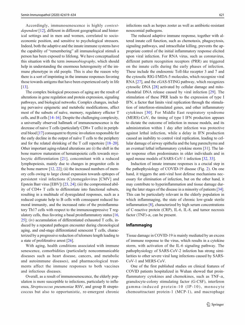
The present SARS-CoV-2 pandemic is posing an unprece-dented healthcare and socio-economic burden worldwide.SARS-CoV-2 hits greatly the older and more fragile fractionof the population, boosting the evergreen issue of vaccinationin elderly people. In Europe, as of week 39/2020, SARS-CoV-2 infection was reported in over 5.7 million people; of those,about 45% were aged 60 or more, while more than 90% of the235,000 reported deaths occurred in this age group.Strikingly, people aged 80 or more accounted for more than50% of the reported deaths, with a median age at death of 81years (https://www.euro.who.int/en/health-topics/health-emergencies/coronavirus-covid-19/weekly-surveillance-report; Fig. 1). These figures are in line with estimateselaborated from the epidemiological data collected in Chinaat the beginning of the outbreak, which reported an adjustedcase fatality ratio of 9.5% in the ≥ 60 age population [1]. Themale to female ratio of SARS-CoV-2 reported cases is around
This article is a contribution to the special issue on: Immunosenescence:New Biomedical Perspectives - Guest Editors: Claudio Franceschi,Aurelia Santoro and Miriam Capri
* Donata [email protected]
1 Laboratory of Molecular Microbiology and Biotechnology(LA.M.M.B.), Department of Medical Biotechnologies, Universityof Siena, Siena, Italy
2 Clinical Chemistry, Department of Laboratory Medicine, KarolinskaInstitute at Huddinge University Hospital, SE-17177 Stockholm, Sweden
3 Department of Experimental, Diagnostic and Specialty Medicine(DIMES), University of Bologna, 40139 Bologna, Italy
4 Interdepartmental Centre ‘L. Galvan’ (CIG), University of Bologna,Via G. Petroni 26, 40139 Bologna, Italy
5 GSK, Siena, Italy6 vAMRes Lab, Toscana Life Sciences, Siena, Italy7 Faculty of Medicine, Imperial College, London, UK8 Lobachevsky State University, Nizhny Novgorod, Russia
https://doi.org/10.1007/s00281-020-00821-0
/ Published online: 6 November 2020
Seminars in Immunopathology (2020) 42:619–634

0.86, while the M:F ratio of deaths is around 1.38, suggestingthat, despite being more frequently infected, females are morecapable of dealing with the infection [2, 3].
It is widely reported that most deaths occurred among pa-tients with at least one underlying disease, such as hyperten-sion [4] and diabetes mellitus [5]. A meta-analysis of sevenclinical studies performed in China identified chronic obstruc-tive pulmonary disease (COPD), cardiovascular disease, andhypertension as risk factors for severe disease and intensitycare unit (ICU) admission [6]. Analysis of risk factors associ-ated with more than ten thousand deaths by COVID-19 in theUK confirmed that age was linearly correlated with risk ofdeath and that obesity, diabetes, severe asthma, respiratorydisease, neurological disease (including stroke), recent (<5 years) hematological malignancy, and recent (< 1 year) can-cer diagnosis were all associated with higher death risk. As forhypertension, the hazard risk was higher only for the popula-tion < 70 years old, even if hypertension itself was stronglyassociated with other risk factors such as obesity and diabetes[7]. Importantly, these epidemiological studies identify thecategories of subjects who are at higher risk of developingsevere SARS-CoV-2 infection and that should be prioritizedin vaccine administration.
Themassive effort for the development of a vaccine againstSARS-CoV-2 could be frustrated by the poor responsivenessto vaccination that characterizes a large proportion of the el-derly population. In this rush against the time, we risk to pay adear toll for the lack of knowledge in the response to vaccina-tion of the elderly, a well-known issue, neglected notwith-standing its evident urgency and the annual reproposalthrough the seasonal influenza epidemic above all.Interestingly, there is a consistent overlap between the factorshampering vaccination effectiveness in the elderly and thosethat boost the virulence and worsen the prognosis of SARS-CoV-2 infection.
A common characteristic of the elderly people is the onsetof a sterile low-grade increase of the basal inflammatory statenamed “inflammaging,” which is considered a universal etio-logical agent of most of the age-related diseases [8]. It is likelythat some specific components of the inflammaging pheno-type could both influence vaccination efficacy and then
increase the risk of the early massive production of inflamma-tory cytokines, termed the “cytokine storm syndrome.” This isa condition reported in severe COVID-19 cases during whichthe patient’s immune system spins out of control and startsdamaging healthy organs owing to the increased vascular per-meability, vascular paralysis, and hypovolemic shock [9].
Angiotensin-converting enzyme 2 (ACE2) has been iden-tified as the receptor for SARS-CoV-2, and it has been sug-gested that differential levels of ACE2 in the cardiac and pul-monary tissues of younger versus older adults may be at leastpartially responsible for the spectrum of disease virulence ob-served among patients with COVID-19 [10].
Here, we analyze the different aspects that tackle SARS-CoV-2 vaccination in the elderly population, considering im-munologic, genetic, and socio-economic factors that impacton the age-related changes of immune responses. A view ofthe current available vaccine platforms with a special focus onthe clinical trials including older adults is reported.
How the elderly condition can affectCOVID-19 disease progressionand the response to vaccination
Immunosenescence
For many reasons, it is difficult to clearly define whatimmunosenescence is: (i) immunosenescence is quite com-plex and involves cellular and molecular changes occurringlifelong (from newborns to centenarians) in both the innateand the adaptive immune systems; (ii) these changes can beat the same time detrimental and beneficial/adaptive [11]; (iii)it is difficult to identify a unique common marker ofimmunosenescence, due to the overwhelming number of bio-logical and non-biological factors that can impinge lifelong onthe immune system of each individual; (iv) the changes oc-curring with age in the immune system are deeply correlatedwith the profound environmental, epidemiological, lifestyle,societal, medical, and public health changes, including vacci-nation policies and practices, that occurred in the last century.
Fig. 1 Distribution of COVID-19cases and deaths by age group.Frequency of COVID-19 cases(upper diagram) and deaths(lower diagram) among differentage ranges (colored boxes) inEurope, estimated in July 2020
620 Semin Immunopathol (2020) 42:619–634
Accordingly, immunosenescence is highly context-dependent [12], different in different geographical and histor-ical settings and in men and women, correlated to socio-economic position, and sensitive to psychological stressors.Indeed, both the adaptive and the innate immune systems havethe capability of “remembering” all immunological stimuli aperson has been exposed to lifelong. We have conceptualizedthis situation with the term immunobiography, which shouldhelp in understanding the enormous heterogeneity of the im-mune phenotype in old people. This is also the reason whythere is a sort of imprinting in the immune responses favoringthose towards antigens that have been experienced early in life[13].
The complex biological processes of aging are the result ofalterations in gene regulation and protein expression, signalingpathways, and biological networks. Complex changes, includ-ing pervasive epigenetic and metabolic modifications, affectmost of the subsets of naïve, memory, regulatory effector Tcells, and B cells [14–16]. Despite the challenging complexity,a universally observed hallmark of immunosenescence is thedecrease of naive T cells (particularly CD8+ T cells) in periph-eral blood [17] consequent to thymic involution responsible forthe early decline in the output of naïve T cells to the peripheryand for the related shrinking of the T cell repertoire [18–20].Other important aging-related alterations are (i) the shift in thebone marrow maturation of hematopoietic cells towards mye-locytic differentiation [21], concomitant with a reducedlymphopoiesis, mainly due to changes in progenitor cells inthe bone marrow [12, 22]; (ii) the increased numbers of mem-ory cells owing to large clonal expansion towards epitopes ofpersistent viral infections (Cytomegalovirus [CMV] andEpstein Barr virus [EBV]) [23, 24]; (iii) the compromised abil-ity of CD4+ T cells to differentiate into functional subsets,resulting in a multitude of dysregulated responses, such as areduced cognate help to B cells with consequent reduced hu-moral immunity, and the increased ratio of the proinflamma-tory Th17 cells with respect to the immunosuppressive T reg-ulatory cells, thus favoring a basal proinflammatory status [16,25]; (iv) accumulation of differentiated exhausted T cells, in-duced by a repeated pathogen encounter during chronologicalaging, and end-stage differentiated senescent T cells, charac-terized by a progressive reduction of telomere length leading toa state of proliferative arrest [26].
With aging, health conditions associated with immunesenescence, comorbidities (particularly noncommunicablediseases such as heart disease, cancers, and metabolicand autoimmune diseases), and pharmacological treat-ments affect the immune responses to both vaccinesand infectious diseases.
Overall, as a result of immunosenescence, the elderly pop-ulation is more susceptible to infections, particularly to influ-enza, Streptococcus pneumoniae RSV, and group B strepto-coccus but also to opportunistic, re-emergent chronic
infections such as herpes zoster as well as antibiotic-resistantnosocomial pathogens.
The reduced adaptive immune response, together with al-tered innate cell function, such as chemotaxis, phagocytosis,signaling pathways, and intracellular killing, prevents the ap-propriate control of the initial inflammatory response elicitedupon viral infection. For RNA virus, such as coronavirus,different pattern recognition receptors (PRR) are triggeredon the innate cells during the early phases of infection.These include the endosomic Toll-like receptor 3 and 7 andthe cytosolic RIG-I/MDA-5 molecules, which recognize viralRNA [27], and the cGAS-STING pathway, which recognizescytosolic DNA [28] activated by cellular damage and mito-chondrial DNA release caused by viral infection [29]. Thestimulation of these PRR leads to the expression of type IIFN, a factor that limits viral replication through the stimula-tion of interferon-stimulated genes, and other inflammatorycytokines [30]. For Middle East respiratory syndrome(MERS)-CoV, the timing of type I IFN production appearsto dictate the outcome of infection in mouse models, and itsadministration within 1 day after infection was protectiveagainst lethal infection, while a delay in IFN productioncaused an inability to control viral replication, leading to cel-lular damage of airway epithelia and the lung parenchyma andan eventual lethal inflammatory cytokine storm [31]. The lat-ter response often predominates in older individuals and inaged mouse models of SARS-CoV-1 infection [32, 33].
Induction of innate immune responses is a crucial step inthe pathophysiology of COVID-19 disease (Fig. 2). On onehand, it triggers the anti-viral host defense mechanisms nec-essary for elimination of infection, but on the other hand, itmay contribute to hyperinflammation and tissue damage dur-ing the later stages of the disease in a minority of patients [34].This can be particularly relevant in the elderly population inwhich inflammaging, the state of chronic low-grade sterileinflammation [8], characterized by high serum concentrationsof C-reactive protein (CRP), IL-6, IL-8, and tumor necrosisfactor (TNF)-α, can be present.
Inflammaging
Tissue damage in COVID-19 is mainly mediated by an excessof immune response to the virus, which results in a cytokinestorm, with activation of the IL-6 signaling pathway. Thepathophysiology of SARS-CoV-2 infection has strong simi-larities to other severe viral lung infections caused by SARS-CoV-1 and MERS-CoV.
One of the first published studies on clinical features ofCOVID patients hospitalized in Wuhan showed that proin-flammatory cytokines and chemokines, such as TNF-α,granulocyte-colony stimulating factor (G-CSF), interferongamma- induced p ro t e i n -10 ( IP -10 ) , monocy t echemoattractant protein-1 (MCP-1), and macrophage
621Semin Immunopathol (2020) 42:619–634

inflammatory proteins 1-α (MIP-1α), were significantlyhigher in patients admitted to the intensive care unit (ICU)compared to those who were not in ICU [35]. Immune pathol-ogy in the form of vascular and cutaneous lesions has alsobeen widely reported [36, 37]. The role of a dysregulatedinflammatory response was proven in an animal model ofSARS-CoV-1 infection using aged macaques. Aged animalsare more prone to develop severe disease and activate morereadily the innate response, in particular the NF-kB pathwayand proinflammatory cytokines such as IL-8 and IL-1β, whilenot inducing significantly IFN-β response. The innate immu-nity activation is not due to the viral load, which is comparableamong young and aged macaques [38].
Transcriptomic analysis performed in samples from sub-jects with severe COVID-19 revealed the presence of lowlevels of type I and type III interferon genes together withelevated levels of proinflammatory cytokines andchemokines, such as IL-6, IL1RA, CCL2, CCL8 CXCL2,CXCL8, CXCL9, and CXCL16 [39].
Which type of cells elicits this cytokine storm and the vi-rological mechanisms behind this inflammatory reaction arestill unclear [40]. Lung epithelial cells, alveolar macrophages,
dendritic cells, and endothelial cells can effectively release theproinflammatory cytokines and chemokines, thus attractingmonocytes, macrophages, and T cells to the site of infection[41]. The overproduction of proinflammatory cytokines in thelungs can damage the tissue infrastructure, recruit macro-phages that infiltrate air spaces, and generate the respiratoryfailure from acute respiratory distress syndrome (ARDS),which is recognized as the leading cause of mortality.Meanwhile, the direct attack on other organs by disseminatedSARS-CoV-2, the immune pathogenesis caused by the sys-temic cytokine storm, and the microcirculation dysfunctionstogether may lead tomulti-organ damage, even though wheth-er SARS-CoV-2 can directly target organs other than the lungand how it can happen are aspects that need to be furtherinvestigated [40] (Fig. 2).
Together with the hyperinflammatory response, a signifi-cant lymphopenia, mainly related to CD4+ T and CD8+ Tcells, which correlates with the severity of viral infection,was reported [42–44]. The causes of this adaptive immunitysuppression are still unclear. Pulmonary recruitment of im-mune cells from the blood and the infiltration of lymphocytesinto the airways may explain the reduction in blood. The well-
Fig. 2 Possible mechanisms of SARS-CoV-2 immunopathology.Systemic and local (lung) immune responses and their pathologicalrole, following SARS-CoV-2 entry into the host are schematicallyrepresented. Induction of innate immune responses is a crucial step inthe pathophysiology of COVID-19 disease, contributing tohyperinflammation and tissue damage during the later stages of thedisease. Infiltration of immune cells in the lungs causes overproductionof proinflammatory cytokines, which eventually damages the lunginfrastructure, accumulation of macrophages in the air spaces anddiffuse alveolar damage leading to acute respiratory distress syndrome
(ARDS). Furthermore, elevated levels of circulating proinflammatorycytokines can cause septic shock and multi-organ dysfunction. Togetherwith the hyperinflammatory response, overt disseminated intravascularcoagulation has been reported and a significant lymphopenia, mainlyrelated to CD4+ T and CD8+ T cells, has been observed, possibly dueto pulmonary recruitment of lymphocytes from the blood. A possibleimmunopathological role can be mediated by non-neutralizingantibodies produced by B cells, which may enhance SARS-CoV-2infection through antibody-dependent enhancement (ADE), furtherexacerbating organ damage
622 Semin Immunopathol (2020) 42:619–634

known age-related alteration of the immune function of T celland B cells could lead to insufficient control of viral replica-tion, thus increasing the macrophage infiltration and the lunginjury (Fig. 2).
Finally, a possible immunopathological role can be medi-ated by non-neutralizing antibodies produced by B cells thatmay enhance SARS-CoV-2 infection through antibody-dependent enhancement (ADE), further exacerbating organdamage. It has recently been shown that SARS-CoV-1 andthe MERS-CoV take advantage of non- or subneutralizingantibodies and enter cells via surface CD32a receptors, anFc receptor expressed on the surfaces of monocytes and alve-olar macrophages. The antibody-CD32 interaction facilitatesviral entry and infection, and activates intracellular signalingto upregulate proinflammatory cytokines [45].
The complex and still unclear immunopathological mech-anisms of SARS-CoV-2 infection, together with the progres-sive age-related decline of innate and adaptive immune re-sponses, and the lack of a clear correlate of protection, makethe design of vaccination strategies for older people extremelychallenging (Fig. 3).
Biological age
An emerging class of instruments in the aging research is thedevelopment of markers capable of assessing the speed of theaging process. Age is a major risk factor for a high number ofdiseases, and in general, it affects the fitness of each individ-ual, including the capability of responding to vaccine admin-istration and counteracting a severe infection [46]. However, itis also evident that the elderly population is extremely hetero-geneous, so while chronological age is useful to identify
macroscopic risk classes, it is poorly informative within ageclasses to get individual information. Biological age is thususeful to evaluate clinical parameters and health risks on thebasis of the individual aging pace, which tend to be moreheterogeneous in the elderly population. Several establishedbiological age markers have been generated based on bothclassical anthropometric, clinical, and biochemical parametersas well as on innovative molecular characterizations such asDNA methylation and the composition of the N-glycan shellof circulating proteins [47]. Such biomarkers have shown in anumber of studies that the aging pace is higher in the vastmajority of the different elderly conditions, thus demonstrat-ing that biological age assessment should be a critical infor-mation in a broad spectrum of clinical practices and in thedevelopment of strategies to tackle healthcare burden andemergencies. The detailed description of available biologicalage markers is out of the scope of the present manuscript, andan extensive overview is available in the review by Jylhäväet al. [46]. To date, biological age has not been assessed in theSARS-CoV-2 clinical setting, but it is noteworthy that biolog-ical age has been associated with all the most important riskfactors related to a poor prognosis of SARS-CoV-2 infection.The field of elderly vaccination could benefit from biologicalage information, but also in this case, the available data arerare. In a study from Gensous et al. [48], the whole genomemethylation profile of PBMC was assessed in a group of vol-unteers of different ages who underwent influenza vaccina-tion. The relationship between the vaccination response andthe methylation profile was studied. While no difference interms of biological age emerged in the study, an age-dependent epigenetic remodeling emerged in elder non-re-sponders. The study is limited owing to the very low number
Fig. 3 Challenges for thedevelopment of a SARS-CoV-2vaccine for elderly people.Schematic interconnectionbetween the main immunemechanisms elicited by thevaccination process, with thepeculiarity of the elderly immunesystem—affected by bothinflammaging andimmunosenescence—and the stillundefined correlates of protectionfrom SARS-CoV-2 infection. Thecomplex and still unclearimmunopathological mechanismsof SARS-CoV-2 infection,together with the progressive age-related decline of innate andadaptive immune responses, andthe lack of a clear correlate ofprotection make the design ofvaccination strategies for olderpeople extremely challenging
623Semin Immunopathol (2020) 42:619–634
of analyzed subjects but confirmed that DNA methylation isan informative instrument to be exploited in vaccination stud-ies and strategies.
Immunobiography
Immunobiography refers to the comprehensive immunologi-cal, clinical, socio-economic, and geographical history of eachindividual, and accounts for the large heterogeneity observedin the elderly regarding their health status, mirrored by theirlarge individual variation in the responsiveness to vaccines. Amajor advantage of immunobiography is that it incorporatesthe most advanced conceptualization of immunosenescencewhich, according to the most recent literature [49], has to beconsidered as a context- and population-dependent phenome-non. Accordingly, in order to be properly interpreted, age-related changes of immune parameters occurring in an elderlyperson necessitate a variety of other additional data regardingsex/gender, demographic cohort, population/country, individ-ual immunological history, anthropometric parameters, socio-economic status and education, CMV serostatus, morbidityand co-morbidity, among others. It is of critical importancetaking in consideration the elderly vulnerability to direct therational design of vaccines designed for this target population.
Gender
Gender is a critical issue in both vaccination of the elderly andin the SARS-Cov-2 pandemic. The pandemic epidemiologicaldata show clearly that the risk of severe disease and mortalityis sharply higher in men than in women.Men’s hospitalizationexceeds women by about 50%, indicating a significantlyhigher susceptibility of men towards severe SARS-CoV-2 in-fection. Available data show that men outnumber women 2 to4 times in terms of ICU admissions [50–52]. These numbersare concordant with the fatality rate that ranges between 1.2and 1.4 men deaths for one women death. Moreover, thisunbalanced pattern is mirrored by the vaccine uptake, re-sponses, and outcome in older-aged individuals. Elderlywomen are indeed more responsive than men for several vac-cine protocols recommended in older-aged individuals such asthose against influenza, tetanus, pertussis, shingles, and pneu-mococcal infections [53]. On the other hand, an influenzavaccination study reported that aged men antibodies hadhigher affinity than those produced by women. Moreover,men seem to respond better to pneumococcal vaccination intwo independent studies [54, 55]. There is an impaired vacci-nation response in both old men and women with sex-specificweaknesses. The most striking data, however, is related toinfection and all-cause mortality: indeed, in a number of re-ports, vaccine administration produces a sharper decrease ofspecific and all-cause mortality in vaccinated women com-pared to men, indicating that women have higher benefit from
vaccination in the elderly [56–58]. These data indicate theneed to consider sex-specific vaccination protocols for theelderly population [58, 59] and that the lack of such instru-ments could be critical in the SARS-CoV-2 pandemic sinceold men are both the most susceptible to severe SARS-CoV-2infection and are those less likely protected by a possibleSARS-CoV-2 vaccine [60–62].
Microbiota
Another factor that could affect vaccine response is the intes-tinal microbiota that plays a crucial rule in the regulation of theimmune system and is highly affected by age [63–66].Microbial community composition indeed is influenced byage, environmental and socio-economic factors, diet, gender,chronic infections, immunosuppressive chemotherapy, antibi-otic treatment, or probiotic use [64, 67–69]. The improvementin the nucleic acid sequencing obtained in the last 15 years hitsmassively the microbiological research and promotes the anal-ysis of heterogeneous microbiological ecosystems such asthose that reside in humans. The characterization of such eco-logical niches opens to the new conceptualization of humansas metaorganisms (organisms composed of different organ-isms) to stress the tight interdependencies between the hostand the microbiological species residing in different anatom-ical districts.
Gut microbiota changes with age and that is likely an im-portant contributor and modulator of the inflammaging phe-notype [70, 71]. Elderly people have less diverse gut micro-biota and reduced beneficial microorganisms [72]. The gener-al imbalance of gut microbiota, called “dysbiosis,” is associ-ated with both frailty, a geriatric syndrome leading to in-creased vulnerability for adverse health outcomes, and sys-temic inflammation. Since a hyperinflammation status hasbeen observed in most severe cases of SARS-CoV-2 infec-tion, it is possible that gut dysbiosis may influence the clinicalmanifestation in COVID-19 infection [73, 74].
Interestingly, the gut microbiota has been shown to alsoaffect pulmonary health through a bidirectional cross-talk be-tween the gut microbiota and the lungs [75]. Along this “gut-lung axis,” microbial products can reach the lung throughblood and modulate pulmonary immune responses [76], whileinflammation processes occurring in the lung can impact onthe gut microbiota [77]. Some studies have demonstrated thatrespiratory infections are associated with a change in the com-position of the gut microbiota [78] and the antibiotic treatmentof mice for removing some gut bacteria has led to increasedsusceptibility to influenza virus infection in the lungs [79].Since one of the severe clinical manifestations of COVID-19is pneumonia and progression to acute respiratory distresssyndrome (ARDS), especially in elderly and immune-compromised patients [80], it can be speculated that SARS-Cov-2 infection can affect this gut-lung cross-talk which
624 Semin Immunopathol (2020) 42:619–634

might influence the outcome of the clinical manifestation [81].Moreover, even though respiratory symptoms represent theprincipal clinical presentation of COVID-19, clinical evidencesuggests that the intestine may be another viral target organ.Indeed, a high expression of ACE2 has been observed in thebrush border of intestinal enterocytes [82] and, using a humansmall intestinal organoid system, it has been demonstrated thatSARS-CoV-2 readily replicates into the enterocytes, resultingin the production of large amounts of infective virus particles[83]. Some reports show that SARS-CoV-2 RNA can be de-tected in the stool of some patients of COVID-19 [84, 85], andpatients often present gastrointestinal symptoms such as diar-rhea, vomiting, and abdominal pain [86]. Therefore, the char-acterization of the gut microbiota in patients with activeSARS-CoV-2 intestinal infection could represent a strikingaspect to investigate.
These considerations on inflammaging, immunobiography,biological age, gender, and microbiota pertain to every vacci-nation strategy, but are particularly relevant for the develop-ment of vaccines against SARS-CoV-2 since it more seriouslyaffects the elderly population and immunopathology is a crucialfactor for the severity disease.
Need for the design of a SARS-CoV-2vaccination strategies tailored for the elderly
SARS-CoV-2 vaccines are urgently needed, and their designshould take into consideration that the elderly population isthe main target population for vaccination. While older adultsare most likely to be severely affected by COVID-19, theyalso may be less responsive to vaccination. Efficacy of vacci-nation in the elderly is indeed strongly reduced compared tothat of younger adults [87, 88]. SARS-CoV-2 vaccinationstrategies, tailored for the elderly, should take into consider-ation the delicate balance between immunosenescence/inflammaging and the immunopathological aspects of theCOVID-19 disease (Fig. 3). Vaccine adjuvants and vectorsshould be specifically designed for stimulating the elderlyimmune system without exacerbating the inflammatory status[87]. Despite these considerations, the elderly are rarely in-cluded in vaccine clinical trials; in the last decades, the vastmajority of randomized control trials did not include olderadults and in particular frail older adults who are mostly atrisk. We currently do not have full knowledge on the mecha-nisms of immunity to protect this population from SARS-CoV-2 [10].
The development of a SARS-CoV-2 vaccine is extremelychallenging, since we are faced with a novel virus, justemerged in humans, and correlates of protection have notyet been fully identified, even though the induction of neutral-izing antibodies is presumed to be a crucial target for an ef-fective vaccination (Fig. 3).
Protection in older individuals against influenza virus ap-pears to require higher neutralization titers than in youngerindividuals [89], and this issue might need to be addressedfor SARS-CoV-2. The knowledge obtained from the vaccinedevelopment efforts for MERS and SARS-CoV-1 can be ofhigh value for SARS-CoV-2, although no vaccines are li-censed for these coronavirus strains [90].
Memory CD4+ T cells, induced by infections with other co-ronavirus and capable of responding to SARS-CoV-2, have beendetected in 20–50% of SARS-CoV-2 unexposed donors [91,92]. The characterization of these cross-reactive T cells in theelderly and their impact on the immunogenicity of vaccine can-didates should be taken into consideration in the ongoingCOVID-19 vaccination studies. SARS-CoV-2 vaccine candi-dates based on different vaccine platforms have been developed,and about 140 candidates have been tested in pre-clinical exper-iments, according to theWHO landscape documents of COVID-19 candidate vaccines (https://www.who.int/publications/m/item/draft-landscape-of-covid-19-candidate-vaccines) (Fig. 4).Information on the specific SARS-CoV-2 molecules selected asvaccine antigens is limited, even though most candidates aim toelicit neutralizing antibodies against the spike (S) protein and itsreceptor-binding domain (RBD), as already performed with theSARS andMERSvaccines. Awide range of both innovative andtraditional technology platforms has been deployed, includingnucleic acid (DNA and RNA), recombinant viral vectors (repli-cating and non-replicating), recombinant protein combined withadjuvants, and live attenuated or inactivated virus [93]. Some ofthese platforms were already tested in human studies for SARS-CoV-1 virus, such as inactivated virus, DNA and soluble S pro-teins [94–96], or for MERS-CoV [97].
The most advanced candidates for SARS-CoV-2 enteredin human clinical testing with unprecedented rapidity em-ploy nucleic acid (both mRNA and DNA), recombinantvaccine vectors (human or chimpanzee Adenovirus vec-tors), subunit S protein combined or not with different ad-juvants, and inactivated SARS-CoV-2 virus. Other novelplatforms based on the use of synthetic modified antigenpresenting cells (APC) or cytotoxic T lymphocytes are alsounder study (Fig. 4). The platforms using mRNA, non-replicating viral vectors, and inactivated SARS-CoV-2 vi-rus have already reached the clinical trial phase III. Some ofthe different platforms used may be tailored for specificpopulation subtypes, such as the elderly, children, pregnantwomen, or immunocompromised patients [98]. In this re-gard, some of the ongoing clinical studies have specificallytaken into consideration the older population, by includingvaccination arms with people aged > 60 years. A schematicdiagram of the ongoing phase I and II clinical trials thathave included older adults is reported in Fig. 5. Enrollingolder adult volunteers will help to better understand vacci-nation outcomes among the older population, who are mostat risk of complications from COVID-19.
625Semin Immunopathol (2020) 42:619–634

The ongoing clinical studies based on mRNA technology(mRNA-1273 fromModerna n. NCT04283461, and BNT162from Biontech SE, n. NCT04368728) aim to evaluate thesafety, tolerability, immunogenicity, and potential efficacyof different SARS-CoV-2 RNA vaccine candidates in theadult population, with a specific attention to older people(N.-N. Releases. NIH clinical trial of investigational vaccinefor COVID-19 begins. 2020. https://www.nih.gov/news-events/news-releases/nih-clinical-trial-investigational-vaccine-covid-19-begins). The lipid nanoparticle-encapsulated mRNA-1273 vaccine, which encodes for thefull-length S protein, is currently evaluated in a dose-rangingstudy in the adult population (18–55 years old), and in partic-ipants from 56 to 70 and > 71 years of age (Fig. 5). Similarly,the large dose-finding study with the BNT162 biological com-ponent (7600 estimated participants) based on the administra-tion of mRNA coding for the full-length S protein, or for thetwo smaller receptor-binding domains, is going to test theimmunogenicity in adults (18–55 years) and older adults(56–85 years).
An ongoing phase I/IIa trial (n. NCT04447781) is alsoaimed at evaluating the safety, tolerability, and immunological
profile of the INO-4800 vaccine that, exploiting the DNAtechnology, contains a plasmid encoding the full-length S gly-coprotein. The INO-4800 vaccine is administered by intrader-mal injection followed by electroporation in healthy adultsaged 19 to 64 years.
Another platform that is currently specifically tested inolder people is based on the Adenovirus type 5 vector thatencodes the S protein from the SARS-CoV-2 strain (trials n.2020-001228-32; PACTR202006922165132; NCT0439814;ChiCTR2000031781 and NCT04400838; Fig. 5). Differentstudies are ongoing, and one conducted in Canada is a dose-escalation designed study, from the younger adults (18 to <55) to the older adults (65 to < 85). Another huge phase 2/3study (n. NCT04400838) is aimed at determining the efficacy,safety, and immunogenicity of the candidate COVID-19 vac-cine based on the chimpanzee adenovirus vector (ChAdOx1nCoV-19) in healthy UK volunteers, specifically divided inadults (18–55 years old), elderly (over the age of 56), andchildren (5–12 years old). The ChAdOx1 platform has alreadybeen shown to be effective in the established rhesus macaquemodel of SARS-CoV-2 infection [99]. In this pre-clinicalstudy, a single dose of ChAdOx1 nCoV-19 has protected six
Fig. 4 SARS-CoV-2 vaccinecandidates based on differentvaccine platforms. Schematicrepresentation of the differentvaccine platforms used fordeveloping SARS-CoV-2vaccines. These include nucleicacid (both mRNA and DNA);subunit S protein with differentadjuvants; non-replicating viralvectors (such as Adenovirus);inactivated SARS-CoV-2 virusalone or combined withadjuvants; live SARS-CoV-2attenuated virus; virus-likeparticles and replicating viralvectors (such as Measles virus,Influenza virus, Vesicularstomatitis virus, and others).About 140 vaccine candidates arecurrently involved in pre-clinicalstudies, while 35 vaccinecandidates are worldwide testedin clinical studies, and some ofthem (indicated with *) havealready reached the phase III. Foreach platform, the number ofongoing clinical or pre-clinicalstudies is reported. Data arereferred to the WHO report,updated to 17 September 2020
626 Semin Immunopathol (2020) 42:619–634

rhesus macaques from pneumonia caused by the virus [100].Moreover, the ChAdOx1 has been used to develop investiga-tional vaccines against several pathogens, including the close-ly related coronavirus responsible for the MERS [101].Adenovirus-based vectors are characterized by a broad rangeof tissue tropism that covers both respiratory and gastrointes-tinal epithelium, the two main sites that express the ACE-2receptor of SARS-CoV-2, even though a possibleimmunodominance mediated by vector genes rather than thetransgenes should always be considered [102].
Using the traditional recombinant protein technology toexpress the spike protein, a trial sponsored by CloverBiopharmaceuticals AUS Pty Ltd. (n. NCT04405908) isassessing the safety, reactogenicity, and immunogenicity ofmultiple doses of SCB-2019 administered with AS03 adju-vant, or with CpG 1018 plus alum adjuvants. Data will beseparately analyzed on adult (18 to 54 years of age) and elder-ly (55–75 years of age) healthy subjects enrolled in the study.In another study, the S protein has been administered with theAdvax adjuvant (n. NCT04453852), a potent and safeimmunopotentiator composed of delta inulin [103].
Four trials are testing in the elderly population theinactivated SARS-CoV-2 virus (n. NCT04456595;ChiCTR2000031809; ChiCTR2000032459), and one of thesehas been specifically performed only in people > 60 years (nNCT04383574; Fig. 5).
Numerous other vaccine developers have indicated plans toinitiate human testing in 2020. Despite the several vaccinecandidates (Fig. 4), challenges including the need for optimiz-ing antigen design and adjuvant formulation define the num-ber of doses needed, induce the optimal immune responsewithout exacerbating the inflammatory and antibody-
dependent response involved in possible lung disease, andfully define correlates of protection and duration of immuneresponses have to be considered [104].
Finally, a general consideration for the SARS-CoV-2 vac-cine development regards safety issues that could arise withCOVID-19 vaccines developed under the strong pressure ofthe pandemic situation. Animal studies on vaccines for SARS-CoV-1 and MERS-CoV report possible adverse effects medi-ated by vaccine-induced antibodies that have poor or no neu-tralizing activity [105]. Safety and efficacy are two indissolu-ble properties of a vaccine to be administered to billions ofpeople globally and need to be accurately evaluated for everySARS-CoV-2 candidate.
Systems biology and integrative analysis
The efforts in the development of COVID-19 vaccinescan benefit from the availability of most advanced toolsand high-throughput technologies to decipher the effec-tive immune responses in the older population and thecorrelates of protection. Recent advances in systems bi-ology integrating clinical, immunologic, and omics datacan help to identify stable and robust markers of vac-cine response and move towards a better understandingof SARS-CoV-2 vaccine responses in the elderly.Machine/statistical learning applied to multi-omics datafrom clinical studies promises to revolutionize vaccinedevelopment by illuminating the mechanistic drivers ofprotective immunity. The high-performance data acqui-sition methods in molecular and cellular biology pushthe field of bioinformatics for the development and use
Fig. 5 Ongoing clinical trials of COVID-19 vaccines specificallyincluding the elderly population. Schematic representation of clinicalstudies specifically including older people in the selection criteria ofvolunteers. The platform used for each clinical trial is shown on the left.
The identifier number of the clinical trial and the number of volunteersincluded (in brackets) are reported on the right. Bars represent thepartition of volunteers according to the age range. Data are updated to8th July 2020
627Semin Immunopathol (2020) 42:619–634

of tools that manage and integrate the different levels ofbiological complexity.
Application of the immunobiography approach could in-form the stratification of elderly subjects and guide the imple-mentation of vaccination strategies designed for specific el-derly population clusters [87]. Mathematical modeling allowsthe combination of different networks involved in biologicalaging such as epigenetic networks, cell-cell networks, andpopulation genetics and can allow to generate hypothesis onresponse to treatment or vaccination [106]. Recent progress inmathematical modeling can be utilized to generate biomarkermodels for prediction of disease and also response to vaccina-tion taking into consideration biological age.
Currently, computational models have been applied toimmunology data, for example, for the analysis of ahigh-dimensional dataset in vaccination studies [107,108], but these models are limited to particular aspects[109, 110]. There is the potential for these models tobecome more sophisticated and to predict how responsesto pathogens and vaccines are affected by pre-disposingfactors [111, 112]. The systems vaccinology approachhas been applied to characterize the immune responseto different vaccines providing the proof-of-concept ev-idence of the capacity of systems approaches to delin-eate “molecular signatures” predictive of vaccine re-sponses [113–131]. This approach has also been appliedto identify molecular signatures induced by immuniza-tion with the rVSV-ZEBOV Ebola vaccine, recently ap-proved for human use. Systems analysis has been con-ducted integrating clinical, immunologic, and omics datain clinical trials with different doses and in differentcontinents (Vianello et al. 2020 submitted, Santoroet al. 2020 submitted).
Despite the great efforts made, unfortunately, manyof the most useful clinical and multi-omics datasetsare siloed in local databases to protect participant priva-cy and data confidentiality. Creation of secure, FAIR-compliant, federated learning databases in which predic-tive biological and mathematical models based on AI/machine/statistical learning can be developed, refined,and tested on distributed datasets would have an enor-mous impact in suppor t ing a ra t ional vaccinedevelopment.
Concluding remarks
SARS-CoV-2 vaccines are urgently needed, and their designshould take into consideration that the elderly are the maintarget population for vaccination. The pandemic is stimulatingthe research on vaccine development, and this should be atremendous opportunity to specifically include age and genderas critical factors for vaccination approaches and
effectiveness.While older adults are most likely to be severelyaffected by COVID-19, they also may be less responsive tovaccination. In the ongoing tremendous efforts for COVID-19vaccine development, only a limited number of clinical trialshave included the older fraction of the population in the studydesign, and the platforms used are not specifically designedconsidering the peculiarity of the elderly immune system.Indeed, vaccination strategies tailored for the SARS-CoV-2infection in the elderly should take into consideration the del-icate balance of immunosenescence and inflammaging withthe immunopathological aspects of the SARS-CoV-2 infec-tion, such as the cytokine storm reported in severe COVID-19. Therefore, the possible overlap between the factors ham-pering vaccination effectiveness in the elderly and those thatboost the virulence and worsen the prognosis of SARS-CoV-2infection should be carefully taken into consideration. Thus,vaccine formulations, such as adjuvants and vectors, shouldbe specifically designed for stimulating the elderly immunesystem without exacerbating the inflammatory status. The on-going efforts in COVID-19 vaccine development should fullyexploit the availability of high-throughput technologies andrecent advances in systems biology to decipher the effectiveimmune responses in the older population and identify corre-lates of protection to guide towards SARS-CoV-2 vaccinestrategies optimally designed to protect the older population.
Code availability Not applicable
Authors’ contributions AC, PG, and DM, FS drafted the work; DM, RR,and CF revised it critically for important intellectual content; all the au-thors approved the version to be published.
Funding Open access funding provided by Università degli Studi diSiena within the CRUI-CARE Agreement. This work was supported byCommission of the European Communities, Horizon 2020 FrameworkProgramme, grant number 730964 (TRANSVAC2), and RussianMinistry of Science and Education Agreement No. 075-15-2020-808.
Data availability Not applicable
Compliance with ethical standards
Conflict of interest The authors declare that they have no conflict ofinterest.
Consent for publication The authors are responsible for the correctnessof the statements provided in the manuscript.
Ethics approval Not applicable
Consent to participate Not applicable
Open Access This article is licensed under a Creative CommonsAttribution 4.0 International License, which permits use, sharing, adap-tation, distribution and reproduction in any medium or format, as long asyou give appropriate credit to the original author(s) and the source, pro-vide a link to the Creative Commons licence, and indicate if changes were
628 Semin Immunopathol (2020) 42:619–634
made. The images or other third party material in this article are includedin the article's Creative Commons licence, unless indicated otherwise in acredit line to the material. If material is not included in the article'sCreative Commons licence and your intended use is not permitted bystatutory regulation or exceeds the permitted use, you will need to obtainpermission directly from the copyright holder. To view a copy of thislicence, visit http://creativecommons.org/licenses/by/4.0/.
References
1. Verity R, Okell LC, Dorigatti I, Winskill P, Whittaker C, Imai N,Cuomo-Dannenburg G, Thompson H, Walker PGT, Fu H, DigheA, Griffin JT, Baguelin M, Bhatia S, Boonyasiri A, Cori A,Cucunubá Z, FitzJohn R, Gaythorpe K, Green W, Hamlet A,Hinsley W, Laydon D, Nedjati-Gilani G, Riley S, van Elsland S,Volz E, Wang H, Wang Y, Xi X, Donnelly CA, Ghani AC,Ferguson NM (2020) Estimates of the severity of coronavirusdisease 2019: a model-based analysis. Lancet Infect. Dis 20:669–677. https://doi.org/10.1016/S1473-3099(20)30243-7
2. Klein SL, Flanagan KL (2016) Sex differences in immune re-sponses. Nat. Rev. Immunol. 16:626–638. https://doi.org/10.1038/nri.2016.90
3. Vom Steeg LG, Klein SL (2016) SeXX matters in infectious dis-ease pathogenesis. PLoS Pathog 12:e1005374. https://doi.org/10.1371/journal.ppat.1005374
4. Feng Y, Ling Y, Bai T, Xie Y, Huang J, Li J, Xiong W, Yang D,Chen R, Lu F, Lu Y, Liu X, Chen Y, Li X, Li Y, Summah HD, LinH, Yan J, Zhou M, Lu H, Qu J (2020) COVID-19 with differentseverities: a multicenter study of clinical features. Am J RespirCrit Care Med 201:1380–1388. https://doi.org/10.1164/rccm.202002-0445OC
5. Rastad H, Karim H, Ejtahed H-S, Tajbakhsh R, Noorisepehr M,Babaei M, Azimzadeh M, Soleimani A, Inanloo SH, ShafiabadiHassani N, Rasanezhad F, Shahrestanaki E, Khodaparast Z,Golami H, Qorbani M (2020) Risk and predictors of in-hospitalmortality fromCOVID-19 in patients with diabetes and cardiovas-cular disease. Diabetol Metab Syndr 12:57. https://doi.org/10.1186/s13098-020-00565-9
6. Jain V, Yuan J-M (2020) Predictive symptoms and comorbiditiesfor severe COVID-19 and intensive care unit admission: a system-atic review andmeta-analysis. Public Health, Int. J. https://doi.org/10.1007/s00038-020-01390-7
7. Williamson EJ, Walker AJ, Bhaskaran K, Bacon S, Bates C,Morton CE, Curtis HJ, Mehrkar A, Evans D, Inglesby P,Cockburn J, McDonald HI, MacKenna B, Tomlinson L,Douglas IJ, Rentsch CT, Mathur R, Wong AYS, Grieve R,Harrison D, Forbes H, Schultze A, Croker R, Parry J, Hester F,Harper S, Perera R, Evans SJW, Smeeth L, Goldacre B (2020)OpenSAFELY: factors associated with COVID-19 death in 17million patients. Nature 584:1–11. https://doi.org/10.1038/s41586-020-2521-4
8. Franceschi C, Bonafè M, Valensin S, Olivieri F, De Luca M,Ottaviani E, De Benedictis G (2000) Inflamm-aging. An evolu-tionary perspective on immunosenescence. Ann. N. Y. Acad. Sci908:244–254. https://doi.org/10.1111/j.1749-6632.2000.tb06651.x
9. Jose RJ, Manuel A (2020) COVID-19 cytokine storm: the inter-play between inflammation and coagulation. Lancet Respir, Med.https://doi.org/10.1016/S2213-2600(20)30216-2
10. Koff WC, Williams MA (2020) Covid-19 and immunity in agingpopulations - a new research agenda. N Engl J Med 383:804–805.https://doi.org/10.1056/NEJMp2006761
11. Fulop T, Larbi A, Dupuis G, Le Page A, Frost EH, Cohen AA,Witkowski JM, Franceschi C (2017) Immunosenescence andinflamm-aging as two sides of the same coin: friends or foes?Front Immunol 8:1960. https://doi.org/10.3389/fimmu.2017.01960
12. Ogawa T, Kitagawa M, Hirokawa K (2000) Age-related changesof human bone marrow: a histometric estimation of proliferativecells, apoptotic cells, T cells, B cells and macrophages. MechAgeing Dev 117:57–68. https://doi.org/10.1016/s0047-6374(00)00137-8
13. Franceschi C, Salvioli S, Garagnani P, de Eguileor M, Monti D,Capri M (2017) Immunobiography and the heterogeneity of im-mune responses in the elderly: a focus on Inflammaging andtrained immunity. Front Immunol 8:982. https://doi.org/10.3389/fimmu.2017.00982
14. Akbar AN, Henson SM, Lanna A (2016) Senescence of T lym-phocytes: implications for enhancing human immunity. TrendsImmunol 37:866–876. https://doi.org/10.1016/j.it.2016.09.002
15. Johnson SA, Cambier JC (2004) Ageing, autoimmunity and ar-thritis: senescence of the B cell compartment – implications forhumoral immunity. Arthritis Res Ther 6:131–139. https://doi.org/10.1186/ar1180
16. Schmitt V, Rink L, Uciechowski P (2013) The Th17/Treg balanceis disturbed during aging. Exp Gerontol 48:1379–1386. https://doi.org/10.1016/j.exger.2013.09.003
17. Fagnoni FF, Vescovini R, Passeri G, Bologna G, Pedrazzoni M,Lavagetto G, Casti A, Franceschi C, Passeri M, Sansoni P (2000)Shortage of circulating naive CD8(+) T cells provides new in-sights on immunodeficiency in aging. Blood. 95:2860–2868
18. Goronzy JJ, Weyand CM (2017) Successful and maladaptive Tcell aging. Immunity. 46:364–378. https://doi.org/10.1016/j.immuni.2017.03.010
19. Thomas R, Wang W, Su DM (2020) Contributions of age-relatedthymic involution to immunosenescence and inflammaging.Immun. Ageing 17. https://doi.org/10.1186/s12979-020-0173-8
20. Wack A, Cossarizza A, Heltai S, Barbieri D, D’Addato S,Fransceschi C, Dellabona P, Casorati G (1998) Age-related mod-ifications of the human alphabeta T cell repertoire due to differentclonal expansions in the CD4+ and CD8+ subsets. Int Immunol10:1281–1288. https://doi.org/10.1093/intimm/10.9.1281
21. Kovtonyuk LV, Fritsch K, Feng X, Manz MG, Takizawa H(2016) Inflamm-aging of hematopoiesis, hematopoietic stem cells,and the bone marrow microenvironment. Front Immunol 7:502.https://doi.org/10.3389/fimmu.2016.00502
22. Linton PJ, Dorshkind K (2004) Age-related changes in lympho-cyte development and function. Nat Immunol 5:133–139. https://doi.org/10.1038/ni1033
23. Tu W, Rao S (2016) Mechanisms underlying T cellimmunosenescence: aging and cytomegalovirus infection.Front. Microbiol 7. https://doi.org/10.3389/fmicb.2016.02111
24. Vescovini R, Telera A, Fagnoni FF, Biasini C, Medici MC,Valcavi P, di Pede P, Lucchini G, Zanlari L, Passeri G, Zanni F,Chezzi C, Franceschi C, Sansoni P (2004) Different contributionof EBV and CMV infections in very long-term carriers to age-related alterations of CD8+ T cells. Exp Gerontol 39:1233–1243.https://doi.org/10.1016/j.exger.2004.04.004
25. Bektas A, Schurman SH, Sen R, Ferrucci L (2017) Human T cellimmunosenescence and inflammation in aging. J Leukoc Biol102:977–988. https://doi.org/10.1189/jlb.3RI0716-335R
26. Pangrazzi L, Weinberger B (2020) T cells, aging and senescence.Exp Gerontol 134:110887. https://doi.org/10.1016/j.exger.2020.110887
27. Züst R, Cervantes-Barragan L, Habjan M, Maier R, Neuman BW,Ziebuhr J, Szretter KJ, Baker SC, Barchet W, Diamond MS,Siddell SG, Ludewig B, Thiel V (2011) Ribose 2’-O-methylationprovides a molecular signature for the distinction of self and non-
629Semin Immunopathol (2020) 42:619–634
self mRNA dependent on the RNA sensor Mda5. Nat Immunol12:137–143. https://doi.org/10.1038/ni.1979
28. Sun L, XingY, ChenX, ZhengY,YangY, Nichols DB, ClementzMA, Banach BS, Li K, Baker SC, Chen Z (2012) Coronaviruspapain-like proteases negatively regulate antiviral innate immuneresponse through disruption of STING-mediated signaling. PLoSONE 7. https://doi.org/10.1371/journal.pone.0030802
29. Sun B, Sundström KB, Chew JJ, Bist P, Gan ES, Tan HC, GohKC, Chawla T, Tang CK, Ooi EE (2017) Dengue virus activatescGAS through the release of mitochondrial DNA. Sci Rep 7:3594.https://doi.org/10.1038/s41598-017-03932-1
30. Prompetchara E, Ketloy C, Palaga T (2020) Immune responses inCOVID-19 and potential vaccines: lessons learned from SARSand MERS epidemic. Asian Pac. J. Allergy Immunol 38:1–9.https://doi.org/10.12932/AP-200220-0772
31. Channappanavar R, Fehr AR, Zheng J, Wohlford-Lenane C,Abrahante JE, Mack M, Sompallae R, McCray PB, MeyerholzDK, Perlman S (2019) IFN-I response timing relative to virusreplication determines MERS coronavirus infection outcomes. JClin Invest 129:3625–3639. https://doi.org/10.1172/JCI126363
32. Rockx B, Baas T, Zornetzer GA, Haagmans B, Sheahan T,Frieman M, Dyer MD, Teal TH, Proll S, van den Brand J, BaricR, Katze MG (2009) Early upregulation of acute respiratory dis-tress syndrome-associated cytokines promotes lethal disease in anaged-mouse model of severe acute respiratory syndrome corona-virus infection. J Virol 83:7062–7074. https://doi.org/10.1128/JVI.00127-09
33. Huang K-J, Su I-J, Theron M, Wu Y-C, Lai S-K, Liu C-C, Lei H-Y (2005) An interferon-gamma-related cytokine storm in SARSpatients. J Med Virol 75:185–194. https://doi.org/10.1002/jmv.20255
34. Netea MG, Giamarellos-Bourboulis EJ, Domínguez-Andrés J,Curtis N, van Crevel R, van de Veerdonk FL, Bonten M (2020)Trained immunity: a tool for reducing susceptibility to and theseverity of SARS-CoV-2 infection. Cell. 181:969–977. https://doi.org/10.1016/j.cell.2020.04.042
35. Huang C, Wang Y, Li X, Ren L, Zhao J, Hu Y, Zhang L, Fan G,Xu J, Gu X, Cheng Z, Yu T, Xia J, Wei Y, WuW, Xie X, YinW,Li H, LiuM, Xiao Y, Gao H, Guo L, Xie J,Wang G, Jiang R, GaoZ, Jin Q, Wang J, Cao B (2020) Clinical features of patients in-fected with 2019 novel coronavirus in Wuhan, China. LancetLond Engl 395:497–506. https://doi.org/10.1016/S0140-6736(20)30183-5
36. Verdoni L, Mazza A, Gervasoni A, Martelli L, Ruggeri M,Ciuffreda M, Bonanomi E, D’Antiga L (2020) An outbreak ofsevere Kawasaki-like disease at the Italian epicentre of theSARS-CoV-2 epidemic: an observational cohort study. Lancet395:1771–1778. https://doi.org/10.1016/S0140-6736(20)31103-X
37. C.G. Casas, A. Català, G.C. Hernández, P. Rodríguez-Jiménez, D.Fernández-Nieto, A.R.-V. Lario, I.N. Fernández, R. Ruiz-Villaverde, D. Falkenhain-López, M.L. Velasco, J. García-Gavín, O. Baniandrés, C. González-Cruz, V. Morillas-Lahuerta,X. Cubiró, I.F. Nart, G. Selda-Enriquez, J. Romaní, X. Fustà-Novell, A. Melian-Olivera, M.R. Riesco, P. Burgos-Blasco, J.S.Ortigosa, M.F. Rodriguez, I. García-Doval, Classification of thecutaneous manifestations of COVID-19: a rapid prospective na-tionwide consensus study in Spain with 375 cases, Br. J.Dermatol. n/a (n.d.). https://doi.org/10.1111/bjd.19163
38. Smits SL, de Lang A, van den Brand JMA, Leijten LM, vanIJcken WF, Eijkemans MJC, van Amerongen G, Kuiken T,Andeweg AC, Osterhaus ADME, Haagmans BL (2010)Exacerbated innate host response to SARS-CoV in aged non-human primates. PLoS Pathog 6. https://doi.org/10.1371/journal.ppat.1000756
39. D. Blanco-Melo, B.E. Nilsson-Payant, W.-C. Liu, S. Uhl, D.Hoagland, R. Møller, T.X. Jordan, K. Oishi, M. Panis, D. Sachs,T.T. Wang, R.E. Schwartz, J.K. Lim, R.A. Albrecht, B.R.tenOever, Imbalanced host response to SARS-CoV-2 drives de-velopment of COVID-19, Cell. 181 (2020) 1036–1045.e9. https://doi.org/10.1016/j.cell.2020.04.026
40. Li H, Liu L, Zhang D, Xu J, Dai H, Tang N, Su X, Cao B (2020)SARS-CoV-2 and viral sepsis: observations and hypotheses.Lancet 395:1517–1520. https://doi.org/10.1016/S0140-6736(20)30920-X
41. Tay MZ, Poh CM, Rénia L, MacAry PA, Ng LFP (2020) Thetrinity of COVID-19: immunity, inflammation and intervention.Nat Rev Immunol 20:363–374. https://doi.org/10.1038/s41577-020-0311-8
42. Li S, Jiang L, Li X, Lin F, Wang Y, Li B, Jiang T, An W, Liu S,Liu H, Xu P, Zhao L, Zhang L, Mu J, Wang H, Kang J, Li Y,Huang L, Zhu C, Zhao S, Lu J, Ji J, Zhao J (2020) Clinical andpathological investigation of patients with severe COVID-19. JCIInsight 5. https://doi.org/10.1172/jci.insight.138070
43. Liu J, Li S, Liu J, Liang B,Wang X,Wang H, LiW, Tong Q, Yi J,Zhao L, Xiong L, Guo C, Tian J, Luo J, Yao J, Pang R, Shen H,Peng C, Liu T, Zhang Q, Wu J, Xu L, Lu S, Wang B, Weng Z,Han C, Zhu H, Zhou R, Zhou H, Chen X, Ye P, Zhu B, Wang L,Zhou W, He S, He Y, Jie S, Wei P, Zhang J, Lu Y, Wang W,Zhang L, Li L, Zhou F,Wang J, Dittmer U, LuM, Hu Y, Yang D,Zheng X (2020) Longitudinal characteristics of lymphocyte re-sponses and cytokine profiles in the peripheral blood of SARS-CoV-2 infected patients. EBioMedicine 55:102763. https://doi.org/10.1016/j.ebiom.2020.102763
44. Liu X, Zhang R, He G (2020) Hematological findings in corona-virus disease 2019: indications of progression of disease. AnnHematol 99:1421–1428. https://doi.org/10.1007/s00277-020-04103-5
45. Iwasaki A, Yang Y (2020) The potential danger of suboptimalantibody responses in COVID-19. Nat. Rev. Immunol. 20:339–341. https://doi.org/10.1038/s41577-020-0321-6
46. Jylhävä J, Pedersen NL, Hägg S (2017) Biological age predictors.EBioMedicine. 21:29–36. https://doi.org/10.1016/j.ebiom.2017.03.046
47. Franceschi C, Garagnani P, Morsiani C, Conte M, Santoro A,Grignolio A, Monti D, Capri M, Salvioli S (2018) The continuumof aging and age-related diseases: common mechanisms but dif-ferent rates. Front. Med 5. https://doi.org/10.3389/fmed.2018.00061
48. Gensous N, Franceschi C, Blomberg BB, Pirazzini C, Ravaioli F,Gentilini D, Di Blasio AM, Garagnani P, Frasca D, Bacalini MG(2018) Responders and non-responders to influenza vaccination: aDNAmethylation approach on blood cells. Exp Gerontol 105:94–100. https://doi.org/10.1016/j.exger.2018.01.019
49. Pawelec G (2017) Does the human immune system ever reallybecome “senescent”? F1000Research 6. https://doi.org/10.12688/f1000research.11297.1
50. Grasselli G, Zangrillo A, Zanella A, Antonelli M, Cabrini L,Castelli A, Cereda D, Coluccello A, Foti G, Fumagalli R, IottiG, Latronico N, Lorini L, Merler S, Natalini G, Piatti A, RanieriMV, Scandroglio AM, Storti E, Cecconi M, Pesenti A (2020)Baseline characteristics and outcomes of 1591 patients infectedwith SARS-CoV-2 admitted to ICUs of the Lombardy region,Italy. JAMA. 323:1574–1581. https://doi.org/10.1001/jama.2020.5394
51. Mitra AR, Fergusson NA, Lloyd-Smith E, Wormsbecker A,Foster D, Karpov A, Crowe S, Haljan G, Chittock DR, KanjiHD, Sekhon MS, Griesdale DEG (2020) Baseline characteristicsand outcomes of patients with COVID-19 admitted to intensivecare units in Vancouver, Canada: a case series. CMAJ Can Med
630 Semin Immunopathol (2020) 42:619–634

Assoc J J Assoc Medicale Can 192:E694–E701. https://doi.org/10.1503/cmaj.200794
52. S. Richardson, J.S. Hirsch, M. Narasimhan, J.M. Crawford, T.McGinn, K.W. Davidson, and the Northwell COVID-19Research Consortium, D.P. Barnaby, L.B. Becker, J.D. Chelico,S.L. Cohen, J. Cookingham, K. Coppa, M.A. Diefenbach, A.J.Dominello, J. Duer-Hefele, L. Falzon, J. Gitlin, N. Hajizadeh,T.G. Harvin, D.A. Hirschwerk, E.J. Kim, Z.M. Kozel, L.M.Marrast, J.N. Mogavero, G.A. Osorio, M. Qiu, T.P. Zanos,Presenting characteristics, comorbidities, and outcomes among5700 patients hospitalized with COVID-19 in the New YorkCity area, JAMA. (2020). https://doi.org/10.1001/jama.2020.6775
53. Fink AL, Klein SL (2015) Sex and gender impact immune re-sponses to vaccines among the elderly. Physiol Bethesda MD30:408–416. https://doi.org/10.1152/physiol.00035.2015
54. Brandão AP, de Oliveira TC, de Cunto Brandileone MC,Gonçalves JE, Yara TI, Simonsen V (2004) Persistence of anti-body response to pneumococcal capsular polysaccharides in vac-cinated long term-care residents in Brazil. Vaccine 23:762–768.https://doi.org/10.1016/j.vaccine.2004.07.024
55. Goldblatt D, Southern J, Andrews N, Ashton L, Burbidge P,Woodgate S, Pebody R, Miller E (2009) The immunogenicity of7-valent pneumococcal conjugate vaccine versus 23-valent poly-saccharide vaccine in adults aged 50-80 years. Clin Infect Dis OffPubl Infect Dis Soc Am 49:1318–1325. https://doi.org/10.1086/606046
56. Fleming DM, Watson JM, Nicholas S, Smith GE, Swan AV(1995) Study of the effectiveness of influenza vaccination in theelderly in the epidemic of 1989-90 using a general practice data-base. Epidemiol Infect 115:581–589. https://doi.org/10.1017/s095026880005874x
57. Nichol KL, Nordin JD, Nelson DB, Mullooly JP, Hak E (2007)Effectiveness of influenza vaccine in the community-dwelling el-derly. N Engl J Med 357:1373–1381. https://doi.org/10.1056/NEJMoa070844
58. Vila-Córcoles A, Rodriguez T, de Diego C, Ochoa O, ValdiviesoA, Salsench E, Ansa X, Badía W, Saún N, EPIVAC Study Group(2007) Effect of influenza vaccine status on winter mortality inSpanish community-dwelling elderly people during 2002-2005influenza periods. Vaccine 25:6699–6707. https://doi.org/10.1016/j.vaccine.2007.07.015
59. Wang C-S, Wang S-T, Chou P (2002) Efficacy and cost-effectiveness of influenza vaccination of the elderly in a denselypopulated and unvaccinated community. Vaccine 20:2494–2499.https://doi.org/10.1016/s0264-410x(02)00181-0
60. Furman D, Hejblum BP, Simon N, Jojic V, Dekker CL, ThiébautR, Tibshirani RJ, Davis MM (2014) Systems analysis of sex dif-ferences reveals an immunosuppressive role for testosterone in theresponse to influenza vaccination. Proc Natl Acad Sci U S A 111:869–874. https://doi.org/10.1073/pnas.1321060111
61. Talaat KR, Greenberg ME, Lai MH, Hartel GF, Wichems CH,Rockman S, Jeanfreau RJ, Ghosh MR, Kabongo ML, GittlesonC, Karron RA (2010) A single dose of unadjuvanted novel 2009H1N1 vaccine is immunogenic and well tolerated in young andelderly adults. J Infect Dis 202:1327–1337. https://doi.org/10.1086/656601
62. Khurana S, Verma N, Talaat KR, Karron RA, Golding H (2012)Immune response followingH1N1pdm09 vaccination: differencesin antibody repertoire and avidity in young adults and elderlypopulations stratified by age and gender. J Infect Dis 205:610–620. https://doi.org/10.1093/infdis/jir791
63. Ciabattini A, Olivieri R, Lazzeri E, Medaglini D (2019) Role ofthe microbiota in the modulation of vaccine immune responses.Front. Microbiol 10. https://doi.org/10.3389/fmicb.2019.01305
64. Zimmermann P, Curtis N (2018) The influence of the intestinalmicrobiome on vaccine responses. Vaccine 36:4433–4439.https://doi.org/10.1016/j.vaccine.2018.04.066
65. Hagan T, Cortese M, Rouphael N, Boudreau C, Linde C, MaddurMS, Das J, Wang H, Guthmiller J, Zheng N-Y, Huang M,Uphadhyay AA, Gardinassi L, Petitdemange C, McCulloughMP, Johnson SJ, Gill K, Cervasi B, Zou J, Bretin A, Hahn M,Gewirtz AT, Bosinger SE, Wilson PC, Li S, Alter G, Khurana S,Golding H, Pulendran B (2019) Antibiotics-driven gutmicrobiome perturbation alters immunity to vaccines in humans.Cell 178:1313–1328.e13. https://doi.org/10.1016/j.cell.2019.08.010
66. de Jong SE, Olin A, Pulendran B (2020) The impact of themicrobiome on immunity to vaccination in humans. Cell HostMicrobe 28:169–179. https://doi.org/10.1016/j.chom.2020.06.014
67. Conlon MA, Bird AR (2015) The impact of diet and lifestyle ongut microbiota and human health. Nutrients 7:17–44. https://doi.org/10.3390/nu7010017
68. Becattini S, Taur Y, Pamer EG (2016) Antibiotic-induced changesin the intestinal microbiota and disease. Trends Mol Med 22:458–478. https://doi.org/10.1016/j.molmed.2016.04.003
69. Vemuri R, Sylvia KE, Klein SL, Forster SC, Plebanski M, Eri R,Flanagan KL (2019) The microgenderome revealed: sex differ-ences in bidirectional interactions between the microbiota, hor-mones, immunity and disease susceptibil i ty. SeminImmunopathol 41:265–275. https://doi.org/10.1007/s00281-018-0716-7
70. Ferrucci L, Fabbri E (2018) Inflammageing: chronic inflammationin ageing, cardiovascular disease, and frailty. Nat Rev Cardiol 15:505–522. https://doi.org/10.1038/s41569-018-0064-2
71. Franceschi C, Garagnani P, Parini P, Giuliani C, Santoro A (2018)Inflammaging: a new immune–metabolic viewpoint for age-related diseases. Nat Rev Endocrinol 14:576–590. https://doi.org/10.1038/s41574-018-0059-4
72. Nagpal R, Mainali R, Ahmadi S, Wang S, Singh R, Kavanagh K,Kitzman DW, Kushugulova A, Marotta F, Yadav H (2018) Gutmicrobiome and aging: physiological and mechanistic insights.Nutr Healthy Aging 4:267–285. https://doi.org/10.3233/NHA-170030
73. Anand S, Mande SS (2018) Diet, microbiota and gut-lung con-nection. Front. Microbiol 9. https://doi.org/10.3389/fmicb.2018.02147
74. Chen X, Mao G, Leng SX (2014) Frailty syndrome: an overview.Clin Interv Aging 9:433–441. https://doi.org/10.2147/CIA.S45300
75. Keely S, Talley NJ, Hansbro PM (2012) Pulmonary-intestinalcross-talk in mucosal inflammatory disease. Mucosal Immunol5:7–18. https://doi.org/10.1038/mi.2011.55
76. Trompette A, Gollwitzer ES, Yadava K, Sichelstiel AK, SprengerN, Ngom-Bru C, Blanchard C, Junt T, Nicod LP, Harris NL,Marsland BJ (2014) Gut microbiota metabolism of dietary fiberinfluences allergic airway disease and hematopoiesis. NatMed 20:159–166. https://doi.org/10.1038/nm.3444
77. Dumas A, Bernard L, Poquet Y, Lugo-Villarino G, Neyrolles O(2018) The role of the lung microbiota and the gut–lung axis inrespiratory infectious diseases. Cell Microbiol 20:e12966. https://doi.org/10.1111/cmi.12966
78. Groves HT, Higham SL, Moffatt MF, Cox MJ, Tregoning JS(2020) Respiratory viral infection alters the gut microbiota byinducing inappetence. MBio 11. https://doi.org/10.1128/mBio.03236-19
79. Looft T, Allen HK (2012) Collateral effects of antibiotics onmam-malian gut microbiomes. Gut Microbes 3:463–467. https://doi.org/10.4161/gmic.21288
631Semin Immunopathol (2020) 42:619–634

80. Lake MA (2020) What we know so far: COVID-19 current clin-ical knowledge and research. Clin Med 20:124–127. https://doi.org/10.7861/clinmed.2019-coron
81. Dhar D, Mohanty A (2020) Gut microbiota and Covid-19- possi-ble link and implications. Virus Res 285:198018. https://doi.org/10.1016/j.virusres.2020.198018
82. Qi F, Qian S, Zhang S, Zhang Z (2020) Single cell RNA sequenc-ing of 13 human tissues identify cell types and receptors of humancoronaviruses. Biochem Biophys Res Commun 526:135–140.https://doi.org/10.1016/j.bbrc.2020.03.044
83. Lamers MM, Beumer J, van der Vaart J, Knoops K, Puschhof J,Breugem TI, Ravelli RBG, van Schayck JP,MykytynAZ, DuimelHQ, van Donselaar E, Riesebosch S, Kuijpers HJH, Schipper D,van deWeteringWJ, de GraafM, KoopmansM, Cuppen E, PetersPJ, Haagmans BL, Clevers H (2020) SARS-CoV-2 productivelyinfects human gut enterocytes. Science. 369:50–54. https://doi.org/10.1126/science.abc1669
84. Wu Y, Guo C, Tang L, Hong Z, Zhou J, Dong X, Yin H, Xiao Q,Tang Y, Qu X, Kuang L, Fang X, Mishra N, Lu J, Shan H, JiangG, Huang X (2020) Prolonged presence of SARS-CoV-2 viralRNA in faecal samples. Lancet Gastroenterol Hepatol 5:434–435. https://doi.org/10.1016/S2468-1253(20)30083-2
85. Cheung KS, Hung IFN, Chan PPY, Lung KC, Tso E, Liu R, NgYY, Chu MY, Chung TWH, Tam AR, Yip CCY, Leung K-H,Fung AY-F, Zhang RR, Lin Y, Cheng HM, Zhang AJX, K.K.W.To, Chan K-H, Yuen K-Y, Leung WK (2020) Gastrointestinalmanifestations of SARS-CoV-2 infection and virus load in fecalsamples from a Hong Kong cohort: systematic review and meta-analysis. Gastroenterology. 159:81–95. https://doi.org/10.1053/j.gastro.2020.03.065
86. Gu J, Han B, Wang J (2020) COVID-19: gastrointestinal mani-festations and potential fecal-oral transmission. Gastroenterology.158:1518–1519. https://doi.org/10.1053/j.gastro.2020.02.054
87. Ciabattini A, Nardini C, Santoro F, Garagnani P, Franceschi C,Medaglini D (2018) Vaccination in the elderly: the challenge ofimmune changes with aging. Semin Immunol 40:83–94. https://doi.org/10.1016/j.smim.2018.10.010
88. Osterholm MT, Kelley NS, Sommer A, Belongia EA (2012)Efficacy and effectiveness of influenza vaccines: a systematic re-view and meta-analysis. Lancet Infect Dis 12:36–44. https://doi.org/10.1016/S1473-3099(11)70295-X
89. Benoit A, Beran J, Devaster J-M, Esen M, Launay O, Leroux-Roels G, McElhaney JE, Oostvogels L, van Essen GA, GaglaniM, Jackson LA, Vesikari T, Legrand C, Tibaldi F, Innis BL, DewéW (2015) Hemagglutination inhibition antibody titers as a corre-late of protection against seasonal A/H3N2 influenza disease.Open Forum Infect. Dis 2. https://doi.org/10.1093/ofid/ofv067
90. Padron-Regalado E (2020) Vaccines for SARS-CoV-2: lessonsfrom other coronavirus strains. Infect Dis Ther 9:255–274.https://doi.org/10.1007/s40121-020-00300-x
91. Mateus J, Grifoni A, Tarke A, Sidney J, Ramirez SI, Dan JM,Burger ZC, Rawlings SA, Smith DM, Phillips E, Mallal S,Lammers M, Rubiro P, Quiambao L, Sutherland A, Yu ED, daAntunes R, S, Greenbaum J, Frazier A, Markmann AJ,Premkumar L, de Silva A, Peters B, Crotty S, Sette A, WeiskopfD (2020) Selective and cross-reactive SARS-CoV-2 T cell epi-topes in unexposed humans. Science. https://doi.org/10.1126/science.abd3871
92. Le Bert N, Tan AT, Kunasegaran K, Tham CYL, Hafezi M, ChiaA, Chng MHY, Lin M, Tan N, Linster M, Chia WN, Chen MI-C,Wang L-F, Ooi EE, Kalimuddin S, Tambyah PA, Low JG-H, TanY-J, Bertoletti A (2020) SARS-CoV-2-specific T cell immunity incases of COVID-19 and SARS, and uninfected controls. Nature.584:457–462. https://doi.org/10.1038/s41586-020-2550-z
93. Amanat F, Krammer F (2020) SARS-CoV-2 vaccines: status re-port. Immunity 52:583–589. https://doi.org/10.1016/j.immuni.2020.03.007
94. Lin J, Zhang J-S, Su N, Xu J, Wang N, Chen J, Chen X, Liu Y,GaoHH, Jia Y, Liu Y, Sun R,WangX, YuD, Hai R, GaoQ, NingY, Wang H, Li M, Kan B, Dong G, An Q, Wang Y-Q, Han JY,Qin C, Yin W, Dongs X-P (2007) Safety and immunogenicityfrom a phase I trial of inactivated severe acute respiratory syn-drome coronavirus vaccine. Antivir. Ther
95. Martin JE, Louder MK, Holman LA, Gordon IJ, Enama ME,Larkin BD, Andrews CA, Vogel L, Koup RA, Roederer M,Bailer RT, Gomez PL, Nason M, Mascola JR, Nabel GJ,Graham BS (2008) VRC 301 study team, a SARS DNA vaccineinduces neutralizing antibody and cellular immune responses inhealthy adults in a phase I clinical trial. Vaccine. 26:6338–6343.https://doi.org/10.1016/j.vaccine.2008.09.026
96. NIH clinical trial of investigational vaccine for COVID-19 begins,Natl. Inst. Health NIH. (2020). https://www.nih.gov/news-events/news-releases/nih-clinical-trial-investigational-vaccine-covid-19-begins (accessed June 3, 2020)
97. Modjarrad K, Roberts CC, Mills KT, Castellano AR, Paolino K,Muthumani K, Reuschel EL, RobbML, Racine T, OhM, LamarreC, Zaidi FI, Boyer J, Kudchodkar SB, Jeong M, Darden JM, ParkYK, Scott PT, Remigio C, ParikhAP,WiseMC, Patel A, DuperretEK, Kim KY, Choi H, White S, Bagarazzi M, May JM, Kane D,Lee H, Kobinger G, Michael NL, Weiner DB, Thomas SJ,Maslow JN (2019) Safety and immunogenicity of an anti-Middle East respiratory syndrome coronavirus DNA vaccine: aphase 1, open-label, single-arm, dose-escalation trial. LancetInfect. Dis 19:1013–1022. https://doi.org/10.1016/S1473-3099(19)30266-X
98. Le TT, Andreadakis Z, Kumar A, Román RG, Tollefsen S, SavilleM, Mayhew S (2020) The COVID-19 vaccine development land-scape. Nat Rev Drug Discov 19:305–306. https://doi.org/10.1038/d41573-020-00073-5
99. Munster VJ, Feldmann F, Williamson BN, van Doremalen N,Pérez-Pérez L, Schulz J, Meade-White K, Okumura A, CallisonJ, Brumbaugh B, Avanzato VA, Rosenke R, Hanley PW,Saturday G, Scott D, Fischer ER, de Wit E (2020) Respiratorydisease in rhesus macaques inoculated with SARS-CoV-2.Nature 585:1–7. https://doi.org/10.1038/s41586-020-2324-7
100. van Doremalen N, Lambe T, Spencer A, Belij-Rammerstorfer S,Purushotham JN, Port JR, Avanzato V, Bushmaker T, FlaxmanA,Ulaszewska M, Feldmann F, Allen ER, Sharpe H, Schulz J,Holbrook M, Okumura A, Meade-White K, Pérez-Pérez L,Bissett C, Gilbride C, Williamson BN, Rosenke R, Long D,Ishwarbhai A, Kailath R, Rose L, Morris S, Powers C, LovaglioJ, Hanley PW, Scott D, Saturday G, deWit E, Gilbert SC,MunsterVJ (2020) ChAdOx1 nCoV-19 vaccination prevents SARS-CoV-2 pneumonia in rhesus macaques. Nature 586:578–582. https://doi.org/10.1038/s41586-020-2608-y.
101. Yong CY, Ong HK, Yeap SK, Ho KL, Tan WS (2019) Recentadvances in the vaccine development against Middle East respira-tory syndrome-coronavirus. Front. Microbiol 10. https://doi.org/10.3389/fmicb.2019.01781
102. Tu Y-F, Chien C-S, Yarmishyn AA, Lin Y-Y, Luo Y-H, Lin Y-T,Lai W-Y, Yang D-M, Chou S-J, Yang Y-P, Wang M-L, Chiou S-H (2020) A review of SARS-CoV-2 and the ongoing clinicaltrials. Int J Mol Sci 21:2657. https://doi.org/10.3390/ijms21072657
103. Petroski N (2017) Chapter 10 - Advax adjuvant: a potent and safeimmunopotentiator composed of delta inulin. In: Schijns VEJC,O’Hagan DT (eds) Immunopotentiators in modern vaccines,Second edn. Academic Press, pp 199–210. https://doi.org/10.1016/B978-0-12-804019-5.00010-4
632 Semin Immunopathol (2020) 42:619–634

104. Lurie N, Saville M, Hatchett R, Halton J (2020) DevelopingCovid-19 vaccines at pandemic speed. N Engl J Med 382:1969–1973. https://doi.org/10.1056/NEJMp2005630
105. Dandekar AA, Perlman S (2005) Immunopathogenesis of corona-virus infections: implications for SARS. Nat. Rev. Immunol. 5:917–927. https://doi.org/10.1038/nri1732
106. Whitwell HJ, Bacalini MG, Blyuss O, Chen S, Garagnani P,Gordleeva SY, Jalan S, Ivanchenko M, Kanakov O, KustikovaV, Mariño IP, Meyerov I, Ullner E, Franceschi C, Zaikin A(2020) The human body as a super network: digital methods toanalyze the propagation of aging. Front. Aging Neurosci 12.https://doi.org/10.3389/fnagi.2020.00136
107. Lucchesi S, Furini S, Medaglini D, Ciabattini A (2020) Frombivariate to multivariate analysis of cytometric data: overview ofcomputational methods and their application in vaccination stud-ies. Vaccines. 8:138. https://doi.org/10.3390/vaccines8010138
108. Lucchesi S, Nolfi E, Pettini E, Pastore G, Fiorino F, Pozzi G,Medaglini D, Ciabattini A (2020) Computational analysis ofmultiparametric flow cytometric data to dissect B cell subsets invaccine studies. Cytometry A 97:259–267. https://doi.org/10.1002/cyto.a.23922
109. Chakraborty AK (2017) A perspective on the role of computation-al models in immunology. Annu Rev Immunol 35:403–439.https://doi.org/10.1146/annurev-immunol-041015-055325
110. Pappalardo F, Flower D, Russo G, Pennisi M, Motta S (2015)Computational modelling approaches to vaccinology. PharmacolRes 92:40–45. https://doi.org/10.1016/j.phrs.2014.08.006
111. Arvaniti E, Claassen M (2017) Sensitive detection of rare disease-associated cell subsets via representation learning. Nat Commun8:14825. https://doi.org/10.1038/ncomms14825
112. Strickland J, Zang Q, Paris M, Lehmann DM, Allen D, Choksi N,Matheson J, Jacobs A, Casey W, Kleinstreuer N (2017)Multivariate models for prediction of human skin sensitizationhazard. J Appl Toxicol JAT 37:347–360. https://doi.org/10.1002/jat.3366
113. Querec TD, Akondy RS, Lee EK, CaoW, Nakaya HI, Teuwen D,Pirani A, Gernert K, Deng J, Marzolf B, Kennedy K, Wu H,Bennouna S, Oluoch H, Miller J, Vencio RZ, Mulligan M,Aderem A, Ahmed R, Pulendran B (2009) Systems biology ap-proach predicts immunogenicity of the yellow fever vaccine inhumans. Nat Immunol 10:116–125. https://doi.org/10.1038/ni.1688
114. Gaucher D, Therrien R, Kettaf N, Angermann BR, Boucher G,Filali-Mouhim A, Moser JM, Mehta RS, Drake DR, Castro E,Akondy R, Rinfret A, Yassine-Diab B, Said EA, Chouikh Y,Cameron MJ, Clum R, Kelvin D, Somogyi R, Greller LD,Balderas RS, Wilkinson P, Pantaleo G, Tartaglia J, Haddad EK,Sékaly R-P (2008) Yellow fever vaccine induces integratedmultilineage and polyfunctional immune responses. J Exp Med205:3119–3131. https://doi.org/10.1084/jem.20082292
115. Nakaya HI, Clutterbuck E, Kazmin D, Wang L, Cortese M,Bosinger SE, Patel NB, Zak DE, Aderem A, Dong T, DelGiudice G, Rappuoli R, Cerundolo V, Pollard AJ, Pulendran B,Siegrist C-A (2016) Systems biology of immunity to MF59-adjuvanted versus nonadjuvanted trivalent seasonal influenza vac-cines in early childhood. Proc Natl Acad Sci U S A 113:1853–1858. https://doi.org/10.1073/pnas.1519690113
116. Bucasas KL, Franco LM, Shaw CA, Bray MS,Wells JM, Niño D,Arden N, Quarles JM, Couch RB, Belmont JW (2011) Early pat-terns of gene expression correlate with the humoral immune re-sponse to influenza vaccination in humans. J Infect Dis 203:921–929. https://doi.org/10.1093/infdis/jiq156
117. Furman D, Jojic V, Kidd B, Shen-Orr S, Price J, Jarrell J, Tse T,Huang H, Lund P, Maecker HT, Utz PJ, Dekker CL, Koller D,Davis MM (2013) Apoptosis and other immune biomarkers
predict influenza vaccine responsiveness. Mol Syst Biol 9:659.https://doi.org/10.1038/msb.2013.15
118. Nakaya HI, Hagan T, Duraisingham SS, Lee EK, Kwissa M,Rouphael N, Frasca D, Gersten M, Mehta AK, Gaujoux R, LiG-M, Gupta S, Ahmed R, Mulligan MJ, Shen-Orr S, BlombergBB, Subramaniam S, Pulendran B (2015) Systems analysis ofimmunity to influenza vaccination across multiple years and indiverse populations reveals shared molecular signatures.Immunity. 43:1186–1198. https://doi.org/10.1016/j.immuni.2015.11.012
119. Li S, Rouphael N, Duraisingham S, Romero-Steiner S, Presnell S,Davis C, Schmidt DS, Johnson SE, Milton A, Rajam G, Kasturi S,Carlone GM, Quinn C, Chaussabel D, Palucka AK, Mulligan MJ,Ahmed R, Stephens DS, Nakaya HI, Pulendran B (2014)Molecular signatures of antibody responses derived from a sys-tems biology study of five human vaccines. Nat Immunol 15:195–204. https://doi.org/10.1038/ni.2789
120. Li S, Sullivan NL, Rouphael N, Yu T, Banton S, Maddur MS,McCausland M, Chiu C, Canniff J, Dubey S, Liu K, Tran V,Hagan T, Duraisingham S, Wieland A, Mehta AK, Whitaker JA,Subramaniam S, Jones DP, Sette A, Vora K, Weinberg A,Mulligan MJ, Nakaya HI, Levin M, Ahmed R, Pulendran B(2017) Metabolic phenotypes of response to vaccination inhumans. Cell 169:862–877.e17. https://doi.org/10.1016/j.cell.2017.04.026
121. QiQ,CavanaghMM,SauxSL,Wagar LE,Mackey S,Hu J,MaeckerH, SwanGE,DavisMM,Dekker CL, Tian L,WeyandCM,GoronzyJJ (2016)Defective Tmemory cell differentiation after varicella zostervaccination in older individuals. PLoS Pathog 12:e1005892. https://doi.org/10.1371/journal.ppat.1005892
122. van den Berg RA, Coccia M, BallouWR, Kester KE, OckenhouseCF, Vekemans J, Jongert E, Didierlaurent AM, van der Most RG(2017) Predicting RTS,S vaccine-mediated protection fromtranscriptomes in a malaria-challenge clinical trial. Front.Immunol 8. https://doi.org/10.3389/fimmu.2017.00557
123. Kazmin D, Nakaya HI, Lee EK, Johnson MJ, van der Most R, vanden Berg RA, BallouWR, Jongert E,Wille-Reece U, Ockenhouse C,Aderem A, Zak DE, Sadoff J, Hendriks J, Wrammert J, Ahmed R,Pulendran B (2017) Systems analysis of protective immune responsesto RTS,S malaria vaccination in humans. Proc Natl Acad Sci U S A114:2425–2430. https://doi.org/10.1073/pnas.1621489114
124. Vahey MT, Wang Z, Kester KE, Cummings J, Heppner DG, NauME, Ofori-Anyinam O, Cohen J, Coche T, Ballou WR,Ockenhouse CF (2010) Expression of Genes Associated withImmunoproteasome Processing of Major HistocompatibilityComplex Peptides Is Indicative of Protection with AdjuvantedRTS,S Malaria Vaccine. J Infect Dis 201:580–589. https://doi.org/10.1086/650310
125. Reif DM, Motsinger-Reif AA, McKinney BA, Rock MT, CroweJE, Moore JH (2009) Integrated analysis of genetic and proteomicdata identifies biomarkers associated with adverse events follow-ing smallpox vaccination. Genes Immun 10:112–119. https://doi.org/10.1038/gene.2008.80
126. Medaglini D, Siegrist C-A (2017) Immunomonitoring of humanresponses to the rVSV-ZEBOV Ebola vaccine. Curr Opin Virol23:88–94. https://doi.org/10.1016/j.coviro.2017.03.008
127. Medaglini D, Harandi AM, Ottenhoff THM, Siegrist C-A, VSV-Ebovac Consortium (2015) Ebola vaccine R&D: filling theknowledge gaps. Sci. Transl. Med 7:317ps24. https://doi.org/10.1126/scitranslmed.aad3106
128. Medaglini D, Santoro F, Siegrist C-A (2018) Correlates of vaccine-induced protective immunity against Ebola virus disease. SeminImmunol 39:65–72. https://doi.org/10.1016/j.smim.2018.07.003
129. Rechtien A, Richert L, Lorenzo H, Martrus G, HejblumB, DahlkeC, Kasonta R, Zinser M, Stubbe H, Matschl U, Lohse A, KrählingV, Eickmann M, Becker S, VEBCON Consortium, Thiébaut R,
633Semin Immunopathol (2020) 42:619–634

Altfeld M, Addo MM (2017) Systems vaccinology identifies anearly innate immune signature as a correlate of antibody responsesto the Ebola vaccine rVSV-ZEBOV. Cell Rep 20:2251–2261.https://doi.org/10.1016/j.celrep.2017.08.023
130. Zak DE, Andersen-Nissen E, Peterson ER, Sato A, HamiltonMK,Borgerding J, Krishnamurty AT, Chang JT, Adams DJ, HensleyTR, Salter AI, Morgan CA, Duerr AC, Rosa SCD, Aderem A,McElrath MJ (2012) Merck Ad5/HIV induces broad innate im-mune activation that predicts CD8+ T-cell responses but is atten-uated by preexisting Ad5 immunity. Proc Natl Acad Sci 109:E3503–E3512. https://doi.org/10.1073/pnas.1208972109
131. Hoek KL, Samir P, Howard LM,NiuX, Prasad N, Galassie A, LiuQ, Allos TM, Floyd KA, Guo Y, Shyr Y, Levy SE, Joyce S,Edwards KM, Link AJ (2015) A cell-based systems biology as-sessment of human blood to monitor immune responses after in-fluenza vaccination. PLoS One 10:e0118528. https://doi.org/10.1371/journal.pone.0118528
Publisher’s note Springer Nature remains neutral with regard to jurisdic-tional claims in published maps and institutional affiliations.
634 Semin Immunopathol (2020) 42:619–634