Severe COVID-19 and Sepsis: Immune Pathogenesis and ...
13
microorganisms Review Severe COVID-19 and Sepsis: Immune Pathogenesis and Laboratory Markers Mai M. Zafer 1 , Hadir A. El-Mahallawy 2 and Hossam M. Ashour 3,4, * Citation: Zafer, M.M.; El-Mahallawy, H.A.; Ashour, H.M. Severe COVID-19 and Sepsis: Immune Pathogenesis and Laboratory Markers. Microorganisms 2021, 9, 159. https://doi.org/ 10.3390/microorganisms9010159 Received: 11 November 2020 Accepted: 29 December 2020 Published: 12 January 2021 Publisher’s Note: MDPI stays neu- tral with regard to jurisdictional clai- ms in published maps and institutio- nal affiliations. Copyright: © 2021 by the authors. Li- censee MDPI, Basel, Switzerland. This article is an open access article distributed under the terms and con- ditions of the Creative Commons At- tribution (CC BY) license (https:// creativecommons.org/licenses/by/ 4.0/). 1 Department of Microbiology and Immunology, Faculty of Pharmacy, Ahram Canadian University (ACU), 6th of October 12566, Egypt; [email protected] 2 Department of Clinical Pathology, National Cancer Institute, Cairo University, Cairo 11796, Egypt; [email protected] 3 Department of Integrative Biology, College of Arts and Sciences, University of South Florida, St. Petersburg, FL 33701, USA 4 Department of Microbiology and Immunology, Faculty of Pharmacy, Cairo University, Cairo 11562, Egypt * Correspondence: [email protected] Abstract: The ongoing outbreak of the novel coronavirus disease 2019 (COVID-19), induced by severe acute respiratory syndrome coronavirus 2 (SARS-CoV-2), has taken a significant toll on people and countries all over the world. The pathogenesis of COVID-19 has not been completely eluci- dated yet. This includes the interplay between inflammation and coagulation which needs further investigation. The massive production of proinflammatory cytokines and chemokines results in the so-called cytokine storm, leading to plasma leakage, vascular hyperpermeability, and disseminated vascular coagulation. This is usually accompanied by multiorgan failure. The extensive changes in the serum levels of cytokines are thought to play a crucial role in the COVID-19 pathogenesis. Additionally, the viral load and host inflammation factors are believed to have a significant role in host damage, particularly lung damage, from SARS-CoV-2. Interestingly, patients exhibit quantitative and qualitative differences in their immune responses to the virus, which can impact the clinical manifestation and outcomes of COVID-19. There needs to be a better understanding of the dynamic events that involve immune responses, inflammatory reactions, and viral replication in the context of the COVID-19 infection. Here, we discuss the main aspects of COVID-19 pathogenesis while supporting the hypothesis that inflammatory immune responses are involved in the progression of the disease to a more critical and fatal phase. We also explore the similarities and differences between severe COVID-19 and sepsis. A deeper understanding of the COVID-19 clinical picture as it relates to better-known conditions such as sepsis can provide useful clues for the management, prevention, and therapy of the disease. Keywords: COVID-19; sepsis; cytokines; SARS-CoV-2 1. Introduction Coronavirus disease-2019 (COVID-19) is an infectious disease that has resulted in a catastrophe worldwide. It is caused by severe acute respiratory syndrome coronavirus 2 (SARS-CoV-2), which causes significant infection-related morbidity and mortality. It was first identified in Wuhan, China and has disseminated to other countries causing an ongoing pandemic [1]. Like most infections, the virulence of SARS-CoV-2 occurs on a continuum: some individuals who attain the infection are asymptomatic, others experience mild disease, whereas a small subset of individuals progress to serious or life-threatening COVID-19 [2]. In addition to genetic variation, advanced age, and comorbidities can increase risk and lead to poorer clinical outcomes [3]. This highly-vulnerable subset of patients with COVID-19 has exceptionally alarming laboratory and clinical biomarkers that include very high serum ferritin and D-dimer levels, lymphopenia, hepatic dysfunction, thrombotic tendency, disseminated intravascular coagulation (DIC), and inflammatory Microorganisms 2021, 9, 159. https://doi.org/10.3390/microorganisms9010159 https://www.mdpi.com/journal/microorganisms
Transcript of Severe COVID-19 and Sepsis: Immune Pathogenesis and ...
microorganisms
Review
Severe COVID-19 and Sepsis: Immune Pathogenesis andLaboratory Markers
Mai M. Zafer 1 , Hadir A. El-Mahallawy 2 and Hossam M. Ashour 3,4,*
�����������������
Citation: Zafer, M.M.;
El-Mahallawy, H.A.; Ashour, H.M.
Severe COVID-19 and Sepsis:
Immune Pathogenesis and
Laboratory Markers. Microorganisms
2021, 9, 159. https://doi.org/
10.3390/microorganisms9010159
Received: 11 November 2020
Accepted: 29 December 2020
Published: 12 January 2021
Publisher’s Note: MDPI stays neu-
tral with regard to jurisdictional clai-
ms in published maps and institutio-
nal affiliations.
Copyright: © 2021 by the authors. Li-
censee MDPI, Basel, Switzerland.
This article is an open access article
distributed under the terms and con-
ditions of the Creative Commons At-
tribution (CC BY) license (https://
creativecommons.org/licenses/by/
4.0/).
1 Department of Microbiology and Immunology, Faculty of Pharmacy, Ahram Canadian University (ACU),6th of October 12566, Egypt; [email protected]
2 Department of Clinical Pathology, National Cancer Institute, Cairo University, Cairo 11796, Egypt;[email protected]
3 Department of Integrative Biology, College of Arts and Sciences, University of South Florida,St. Petersburg, FL 33701, USA
4 Department of Microbiology and Immunology, Faculty of Pharmacy, Cairo University, Cairo 11562, Egypt* Correspondence: [email protected]
Abstract: The ongoing outbreak of the novel coronavirus disease 2019 (COVID-19), induced bysevere acute respiratory syndrome coronavirus 2 (SARS-CoV-2), has taken a significant toll on peopleand countries all over the world. The pathogenesis of COVID-19 has not been completely eluci-dated yet. This includes the interplay between inflammation and coagulation which needs furtherinvestigation. The massive production of proinflammatory cytokines and chemokines results in theso-called cytokine storm, leading to plasma leakage, vascular hyperpermeability, and disseminatedvascular coagulation. This is usually accompanied by multiorgan failure. The extensive changesin the serum levels of cytokines are thought to play a crucial role in the COVID-19 pathogenesis.Additionally, the viral load and host inflammation factors are believed to have a significant role inhost damage, particularly lung damage, from SARS-CoV-2. Interestingly, patients exhibit quantitativeand qualitative differences in their immune responses to the virus, which can impact the clinicalmanifestation and outcomes of COVID-19. There needs to be a better understanding of the dynamicevents that involve immune responses, inflammatory reactions, and viral replication in the contextof the COVID-19 infection. Here, we discuss the main aspects of COVID-19 pathogenesis whilesupporting the hypothesis that inflammatory immune responses are involved in the progression ofthe disease to a more critical and fatal phase. We also explore the similarities and differences betweensevere COVID-19 and sepsis. A deeper understanding of the COVID-19 clinical picture as it relatesto better-known conditions such as sepsis can provide useful clues for the management, prevention,and therapy of the disease.
Keywords: COVID-19; sepsis; cytokines; SARS-CoV-2
1. Introduction
Coronavirus disease-2019 (COVID-19) is an infectious disease that has resulted in acatastrophe worldwide. It is caused by severe acute respiratory syndrome coronavirus 2(SARS-CoV-2), which causes significant infection-related morbidity and mortality. It wasfirst identified in Wuhan, China and has disseminated to other countries causing anongoing pandemic [1]. Like most infections, the virulence of SARS-CoV-2 occurs on acontinuum: some individuals who attain the infection are asymptomatic, others experiencemild disease, whereas a small subset of individuals progress to serious or life-threateningCOVID-19 [2]. In addition to genetic variation, advanced age, and comorbidities canincrease risk and lead to poorer clinical outcomes [3]. This highly-vulnerable subset ofpatients with COVID-19 has exceptionally alarming laboratory and clinical biomarkers thatinclude very high serum ferritin and D-dimer levels, lymphopenia, hepatic dysfunction,thrombotic tendency, disseminated intravascular coagulation (DIC), and inflammatory
Microorganisms 2021, 9, 159. https://doi.org/10.3390/microorganisms9010159 https://www.mdpi.com/journal/microorganisms

Microorganisms 2021, 9, 159 2 of 13
bursts that result in multiple-organ failure (MOF) and ultimately death. Additionally, mod-erate and severe COVID-19 cases present predominantly with respiratory pathology thatincludes alveolar damage, acute respiratory distress syndrome (ARDS), and reduced oxy-gen saturation [4]. Severe COVID-19 cases can trigger severe inflammatory responses [4]and patients may end up with septic shock or MOF. The possible causes of this sepsis-likepicture might be a dysregulated immune response incapable of controlling the productionof excessive amounts of cytokines and chemokines. Alternatively, it could be due to a sec-ondary bacterial infection of the damaged alveoli. Increased cytokine secretion, includingIL-2, IL-4, IL-6, IL-10, tumor necrosis factor (TNF)-α, and IFN-γ, in COVID-19 patients hasbeen reported [5]. Genetic and host factors play major roles in viral infections [6]. In thiscontext, the host–pathogen relationship, including correlates of immune dysregulation,such as over-production of pro-inflammatory mediators and pro-inflammatory cytokinesthat might promote COVID-19 disease progression, must be well characterized [7]. In-fection by SARS-CoV-2 involves the action of the virus spike protein (S), which involvesangiotensin-converting enzyme 2 (ACE2) as the entry receptor (a metallopeptidase onthe membrane of many target host cells) [8], and the engagement of the cellular serineprotease TMPRSS2, allowing S2 to facilitate the fusion of the virus envelope with the cellmembrane, which facilitates viral RNA entry into the cytoplasm of the target host cells [9].Although the etiological agent of this highly contagious disease was quickly identified, ourknowledge of the virus remains incomplete and inadequate. Further studies are neededto better understand COVID-19 pathogenesis. More data will help avoid misconceptionsabout the virus and improve prognosis and chances of success in management of thedisease. In this review, we focus on COVID-19 pathogenesis and its laboratory markers.We also reflect on the similarities and differences between patients with COVID-19 andpatients with sepsis.
2. The Dynamic Changes in Cytokine Response
Most patients with COVID-19 are not hospitalized as they are either asymptomaticor clinically present with a mild upper respiratory tract-like illness (Figure 1). The clini-cal symptoms that lead to hospitalization include life-threatening acute respiratory syn-drome and pulmonary deterioration, which are the main clinical manifestations of severeCOVID-19. This typically happens in patients with high levels of proinflammatory cy-tokines, in which a cytokine storm is believed to initiate disease pathogenesis [10]. Amongthe minority of patients that require hospitalization, the percentage of patients that expe-rience cytokine storms is undetermined. Therefore, it is hard to assess the mortality ofCOVID-19-related cytokine storms. The potent cytokine release mechanisms in responseto infections with SARS-CoV-2 appear to be leading to acute lung injury (ALI), ARDS,marked coagulopathy, and MOF in patients with COVID-19, which frequently requireintensive-care support [11]. An attempt to correlate serum cytokine concentrations for over1500 COVID-19 hospitalized patients with disease outcomes revealed that levels of IL-6,IL-8, and TNF correlated with disease outcome and mortality [12]. The cytokine levels werealso elevated at the time of hospitalization [12]. The consequences of excessive secretion ofIL-6 include activation of the coagulation pathway, increase in vascular permeability, andreduced cardiac function; all these factors contributed to the poor prognosis and diseaseseverity [13]. In one study, the proinflammatory cytokines stayed elevated throughout thedisease course except if patients were treated with steroids or remdesivir, which resultedin reduced levels of circulating IL-6 [14]. In another study, T cell numbers in COVID-19 pa-tients were negatively correlated with patient survival and with serum IL-6, IL-10, andTNF-α concentrations [15].

Microorganisms 2021, 9, 159 3 of 13Microorganisms 2021, 9, x FOR PEER REVIEW 3 of 15
Figure 1. The clinical spectrum of coronavirus disease 2019 (COVID-19) ranges from no or mild symptoms that do not
require hospitalizations to severe pneumonia and respiratory failure that can lead to death, especially in aging patients
and patients with comorbidities.
3. Pathogenesis of COVID-19 in Light of the Damage–Response Framework
The damage–response framework (DRF) is an integrated, flexible, dynamic theory of
microbial pathogenesis and infectious diseases that considers the host damage a signifi-
cant outcome of host–microbe interactions [16]. The DRF provided valuable insights into
microbial pathogenesis by shifting the attention from a complete focus on either the host
or the microbe to investigating the consequences of microbe–host interaction in the con-
text of infectious diseases. This interaction between the same pair of microbes and hosts
can be mutually beneficial at times (commensal relationship) and pathogenic at other
times [10].
In the DRF, the host–microbe interaction can be represented by a parabolic relation-
ship between damage in the host and the host’s immune response. In this schema, the
host–microbe interaction is represented by a point on the parabola corresponding to a
certain amount of damage plotted on the Y-axis as a function of the immune response [17].
The position of the patients on the left-hand side of the DRF parabola is indicative that
those who have comorbid conditions may have an impaired host’s immune response and
thus damage can be caused in the host. The right-hand side of the parabola is indicative
that some infectious diseases can be the product of aggressive immune responses [18]. The
microbe–host relationship includes correlates of immune protection, such as virus-specific
Figure 1. The clinical spectrum of coronavirus disease 2019 (COVID-19) ranges from no or mild symptoms that do notrequire hospitalizations to severe pneumonia and respiratory failure that can lead to death, especially in aging patients andpatients with comorbidities.
3. Pathogenesis of COVID-19 in Light of the Damage–Response Framework
The damage–response framework (DRF) is an integrated, flexible, dynamic theory ofmicrobial pathogenesis and infectious diseases that considers the host damage a significantoutcome of host–microbe interactions [16]. The DRF provided valuable insights intomicrobial pathogenesis by shifting the attention from a complete focus on either the host orthe microbe to investigating the consequences of microbe–host interaction in the context ofinfectious diseases. This interaction between the same pair of microbes and hosts can bemutually beneficial at times (commensal relationship) and pathogenic at other times [10].
In the DRF, the host–microbe interaction can be represented by a parabolic relation-ship between damage in the host and the host’s immune response. In this schema, thehost–microbe interaction is represented by a point on the parabola corresponding to acertain amount of damage plotted on the Y-axis as a function of the immune response [17].The position of the patients on the left-hand side of the DRF parabola is indicative thatthose who have comorbid conditions may have an impaired host’s immune response andthus damage can be caused in the host. The right-hand side of the parabola is indicativethat some infectious diseases can be the product of aggressive immune responses [18].The microbe–host relationship includes correlates of immune protection, such as virus-specific antibodies that control disease [19] and correlates of immune dysregulation, suchas proinflammatory cytokine overexpression that may promote disease [20]. Consideringdifferent correlates provides a roadmap into the understanding of COVID-19 pathogenesisand clinical manifestations. The initial syndrome of SARS-CoV-2 is characterized by feverand respiratory symptoms. This indicates an immune response that is trying to controlthe viral infection. Asymptomatic transmission may indicate low viral loads or immune

Microorganisms 2021, 9, 159 4 of 13
responses that do not provoke clinical signs, symptoms, or damage to the host. Asymp-tomatic carriers can still disseminate the virus, leading to infections in more susceptiblepeople who will eventually develop clinical disease symptoms [21,22]. Thus, they cancontribute to silent epidemics, while at the same time contributing to the establishment ofherd immunity [21,22].
Aging is usually accompanied with weaker immunity [23]. COVID-19 occurrencein the elderly population and/or patients who already have numerous comorbiditiescan result in very high risks of severe disease and death [10]. The immune responseof patients having comorbidities might be weaker and less effective in containing thevirus. Respiratory diseases and cardiovascular diseases, hypertension, diabetes, anddiseases affecting the immune system are associated with significant morbidity and mor-tality [24]. As COVID-19 progresses, it can lead to hyper-inflammatory responses andcytokine storms [25]. Given that IL-6 is one of the key cytokines that are correlated withdisease progression and severity, IL-6 inhibitors may be of benefit to patients with severeCOVID-19 [26]. In this context, immunosuppressive drugs might be valuable. Conversely,a recent study suggested that an IL-6 receptor blockade may not be a successful treat-ment strategy in patients who are moderately or severely ill with COVID-19, as it didnot prevent the most severe disease consequences [27]. The discrepancy might be dueto individual differences in host immune responses to the COVID-19 infection. Hence,more studies on different patient groups can better clarify the exact role of elevated IL-6 serum concentration in disease progression. In patients with COVID-19 with elevatedinflammatory cytokines, post-mortem pathology reports have shown tissue necrosis andinterstitial macrophage and monocyte infiltrations in the lung, heart, and gastrointestinalmucosa [28,29]. In addition, severe lymphopenia with functionally exhausted T cells [15]and dysregulated T cells responses [30] are frequently seen in severe cases of COVID-19.Although the T cell activation pattern has emerged as a hallmark of acute COVID-19 inearly stages [31,32], several studies reported dysregulated T cell activation in critically illpatients and ultimately T cell exhaustion [33]. Taken together, results pose hard questionsabout the state of exhaustion of T cells in patients with severe hyperinflammation and itsrelation to the increased T-cell activation in earlier stages of COVID-19 immune responses.
4. Cytokine Storm Syndrome
Cytokine storm syndrome (CSS), cytokine release syndrome, or hypercytokinemia is acytokine-mediated systemic inflammatory response characterized by an overwhelmingrelease of proinflammatory mediators provoked by a variety of conditions that havedifferent etiologies and outcomes [34]. The host’s immune response to any infection isnot only a simple, quick process but is the outcome of complex steps that evolve withtime and space and include different cell types [35]. The production of inflammatorycytokines can trigger new cytokine release, which can consequently cause organ damageduring severe COVID-19 infections. It appears that when a certain response thresholdis eventually reached, sepsis (severe clinical syndrome) can be detected. This can highlyinfluence the morbidity and mortality rates [35]. The clinical consequences of CSS includea presentation of persistent fever, lymphadenopathy, cytopenia, elevated triglycerides andferritin with accompanying hepatosplenomegaly, progressive organ failure, and eventuallydeath if the high cytokine levels persist and remain uncontrolled over time [36]. Fromthis perspective, respiratory failure is the most noticeable symptom, but the heart andcentral nervous system are also impacted [37]. In the context of the process, a wide rangeof proinflammatory cytokines are secreted in an unrestrained way from both arms of theimmune system. These include interferon (IFN)-γ, TNF, IL-1, IL-6, and IL-18, and theydirectly lead to the formation of the dreaded cytokine storm [38] (Figure 2).

Microorganisms 2021, 9, 159 5 of 13
Microorganisms 2021, 9, x FOR PEER REVIEW 5 of 15
inflammatory cytokines are secreted in an unrestrained way from both arms of the im-
mune system. These include interferon (IFN)-γ, TNF, IL-1, IL-6, and IL-18, and they di-
rectly lead to the formation of the dreaded cytokine storm [38] (Figure 2).
Figure 2. Imbalance between antiviral and proinflammatory responses. Insufficient or inappropriately-timed activation of
interferon signaling may play a significant role in disease progression to severe COVID-19.
Accumulating evidence suggests that cytokine storms are salient features of the most
severe cases of COVID-19. In the context of COVID-19, high concentrations of peripheral
blood immune mediators have been detected including monokine induced by gamma in-
terferon (MIG), IL-6, IL-9, IL-1β, macrophage inflammatory protein 2-alpha (MIP2-α),
TNF-α, IL-2, interleukin-1 receptor antagonists (IL-1RA), IL-7, macrophage inflammatory
protein 1-alpha (MIP1-α), MIP1-β, interferon gamma-induced protein 10 (IP-10), IL-8,
basic fibroblast growth factor (bFGF), monocyte chemoattractant protein-1 (MCP-1), gran-
ulocyte-colony stimulating factor (G-CSF/GCSF), IFN-γ, granulocyte-macrophage colony-
stimulating factor (GM-CSF), platelet-derived growth factor (PDGF), IL-10, and vascular
Figure 2. Imbalance between antiviral and proinflammatory responses. Insufficient or inappropriately-timed activation ofinterferon signaling may play a significant role in disease progression to severe COVID-19.
Accumulating evidence suggests that cytokine storms are salient features of themost severe cases of COVID-19. In the context of COVID-19, high concentrations ofperipheral blood immune mediators have been detected including monokine inducedby gamma interferon (MIG), IL-6, IL-9, IL-1β, macrophage inflammatory protein 2-alpha(MIP2-α), TNF-α, IL-2, interleukin-1 receptor antagonists (IL-1RA), IL-7, macrophageinflammatory protein 1-alpha (MIP1-α), MIP1-β, interferon gamma-induced protein 10(IP-10), IL-8, basic fibroblast growth factor (bFGF), monocyte chemoattractant protein-1 (MCP-1), granulocyte-colony stimulating factor (G-CSF/GCSF), IFN-γ, granulocyte-macrophage colony-stimulating factor (GM-CSF), platelet-derived growth factor (PDGF),IL-10, and vascular endothelial growth factor (VEGF). IL-2, IL-6, IL-7, IP-10, IL-2R, IL-10,TNF-α, MIP1-α, MCP-1, and GSCF levels are positively correlated with disease severity [7].
5. Clinical and Laboratory Biomarkers in COVID-19 and Sepsis
Several abnormalities in patients with COVID-19 are observed using routine labora-tory investigations. A meta-analysis of 19 observational studies involving almost 3000 pa-tients with confirmed COVID-19 showed that the most common laboratory findings re-ported were decreased serum albumin (76% prevalence), increased C-reactive protein (CRP)

Microorganisms 2021, 9, 159 6 of 13
(58%), increased lactate dehydrogenase (LDH) (57%), lymphopenia (43%), and elevatederythrocyte sedimentation rate (ESR) (42%) [39]. Similar abnormalities in the laboratoryfindings of patients with COVID-19 were reported in a review of eight smaller studies:lymphopenia (35–75%); increased CRP (75–93%), LDH (27–92%), and ESR (up to 85% ofcases); low concentrations of serum albumin (50–98%); elevated D-dimer (36–43%); and lowhemoglobin (41–50%) [40]. In-hospital deaths were significantly associated with elevatedCRP, advanced age, and impaired renal function [41]. A recent study showed that reducedserum albumin levels were associated with increased mortality in COVID-19 patients [42].Other laboratory parameters reported in COVID-19 patients were increased neutropenia,total bilirubin, creatinine, cardiac troponin, prothrombin time (PT), and procalcitonin(PCT) [43].
Baseline serum ferritin level (≥ 500 ng/mL) is a prognostic marker of severe andlethal COVID-19 and an independent risk factor for disease severity and bilateral lunginfiltrations [44]. Ferritin is a major mediator of immune dysregulation, particularlyin cases of excessive hyperferritinemia through pro-inflammatory effects contributingto the cytokine storm [45]. Hyperferritinemia has shown a positive correlation withlevels of CRP (p < 0.0001) and a negative correlation with lymphocyte counts (p < 0.03).High serum ferritin levels are significantly higher in non-survivors than in survivors [45].An analysis involving 69 patients critically-ill with COVID-19 showed elevated levelsof ferritin compared with patients with mild disease [45]. In line with this, high serumferritin levels were detected upon hospital admission and throughout the hospital stay inpatients who died by COVID-19 [46]. It is believed that there was a non-stop increase inserum ferritin levels in these hospitalized patients to the extent that the median values ofserum ferritin levels after day 16 of hospitalization exceeded the upper limit of detectionof ferritin levels in these patients [46]. As such, elevated serum ferritin is thought to bea crucial factor influencing the severity of COVID-19 [47]. The association of elevatedferritin and COVID-19 disease severity may be related to the cytokine storm or may bedue to tissue damage that is similar to the damage that occurs due to hyperferritinemiain severe sepsis and septic shock. Importantly, ferritin is elevated in both sepsis andCOVID-19. Thus, hyperferritinemia can be a marker of uncontrolled inflammation. It hasbeen proposed that markedly elevated ferritin is a key mediator of immune dysregulationthrough proinflammatory and immunoregulatory activities [48]. Cytokine storms thatare induced in patients critically-ill with COVID-19 can trigger the coagulation cascade,resulting in thrombotic complications [13]. This is clinically significant given that theactivation of the coagulation cascade is a common characteristic of DIC and an indicationof worse clinical outcome [49].
The D-dimer, a fibrin degradation product, is a relatively small protein fragmentproduced when plasmin cleaves fibrin to break down clots. The circulating D-dimerconcentration may be used as a prognostic biomarker to diagnose thrombotic states, in-cluding pulmonary embolism, arterial thrombosis, and DIC [50]. Increased D-dimer wasreported in 35–40% of patients with COVID-19, especially in the elderly and those withcomorbidities. As a prognostic marker, elevated D-dimer was indicative of disease severityand mortality [51]. The correlation between D-dimer levels and disease severity as wellas in-hospital deaths was assessed using the predictive value of D-dimer for in-hospitaldeaths via receiver operating characteristic analysis [52]. In this analysis, D-dimer wasdefined as a continuous variable. The findings suggested that an increased D-dimer levelon hospital admission (>2.14 mg/L) points to a higher risk for in-hospital deaths [52].Accordingly, physicians would be better informed about candidates who are more proneto complications and those who would most likely need prompt intervention [52]. Theincreased D-dimer could be related to DIC or, alternatively, to a sepsis-induced coagulopa-thy. Even though the exact mechanism for the abnormal thrombotic activity is currentlyunknown, studies point to the absence of evidence of the existence of DIC in patientswith elevated D-dimer who require intensive care unit (ICU) admission due to severeCOVID-19 pneumonia [53].
Microorganisms 2021, 9, 159 7 of 13
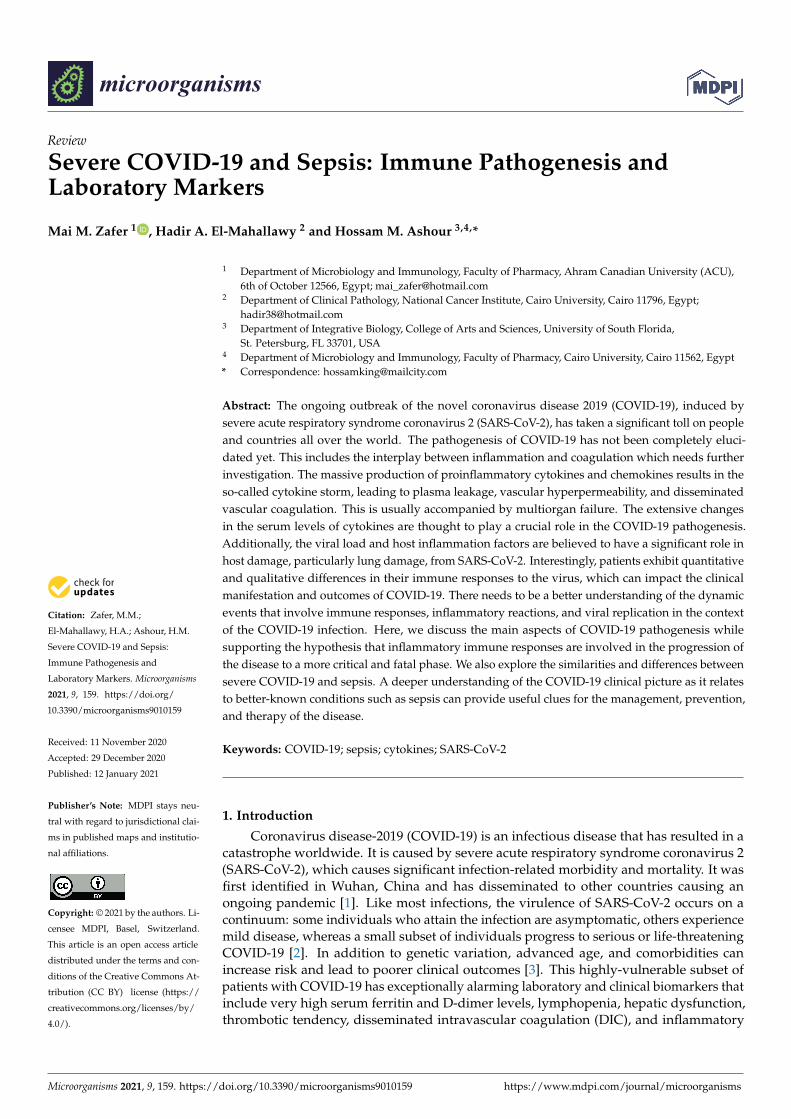
Lymphopenia is caused by severe infections and may be a sign of sepsis. Only a fewviral infections may lead to lymphopenia including advanced human immunodeficiencyvirus (HIV), influenza virus, and viral hepatitis. Other infectious agents that may cause lym-phopenia are tuberculosis, typhoid fever, Campylobacter, malaria, and histoplasmosis [54].Lymphopenia has been related to higher levels of cytokines, especially IL-6 and TNF [15].
Lymphopenia is considered to be an immunological hallmark of COVID-19 infections,but the cause of this association is not entirely clear. It is noteworthy that lymphopeniawas described in patients with Middle East respiratory syndrome (MERS), as MERS-CoVcan directly infect human primary T lymphocytes and induce intrinsic and extrinsic T-cellapoptosis [55]. Lymphopenia was also reported in patients with SARS-CoV as a resultof the involvement of soluble vascular cell adhesion molecule-1 (sVCAM-1), soluble Fasligand (sFasL), and glucocorticoids [56]. Patients with SARS-CoV experienced intensecytokine storms, which induced apoptosis in lymphocytes and monocytes in a mannersimilar to what takes place in patients with MERS [56]. In SARS-CoV-2, the underlyingmechanisms of reduced lymphocyte counts have not yet been delineated. It was recentlysuggested that the cause of lymphopenia can be the SARS-CoV-2-induced activation ofapoptosis in lymphocytes [57,58]. Alternatively, due to the cytokine storm, lymphocytesmay be recruited to the lungs (and other affected organs) leading to their depletion. Thatmight explain why lymphocyte counts are markedly reduced in patients with severeCOVID-19 [59]. Lymphocyte counts can also impact the duration of COVID-19. Lowerlymphocyte counts have been associated with a longer duration of the symptoms ofCOVID-19 [60]. Serial measurements of lymphocyte counts can be used as a prognosticmarker of patient outcomes [61]. Table 1 summarizes the results of many studies thatreported an association between different biomarker levels and severe/poor outcomes inpatients with COVID-19. These poor outcomes can lead to severe disease symptoms, higherhospital admission rates, higher rates of mortality, the need for intensive care support, andthe need for mechanical ventilation.
Table 1. Biomarkers and outcomes in patients hospitalized with COVID-19 (Reference [62]).
Biomarker Number of Studies Total Sample Size(Number of Patients)
Biomarker Associated with HigherRisk of Poor Outcomes in
COVID-19 Patients
Hematological Biomarkers
Platelets 17 3481 Thrombocytopenia
Lymphocytes 28 6449 Low lymphocyte count
Inflammatory Biomarkers
C-reactive Protein (CRP) 20 4843 Elevated CRP
Procalcitonin (PCT) 21 6031 Elevated PCT
Creatine Kinase (CK) 12 1910 Elevated CK
Metabolic Biomarkers
Aspartate Aminotransferase (AST) 32 6383 Elevated AST
Alanine Aminotransferase (ALT) 13 6019 Elevated ALT
Creatinine 19 3635 Elevated creatinine
Lactate Dehydrogenase (LDH) 18 5394 Elevated LDH
Coagulation Biomarkers
D-Dimer 16 4862 Elevated D-dimer

Microorganisms 2021, 9, 159 8 of 13
6. Immune Responses in COVID-19 and Sepsis
Many questions pertinent to the immunopathogenesis of COVID-19 have yet to beanswered. These include the following questions and many others. Can someone bere-infected with COVID-19? How long are the COVID-19 antibodies protective for? Canwe estimate the disease progression and clinical outcomes of patients with COVID-19 byanalyzing their immune profiles shortly after admission? There is currently not enoughevidence to answer any of these questions.
Chu et al. [63] compared the effects of SARS-CoV and SARS-CoV-2 on human lungexplants and revealed that SARS-CoV-2 had a better capacity to replicate in pulmonarytissues and that both viruses can equally infect type-I and type-II pneumocytes and alveolarmacrophages. They demonstrated that SARS-CoV-2 failed to induce IFN-I, IFN-II, andIFN-III, but induced the expression of five other cytokines (IL-6, MCP1, CXCL1, CXCL5,and CXCL10/IP10) [63]. The findings of another study conducted by Blanco-Melo et al.demonstrated that SARS-CoV-2 induced an immune reaction characterized by reduced IFN-I and IFN-III responses and significant induction of various proinflammatory chemokines,IL-1β, IL-6, TNF, and IL1Rα [64].
After viruses enter the host cells, they are recognized by pattern recognition recep-tors (PRRs), which include Toll-like receptors (TLRs) such as TLR7 and TLR8 [64]. Onceengaged, these innate immune receptors can induce interferon regulatory factor (IRF),NF-κB, and AP-1, resulting in the production of the Type-I and -III antiviral interferons andother chemokines [65]. These chemokines attract innate cells such as polymorphonuclearleukocytes, NK cells, monocytes, and dendritic cells [66]. These can, in turn, generateMIG, IP-10, MCP-1, and other chemokines that can recruit lymphocytes that are capableof recognizing antigen presentation of the viral antigens [66]. Importantly, the transitionbetween innate and adaptive immune responses appears to be a serious factor for theclinical progression of SARS-CoV-2 infections. Patients with severe COVID-19 show ex-treme cytokine storms involving the overexpression of IL-2 and IL-6 [67]. Available dataindicate a vigorous innate response followed by an inappropriate switch to the adaptiveresponse, which results in immune system exhaustion during the SARS-CoV-2 diseaseprogression [68]. In sepsis, the functional plasticity of monocytes as they switch from aproinflammatory to an immunosuppressive phenotype has been demonstrated [69]. Lym-phocyte exhaustion was also reported in sepsis [70]. Thus, T-cell exhaustion is a hallmarkthat both sepsis and COVID-19 have in common. These effects can be attributed to theactivation of immune checkpoints (ICs) and their ligands, such as the programmed death-1(PD-1) and PD-L1 axis.
The protective response in COVID-19 is T-cell dependent, with the CD4+ T helper cellsproviding help for B cells to secrete their specific neutralizing antibodies, and the cytotoxicCD8+ T cells capable of directly clearing viral infected cells. It is noteworthy that 80% ofthe infiltrating cells in COVID-19 are CD8+ T cells [71]. Other key relevant changes in theimmune system in the COVID-19 context include lymphocytopenia and a modulation intotal neutrophils. Reported data suggest an association between lymphocytopenia andneutrophilia with COVID-19 disease severity and death [72,73]. An evident decline inlymphocytes has been reported [7,29,59]. In addition, there was a reported decrease in thenumbers of monocytes, eosinophils, and basophils [73] in patients with COVID-19 whoare critically ill (Table 2).

Microorganisms 2021, 9, 159 9 of 13
Table 2. Correlation between immune cells and patients with severe COVID-19 (Reference [74]).
Immune Cells Number ofStudies
Total Sample Size(Number of Patients) Comments
White Blood Cells 25 4278 Significant increase in white blood cell count insevere COVID-19
Neutrophils 182446
(758 severe and1688 non-severe cases)
Significant increase in neutrophil count insevere COVID-19
T Cells 7 637
T cell responses are critical for the clearanceof COVID-19
Delayed T cell response leads to uncontrolledviremia, which drives stronger T cell responses that
could aggravate tissue damage
Cytotoxic T Cells 7 637
Functional impairment is observed in severeCOVID-19 patients
CD8+ T cells express more inhibitory receptors insevere cases
Monocytes 7 1128 Activation of blood monocytes was detected in theperipheral blood of patients with severe COVID-19
7. Final Remarks on COVID-19 and Sepsis
Sepsis is a life-threatening organ dysfunction that results from dysregulated hostresponses to infection and impaired immune homeostasis [75]. Sepsis has high mortalityrates and can be caused by bacteria, fungi, or viruses such as SARS-CoV-2, the causativeagent of COVID-19 [75]. The clinical features of sepsis include uncontrolled systemicinflammation that is characterized by a storm of inflammation associated with the releaseof proinflammatory and anti-inflammatory biomarkers such as IL-6, IL-1, TNF-α, PCT,and CRP [30]. The pathogenesis of sepsis can be explained by the continued activation ofneutrophils, macrophages and monocytes due to the inappropriate regulation of immuneresponses and physiological reactions in response to an infection. In addition, there isdelayed apoptosis of neutrophils, and enhanced necrosis of cells and tissues. The resultingdysregulated immune response is a consequence of the close correlation between the coag-ulation system and the inflammatory response [76]. Severe COVID-19 patients requiringaggressive intensive support frequently present with MOF that involves hypotension andshock, acute respiratory failure, acute kidney injury, and coagulation abnormalities.
There are striking similarities between patients with COVID-19 and those with sepsis.In both cases, cytokines are key players in the hyperinflammation [76]. In both cases,the inflammation leads to the activation of the coagulation cascade, which can cause theactivation of the fibrinolytic system [77]. Abnormal coagulation function is a prominentfeature in severe COVID-19 cases. This includes increased D-dimer and fibrinogen andthe occurrence of venous and arterial thromboembolic events. Possible causes includea direct attack of the virus on the endothelial cells via ACE-2 receptors, cytokine storm,or activation of the coagulation system. Surprisingly, disseminated intravascular coagu-lation has also been reported in patients with COVID-19 [78]. However, DIC associatedwith COVID-19 has a different pathophysiology than that of septic DIC. The COVID-19-associated DIC is characterized by an enhanced fibrinolytic system, whereas septic DICis a DIC with suppressed fibrinolysis [78]. Thus, coagulopathies accompanying severeCOVID-19 can cause thromboembolism, but can also go to the other extreme of induc-ing DIC with a different underlying mechanism than that of sepsis. This might explainthe limitations of D-dimer in detecting all causes of COVID-19-associated coagulationabnormalities. Increased coagulation and elevated D-dimer levels were associated withpoor prognosis in patients with COVID-19 [51]. Additionally, a substantial proportion ofpatients who developed sepsis and severe COVID-19 had comorbidities, including diabetes
Microorganisms 2021, 9, 159 10 of 13
and chronic lung disease [79]. Patients presenting with bacterial sepsis and patients withCOVID-19 had similar patterns of MOF. Since SARS-CoV-2 is an infectious pathogen, it isreasonable to conclude that severe COVID-19 is sepsis that is caused by SARS-CoV-2 ratherthan an uncomplicated inflammatory process [75]. Therapies that prove to be efficaciousagainst COVID-19 should also be successful in treating sepsis through the reduction ofthe inflammatory response and the anticoagulant effect. Since COVID-19 is a multisystemdisease of very high complexity, multiple treatments can be necessary. For example, a com-bination of remdesivir (to avoid viral replication), cytokine blocking agents (to reduce theinflammatory response), and anticoagulants (to control thrombi formation) can prove tobe efficient.
8. Conclusions
Although most patients infected with SARS-CoV-2 develop mild symptoms or arecompletely asymptomatic, severe cases gain wider attention, especially with the highlycontagious ability of this virus [80]. In a manner similar to sepsis, a dysregulated immuneresponse is responsible for the cascade of events occurring in severe COVID-19 cases.This results in a dynamic process that leads to the activation of the adaptive immunesystem including T lymphocytes and B lymphocytes, which can ultimately lead to celland tissue necrosis and organ dysfunction. As mentioned in this review, a surge ofinflammatory cytokines in sepsis and COVID-19 has been reported. It is of note thatferritin is elevated in both COVID-19 and sepsis. It is evident that hyperferritinemia is amarker of immune dysregulation and uncontrolled inflammation. Although lymphopeniais a common finding in both COVID-19 and sepsis, the immune pathology responsiblefor COVID-19 lymphopenia is different than that in sepsis. As the coagulation systemis linked to the inflammatory process, coagulation abnormalities are manifested in bothsevere sepsis and critically-ill patients with COVID-19. The coagulation abnormalitiesaccompanying severe COVID-19 manifest a different underlying mechanism than that ofsepsis. Thus, it appears that SARS-CoV-2 may possess a unique immune pathology. A morecomprehensive understanding of the parameters involved in the development of seriousclinical complications (including high disease severity and lethal outcomes) in patientswith COVID-19 might improve disease progression, lower the existing burden on healthcare facilities, and help us identify better therapeutic solutions. Finally, the lessons learnedfrom this pandemic must be implemented in order to make us better prepared for anyfuture attacks by coronaviruses or any other viruses.
Author Contributions: M.M.Z., H.A.E.-M., and H.M.A. designed, planned, structured, wrote, andrevised the manuscript. The final version has been approved by all authors.
Funding: This research received no external funding.
Institutional Review Board Statement: Not applicable.
Informed Consent Statement: Not applicable.
Data Availability Statement: Data sharing is not applicable to this article.
Conflicts of Interest: The authors declare no conflict of interest.
References1. Sohrabi, C.; Alsafi, Z.; O’Neill, N.; Khan, M.; Kerwan, A.; Al-Jabir, A.; Iosifidis, C.; Agha, R. World Health Organization declares
global emergency: A review of the 2019 novel coronavirus (COVID-19). Int. J. Surg. 2020, 76, 71–76. [CrossRef] [PubMed]2. Ayres, J.S. A metabolic handbook for the COVID-19 pandemic. Nat. Metab. 2020, 2, 572–585. [CrossRef] [PubMed]3. Wingfield, T.; Cuevas, L.E.; MacPherson, P.; Millington, K.A.; Squire, S.B. Tackling two pandemics: A plea on World Tuberculosis
Day. Lancet Respir. Med. 2020, 8, 536–538. [CrossRef]4. Batah, S.S.; Fabro, A.T. Pulmonary pathology of ARDS in COVID-19: A pathological review for clinicians. Respir. Med. 2020,
176, 106239. [CrossRef]5. Song, C.-Y.; Xu, J.; He, J.-Q.; Lu, Y.-Q. Immune dysfunction following COVID-19, especially in severe patients. Sci. Rep. 2020, 10,
1–11. [CrossRef]

Microorganisms 2021, 9, 159 11 of 13
6. Rouse, B.T.; Sehrawat, S. Immunity and Immunopathology to Viruses: What Decides the Outcome? Rev. Immunol. 2010, 7,514–526.
7. Huang, C.; Wang, Y.; Li, X.; Ren, L.; Zhao, J.; Hu, Y.; Zhang, L.; Fan, G.; Xu, J.; Gu, X.; et al. Clinical features of patients infectedwith 2019 novel coronavirus in Wuhan, China. Lancet 2020, 395, 497–506. [CrossRef]
8. Li, W.; Moore, M.J.; Vasilieva, N.; Sui, J.; Wong, S.K.; Berne, M.A.; Somasundaran, M.; Sullivan, J.L.; Luzuriaga, K.;Greenough, T.C.; et al. Angiotensin-converting enzyme 2 is a functional receptor for the SARS coronavirus. Nature 2003, 426,450–454. [CrossRef]
9. Walls, A.C.; Park, Y.-J.; Tortorici, M.A.; Wall, A.; McGuire, A.T.; Veesler, D. Structure, Function, and Antigenicity of the SARS-CoV-2 Spike Glycoprotein. Cell 2020, 181, 281–292.e6. [CrossRef]
10. Pirofski, L.-A.; Casadevall, A. Pathogenesis of COVID-19 from the Perspective of the Damage-Response Framework. mBio2020, 11. [CrossRef]
11. Tay, M.Z.; Poh, C.M.; Rénia, L.; Macary, P.A.; Ng, L.F.P. The trinity of COVID-19: Immunity, inflammation and intervention. Nat.Rev. Immunol. 2020, 20, 363–374. [CrossRef] [PubMed]
12. Del Valle, D.M.; Kim-Schulze, S.; Hsin-hui, H.; Beckmann, N.D.; Nirenberg, S.; Wang, B.; Lavin, Y.; Swartz, T.; Madduri, D.;Stock, A.; et al. An Inflammatory Cytokine Signature Helps Predict COVID-19 Severity and Death. Nat. Med. 2020, 26, 1636–1643.[CrossRef] [PubMed]
13. Liu, B.; Li, M.; Zhou, Z.; Guan, X.; Xiang, Y. Can we use interleukin-6 (IL-6) blockade for coronavirus disease 2019 (COVID-19)-induced cytokine release syndrome (CRS)? J. Autoimmun. 2020, 111, 102452. [CrossRef] [PubMed]
14. Buszko, M.; Park, J.-H.; Verthelyi, D.; Sen, R.; Young, H.A.; Rosenberg, A.S. The dynamic changes in cytokine responses inCOVID-19: A snapshot of the current state of knowledge. Nat. Immunol. 2020, 21, 1146–1151. [CrossRef]
15. Diao, B.; Wang, C.; Tan, Y.; Chen, X.; Liu, Y.; Ning, L.; Chen, L.; Li, M.; Liu, Y.; Wang, G.; et al. Reduction and FunctionalExhaustion of T Cells in Patients with Coronavirus Disease 2019 (COVID-19). Front. Immunol. 2020, 11, 827. [CrossRef]
16. Casadevall, A.; Pirofski, L.-A. Host-Pathogen Interactions: Redefining the Basic Concepts of Virulence and Pathogenicity. Infect.Immun. 1999, 67, 3703–3713. [CrossRef]
17. Casadevall, A.; Pirofski, L.A. The Damage-Response Framework of Microbial Pathogenesis. Nat. Rev. Microbiol. 2003, 1, 17–24.[CrossRef]
18. Pirofski, L.A.; Casadevall, A. The Damage–Response Framework as a Tool for the Physician-Scientist to Understand thePathogenesis of Infectious Diseases. J. Infect. Dis. 2018, 218, S7–S11. [CrossRef]
19. Ju, B.; Zhang, Q.; Ge, J.; Wang, R.; Sun, J.; Ge, X.; Yu, J.; Shan, S.; Zhou, B.; Song, S.; et al. Human neutralizing antibodies elicitedby SARS-CoV-2 infection. Nat. Cell Biol. 2020, 584, 115–119. [CrossRef]
20. Mehta, P.; McAuley, D.F.; Brown, M.; Sanchez, E.; Tattersall, R.S.; Manson, J.J. COVID-19: Consider cytokine storm syndromesand immunosuppression. Lancet 2020, 395, 1033–1034. [CrossRef]
21. Raoult, D.; Zumla, A.; Locatelli, F.; Ippolito, G.; Kroemer, G. Coronavirus infections: Epidemiological, clinical and immunologicalfeatures and hypotheses. Cell Stress 2020, 4, 66–75. [CrossRef] [PubMed]
22. Li, R.; Pei, S.; Chen, B.; Song, Y.; Zhang, T.; Yang, W.; Shaman, J. Substantial undocumented infection facilitates the rapiddissemination of novel coronavirus (SARS-CoV-2). Science 2020, 368, 489–493. [CrossRef] [PubMed]
23. Casadevall, A.; Pirofski, L.-A. What Is a Host? Attributes of Individual Susceptibility. Infect. Immun. 2017, 86. [CrossRef][PubMed]
24. Ejaz, H.; Alsrhani, A.; Zafar, A.; Javed, H.; Junaid, K.; Abdalla, A.E.; Abosalif, K.O.; Ahmed, Z.; Younas, S. COVID-19 andcomorbidities: Deleterious impact on infected patients. J. Infect. Public Health 2020, 13, 1833–1839. [CrossRef]
25. Henderson, L.A.; Canna, S.; Schulert, G.S.; Volpi, S.; Lee, P.Y.; Kernan, K.F.; Caricchio, R.; Mahmud, S.; Hazen, M.M.;Halyabar, O.; et al. On the Alert for Cytokine Storm: Immunopathology in COVID-19. Arthritis Rheumatol. 2020, 72, 1059–1063.[CrossRef]
26. Atal, S.; Fatima, Z. IL-6 Inhibitors in the Treatment of Serious COVID-19: A Promising Therapy? Pharm. Med. 2020, 34, 223–231.[CrossRef]
27. Stone, J.H.; Frigault, M.J.; Serling-Boyd, N.J.; Fernandes, A.D.; Harvey, L.; Foulkes, A.S.; Horick, N.K.; Healy, B.C.; Shah, R.;Bensaci, A.M.; et al. Efficacy of Tocilizumab in Patients Hospitalized with Covid-19. N. Engl. J. Med. 2020, 383, 2333–2344.[CrossRef]
28. Yao, X.H.; Li, T.Y.; He, Z.C.; Ping, Y.F.; Liu, H.W.; Yu, S.C.; Mou, H.M.; Wang, L.H.; Zhang, H.R.; Fu, W.J.; et al. A pathologicalreport of three COVID-19 cases by minimally invasive autopsies. Chin. J. Pathol. 2020, 49, E009.
29. Xu, Z.; Shi, L.; Wang, Y.; Zhang, J.; Huang, L.; Zhang, C.; Liu, S.; Zhao, P.; Liu, H.; Zhu, L.; et al. Pathological findings ofCOVID-19 associated with acute respiratory distress syndrome. Lancet Respir. Med. 2020, 8, 420–422. [CrossRef]
30. Bellinvia, S.; Edwards, C.J.; Schisano, M.; Banfi, P.; Fallico, M.; Murabito, P. The unleashing of the immune system in COVID-19 andsepsis: The calm before the storm? Inflamm. Res. 2020, 69, 757–763. [CrossRef]
31. Weiskopf, D.; Schmitz, K.S.; Raadsen, M.P.; Grifoni, A.; Okba, N.M.; Endeman, H.; Akker, J.P.V.D.; Molenkamp, R.; Koopmans, M.;Van Gorp, E.C.M.; et al. Phenotype and kinetics of SARS-CoV-2-specific T cells in COVID-19 patients with acute respiratorydistress syndrome. Sci. Immunol. 2020, 5, 2071. [CrossRef] [PubMed]

Microorganisms 2021, 9, 159 12 of 13
32. Wilk, A.J.; Rustagi, A.; Zhao, N.Q.; Roque, J.; Martínez-Colón, G.J.; McKechnie, J.L.; Ivison, G.T.; Ranganath, T.; Vergara, R.;Hollis, T.; et al. A single-cell atlas of the peripheral immune response in patients with severe COVID-19. Nat. Med. 2020, 26,1070–1076. [CrossRef] [PubMed]
33. Saeidi, A.; Zandi, K.; Cheok, Y.Y.; Saeidi, H.; Wong, W.F.; Lee, C.Y.Q.; Cheong, H.C.; Yong, Y.K.; Larsson, M.; Shankar, E.M. T-cellexhaustion in chronic infections: Reversing the state of exhaustion and reinvigorating optimal protective immune responses.Front. Immunol. 2018, 9, 2569. [CrossRef] [PubMed]
34. Behrens, E.M. Cytokines in Cytokine Storm Syndrome. In Cytokine Storm Syndrome; Springer: Berlin/Heidelberg, Germany, 2019;pp. 197–207.
35. Chousterman, B.; Swirski, F.K.; Weber, G.F. Cytokine storm and sepsis disease pathogenesis. Semin. Immunopathol. 2017, 39,517–528. [CrossRef]
36. Murthy, H.; Iqbal, M.; Chavez, J.C.; Kharfan-Dabaja, M.A. Cytokine Release Syndrome: Current Perspectives. Immunotargets Ther.2019, 8, 43–52. [CrossRef]
37. Mangalmurti, N.; Hunter, C. Cytokine Storms: Understanding COVID-19. Immunity 2020, 53, 19–25. [CrossRef]38. Shimabukuro-Vornhagen, A.; Gödel, P.; Subklewe, M.; Stemmler, H.J.; Schlößer, H.A.; Schlaak, M.; Kochanek, M.; Böll, B.; Von
Bergwelt-Baildon, M.S. Cytokine release syndrome. J. Immunother. Cancer 2018, 6, 56. [CrossRef]39. Rodriguez-Morales, A.J.; Cardona-Ospina, J.A.; Gutiérrez-Ocampo, E.; Villamizar-Peña, R.; Holguin-Rivera, Y.; Escalera-
Antezana, J.P.; Alvarado-Arnez, L.E.; Bonilla-Aldana, D.K.; Franco-Paredes, C.; Henao-Martinez, A.F.; et al. Clinical, Laboratoryand Imaging Features of COVID-19: A Systematic Review and Meta-Analysis. Travel Med. Infect Dis. 2020, 34. [CrossRef]
40. Lippi, G.; Plebani, M. Laboratory abnormalities in patients with COVID-2019 infection. Clin. Chem. Lab. Med. 2020, 58, 1131–1134.[CrossRef]
41. Di Castelnuovo, A.; Bonaccio, M.; Costanzo, S.; Gialluisi, A.; Antinori, A.; Berselli, N.; Blandi, L.; Bruno, R.; Cauda, R.;Guaraldi, G.; et al. Common cardiovascular risk factors and in-hospital mortality in 3894 patients with COVID-19: Survivalanalysis and machine learning-based findings from the multicentre Italian CORIST Study. Nutr. Metab. Cardiovasc. Dis. 2020, 30,1899–1913. [CrossRef]
42. Violi, F.; Cangemi, R.; Romiti, G.F.; Ceccarelli, G.; Oliva, A.; Alessandri, F.; Pirro, M.; Pignatelli, P.; Lichtner, M.; Carraro, A.; et al.Is Albumin Predictor of Mortality in COVID-19? Antioxid. Redox Signal. 2020, 2020, 8142. [CrossRef]
43. Lippi, G.; Plebani, M. The Critical Role of Laboratory Medicine during Coronavirus Disease 2019 (COVID-19) and Other ViralOutbreaks. Clin. Chem. Lab. Med. 2020, 58, 1063–1069. [CrossRef] [PubMed]
44. Henry, B.M.; De Oliveira, M.H.S.; Benoit, S.; Plebani, M.; Lippi, G. Hematologic, biochemical and immune biomarker abnormalitiesassociated with severe illness and mortality in coronavirus disease 2019 (COVID-19): A meta-analysis. Clin. Chem. Lab. Med.2020, 58, 1021–1028. [CrossRef] [PubMed]
45. Vargas-Vargas, M.; Cortés-Rojo, C. Ferritin Levels and COVID-19. Rev Panam Salud Publica. 2020, 44, e72. [CrossRef] [PubMed]46. Zhou, F.; Yu, T.; Du, R.; Fan, G.; Liu, Y.; Liu, Z.; Xiang, J.; Wang, Y.; Song, B.; Gu, X.; et al. Clinical course and risk factors
for mortality of adult inpatients with COVID-19 in Wuhan, China: A retrospective cohort study. Lancet 2020, 395, 1054–1062.[CrossRef]
47. Liu, T.; Zhang, J.; Yang, Y.; Zhang, L.; Ma, H.; Li, Z.; Zhang, J.; Cheng, J.; Zhang, X.; Wu, G.; et al. The Potential Role of IL-6 inMonitoring Coronavirus Disease 2019. SSRN Electron. J. 2020. [CrossRef]
48. Kernan, K.F.; Carcillo, J.A. Hyperferritinemia and inflammation. Int. Immunol. 2017, 29, 401–409. [CrossRef]49. Paliogiannis, P.; Mangoni, A.A.; Dettori, P.; Nasrallah, G.K.; Pintus, G.; Zinellu, A. D-Dimer Concentrations and COVID-19 Sever-
ity: A Systematic Review and Meta-Analysis. Front. Public Health 2020, 8, 432. [CrossRef]50. Olson, J.D. D-dimer: An Overview of Hemostasis and Fibrinolysis, Assays, and Clinical Applications. In Advances in Clinical
Chemistry; Academic Press Inc.: Cambridge, MA, USA, 2015; Volume 69, pp. 1–46.51. Tang, N.; Li, D.; Wang, X.; Sun, Z. Abnormal Coagulation parameters are associated with poor prognosis in patients with novel
coronavirus pneumonia. J. Thromb. Haemost. 2020, 18, 844–847. [CrossRef]52. Yao, Y.; Cao, J.; Wang, Q.; Shi, Q.; Liu, K.; Luo, Z.; Chen, X.; Chen, S.; Yu, K.; Huang, Z.; et al. D-dimer as a biomarker for disease
severity and mortality in COVID-19 patients: A case control study. J. Intensiv. Care 2020, 8, 1–11. [CrossRef]53. Sakka, M.; Connors, J.; Hékimian, G.; Martin-Toutain, I.; Crichi, B.; Colmegna, I.; Bonnefont-Rousselot, D.; Farge, D.; Frère, C.
Association between D-Dimer levels and mortality in patients with coronavirus disease 2019 (COVID-19): A systematic reviewand pooled analysis. JMV-J. Médecine Vasc. 2020, 45, 268–274. [CrossRef] [PubMed]
54. Herbinger, K.-H.; Hanus, I.; Beissner, M.; Berens-Riha, N.; Kroidl, I.; Von Sonnenburg, F.; Löscher, T.; Hoelscher, M.;Nothdurft, H.D.; Schunk, M. Lymphocytosis and Lymphopenia Induced by Imported Infectious Diseases: A ControlledCross-Sectional Study of 17,229 Diseased German Travelers Returning from the Tropics and Subtropics. Am. J. Trop. Med. Hyg.2016, 94, 1385–1391. [CrossRef] [PubMed]
55. Chu, H.; Zhou, J.; Wong, B.H.-Y.; Li, C.; Chan, J.F.-W.; Cheng, Z.-S.; Yang, D.; Wang, D.; Lee, A.C.-Y.; Li, C.; et al. Middle EastRespiratory Syndrome Coronavirus Efficiently Infects Human Primary T Lymphocytes and Activates the Extrinsic and IntrinsicApoptosis Pathways. J. Infect. Dis. 2016, 213, 904–914. [CrossRef]
56. Chan, P.K.; Chen, G.G. Mechanisms of Lymphocyte Loss in SARS Coronavirus Infection. Hong Kong Med. J. 2008, 14, 21–26.[PubMed]

Microorganisms 2021, 9, 159 13 of 13
57. Xiong, Y.; Liu, Y.; Cao, L.; Wang, D.; Guo, M.; Jiang, A.; Guo, D.; Hu, W.; Yang, J.; Tang, Z.; et al. Transcriptomic characteristicsof bronchoalveolar lavage fluid and peripheral blood mononuclear cells in COVID-19 patients. Emerg. Microbes Infect. 2020, 9,761–770. [CrossRef] [PubMed]
58. Pontelli, M.C.; Castro, I.A.; Martins, R.B.; Veras, F.P.; La Serra, L.; Nascimento, D.C.; Cardoso, R.S.; Rosales, R.; Lima, T.M.;Souza, J.P.; et al. Infection of human lymphomononuclear cells by SARS-CoV-2. bioRxiv 2020. [CrossRef]
59. Zheng, M.; Gao, Y.; Wang, G.; Song, G.; Liu, S.; Sun, D.; Xu, Y.; Tian, Z. Functional exhaustion of antiviral lymphocytes inCOVID-19 patients. Cell. Mol. Immunol. 2020, 17, 533–535. [CrossRef]
60. D’Ardes, D.; Pontolillo, M.; Esposito, L.; Masciarelli, M.; Boccatonda, A.; Rossi, I.; Bucci, M.; Guagnano, M.T.; Claudio, U.;Santilli, F.; et al. Duration of COVID-19: Data from an Italian Cohort and Potential Role for Steroids. Microorganisms 2020, 8, 1327.[CrossRef]
61. Wang, D.; Hu, B.; Hu, C.; Zhu, F.; Liu, X.; Zhang, J.; Wang, B.; Xiang, H.; Cheng, Z.; Xiong, Y.; et al. Clinical Characteristics of138 Hospitalized Patients With 2019 Novel Coronavirus–Infected Pneumonia in Wuhan, China. JAMA 2020, 323, 1061–1069.[CrossRef]
62. Malik, P.; Patel, U.; Mehta, D.; Patel, N.; Kelkar, R.; Akrmah, M.; Gabrilove, J.L.; Sacks, H. Biomarkers and Outcomes ofCOVID-19 Hospitalisations: Systematic Review and Meta-Analysis. BMJ Evid.-Based Med. 2020. [CrossRef]
63. Chu, H.; Chan, J.F.; Wang, Y.; Yuen, T.T.; Chai, Y.; Hou, Y.; Shuai, H.; Yang, D.; Hu, B.; Huang, X.; et al. Comparative Replicationand Immune Activation Profiles of SARS-CoV-2 and SARS-CoV in Human Lungs: An Ex Vivo Study with Implications for thePathogenesis of COVID-19. Clin. Infect Dis. 2020, 71, 1400–1409. [CrossRef] [PubMed]
64. Blanco-Melo, D.; Nilsson-Payant, B.E.; Liu, W.-C.; Uhl, S.; Hoagland, D.; Møller, R.; Jordan, T.X.; Oishi, K.; Panis, M.;Sachs, D.; et al. Imbalanced Host Response to SARS-CoV-2 Drives Development of COVID-19. Cell 2020, 181, 1036–1045.e9.[CrossRef] [PubMed]
65. Fung, T.S.; Liu, D.X. Human Coronavirus: Host-Pathogen Interaction. Annu. Rev. Microbiol. 2019, 73, 529–557. [CrossRef][PubMed]
66. García, L.F. Immune Response, Inflammation, and the Clinical Spectrum of COVID-19. Front. Immunol. 2020, 11, 1441. [CrossRef]67. Liu, J.; Li, S.; Liu, J.; Liang, B.; Wang, X.; Wang, H.; Li, W.; Tong, Q.; Yi, J.; Zhao, L.; et al. Longitudinal characteristics of lymphocyte
responses and cytokine profiles in the peripheral blood of SARS-CoV-2 infected patients. EBio Med. 2020, 55, 102763. [CrossRef]68. López-Collazo, E.; Avendaño-Ortiz, J.; Martín-Quirós, A.; Aguirre, L.A. Immune Response and COVID-19: A mirror image of
Sepsis. Int. J. Biol. Sci. 2020, 16, 2479–2489. [CrossRef]69. Shalova, I.N.; Lim, J.Y.; Chittezhath, M.; Zinkernagel, A.S.; Beasley, F.; Hernández-Jiménez, E.; Toledano, V.; Cubillos-Zapata, C.;
Rapisarda, A.; Chen, J.; et al. Human Monocytes Undergo Functional Re-programming during Sepsis Mediated by Hypoxia-Inducible Factor-1α. Immunity 2015, 42, 484–498. [CrossRef]
70. Chang, K.C.; Svabek, C.; Vazquez-Guillamet, C.; Sato, B.; Rasche, D.P.; Wilson, S.; Robbins, P.; Ulbrandt, N.D.; Suzich, J.A.;Green, J.M.; et al. Targeting the programmed cell death 1: Programmed cell death ligand 1 pathway reverses T cell exhaustion inpatients with sepsis. Crit. Care 2014, 18, R3. [CrossRef]
71. Li, G.; Fan, Y.; Lai, Y.; Han, T.; Li, Z.; Zhou, P.; Pan, P.; Wang, W.; Hu, D.; Liu, X.; et al. Coronavirus infections and immuneresponses. J. Med. Virol. 2020, 92, 424–432. [CrossRef]
72. Zhang, B.; Zhou, X.; Zhu, C.; Song, Y.; Feng, F.; Qiu, Y.; Feng, J.; Jia, Q.; Song, Q.; Zhu, B.; et al. Immune Phenotyping Based on theNeutrophil-to-Lymphocyte Ratio and IgG Level Predicts Disease Severity and Outcome for Patients With COVID-19. Front. Mol.Biosci. 2020, 7, 157. [CrossRef]
73. Qin, C.; Zhou, L.; Hu, Z.; Zhang, S.; Yang, S.; Tao, Y.; Xie, C.; Ma, K.; Shang, K.; Wang, W.; et al. Dysregulation of ImmuneResponse in Patients With Coronavirus 2019 (COVID-19) in Wuhan, China. Clin. Infect Dis. 2020, 71, 762–768. [CrossRef]
74. Feng, X.; Li, S.; Sun, Q.J.; Bo Chen, Z.; Xiong, M.; Cao, G. Immune-Inflammatory Parameters in COVID-19 Cases: A SystematicReview and Meta-Analysis. Front. Med. 2020, 7, 301. [CrossRef]
75. Yataco, A.O.; Simpson, S.Q. Coronavirus Disease 2019 Sepsis: A Nudge toward Antibiotic Stewardship. Chest 2020, 158, 1833–1834.[CrossRef]
76. Stearns-Kurosawa, D.; Osuchowski, M.F.; Valentine, C.; Kurosawa, S.; Remick, D.G. The Pathogenesis of Sepsis. Annu. Rev. Pathol.Mech. Dis. 2011, 6, 19–48. [CrossRef]
77. Bouck, E.; Denorme, F.; Holle, L.A.; Middleton, E.A.; Blair, A.M.; Laat, B.; Schiffman, J.D.; Yost, C.C.; Rondina, M.T.;Wolberg, A.S.; et al. COVID-19 and Sepsis Are Associated With Different Abnormalities in Plasma Procoagulant and Fibri-nolytic Activity. Arterioscler Thromb. Vasc. Biol. 2021, 41, 401–414. [CrossRef]
78. Beltrán-García, J.; Osca-Verdegal, R.; Pallardó, F.V.; Ferreres, J.; Rodríguez, M.; Mulet, S.; Ferrando-Sánchez, C.; Carbonell, N.;García-Giménez, J.L. Sepsis and Coronavirus Disease 2019: Common Features and Anti-Inflammatory Therapeutic Approaches.Crit. Care Med. 2020, 48, 1841–1844. [CrossRef]
79. Almalki, Z.S.; Khan, M.F.; Almazrou, S.; Alanazi, A.S.; Iqbal, M.S.; Alqahtani, A.; Alghamdi, S.; Alahmari, A.K. ClinicalCharacteristics and Outcomes Among COVID-19 Hospitalized Patients with Chronic Conditions: A Retrospective Single-CenterStudy. J. Multidiscip. Healthc. 2020, 13, 1089–1097. [CrossRef]
80. Ashour, H.M.; Elkhatib, W.F.; Rahman, M.; Elshabrawy, H.A. Insights into the recent 2019 novel coronavirus (SARS-CoV-2) inlight of past human coronavirus outbreaks. Pathogens 2020, 9, 186.


![Modelling severe Staphylococcus aureus sepsis in …...with Staphylococcus aureus sepsis [7]. The pathogenesis of sepsis-related liver dysfunction is however not well understood [2,](https://static.fdocuments.in/doc/165x107/5f591cff3f9e5c1a6f6fc6fe/modelling-severe-staphylococcus-aureus-sepsis-in-with-staphylococcus-aureus.jpg)