SEUD 2015 Prognostic factors of ART outcome in a...
15
Chloé Maignien, Resident C. Maignien, P. Santulli, D. Korb, L. Marcellin, V. Gayet, V. Blanchet, J. Gonnot, D. de Ziegler, C. Chapron Université Paris Descartes, Sorbonne Paris Cité, Faculté de médecine, AP-HP, Cochin Saint Vincent de Paul, Department of Gynecology Obstetrics II and Reproductive Medicine, Paris, France Inserm, Unité de recherche U1016 – équipe Pr F. Batteux, Institut Cochin, Paris, France SEUD 2015 Society Of Endometriosis and Uterine Disorders Prognostic factors of ART outcome in a continuous series of 359 endometriosis patients
Transcript of SEUD 2015 Prognostic factors of ART outcome in a...

Chloé Maignien, Resident
C. Maignien, P. Santulli, D. Korb, L. Marcellin, V. Gayet, V. Blanchet, J. Gonnot, D. de Ziegler, C. Chapron
Université Paris Descartes, Sorbonne Paris Cité, Faculté de médecine, AP-HP,
Cochin Saint Vincent de Paul, Department of Gynecology Obstetrics II and Reproductive Medicine, Paris, France
Inserm, Unité de recherche U1016 – équipe Pr F. Batteux, Institut Cochin, Paris,
France
SEUD 2015
Society Of Endometriosis
and Uterine Disorders
Prognostic factors of ART outcome in a continuous series of 359 endometriosis patients

Endometriosis and IVF : conflicting evidence in the literature
Hamdan et al. Obstet Gynecol (2015)
Barbosa et al. Ultrasound Obstet Gynecol (2014)
Stage I/II vs controls
Stage III/IV vs controls
Live birth (OR, 95% CI)
0.96 (0.82 - 1.12) 0.77 (0.64 – 0.92)
Clinical pregnancy (OR, 95% CI)
0.84 (0.69 – 1.03) 0.60 (0.44 – 0.81)

Endometriosis and IVF : conflicting evidence in the literature
Why?
OMA SUP DIE
SUP = superficial endometriosis; OMA = endometrioma; DIE = deep infiltrating endometriosis
Implantation theory
Impact of surgery ?
Heterogeneous populations
Impact of adenomyosis ? ! Benaglia et al. (2014): similar implantation
rates ! Vercellini et al. (2014): negative impact on
clinical pregnancy and implantation Benaglia et al. Reprod Biomed Online (2014)
Vercellini et al. Human Reprod (2014)

Objective of the study
To evaluate the IVF/ICSI outcomes in a continuous series of endometriosis
(OSIS) patients
- Clinical pregnancy
rates - Live birth
rates
Outcomes according to
the PHENOTYPE
Prognostic factors of
ART outcomes

Methods (1)
TVS = transvaginal sonography; MRI = magnetic resonance imaging; USL = uterosacral ligament
June 2005 February 2013 Observational cohort study
SUP
OMA
DIE: - USL
- Vagina - Bladder
- Intestine - Ureter
Severity
359 OSIS patients
Previous surgery for OSIS
Histological diagnosis
No history of surgery "
Imaging criteria (TVS, MRI)
Radiological diagnosis

Methods (2)
Controlled Ovarian Hyperstimulation IVF/ICSI
General characteristics:
# Age # Weight # Height # BMI # Gravidity # Parity # Previous surgery for OSIS and/or OMA
Outcomes :
# Number of oocytes # Number of embryos obtained/tranfered # Clinical pregnancies # Live birth
Infertility work-up:
# Duration of prior infertility
# Hysterosalpingography # Cycle day 3 level measurement of FSH,
LH, E2, AMH # Antral follicle count
(AFC) # Semen analysis # Adenomyosis
YES NO
Pregnancy
E2 = estradiol
OSIS phenotype

Baseline characteristics Lesion topography
N = 359
DIE 212 (59.1%) USL 67 (31.6%)
Vagina 10 (4.7%) Bladder 11 (5.2%) Intestine 118 (55.7%) Ureter 6 (2.8%)
SUP 49 (13.6%)
OMA 98 (27,3%)
R 30 (30.6%) L 36 (36.7%) BI 32 (32.7%)
ADENOMYOSIS
145 (40.4%)
ENDOMETRIOSIS
R = right; L = left; BI = bilateral
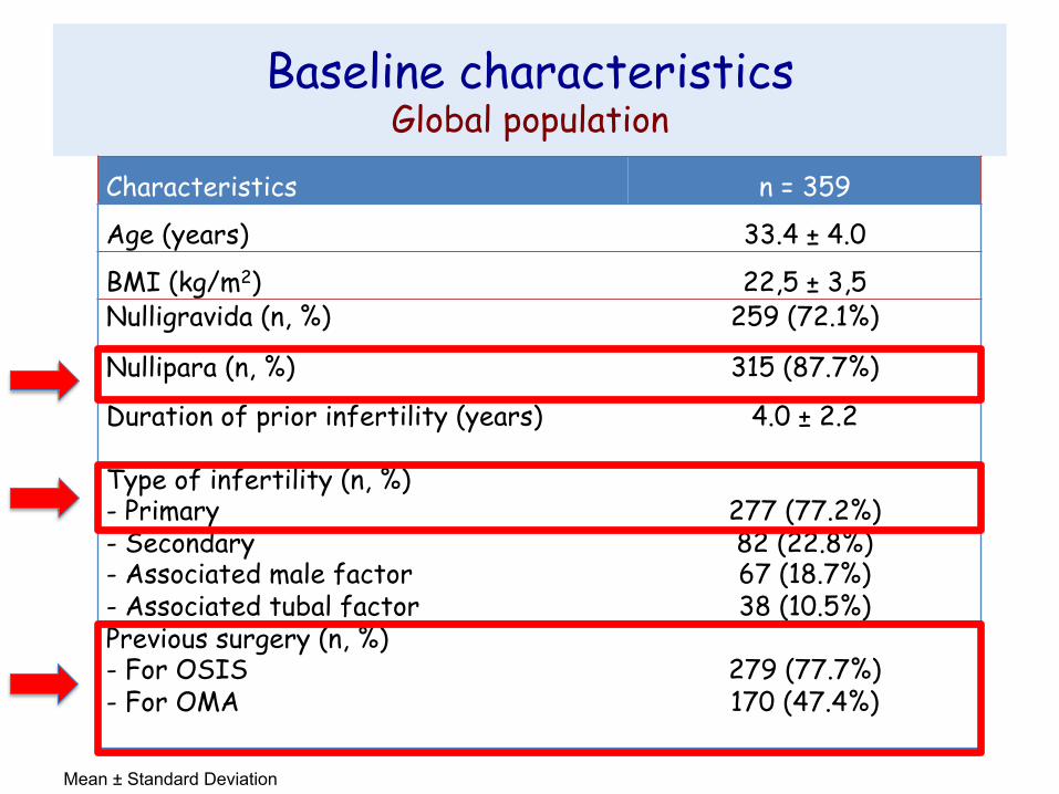
Baseline characteristics Global population
Characteristics n = 359
Age (years) 33.4 ± 4.0
BMI (kg/m2) 22,5 ± 3,5 Nulligravida (n, %) 259 (72.1%)
Nullipara (n, %) 315 (87.7%)
Duration of prior infertility (years)
4.0 ± 2.2
Type of infertility (n, %) - Primary - Secondary - Associated male factor - Associated tubal factor
277 (77.2%) 82 (22.8%) 67 (18.7%) 38 (10.5%)
Previous surgery (n, %) - For OSIS - For OMA
279 (77.7%) 170 (47.4%)
Mean ± Standard Deviation

Pregnancies Global IVF outcomes
Outcomes GLOBAL POPULATION (n = 359)
Number of cycles 720
Number of embryo transfers 500
Number of pregnant women 158 (44%)
Number of live birth 114 (31.8%)
Clinical pregnancy rate per cycle 25.3%
Clinical pregnancy rate per embryo transfer
36.4%
Implantation rate 22.7%
Live birth rate per cycle 15.8%
Live birth rate per embryo transfer 22.8%

Baseline characteristics according to the OSIS phenotype
Characteristics SUP (n = 49)
OMA (n = 98)
DIE (n = 212)
p -value
Age (years) 33.9 ± 3.6 34.1 ± 4.1 33.0 ± 4.0 0.058 BMI (kg/m2) 22.6 ± 3.7 22.0 ± 3.4 22.8 ± 3.6 0.244
Gravidity 0.5 ± 1.0 0.4 ± 0.7 0.4 ± 0.8 0.558
Parity 0.1 ± 0.3 0.1 ± 0.4 0.1 ± 0.4 0.658
Associated male F. 9 (18.4%) 25 (25.5%) 67 (18.7%) 0.113
Associated tubal F. 5 (10.2%) 6 (6.1%) 38 (10.5%) 0.212
Day 3 FSH (IU/L) 8.3 ± 5.4 7.8 ± 2.9 7.5 ± 4.4 0.554
AMH (ng/mL) 2.8 ± 1.8 3.1 ± 2.6 2.6 ± 2.1 0.281
AFC 14.0 ± 8.0 12.0 ± 6.2 11.0 ± 6.4 0.041
Adenomyosis 7 (14.3%) 18 (18.4%) 120 (56.7%) < 0.001
Previous surgery for OSIS
47 (95.9%) 79 (80.6%) 153 (72.2%) 0.212
F = factor

Pregnancies Outcomes according to the OSIS phenotype
SUP (n = 49)
OMA (n = 98)
DIE (n = 212)
p-value
Number of cycles 95 200 425
Cancellation rate 18.9% 29.5% 33.6% 0.018
Clinical pregnancy rate per cycle
30.5% 27.5% 23.1% 0.22
Clinical pregnancy rate per embryo transfer
37.7% 39% 34.8% 0.67
Implantation rate 25% 23.4% 21.6% 0.66
Abortion rate 55.2% 40% 30.6% 0.049
Live birth rate per cycle 13.7% 16.5% 16% 0.82
Live birth rate per embryo transfer
16.9% 23.4% 24.1% 0.40

Prognostic factors of ART outcomes Univariate analysis
Characteristics Women who didn’t became pregnant
Women who became pregnant
p -value
Age > 35 years 90 (44.8%) 54 (34.1%) 0.042 BMI (kg/m2) 22.8 ± 3.7 22.2 ± 3.4 0.174
Gravidity 0.4 ± 0.9 0.3 ± 0.7 0.247
DIE 129 (64.2%) 83 (52.5%) 0.026
Number of DIE lesions 1.2 ± 1.3 0.7 ± 1.0 < 0.001
Intestinal DIE 88 (43.8%) 36 (22.8%) < 0.001
Day 3 FSH (IU/L) 8.4 ± 5.2 6.8 ± 2.2 < 0.001
AMH < 2 ng/mL 103 (51.2%) 42 (26.6%) < 0.001
AFC < 10 101 (50.2%) 43 (27.2%) < 0.001
Associated Adenomyosis 91 (45.3%) 54 (34.2%) 0.033
Previous surgery for OSIS 171 (85.1%) 108 (68.4%) < 0.001
Previous surgery for OMA 108 (53.7%) 62 (39.2%) 0.006

Prognostic factors of ART outcomes Multivariate analysis
OR (95% CI ) p - value
AMH < 2 ng/mL 0.49 (0.3-0.9) 0.014
AFC < 10 0.41 (0.2-0.7) 0.002
Previous surgery for OSIS 0.29 (0.1-0.6) 0.001
Previous surgery for OMA 0.34 (0.2-0.7) 0.002
Intestinal DIE 0.31 (0.2-0.6) < 0.001

Conclusion
! IVF outcomes did not differ according to the phenotypes
! Prognostic factors associated with poorer outcomes
Intestinal DIE
Altered ovarian reserve : • AMH < 2 ng/mL • AFC < 10
Associated adenomyosis
Previous surgery for OSIS/OMA

Gynecology Surgical unit: C Chapron, B Borghese, P Santulli,
H Foulot, MC Lafay-Pillet, A Bourret, G Pierre, M Even, MC Lamau, L Marcellin, P Marzouk Medical unit: A Gompel, G Plu-Bureau, L Maitrot
Reproductive Endocrinology unit: D de Ziegler, P Santulli, V Gayet, P Piertea, FX Aubriot
Intestinal surgery B Dousset, S Gaujoux, M Leconte
Radiology AE Millischer, L Maitrot
Laboratory: Genetic D Vaiman, F Mondon, S Barbaux
Laboratory: Imunulogy
F Batteux, S Chouzenoux C Nicco, C Chéreau, B Weill
Laboratory: Reproductive biology
JP Wolf, V Lange, K Pocate, JM Kuntzman, C Chalas
Statistical unit
F Goffinet, PY Ancel
D. de Ziegler, Professor and Head, Reproductive Endocrinology and Infertility unit, A. Gompel, Professor and Head, Medical Gynecological unit,
C. Chapron, Professor and Chair, Gynecology Obstetrics II and Reproductive Medicine