Settler Describe and explain the 2 features that make an exchange surface efficient (4 marks)
23
Settler Describe and explain the 2 features that make an exchange surface efficient (4 marks)
-
Upload
beverly-paul -
Category
Documents
-
view
214 -
download
0
Transcript of Settler Describe and explain the 2 features that make an exchange surface efficient (4 marks)
Settler
Describe and explain the 2 features that make an exchange surface
efficient (4 marks)
Module 2Exchange and transport
1.2.2 Lungs

Starter Activity
• In pairs talk for 30 seconds on how to breathe

Learning Objectives Success Criteria
• Describe how the features of the lung structure that allow it to be an efficient gas exchange surface
• Outline the mechanism of breathing
• Label the lungs and each features importance(Grade E – D)
• List how the structure of the lungs allows efficient gas exchange(Grade D)
• Describe the features of an efficient exchange surface, with reference to diffusion of oxygen and carbon dioxide across the alveolus(Grade C – B)
• Outline the mechanism of breathing in mammals, with reference to the function of the ribcage, intercostal muscles and diaphragm (Grade B – A)

Trachea
Bronchiole
Pleural membrane
Diaphragm
Rib
Intercostal muscle
Bronchus
add onto your diagram briefly the importance of each structure
LUNGS •Label the lungs and each features importance
(Grade D – C)
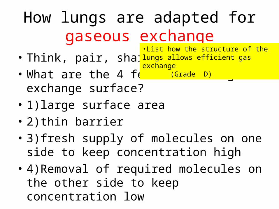
How lungs are adapted for gaseous exchange
• Think, pair, share• What are the 4 features of a good exchange
surface?• 1)large surface area• 2)thin barrier• 3)fresh supply of molecules on one side to
keep concentration high• 4)Removal of required molecules on the other
side to keep concentration low
•List how the structure of the lungs allows efficient gas exchange
(Grade D)

Gas exchange in the alveoli
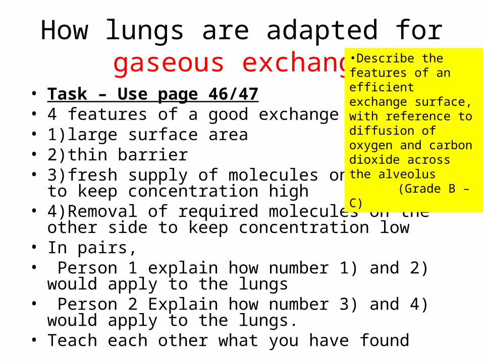
How lungs are adapted for gaseous exchange
• Task – Use page 46/47• 4 features of a good exchange surface?• 1)large surface area• 2)thin barrier• 3)fresh supply of molecules on one side to keep
concentration high• 4)Removal of required molecules on the other side to keep
concentration low• In pairs,• Person 1 explain how number 1) and 2) would apply to the
lungs • Person 2 Explain how number 3) and 4) would apply to the
lungs.• Teach each other what you have found
•Describe the features of an efficient exchange surface, with reference to diffusion of oxygen and carbon dioxide across the alveolus
(Grade B – C)

How they are adapted for exchange
• Large surface Area = more space for molecules to pass through
• Alveoli = 100-300µm• Many of them• Total surface area = 70m2
• Permeable to oxygen and Carbon Dioxide• Plasma membrane allows diffusion of both these
molecules• Thin barrier to reduce diffusion distance

How they are adapted for exchange• Maintaining the diffusion gradient• Steep diffusion gradient is needed• Achieved by the blood transport system and the ventilation
movements• Blood Transport System
– Blood brings Carbon dioxide to the lungs– Carries oxygen away
• Breathing Movements– Replace used air with fresh air– Ensures concentration of oxygen is higher than in the blood– Removes air containing carbon dioxide– Ensure concentration of carbon dioxide is lower than in the blood

What do you know?? - Structure of the lungs
Learning Objectives Success Criteria
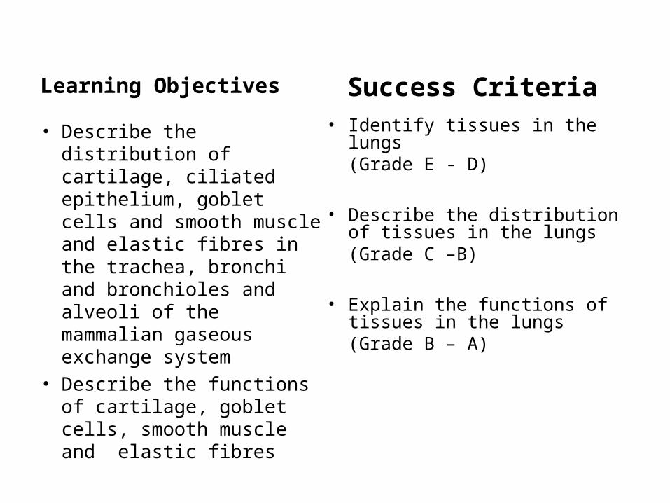
• Describe the distribution of cartilage, ciliated epithelium, goblet cells and smooth muscle and elastic fibres in the trachea, bronchi and bronchioles and alveoli of the mammalian gaseous exchange system
• Describe the functions of cartilage, goblet cells, smooth muscle and elastic fibres
• Identify tissues in the lungs(Grade E - D)
• Describe the distribution of tissues in the lungs(Grade C –B)
• Explain the functions of tissues in the lungs(Grade B – A)
Task
• Lung dissection
•Identify tissues in the lungs(Grade E - D)

Trachea
The trachea is a flexible airway supported by C- rings of cartilage which prevent the trachea
collapsing when the air pressure inside falls.Inner lining is a ciliated epithelium and goblet
cells.– The goblet cells produce mucus which
traps dirt and bacteria.– The cilia move the mucus up to the
throat where it is swallowed.

Bronchi – Similar in structure to trachea only narrower. 2 divisions of the trachea. Larger bronchi are supported by cartilage.
Bronchioles – branching subdivisions of the bronchi. Larger bronchioles may have cartilage, walls are made mainly of smooth muscle and elastic fibres. The muscle enables them to control the flow of air in and out of the alveoli.

Distribution of tissues in the lungsPart of the lung
Cartilage Smooth muscle
Elastic fibres Goblet cells epithelium
Trachea Large c-shaped pieces
Bronchi
Larger Bronchiole
Smaller Bronchiole
Smallest Bronchiole
Alveoli

This means that the rib cage must also be able to change position.
Take your hands and place them flat on your chest just above your hips on each side of your body. Now breathe in and out very deeply. Whilst you do this, watch to see what happens to your hands.
You should notice the following things…..
A mobile ribcage?

Mechanism of breathing
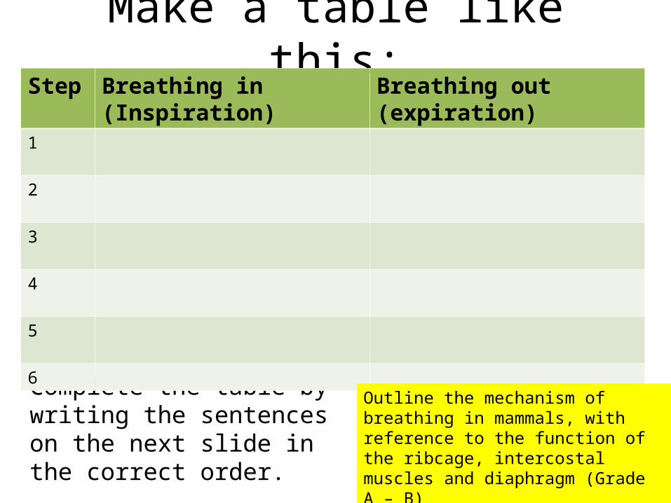
Make a table like this:
Complete the table by writing the sentences on the next slide in the correct order.
Step Breathing in (Inspiration) Breathing out (expiration)1
2
3
4
5
6
Outline the mechanism of breathing in mammals, with reference to the function of the ribcage, intercostal muscles and diaphragm (Grade A – B)
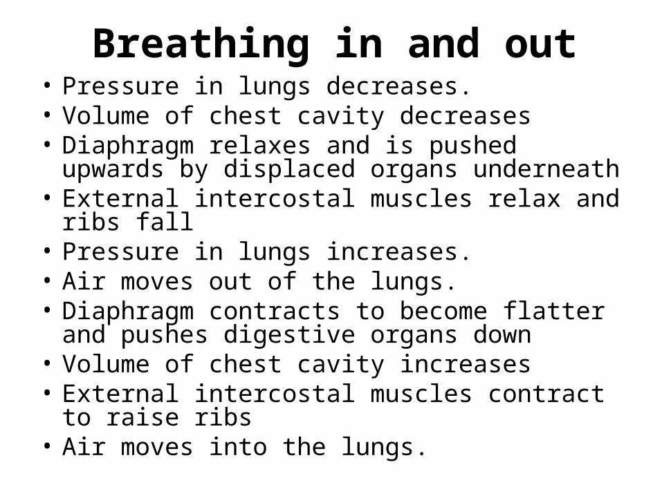
Breathing in and out• Pressure in lungs decreases.• Volume of chest cavity decreases• Diaphragm relaxes and is pushed upwards by
displaced organs underneath• External intercostal muscles relax and ribs fall• Pressure in lungs increases.• Air moves out of the lungs.• Diaphragm contracts to become flatter and
pushes digestive organs down• Volume of chest cavity increases• External intercostal muscles contract to raise ribs• Air moves into the lungs.

Breathing in and outBreathing in Breathing out
1. External intercostal muscles contract to raise ribs
1. External intercostal muscles relax and ribs fall
2. Diaphragm contracts to become flatter and pushes digestive organs down
2. Diaphragm relaxes and is pushed upwards by displaced organs underneath
3. Volume of chest cavity increases
3. Volume of chest cavity decreases
4. Pressure in lungs decreases.
4. Pressure in lungs increases.
5. Air moves into the lungs. 5. Air moves out of the lungs.

Task
• Complete questions 1-3 on p47

Plenary Activity• Alveolus• Intercostal muscles• Inspiration• Expiration• Diaphragm• Squamous Tissue• Capillary• Trachea• Bronchi• Bronchioles• Ribs• Lungs• Surfactant• Volume• Pressure


![Briefly explain how a catalyst works. Marks 2 · PDF file · 2017-03-30• Briefly explain how a catalyst works. Marks 2 . ... Calculate the rate of appearance of N 2O when [NO] =](https://static.fdocuments.in/doc/165x107/5aa28b617f8b9aa0108d559c/briefly-explain-how-a-catalyst-works-marks-2-2017-03-30-briefly-explain-how.jpg)


![[Introduction] Settler colonialism and French Algeriasro.sussex.ac.uk/id/eprint/66063/1/barclay_chopin_evans.pdfIntroduction: Settler colonialism and French Algeria Fiona Barclay,](https://static.fdocuments.in/doc/165x107/6122255653bc2c097d188695/introduction-settler-colonialism-and-french-introduction-settler-colonialism.jpg)