SESSION I –LC-MS · Analysis Metabolites Bioinformatics platform ... Time-of-Flight principle ......
27
SESSION I –LC-MS “Natural products and replication” Prof. Jacques Vervoort
-
Upload
truongliem -
Category
Documents
-
view
215 -
download
3
Transcript of SESSION I –LC-MS · Analysis Metabolites Bioinformatics platform ... Time-of-Flight principle ......

SESSION I –LC-MS
“Natural products and replication”
Prof. Jacques Vervoort

Natural productsCharacterization of the
TOMATO metabolome
•Development and optimization of
LC-hyphenated methods
LC/PDA/MS/NMR
Liquid Chromatography /
Photo Diode Array /
Mass Spectrometry /
Nuclear Magnetic Resonance
•Metabolite profiling
•Metabolite identification
•Development of Metabolite
Databases
•Comparison of varieties, tissues,
developmental stages
Genomics
Transcriptomics
Proteomics
METABOLOMICS

Chemical Complexity
• DNA A G T C
• RNA sequence-basedA G U C
• ProteinA R N D C E Q G H I L K M F P S T W Y V
• Metabolites Chlorophyll a
LycopeneChlorogenic acid
α-D-Glucose

Metabolome

� NMR�
“Chemical fingerprint”� Quantitative� Signal-to-noise
Identification
Hyphenation – integration towards
Identification
• LC-Q-TOF-PDA-MS/MS
– Fast in data acquisition
– High throughput
– Ionization
Identification
(Putative)
• Standard compounds
• Databases
• literature
•Retention time
•UV/Vis spectra
•Accurate mass -> CaHbOcNd
•Fragmentation pattern

LC-PDA-MS
Time10.00 20.00 30.00 40.00 50.00 60.00
%
0
100
Sample
preparation
Biological
system Analytical
platform
Raw
data
Multivariate
Analysis
Metabolites
Bioinformatics
platform
LC-PDA-MS method for metabolite analysis
Chemical DB’s
Literature
Species
information
…
Knowledge
platform
Sofia 0.5g FW tomato 1
m/z608 609 610 611 612 613 614 615 616 617 618 619 620 621 622 623 624 625 626 627 628 629
%
2
611.1537
612.1633
613.1693614.1746
De Vos, Moco and Lommen et al. (2007) Nat Protoc.
Autosampler
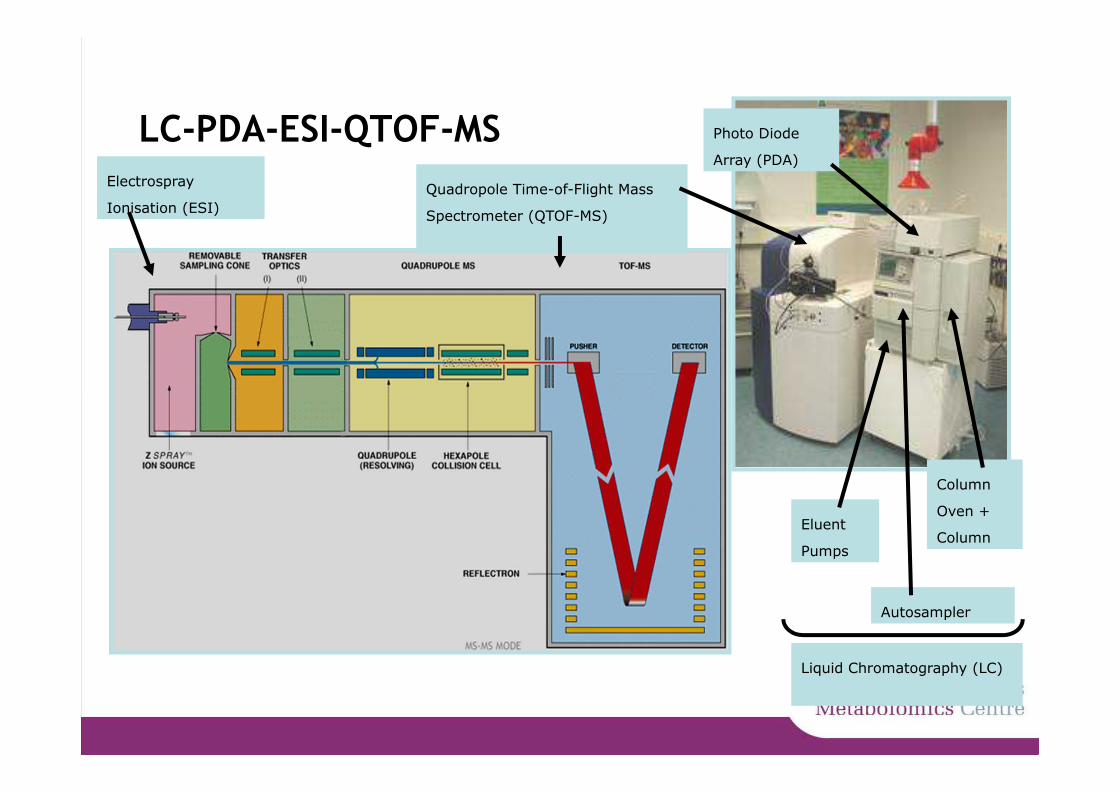
LC-PDA-ESI-QTOF-MS
Eluent
Pumps
Photo Diode
Array (PDA)
Column
Oven +
Column
Quadropole Time-of-Flight Mass
Spectrometer (QTOF-MS)
Liquid Chromatography (LC)
Electrospray
Ionisation (ESI)

Time-of-Flight principle
tD = time that ion drifts at constant velocity
D = drift distance
q = charge
E = electric field strength
sa = distance in E from average initial position of the ion to
drift region
m = mass of the ion
TOF =
Guilhaus (1995) Journal of Mass Spectrometry

Time14.00 16.00 18.00 20.00 22.00 24.00
%
0
LC-PDA-QTOF-MS analysis
• LC– Composition of extract
– Hydrophobicity
– Retention time
� UV� Spectra 240-600 nm / peak� Chromophores - λmax
� MS� Accurate mass / peak� Elemental composition: CaHbOcNd
• MS/MS
– Fragmentation
pattern
– Structural information
nm240 260 280 300 320 340 360 380 400 420 440 460 480 500 520 540 560 580 600
%
0
100
m/z608 609 610 611 612 613 614 615 616 617 618 619 620 621 622 623 624 625 626 627 628 629
%
2
611.1537
612.1633
613.1693614.1746
m/z100 150 200 250 300 350 400 450 500 550 600 650 700 750 800
%
0
100611.1582
303.0492
147.0697
465.1058
304.0555449.1131
466.1044
612.1692
613.1564
Identification
(Putative)
� Standard compounds� Databases, literature
� NMR Identification
AUTOMATION

superpeel6Q-tof
Time5.00 10.00 15.00 20.00 25.00 30.00 35.00 40.00 45.00 50.00 55.00 60.00
%
0
100
M00798 1: TOF MS ES- BPI
1.86e441.80271
26.161315
2.75191
3.20191
23.36609
21.36741
13.63163
18.06427
38.28677
29.59515
32.59433
43.261455 55.76
12153.07564
10.00 11.00 12.00 13.00 14.00 15.00 16.00 17.00 18.00 19.00
13.61163
12.83343
11.98355
11.27341
18.04427
17.66431
14.2535315.76
447
16.65353
19.08427
Ferg
lu
Cafg
lu
Cafq
uin
ic a
cid
II
Cafq
uin
ic a
cid
III
p-C
ouglu
Q-g
lu-r
ham
-apio
s
Q-g
lu-r
ham
Nch
N
Nch-g
lu-g
lu
triC
afq
uin
ic a
cid
Q-g
lu-r
ham
-apio
s-p
-coum
aric
acid
diC
afq
uin
ic a
cid
II
diC
afq
uin
ic a
cid
III
diC
afq
uin
ic a
cid
I
Cafq
uin
ic a
cid
I
LC-PDA-Q-TOF-MS analysis of compounds in tomato
peelphenyla
lanin
e
α-to
matin
α-dehydro
tom
atin
Com
pound I
I
Com
pound I
Com
pound I
II
Lycopero
sid
e A
, B o
r C
Lycoperoside F, G or
esculeoside A
Nch-g
lu
VERY P
OLAR
CO
MPO
UN
DS
VERY A
PO
LAR
CO
MPO
UN
DS
Moco et al. (2006) Plant Phys

From m/z to elemental formula to structure
• Accurate mass spectrometers
• Fast assignment of elemental formulas
• Isomers
– Compounds sharing the same EF
122 hits for C27H30O16
Dictionary of natural compoundshttp://dnp.chemnetbase.comNaringenin-6-C-glucoside Naringenin-4’-O-glucoside Naringenin-7-O-glucoside
Aim
• Develop robust methods for fast and unambiguous identification of metabolites using state-of-the-art technologies
• Two analytical methods:
– Accurate mass MSn
– HPLC-MS-SPE-NMR

1. Accurate mass MSn
• NanoMate (Advion)
– Small sample volumes
– Chip based nano-electrospray
• Ion trap (Thermo)
– Stable fragmentation
– MSn possibilities
• Orbitrap (FT-MS, Thermo)
– Wide dynamic range, high mass resolution
– Accurate mass values (within 2 ppm)

Concept of MSn spectral tree
MS1
MS2
MS3
MS4
MS5
Quercetin-3-O-(2-O-xylopyranosyl-6-O-rhamnopyranosyl)glucopyranoside
Quercetin-triglycoside
MS3 fragment 300
MS5 fragment 227
Quercetin-triglycoside
Q-triglyc MS2
MS3 fragment 300
MS4 fragment 255
MS5 fragment 226
MS4 fragment 271
MS5 fragment 227 MS5 fragment 243 MS5 fragment 271
MS3 fragment 609
File MS4 301 MS4 fragment 343
MS5 fragment 151 MS5 fragment 179 MS5 fragment 297Accurate mass
C7H3O4

Flavonoids chosen as example
• 121 structurally related compounds tested
– Share same backbone
– Different number and sites of modification
• -OH, -CH3, -C6H10O5, etc
– Many isomeric forms:
• Positional
• Stereo
hydroxygenkwanin hispidulin chrysoeriol

Unique fragmentation patterns for positional isomers
• MS3: quercetin fragment ion– major difference in fragment ratios
• 3 and 4’: m/z 179 is highest• 7: m/z 151 is highest
– due to charge localization differences
4’
3’
7
5
3
MS1
MS2
MS3
MS4
MS5
Quercetin-7-O-glucoside
Quercetin-4’-O-glucoside
Quercetin-3-O-glucoside

Spectral trees are highly reproducible
MS1
MS2
MS3
MS4
MS5O
O
OH
OH
6,4’-dihydroxyflavone
3,4’-dihydroxyflavone(4’-hydroxyflavonol)
O
O
OH
HO
OOH
HO O
OH
OH
O
OH
OH
OH
O
OH
Quercetin-3-O-glucoside
Rela
tive I
nte
nsity [
%]
100
80
60
40
20
0
C7H
5O
Positive mode0 months3 months5 months
C7H
5O
2
C8H
5O
2
C12H
9
C13H
9
C12H
11O
C13H
9O
C13H
11O
2
C14H
9O
2
C14H
10O
3
C14H
11O
3
C15H
9O
3
Rela
tive I
nte
nsity [
%]
C8H
5O
C7H
3O
3
C9H
4O
3
C14H
9O
2
C13H
7O
3
C14H
8O
3
C14H
9O
3
100
80
60
40
20
0
Negative mode0 months3 months5 months
Rela
tive I
nte
nsity [
%]
100
80
60
40
20
0
Negative modeNormalized collisionenergy:30%35%40%
C15H
8O
7
C15H
9O
7
Rela
tive I
nte
nsity [
%]
100
80
60
40
20
0
C7H
3O
4
C14H
7O
5
C14H
9O
5
C14H
7O
6
C14H
9O
6
C8H
3O
5
C9H
5O
5
C13H
5O
4
C15H
7O
6

Accurate MSn as powerful discriminative tool
• Reproducible and robust method
– in time, concentration, energy
• Provides structural information
• 121 reference compounds fragmented
– Both ionization modes
• 119 compounds gave unique MSn trees
OH
OH
HO O
OH
OH
OH
OH
HO O
OH
OH
Catechin Epicatechin

Tomato fruit as an example
• Food industry
– Fresh fruit
– Tomato sauce
• Breeding companies
– Genetic modification
– Tomato taste
– Human health

Are there still unknowns?
From Slimestad and Verheul, 2009

2. HPLC-MS-SPE-NMR
SPE cartridge
multipletimes
Trapped
-concentrated--automated-
Dry with nitrogen NMR
threshold

Peak selection
• Complex extracts
• Metabolites of interest
– Differential
• mutant vs wildtype
– Biomarker
• Mixture of compounds
**
*
*
*
*
**
*
* *
* *
****
*
*
*
*
480 490 500 510 520 530 540 550 560 570 580 590 600 610 620
m/z
0
10
20
30
40
50
60
70
80
90
100
Re
lativ
e A
bu
nd
an
ce
C23H21O13
C24H21O15
-CO2
Tomato mutant versus wildtype
m/z
Rela
tive inte
nsity

Structural information from MSn spectral trees
• Parent formula C24H21O15 [M-H]-
• In source: loss of CO2
• Fragmentation spectra:
4’
3’
7
5
3
Structural information:-COOH group (?)
-Loss of hexose
-3-O substituted quercetin
MS1
MS3
MS2
MS4
MS5
-Substituted with hexose and additional C3H2O3 group
m/z
Rel
ativ
e in
tens
ity [%
]
-C6H10O5
C15H9O7
C21H19O12
Rel
ativ
e in
tens
ity [%
]
m/z0
10
20
30
40
50
60
70
80
90
100
C7H3O4
C8H3O5
C14H7O5
C14H7O6
Rel
ativ
e in
tens
ity [%
]
m/z
Reference compound:Quercetin-3-O-substituted

Metabolite identification by NMR
Aromatic signals – correspond to 3-O-substituted quercetin
OR
OOH
HO O
OH
OH

Metabolite identification by NMR
Sugar signals – hexose identified as glucose– glucose is malonylated at 6-OH
RO OH
OH
OH
O
HO
RO OH
OH
OH
O
OH2C
HO
OO

Combination of MSn and NMR
MSn NMR
OH
OOH
HO O
OH
OH
Quercetin-3-O-(6-O-malonyl)glucoside
+
+
Hexose moiety
C3H2O3-group (including –COOH?)
OOH
HO O
OH
OH
O
OH
OH
OH
O
O
OO
HO

Conclusions
• The accurate MSn spectral tree method
– robust and reproducible method
– unique fragmentation patterns
– more structural information than MS/MS
– Worked well for flavonoids, will be tested on more plant and human metabolites
• NMR needed for final structural elucidation
– LC-MS-SPE to trap compounds in crude extract
• MSn-NMR combination: powerful approach