Serum YKL-40 is increased in patients with hepatic …labs.biology.ucsd.edu/pprice/YKL-40...
10
Copyright Q Europeon Association for the Study of the Liver 2000 Journal of Hepatology 2000; 32: 91 l-920 Printed in Denmark . AN rights reserved Mmksgaard . Copenhagen Serum YKL-40 is increased in patients with hepatic fibrosis Julia S. Johansen’, Per Christoffersen4, Soren Moller3, Paul A. Price5, Jens H Charly Garbarsch6 and Flemming Bendtsen2 Henriksen3, Department of Medicine. ‘Division of Rheumatology and 2Department of Gastroenterology, Medical Division, 3Department of Clinical Physiology and Nuclear Medicine, 4Department of Pathology, Hvidovre Hospital, and 61nstitute of Medical Anatomy Section A, The Panum Institute, University of Copenhagen, Denmark; 5Department of Biology 0368, University of California, San Diego, La Jolla, CA, USA Journal of HepatoIogy ISSN 0168-8278 BackgroundAims: YKL-40, a mammalian member of the chitinase family, is a lectin that binds heparin and chitin. The function of YKL-40 is unknown, but it may function in tissue remodelling. The aims of this study were to assess the level of circulating YKL-40 in patients with various kinds and degree of chronic liver disease and its possible relation to liver fibrosis, Metltods: Serum YKL-40 levels were determined by radioimmunoassay in 129 patients with suspected liver disease and related to histological findings and im- munohistochemical staining of YKL-40 in a liver bi- opsy taken simultaneously with the blood sample. Results: The median serum YKL-40 was highest in patients with alcoholic cirrhosis (532 yg/l), in particu- lar in patients with additional alcoholic hepatitis (740 ,@l). Patients with alcoholic cirrhosis, post-hepatitic cirrhosis (425 pg/l) and non-cirrhotic fibrosis (330 ,ugl 1) had significantly higher serum YKL-40 than normal subjects (102 pg/l), patients with fatty liver (195 ,&l) EPATIC H fibrosis is a complex and dynamic process that involves activation of cells producing matrix material, changes in the extracellular matrix compo- nents and tissue remodelling (1). Conventional bio- chemical and serological tests are of little value for di- agnosis of the degree of liver fibrosis and the activity of fibrogenesis, and percutaneous liver biopsy is therefore used to assess the extent of liver fibrosis and fibrogen- esis (1,2). However, a liver biopsy is sometimes of ques- tionable value because of the heterogeneous distri- bution of pathological changes in the liver. For years Received 25 May; revised 8 November; accepted 15 November 1999 Correspondence: Julia S. Johansen, Department of Medi- cine, Division of Rheumatology 232, Hvidovre Hospital, University of Copenhagen, Kettegard Alle 30, DK-2650 Hvidovre, Denmark. Tel: 45 36322839. Fax: 45 36471410. e-mail: [email protected] or patients with viral hepatitis without fibrosis (174 &l). Serum YKL-40 was significantly (p<O.OOl) re- lated to the degree of liver fibrosis with the highest levels in patients with moderate (466 ,ug/l) to severe (676 &l) fibrosis. Serum YKL40 was also increased (p=O.O18) in patients with slight fibrosis (270 &l) compared to patients without fibrosis. Immunohisto- chemical analysis demonstrated positive staining for YKL-40 antigen in areas with fibrosis, particularly areas with active fibrogenesis. YKL-40 staining was never found in hepatocytes. Conclusions: Our study indicates that the increased serum YKL-40 in patients with liver disease of various degree and aetiology seems to reflect fibrosis and fibrogenesis. Key words: Alcoholic liver disease; HC gp-39; Liver fibrogenesis; Liver fibrosis; YKL-40. there has been a search for biochemical or serological markers reflecting fibrotic processes in liver disease. Markers that detect patients with ongoing fibrosis at an early stage, before irreversible damage has de- veloped, would be an important addition to the clini- cian’s diagnostic and prognostic tools (l-3). YKL-405 is a mammalian member of a chitinase family (family 1 %glycosylhydrolases) (4-13). The physiological function of YKL-40 is not known, but the pattern of its expression in normal and diseased states suggests that it could function in remodelling of the extracellular matrix or in tissue inflammation (4 16). YKL-40 mRNA expression is found in human 5 The protein has been termed YKL-40 from its molecular weight (40 kDa) and the one letter code for its three N- terminal amino acids (tyrosine, lysine and leucine) (10). 911
Transcript of Serum YKL-40 is increased in patients with hepatic …labs.biology.ucsd.edu/pprice/YKL-40...

Copyright Q Europeon Association for the Study of the Liver 2000
Journal of Hepatology 2000; 32: 91 l-920 Printed in Denmark . AN rights reserved Mmksgaard . Copenhagen
Serum YKL-40 is increased in patients with hepatic fibrosis
Julia S. Johansen’, Per Christoffersen4, Soren Moller3, Paul A. Price5, Jens H
Charly Garbarsch6 and Flemming Bendtsen2 Henriksen3,
Department of Medicine. ‘Division of Rheumatology and 2Department of Gastroenterology, Medical Division, 3Department of Clinical Physiology and Nuclear Medicine, 4Department of Pathology, Hvidovre Hospital, and 61nstitute of Medical Anatomy Section A, The Panum Institute,
University of Copenhagen, Denmark; 5Department of Biology 0368, University of California, San Diego, La Jolla, CA, USA
Journal of HepatoIogy ISSN 0168-8278
BackgroundAims: YKL-40, a mammalian member of the chitinase family, is a lectin that binds heparin and chitin. The function of YKL-40 is unknown, but it may function in tissue remodelling. The aims of this study were to assess the level of circulating YKL-40 in patients with various kinds and degree of chronic liver disease and its possible relation to liver fibrosis, Metltods: Serum YKL-40 levels were determined by radioimmunoassay in 129 patients with suspected liver disease and related to histological findings and im- munohistochemical staining of YKL-40 in a liver bi- opsy taken simultaneously with the blood sample. Results: The median serum YKL-40 was highest in patients with alcoholic cirrhosis (532 yg/l), in particu- lar in patients with additional alcoholic hepatitis (740 ,@l). Patients with alcoholic cirrhosis, post-hepatitic cirrhosis (425 pg/l) and non-cirrhotic fibrosis (330 ,ugl 1) had significantly higher serum YKL-40 than normal subjects (102 pg/l), patients with fatty liver (195 ,&l)
EPATIC H fibrosis is a complex and dynamic process that involves activation of cells producing matrix
material, changes in the extracellular matrix compo- nents and tissue remodelling (1). Conventional bio- chemical and serological tests are of little value for di- agnosis of the degree of liver fibrosis and the activity of fibrogenesis, and percutaneous liver biopsy is therefore used to assess the extent of liver fibrosis and fibrogen- esis (1,2). However, a liver biopsy is sometimes of ques- tionable value because of the heterogeneous distri- bution of pathological changes in the liver. For years
Received 25 May; revised 8 November; accepted 15 November 1999
Correspondence: Julia S. Johansen, Department of Medi- cine, Division of Rheumatology 232, Hvidovre Hospital, University of Copenhagen, Kettegard Alle 30, DK-2650 Hvidovre, Denmark. Tel: 45 36322839. Fax: 45 36471410. e-mail: [email protected]
or patients with viral hepatitis without fibrosis (174 &l). Serum YKL-40 was significantly (p<O.OOl) re- lated to the degree of liver fibrosis with the highest levels in patients with moderate (466 ,ug/l) to severe (676 &l) fibrosis. Serum YKL40 was also increased (p=O.O18) in patients with slight fibrosis (270 &l) compared to patients without fibrosis. Immunohisto- chemical analysis demonstrated positive staining for YKL-40 antigen in areas with fibrosis, particularly areas with active fibrogenesis. YKL-40 staining was never found in hepatocytes. Conclusions: Our study indicates that the increased serum YKL-40 in patients with liver disease of various degree and aetiology seems to reflect fibrosis and fibrogenesis.
Key words: Alcoholic liver disease; HC gp-39; Liver fibrogenesis; Liver fibrosis; YKL-40.
there has been a search for biochemical or serological markers reflecting fibrotic processes in liver disease. Markers that detect patients with ongoing fibrosis at an early stage, before irreversible damage has de- veloped, would be an important addition to the clini- cian’s diagnostic and prognostic tools (l-3).
YKL-405 is a mammalian member of a chitinase family (family 1 %glycosylhydrolases) (4-13). The physiological function of YKL-40 is not known, but the pattern of its expression in normal and diseased states suggests that it could function in remodelling of the extracellular matrix or in tissue inflammation (4 16). YKL-40 mRNA expression is found in human
5 The protein has been termed YKL-40 from its molecular weight (40 kDa) and the one letter code for its three N- terminal amino acids (tyrosine, lysine and leucine) (10).
911
J. S. Johansen et al.
liver (5). We have recently reported in a small study of patients with chronic liver disease that plasma YKL- 40 is elevated in patients with chronic liver disease and may be related to the degree of liver fibrosis (17). Moreover, YKL-40 is released from the hepato- splanchnic system (17).
The purpose of the present study was to assess whether serum YKL-40 reflects the severity of liver fi- brosis. We measured the serum levels of YKL-40, the aminoterminal propeptide of type III procollagen (PIIINP) and hyaluronan in patients with various liver diseases. We compared the serum YKL-40 values with histological changes in liver biopsies. Especially the de- gree of fibrosis and ongoing fibrogenesis was related to serum YKL-40 and to the degree of immunohisto- chemical YKL-40 staining in the liver biopsy.
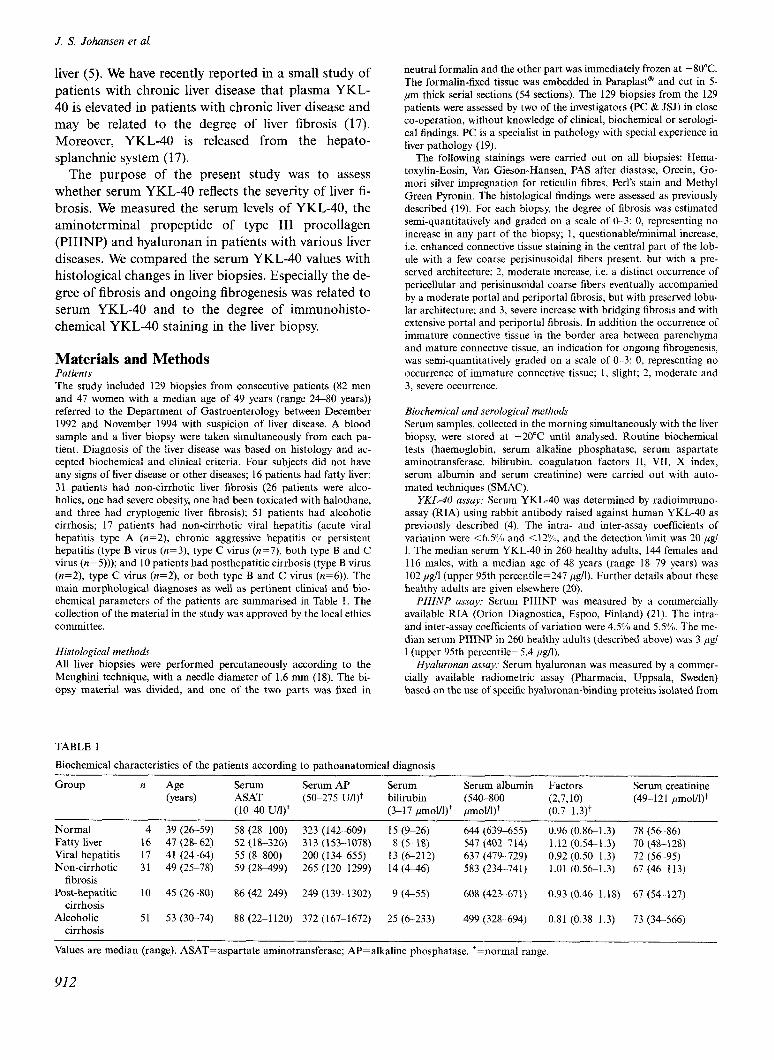
Materials and Methods Patients The study included 129 biopsies from consecutive patients (82 men and 47 women with a median age of 49 years (range 2680 years)) referred to the Department of Gastroenterology between December 1992 and November 1994 with suspicion of liver disease. A blood sample and a liver biopsy were taken simultaneously from each pa- tient. Diagnosis of the liver disease was based on histology and ac- cepted biochemical and clinical criteria. Four subjects did not have any signs of liver disease or other diseases; 16 patients had fatty liver; 31 patients had non-cirrhotic liver fibrosis (26 patients were alco- holics, one had severe obesity, one had been toxicated with halothane, and three had cryptogenic liver fibrosis); 51 patients had alcoholic cirrhosis; 17 patients had non-cirrhotic viral hepatitis (acute viral hepatitis type A (n=2), chronic aggressive hepatitis or persistent hepatitis (type B virus (n=3), type C virus (n=7), both type B and C virus (n=5))); and 10 patients had posthepatitic cirrhosis (type B virus (n=2), type C virus (n=2), or both type B and C virus (n=6)). The main morphological diagnoses as well as pertinent clinical and bio- chemical parameters of the patients are summarised in Table 1. The collection of the material in the study was approved by the local ethics committee.
Histological methods
All liver biopsies were performed percutaneously according to the Menghini technique, with a needle diameter of 1.6 mm (18). The bi- opsy material was divided, and one of the two parts was fixed in
TABLE 1
neutral formalin and the other part was immediately frozen at -80°C.
The formalin-fixed tissue was embedded in Paraplast@ and cut in 5- pm thick serial sections (54 sections). The 129 biopsies from the 129 patients were assessed by two of the investigators (PC & JSJ) in close co-operation, without knowledge of clinical, biochemical or serologi- cal findings. PC is a specialist in pathology with special experience in
liver pathology (19). The following stainings were carried out on all biopsies: Hema-
toxylin-Eosin, Van Gieson-Hansen, PAS after diastase. Orcein, Go- mori silver impregnation for reticulin fibres, Perl’s stain and Methyl Green Pyronin. The histological findings were assessed as previously described (19). For each biopsy, the degree of fibrosis was estimated semi-quantitatively and graded on a scale of G-3: 0, representing no increase in any part of the biopsy; 1, questionable/minimal increase, i.e. enhanced connective tissue staining in the central part of the lob- ule with a few coarse perisinusoidal fibers present, but with a pre- served architecture: 2, moderate increase, i.e. a distinct occurrence of
pericellular and perisinusoidal coarse fibers eventually accompanied by a moderate portal and periportal fibrosis, but with preserved lobu- lar architecture; and 3, severe increase with bridging fibrosis and with extensive portal and periportal fibrosis. In addition the occurrence of immature connective tissue in the border area between parenchyma and mature connective tissue, an indication for ongoing fibrogenesis, was semi-quantitatively graded on a scale of O-3: 0. representing no occurrence of immature connective tissue; I, slight; 2, moderate and 3, severe occurrence.
Biochemical and serological methods
Serum samples. collected in the morning simultaneously with the liver biopsy, were stored at -20°C until analysed. Routine biochemical tests (haemoglobin, serum alkaline phosphatase, serum aspartate aminotransferase. bilirubin, coagulation factors II, VII, X index, serum albumin and serum creatinine) were carried out with auto-
mated techniques (SMAC). YKL-40 assay: Serum YKL-40 was determined by radioimmuno-
assay (RIA) using rabbit antibody raised against human YKL40 as previously described (4). The intra- and inter-assay coefficients of variation were <6.5% and <12%, and the detection limit was 20 pg/ 1. The median serum YKL40 in 260 healthy adults, 144 females and 116 males, with a median age of 48 years (range 18.-79 years) was 102 ,&I (upper 95th percentile=247 ,&I). Further details about these healthy adults are given elsewhere (20).
PIZINP assay: Serum PIIINP was measured by a commercially available RIA (Orion Diagnostica, Espoo, Finland) (21). The intra- and inter-assay coefficients of variation were 4.5% and 5.5%. The me- dian serum PIIINP in 260 healthy adults (described above) was 3 pg/ 1 (upper 95th percentile=5.4 /&l).
Hyaluronan assay: Serum hyaluronan was measured by a commer- cially available radiometric assay (Pharmacia, Uppsala, Sweden) based on the use of specific hyaluronan-binding proteins isolated from
Biochemical characteristics of the patients according to pathoanatomical diagnosis
Group n -4s (years)
4 39 (26-59) 16 47 (2862) 17 41 (24-64) 31 49 (25-78)
10 45 (26-80)
51 53 (30-74)
Serum Serum AP ASAT (50-275 U/l)+ (lo-40 u/l)+
58 (288100) 323 (142609) 52 (18-326) 313 (153-1078)
55 (S-800) 200 (134-655) 59 (288499) 265 (12@1299)
86 (42-249) 249 (139-l 302)
88 (22-1120) 372 (167-1672)
Serum Serum albumin Factors bilirubin (540-800 (2,7,10) (3-17 fimoVl)+ ~molfl)+ (0.7-l .3)+
Normal Fatty liver Viral hepatitis Non-cirrhotic
fibrosis Post-hepatitic
cirrhosis Alcoholic
cirrhosis
Serum creatinine (499121 pmol/l)+
15 (9926) 8 (5-18)
13 (6212) 14 (446)
9 (455)
25 (6233)
644 (639-655) 547 (402-714) 637 (479-729) 583 (234741)
608 (423367 1)
499 (3288694)
0.96 (0.861.3) 1.12 (0.54-1.3) 0.92 (0.5& 1.3) 1.01 (0.56-1.3)
0.93 (0.46-1.18)
0.81 (0.38-1.3)
78 (5686) 70 (48-l 28) 72 (5695) 67 (46113)
67 (54-127)
73 (34566)
Values are median (range). ASAT=aspartate aminotransferase; AP=alkaline phosphatase. +=normal range
912

Serum YKL-40 and liver fibrosis
bovine cartilage. The intra- and inter-assay coefficients of variation were 10% and 8%. The median serum hyaluronan in 247 healthy adults was 28 &l (upper 95th percentile=97 @l) (22).
Immunohistochemical methods for YKL-40 Frozen samples of liver tissue were cut at 5 e and stained routinely with haematoxylin and eosin in order to establish that the tissue was well conserved and neighbouring sections were used for immunolocal- ization of YKL40 antigen using specific antisera. Prior to immuno- staining sections were methanol-fixed at -20°C for 5 min. Conven- tional alkaline phosphatase staining technique for polyclonal anti- bodies was used as previously described (17). Briefly the following steps were included (all performed at room temperature): non-specific binding was blocked by incubation for 5 min with 4% bovine serum albumin (BSA) (Sigma A-4503) in Tris buffered saline (TBS); binding of primary antibody was performed for 30 min with an affinity-puri- fied rabbit polyclonal IgG against human YKL40 diluted in TBS containing 4% BSA (IgG concentration of the YKL40 antibody was 66 &nl). Non-immune rabbit serum (Dako X936, Copenhagen, Demnark) was used as negative controls in the same IgG concen- tration of 66 &ml in TBS containing 4% BSA. The slides were then washed 3 times with TBS and incubated for 30 min with alkaline phosphatase-conjugated swine antibodies to rabbit immunoglobulins (Dako D306) diluted 1:20 in TBS containing 4% BSA, washed twice in TBS and then incubated for 10 min with 0.05 M Tris/HCl, pH 7.6, washed twice with 0.2 M Tris-HCl, pH 9.5 and then incubated for 5 min with 0.75 mg/ml levamisol (Sigma L-9756) in 0.2 M Tris-HCl, pH 9.5. The slides were stained for 20 min with Sigma FASTTM BCIP/NBT tablets (Sigma B-5655) with 0.75 mg/ml levamisol in 0.2 M Tris-HCl, pH 9.5. The colour reaction was stopped by washing in running tap water and the slides were mounted in Glycergel (Dako).
Each liver specimen was microscopically examined blindly and scored for the presence of YKL-40 expression in a scale as follows: score O=no YKL-40 staining; score 1 =scanty YKL40 staining; and score 2=moderate to intense YKL-40 staining.
Statistical analysis The statistical analyses were done with SigmaStat (SPSS, Chicago, IL, USA) and SAS@ (SAS Institute, Cary, NC, USA). Results am given as median and range. Comparison between groups was per- formed by the non-parametric Mann-Whitney rank sum test or the
Serum YKL-40 (ugIL)
5000 -j
.
Serum PMNP @g/L)
Kruskal-Wallis test for unpaired differences. Correlation analysis was based on the Spearman’s rho test. p-values less than 0.05 were con- sidered to be significant. In a multiple regression model serum YKL- 40, PIIINP and hyaluronan were logarithmically transformed and re- lated to the degree of liver fibrosis (treated as an ordinal variable with four categories) through a proportional odds regression model for ordinal data (23).
Results The individual concentrations of serum YKL40, PIIINP and hyaluronan in relation to the various liver diseases, determined by histopathological and clinical criteria, are illustrated in Fig. 1 (a, b and c) and the median levels are given in Table 2. The serum YKL- 40 levels were highest in patients with alcoholic liver cirrhosis (median 532 ,ug/l and 5-fold increased com- pared with the median level of healthy age-matched controls), posthepatitic cirrhosis (425 pg/l) and non- cirrhotic fibrosis (330 &l), and these serum YKL40 levels were significantly (p<O.Ol-p~O.001) higher than serum Y KL-40 values in age-matched controls (102 ,ug/ 1; upper 95th percentile=247 &l), in patients with fatty liver (195 &l), and in patients with chronic viral hepatitis without cirrhosis (174 fig/l). Multiple com- parison between the different groups of patients with liver disease showed that patients with alcoholic cir- rhosis had significantly higher serum YKL40, PIIINP and hyaluronan concentrations than patients with non- cirrhotic fibrosis, viral hepatitis and fatty liver (Table 2) (Kruskal-Wallis one way ANOVA on ranks with Dunn’s method: p<O.O5). A significant difference be- tween alcoholic and posthepatitic cirrhosis was only found in serum PIIINP levels.
serum Hyal”mnan WL)
5WO
1 . . 4500
.
. . . .
Fig. 1. Serum concentrations of YKL-40 (a), PIIINP (b) and hyaluronan (c) in patients with different liver diseases. The bars represent median values. The horizontal lines represent the upper limit (95th percentile) of the normal range of serum YKL-40 (247 pgll), serum PIIINP (5.4 ,ugll) and serum hyaluronan (97 pgll). (IO) patients with alcoholic cirrhosis in combination with alcoholic hepatitis and (A) patients without alcoholic hepatitis. (0) all other patients.
913
J. S. Johansen et al.
TABLE 2
Serum YKL40, PIIINP and hyaluronan concentrations in patients with different liver diseases
Serum YKL-40 Serum PIIINP
(l&l) (L&l)
Serum hyaluronan (U&?/l)
Normal 118* (105-165) 3.9* (3.0-5.0) 25* (25-36) Fatty liver 195* (50-408) 4.9* (1.7-10.1) 26* (21-93) Viral hepatitis 174* (11 l-380) 5.2% (2427.0) 30* (255508, Non-cirrhotic fibrosis 330* (115-967) 6.4* (2615.6) 54* (2552920) Posthepatitic cirrhosis 425 (145-2070) 8.4* (3.2-21.0) 127 (255894) Alcoholic cirrhosis 532 (824850) 17.6 (5.1l70.0) 258 (3&4730)
Values are medians (range). PIIINP=N-terminal propcptide of type III procollagen. Kruskal-Wallis one-way ANOVA on ranks with multiple comparisons, Dunn’s method: *p<O.O5 rs. alcoholic cirrhosis.
Serum YKL40 levels were significantly @=0.014) higher in the subset of patients with alcoholic cirrhosis who also had alcoholic hepatitis (median 740 ,&I; 30 of these 31 patients had elevated serum YKL40 (i.e. above the upper 95th percentile level of the controls, i.e. >247 ,&l)) compared with patients with alcoholic cirrhosis without hepatitis (median 338 ,ug/l; 14 of these 20 patients had elevated serum YKL-40). No dif- ferences between these two patient groups were found in serum PIIINP (17.4 vs. 19.7 ,q/l, p=O.7) or serum hyaluronan (258 vs. 238 pug/l, p=O.3). Sixteen (52%) of the 31 patients with alcoholic cirrhosis in combination with alcoholic hepatitis had severe fibrosis compared to only four (20%) of the patients with alcoholic cirrhosis without alcoholic hepatitis.
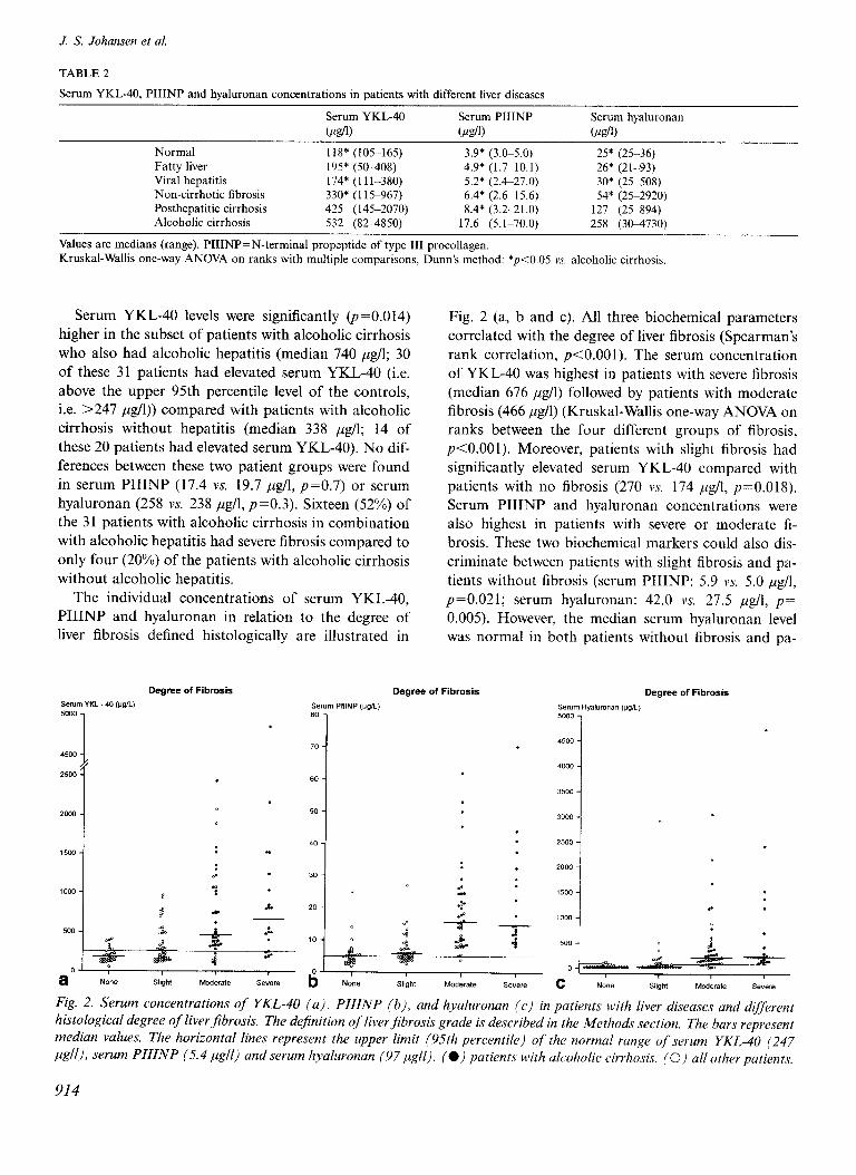
The individual concentrations of serum YKL40, PIIINP and hyaluronan in relation to the degree of liver fibrosis defined histologically are illustrated in
Degree of Fibrosis
serum YKL 40 (I@!_, 5cm 1
Fig. 2 (a, b and c). All three biochemical parameters correlated with the degree of liver fibrosis (Spearman’s rank correlation, p<O.OOl). The serum concentration of YKL-40 was highest in patients with severe fibrosis (median 676 ,q/l) followed by patients with moderate fibrosis (466 ,&l) (Kruskal-Wallis one-way ANOVA on ranks between the four different groups of fibrosis, p<O.OOl). Moreover, patients with slight fibrosis had significantly elevated serum YKL-40 compared with patients with no fibrosis (270 vs. 174 ,ug/l, p=O.OlS). Serum PIIINP and hyaluronan concentrations were also highest in patients with severe or moderate fi- brosis. These two biochemical markers could also dis- criminate between patients with slight fibrosis and pa- tients without fibrosis (serum PIIINP: 5.9 11s. 5.0 lug/l, p=O.O21; serum hyaluronan: 42.0 vs. 27.5 yg/l, p= 0.005). However, the median serum hyaluronan level was normal in both patients without fibrosis and pa-
Degree of Fibrosis Degree of Fibrosis
Fig. 2. Serum concentrations of YKL-40 (a), PIIINP (b), and hyaluronan (c) in patients with liver diseases and different histological degree of liver fibrosis. The definition of liverjibrosis grade is described in the Methods section. The bars represent median values. The horizontal lines represent the upper limit (95th percentile) qf the normal range of serum YKL-40 (247 pgll), serum PIIINP (5.4 pgll) and serum hyaluronan (97 pgll). (e) patients with alcoholic cirrhosis. (0) all other patients.
914
Serum YKL-40 and liver jibrosis
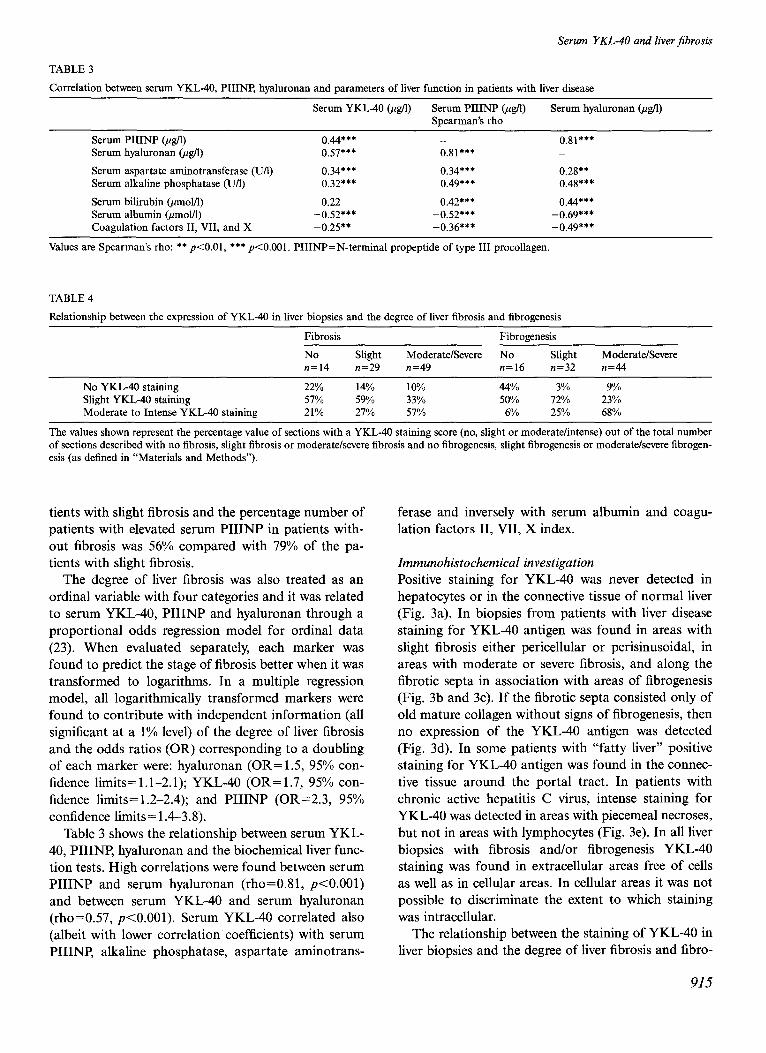
TABLE 3
Correlation between serum YKL-40, PIIINP, hyaluronan and parameters of liver function in patients with liver disease
Serum YKL40 @g/l) Serum PIIINP @g/l) Snearman’s rho
Serum hyaluronan (&l)
Serum PIIINP @g/l) 0.44*** _ o.s1*** Serum hyaluronan @g/l) 0.57*** 0.81*** _
Serum aspartate aminotransferase (U/l) 0.34*** 0.34*** 0.28** Serum alkaline phosphatase (U/l) 0.32*** 0.49*** 0.48***
Serum bilirubin (umol/l) 0.22 0.42*** 0.44*** Serum albumin @mol/l) -0.52*** -0.52*** -0.69*** Coagulation factors II, VII, and X -0.25** -0.36*** -0.49***
Values are Spearman’s rho: ** p<O.Ol, *** p<O.OOl. PIIINP=N-terminal propeptide of type III procollagen.
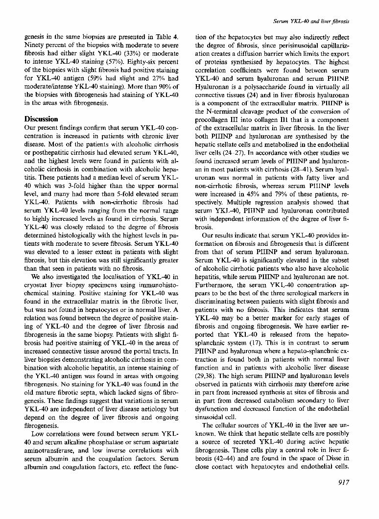
TABLE 4
Relationship between the expression of YKL40 in liver biopsies and the degree of liver fibrosis and fibrogenesis
Fibrosis Fibrogenesis
No Slight Moderate/Severe No Slight n=14 n=29 n=49 n=16 n=32
No YKL40 staining 22% 14% 10% 44% 3% Slight YKL40 staining 57% 59% 33% 50% 72% Moderate to Intense YKL-40 staining 21% 27% 57% 6% 25%
Moderate/Severe n=44
9% 23% 68%
The values shown represent the percentage value of sections with a YKL-40 staining score (no, slight or moderate/intense) out of the total number of sections described with no fibrosis, slight fibrosis or moderate/severe fibrosis and no fibrogenesis, slight fibrogenesis or moderate/severe fibrogen- esis (as defined in “Materials and Methods”).
tients with slight fibrosis and the percentage number of patients with elevated serum PIIINP in patients with- out fibrosis was 56% compared with 79% of the pa- tients with slight fibrosis.
The degree of liver fibrosis was also treated as an ordinal variable with four categories and it was related to serum YKL-40, PIIINP and hyaluronan through a proportional odds regression model for ordinal data (23). When evaluated separately, each marker was found to predict the stage of fibrosis better when it was transformed to logarithms. In a multiple regression model, all logarithmically transformed markers were found to contribute with independent information (all significant at a 1% level) of the degree of liver fibrosis and the odds ratios (OR) corresponding to a doubling of each marker were: hyaluronan (OR= 1.5, 95% con- fidence limits= 1.1-2.1); YKL-40 (OR= 1.7, 95% con- fidence limits= 1.2-2.4); and PIIINP (OR=2.3, 95% confidence limits= 1.4-3.8).
Table 3 shows the relationship between serum YKL- 40, PIIINP hyaluronan and the biochemical liver func- tion tests. High correlations were found between serum PIIINP and serum hyaluronan (rho=O.81, p<O.OOl) and between serum YKL40 and serum hyaluronan (rho=0.57, p<O.OOl). Serum YKL40 correlated also (albeit with lower correlation coefficients) with serum PIIINP, alkaline phosphatase, aspartate aminotrans-
ferase and inversely with serum albumin and coagu- lation factors II, VII, X index.
Immunohistochemical investigation
Positive staining for YKL-40 was never detected in hepatocytes or in the connective tissue of normal liver (Fig. 3a). In biopsies from patients with liver disease staining for YKL-40 antigen was found in areas with slight fibrosis either pericellular or perisinusoidal, in areas with moderate or severe fibrosis, and along the fibrotic septa in association with areas of fibrogenesis (Fig. 3b and 3~). If the fibrotic septa consisted only of old mature collagen without signs of fibrogenesis, then no expression of the YKL-40 antigen was detected (Fig. 3d). In some patients with “fatty liver” positive staining for YKL-40 antigen was found in the connec- tive tissue around the portal tract. In patients with chronic active hepatitis C virus, intense staining for YKL-40 was detected in areas with piecemeal necroses, but not in areas with lymphocytes (Fig. 3e). In all liver biopsies with fibrosis and/or fibrogenesis YKL40 staining was found in extracellular areas free of cells as well as in cellular areas. In cellular areas it was not possible to discriminate the extent to which staining was intracellular.
The relationship between the staining of YKL-40 in liver biopsies and the degree of liver fibrosis and fibro-
915

J. S. Johansen et al.
Fig. 3. Representative light micrographs of immunohistochemical staining of YKL-40 in six cryostat liver biopsies stained with an affinity-purtfiedpolyclonal rabbit antibody against human YKL-40. a) normal liver (serum YKL-40=105 ,ugfl): no staining for YKL-40 in hepatocytes and slight positive staining for YKL-40 is limited to mesenchymal structures within the portal tract. b) non-cirrhotic liver fibrosis (205 ngll): positive staining for YKL-40 in ureas with Jibrosis along the portal tracts. c) alcoholic cirrhosis with jbrogenesis and alcoholic hepatitis (2160 ,ugll): intense staining for YKL-40 in areas with active fibrogenesis and slight staining along the sinusoids. d) inactive alcoholic cirrhosis (532 /&g/l): slight YKL-40 staining on the surface of the,fibrotic septa and no staining inside the septa with mature collagen. e) chronic active hepatitis C virus (175 pgll): staining jbr YKL-40 in ureas with fibrosis along the portal tracts and in ureas with piecemeal necrosis. f) liver necrosis in cc patient with forward failure (490 ,ugll): strong YKL-40 staining in areas with neutrophils and necrosis. All ure magni&ation X250.
916
Serum YKL-40 and liver jibrosis
genesis in the same biopsies are presented in Table 4. Ninety percent of the biopsies with moderate to severe fibrosis had either slight YKL40 (33%) or moderate to intense YKL40 staining (57%). Eighty-six percent of the biopsies with slight fibrosis had positive staining for YKL-40 antigen (59% had slight and 27% had moderate/intense YKL40 staining). More than 90% of the biopsies with fibrogenesis had staining of YKL40 in the areas with fibrogenesis.
Discussion Our present findings confirm that serum YKL40 con- centration is increased in patients with chronic liver disease. Most of the patients with alcoholic cirrhosis or posthepatitic cirrhosis had elevated serum YKL40, and the highest levels were found in patients with al- coholic cirrhosis in combination with alcoholic hepa- titis. These patients had a median level of serum YKL- 40 which was 3-fold higher than the upper normal level, and many had more than 5-fold elevated serum YKL40. Patients with non-cirrhotic fibrosis had serum YKL40 levels ranging from the normal range to highly increased levels as found in cirrhosis. Serum YKL40 was closely related to the degree of fibrosis determined histologically with the highest levels in pa- tients with moderate to severe fibrosis. Serum YKL-40 was elevated to a lesser extent in patients with slight fibrosis, but this elevation was still significantly greater than that seen in patients with no fibrosis.
We also investigated the localisation of YKL40 in cryostat liver biopsy specimens using immunohisto- chemical staining. Positive staining for YKL-40 was found in the extracellular matrix in the fibrotic liver, but was not found in hepatocytes or in normal liver. A relation was found between the degree of positive stain- ing of YKL40 and the degree of liver fibrosis and fibrogenesis in the same biopsy. Patients with slight fi- brosis had positive staining of YKL-40 in the areas of increased connective tissue around the portal tracts. In liver biopsies demonstrating alcoholic cirrhosis in com- bination with alcoholic hepatitis, an intense staining of the YKL40 antigen was found in areas with ongoing fibrogenesis. No staining for YKL-40 was found in the old mature fibrotic septa, which lacked signs of fibro- genesis. These findings suggest that variations in serum YKL40 are independent of liver disease aetiology but depend on the degree of liver fibrosis and ongoing fibrogenesis.
Low correlations were found between serum YKL- 40 and serum alkaline phosphatase or serum aspartate aminotransferase, and low inverse correlations with serum albumin and the coagulation factors. Serum albumin and coagulation factors, etc. reflect the func-
tion of the hepatocytes but may also indirectly reflect the degree of fibrosis, since perisinusoidal capillariz- ation creates a diffusion barrier which limits the export of proteins synthesised by hepatocytes. The highest correlation coefficients were found between serum YKL40 and serum hyaluronan and serum PIIINI? Hyaluronan is a polysaccharide found in virtually all connective tissues (24) and in liver fibrosis hyaluronan is a component of the extracellular matrix. PIIINP is the N-terminal cleavage product of the conversion of procollagen III into collagen III that is a component of the extracellular matrix in liver fibrosis. In the liver both PIIINP and hyaluronan are synthesised by the hepatic stellate cells and metabolised in the endothelial liver cells (24-27). In accordance with other studies we found increased serum levels of PIIINP and hyaluron- an in most patients with cirrhosis (2841). Serum hyal- uronan was normal in patients with fatty liver and non-cirrhotic fibrosis, whereas serum PIIINP levels were increased in 45% and 79% of these patients, re- spectively Multiple regression analysis showed that serum YKL-40, PIIINP and hyaluronan contributed with independent information of the degree of liver fi- brosis.
Our results indicate that serum YKL-40 provides in- formation on fibrosis and fibrogenesis that is different from that of serum PIIINP and serum hyaluronan. Serum YKL40 is significantly elevated in the subset of alcoholic cirrhotic patients who also have alcoholic hepatitis, while serum PIIINP and hyaluronan are not. Furthermore, the serum YKL40 concentration ap- pears to be the best of the three serological markers in discriminating between patients with slight fibrosis and patients with no fibrosis. This indicates that serum YKL40 may be a better marker for early stages of fibrosis and ongoing fibrogenesis. We have earlier re- ported that YKL-40 is released from the hepato- splanchnic system (17). This is in contrast to serum PIIINP and hyaluronan where a hepato-splanchnic ex- traction is found both in patients with normal liver function and in patients with alcoholic liver disease (29,38). The high serum PIIINP and hyaluronan levels observed in patients with cirrhosis may therefore arise in part from increased synthesis at sites of fibrosis and in part from decreased catabolism secondary to liver dysfunction and decreased function of the endothelial sinusoidal cell.
The cellular sources of YKL-40 in the liver are un- known. We think that hepatic stellate cells are possibly a source of secreted YKL-40 during active hepatic fibrogenesis. These cells play a central role in liver fi- brosis (4244) and are found in the space of Disse in close contact with hepatocytes and endothelial cells.
917

J. S. Johansen et al.
Most of the extracellular matrix proteins (collagen% non-collagenous structural glycoproteins, glycosamin- oglycans, proteoglycans, and elastin) and the degradat- ive metalloproteinases are synthesised by the hepatic stellate cells during the development of liver fibrosis (3,4244). Furthermore, the hepatic stellate cells are functionally and morphologically related to cells (42- 44) that have been shown to secrete YKL-40, like smooth muscle cells (12) and myofibroblasts (Johansen JS, personal observation). In some instances YKL40 may also be secreted by macrophages at a late stage of differentiation (7,13,15) and by activated neutrophils (16). We found intense expression of the YKL-40 anti- gen in areas with liver necrosis and alcoholic hepatitis and in these cases YKL-40 may also originate from activated neutrophils and macrophages.
The biological function of YKL-40 is unknown. The protein is produced in a wide variety of cell types and in particular from cells located in tissues with increased remodelling/degradation or inflammation of the extra- cellular matrix (4,5,7,9,11,12,15,16). Due to its chitin- and heparin-binding properties (12,13), YKL-40 may have a function in adhesion of cells to extracellular ma- trix proteins and it may have a role in tissue remodel- ling and cell migration. Chitin is not found in mam- mals, and no studies have been able to demonstrate chitinase or hyaluronidase activity of YKL-40 (5,6,13). Recently, a vertebrate synthase has been identified (45- 47) which is supposed to create short chitin stretches that are essential to initiate hyaluronan synthesis (46). It is possible that YKL-40 recognise hyaluronan pre- cursor as a substrate and interfere with its synthesis, which could affect local hyaluronan levels. One physio- logical ligand for the heparin binding site in YKL-40 could be perlecan, which is the major heparan sulphate proteoglycan of basement membranes and is also ex- pressed in the extracellular matrix (48). Perlecan is known to be involved in cell migration and prolifer- ation and in adhesion of cells to extracellular matrix molecules. Studies have shown that perlecan can store, activate or inactivate growth factors and cytokines which play important roles in fibrogenesis (48). Human hepatic stellate cells and endothelial cells express perle- can, whereas hepatocytes and Kuppfer cells do not (49-51). Immunohistochemical studies have demon- strated staining for perlecan in normal human liver in the sinusoids and the blood vessels of the portal tracts (49,50). In damaged rat liver with cirrhosis, positive perlecan staining was found in the perisinusoidal area, in the fibrotic septa and in necrotic areas (51) i.e. the pattern of positive staining for YKL40 in cirrhotic liver is found in the same areas as perlecan.
In summary, the results of the present study indicate
918
that the serum concentration of YKL-40 may provide new information of the amount of liver fibrosis and ongoing fibrogenesis in patients with liver diseases. Fu- ture longitudinal studies should evaluate if serum YKL-40 in combination with other serological markers, like serum PIIINP, serum hyaluronan and the metalloproteinases, can be of value in the detection of (alcoholic) patients who have a high risk for pro- gression from fatty liver to more severe liver damage, in particular early fibrosis.
Acknowledgements The expert technical assistance of Margit Beth and Vi- beke Karlsen, Department of Pathology, Hvidovre Hospital, Denmark and Birgitte Olsen, Institute of Medical Anatomy Section A, The Panum Institute, University of Copenhagen, Denmark is gratefully ac- knowledged. We also appreciate helpful support from Hanne Hansen, Department of Clinical Physiology and Nuclear Medicine, Hvidovre Hospital, Denmark in the statistical calculations and the preparation of the figures, and from Lene Theil Skovgaard, Department of Biostatistics, University of Copenhagen, Denmark, in the statistical analysis.
The study was supported by grants from the “Dag- mar Marshalls Foundation”, the “Danish Foundation for the Advancement of Medical Science”, the “Danish Hospital Foundation for Medical Research, Region of Copenhagen, The Faroe Islands and Greenland”, “Michaelsen Fonden“ and “Overhege Johan Boserup og Lise Boserups Legat”.
References Bissell DM, Roll J. Connective tissue metabolism and hepatic fibrosis. In: Zakim D, Bayer TD, editors. Hepatology. Philadel- phia. London, Toronto: W.B. Saunders Company; 1990. p. 424.~
44. Friedman SL. The cellular basis of hepatic fibrosis. Mechanisms and treatment strategies. N Engl J Med 1993; 328: 182835. Gressner AM. Liver fibrosis: perspectives in pathobiochemical research and clinical outlook. Eur J Clin Chem Clin Biochem 1991; 29: 2933311. Johanscn JS, Jensen HS, Price PA. A new biochemical marker for joint injury. Analysis of YKL-40 in serum and synovial fluid. Br J Rheumatol 1993; 32: 949955. Hakala BE, White C. Recklies AD. Human cartilage gp-39. a major secretory product of articular chondrocytes and synovial cells, is a mammalian member of a chitinase protein family. J Biol Chem 1993; 268: 25803 10. Hu B, Trinh K, Figueira WE Price PA. Isolation and sequence of a novel human chondrocyte protein related to mammalian members of the chitinase protein family. J Biol Chem 1996: 271: 19415.-m20. Krause SW, Rehli M, Kreutz M, Schwarzfischer L, Paulauskis JD, Andreesen R. Differential screening identifies genetic markers of monocyte to macrophage maturation. J Leukoc Biol 1996; 60: 540.-5. Rejman JJ, Hurley WL. Isolation and characterization of a novel
Serum YKL-40 and liverjibrosis
39 kilodalton whey protein from bovine mammary secretions col- lected during the nonlactating period. Biochem Biophys Res Commun 1988; 150: 329-34.
9. Nyirkos P, Golds EE. Human synovial cells secrete a 39 kDa protein similar to a bovine mammary protein expressed during the non-lactating period. Biochem J 1990; 268: 265-8.
10. Johansen JS, Williamson MK, Rice JS, Price PA. Identification of proteins secreted by human osteoblastic cells in culture. J Bone Miner Res 1992; 7: 501-12.
11. Morrison SW, Leder F! neu and ras initiate murine mammary tumors that share genetic markers generally absent in c-myc and int-2-initiated tumors. Oncogene 1994; 9: 3417-26.
12. Shackelton LM, Mann DM, Millis AJT. Identification of a 38- kDa heparin-binding glycoprotein (gp38k) in differentiating vas- cular smooth muscle cells as a member of a group of proteins associated with tissue remodeling. J Biol Chem 1995; 270: 13076 83.
13. Renkema GH, Boot RG, Au FL, Donker-Koopman WE, Strij- land A, Muijsers AO, et al. Chitotriosidase, a chitinase, and the 39-kDa human cartilage glycoprotein, a chitin-binding lectin, are homologues of family 18 glycosyl hydrolases secreted by human macrophages. Eur J Biochem 1998; 251: 504-9.
14. Verheijden GFM, Rijnders AWM, Bos E, Coenen-de Roo CJJ, Van Staveren CJ, Miltenburg AMM, et al. Human cartilage gly- coprotein-39 as a candidate autoantigen in rheumatoid arthritis. Arthritis Rheum 1997; 40: 1115-25.
15. Rehli M, Krause SW, Andreesen R. Molecular characterization of the gene for human cartilage gp-39 (CHI3Ll), a member of the chitinase protein family and marker for late stage macro- phage differentiation. Genomics 1997; 43: 221-5.
16. Volck B, Price PA, Johansen JS, Serensen 0, Benfield T, Nielsen HJ, et al. YKL-40, a mammalian member of the bacterial chitin- ase family, is a matrix protein of specific granules in human neu- trophils. Proc Assoc Am Assoc Physician 1998; 110: 351-60.
17. Johansen JS, Meller S, Price PA, Bendtsen E Junge J, Garbarsch C, et al. Plasma YKL-40: a new potential marker of fibrosis in patients with alcoholic cirrhosis? Stand J Gastroenterol 1997; 32: 582-90.
18. Menghini G. One-second biopsy of the liver - problems of its clinical application. N Engl J Med 1970; 283: 582-5.
19. Poulsen H, Christoffersen P Atlas of Liver Biopsies. Copen- hagen: Munksgaard; 1979.
20. Johansen JS, Hvolris J, Hansen M, Backer V, Lorenzen I, Price PA. Serum YKL-40 levels in healthy children and adults. Com- parison with serum and synovial fluid levels of YKL-40 in pa- tients with osteoarthritis or trauma of the knee joint. Br J Rheu- matol 1996; 35: 553-9.
21. Risteli J, Niemi S, Trivedi P, Mentausta 0, Mowat AP, Risteli L. Rapid equilibrium radioimmunoassay for the amino-terminal propeptide of human type III procollagen. Clin Chem 1988; 34: 715-8.
22. Brandt R, Hedliif E, &man I, Bucht A, Tengblad A. A con- venient radiometric assay for hyaluronan. Acta Otolaryngol (Stockholm) 1987; 442 (Suppl): 31-5.
23. McCullagh P, Nelder JA. Generalized Linear Models. 2nd ed. London: Chapman and Hall; 1989.
24. Laurent TC, Fraser JRE. Hyaluronan. FASEB J 1992; 6: 2397-
404. 25. Gressner AM, Scafer S. Comparison of sulphated glycosaminog-
lycan and hyaluronate synthesis and secretion in cultured hepato- cytes, fat storing cells, and Kupffer cells. J Clin Chem Clin Bio- them 1989; 27: 141-9.
26. Smedsred B, Pertoft H, Eriksson S, Fraser JRE, Laurent TC. Studies in vitro on the uptake and degradation of sodium hyalu- ronate in rat liver endothelial cells. Biochem J 1984; 223: 617-26.
27. Smedsrsd B. Aminoterminal propeptide of type III procollagen is cleared from the circulation by receptor-mediated endocytosis in liver endothelial cells. Collagen Rel Res 1988; 8: 375-88.
28. Engstrijm-Laurent A, Loof L, Nyberg A, Schrader T Increased serum levels of hyaluronate in liver disease. Hepatology 1985; 5: 638-42.
29. Henriksen JH, Bentsen KD, Laurent TC. Splanchnic and renal extraction of circulating hyaluronan in patients with alcoholic liver disease. J Hepatol 1988; 6: 158-66.
30. Gibson PR, Fraser JRE, Brown TJ, Finch CF, Jones PA, Colman JC, et al. Hemodynamic and liver function predictors of serum hyalmonan in alcoholic liver disease. Hepatology 1992; 15: 1054 9.
31. Guechot J, Poupon RE, Giral P, Balkau B, Giboudeau J, Poupon R. Relationship between procollagen III aminoterminal propep- tide and hyaluronan serum levels and histological fibrosis in pri- mary biliary cirrhosis and chronic viral hepatitis C. J Hepatol 1994; 20: 388-93.
32. Pares A, Deulofeu R, Gimenez A, Caballeria L, Bruguera M, Caballeria J, et al. Serum hyaluronate reflects hepatic fibrogenesis in alcoholic liver disease and is useful as a marker of fibrosis. Hepatology 1996; 24: 1399403.
33. Bentsen KD, Horn T, Risteli J, Risteli L, Engstrcm-Laurent A, Hsrslev-Petersen K, et al. Serum aminoterminal type III procol- lagen peptide and the 7s domain of type IV collagen in patients with alcohol abuse. Relation to ultrastructural fibrosis in the aci- nar zone 3 and to serum hyaluronan. Liver 1987; 7: 339-46.
34. Ramadori G, Zbhrens G, Manns M, Rieder H, Dienes HP, Hess G, et al. Serum hyaluronate and type III procollagen aminoter- minal propeptide concentration in chronic liver disease. Relation- ship to cirrhosis and disease activity. Eur J Clin Invest 1991; 21: 323-30.
35. Hayasaka A, Schuppan D, Ohnishi K, Okuda K, Hahn EG. Serum concentrations of the carboxyterminal cross-linking do- main of procollagen type IV (NCl) and the aminoterminal pro- peptide of procollagen type III (PIIIP) in chronic liver disease. J Hepatol 1990; 10: 17-22.
36. Shahin M, Schuppan D, Waldherr R, Risteli J, Risteli L, Savo- lainen E-R, et al. Serum procollagen peptides and collagen type VI for the assessment of activity and degree of hepatic fibrosis in schistosomiasis and alcoholic liver disease. Hepatology 1992; 15: 637-44.
37. Colombo M, Annoni G, Donato ME Conte D, Martines D, Zar- amella MG, et al. Serum type III procollagen peptide in alcoholic liver disease and idiopathic hemochromatosis: its relationship to hepatic fibrosis, activity of the disease and iron overload. Hep-
atology 1985; 5: 475-9. 38. Bentsen KD, Henriksen JH, Bendtsen E Hsrslev-Petersen K, Lo-
renzen I. Splanchnic and renal extraction of circulating type III procollagen aminoterminal propeptide in patients with normal liver function and in patients with alcoholic cirrhosis. Hepatology
1990; 11: 957-63. 39. Trincher J-C, Hartmann DJ, Pateron D, Laarif M, Gallard I’,
Ville G, et al. Serum type I collagen and N-terminal peptide of type III procollagen in chronic hepatitis: relationship to liver his- tology and conventional liver tests. J Hepatol 1991; 12: 1394.
40. Savolainen ER, Goldberg B, Leo MA, Velez M, Lieber CS. Diag- nostic value of serum procollagen peptide measurements in al- coholic liver disease. Alcohol Clin Exp Res 1984; 8: 38&9.
41. Torres-Salines M, Pares A, Caballeria J, Jimenez W, Heredia D, Bruguera M, et al. Serum procollagen type III peptide as a marker of hepatic fibrogenesis in alcoholic hepatitis. Gastroenter- ology 1986; 90: 1241-6.
42. Pinzani M, Marra F, Carloni V Signal transduction in hepatic stellate cells (review article). Liver 1998; 18: 2-13.
43. Iredale JP. Matrix turnover in fibrogenesis [review]. Hepato-Gas- troenterology 1996; 43: 5671.
44. Mathew J, Geerts A, Burt AD. Pathobiology of hepatic stellate cells [review]. Hepato-Gastroenterology 1996; 43: 72-91.
45. Semino CE, Specht CA, Raimondi A, Robbins PW. Homologs of the Xenopus developmental gene DG42 are present in zebralish and mouse and are involved in the synthesis of Nod-like chitin oligosaccharides during early embryogenesis. Proc Nat1 Acad Sci
USA 1996; 93: 4548-53. 46. Meyer MF, Kreil G. Cells expressing the DG42 gene from early
Xenopus embryos synthesize hyaluronan. Proc Nat1 Acad Sci USA 1996; 93: 4543-7.
919
J. S. Johansen et al.
47. Varki A. Does DG42 synthesize hyaluronan or chitin? A contro- versy about oligosaccharides in vertebrate development. Proc Nat1 Acad Sci USA 1996; 93: 452335.
48. Iozzo RV, Cohen IR, Grlssel S, Murdoch AD. The biology of perlecan: the multifaceted heparan sulphate proteoglycan of basement membranes and pericellular matrices. Biochem J 1994; 302: 62539.
49. Roskams T, Moshage H, de Vos R, Guido D, Yap P, Desmet V Heparan sulfate proteoglycan expression in normal human liver. Hepatology 199.5; 21: 95c-8.
50. Murdoch AD, Liu B, Schwarting R, Tuan RS, Iozzo RV Wide- spread expression of perlecan proteoglycan in basement mem- branes and extracellular matrices of human tissues as detected by a novel monoclonal antibody against domain III and by in situ hybridization. J Histochem Cytochem 1994; 42: 23949.
5 1. Gallai M, Kovalszky I, Knittel T, Neubauer K, Armbrust T, Ra- madori G. Expression of extracellular matrix proteoglycans per- lecan and decorin in carbon-tetrachloride-injured rat liver and in isolated liver cells. Am J Path01 1996: 148: 1463371.
920