Serpentine supravenous streaks induced by 5-fluorouracil
2
sensory deficit that included the ulnar aspect of the third finger and the radial aspect of the fourth finger of the left hand, consistent with the distribution of the median nerve. Neural fibrolipoma (neurolipomatosis, lipofibro- matous hamartoma of nerves, and macrodystrophia lipomatosa) is a rare, slow-growing tumor consist- ing of proliferating fibrofatty tissue surrounding and infiltrating major nerves and their branches. 1 The origin is unknown, although some cases are associated with trauma. Neural fibrolipomas are most often seen during the first 3 decades of life and may sometimes be considered as congenital lesions. Of 26 cases reported by Silverman and Enzinger, 2 there were no patients older than 40 years of age. To our knowledge, our patient represents the oldest reported case of neural fibrolipoma. The median nerve is most often affected, but involvement of the ulnar, radial, peroneal, and cranial nerves and brachial plexus has been described. The usual symptoms of neural fibrolipoma are pain, dimin- ished sensation, and an enlarging mass. Macrodac- tyly occurs in approximately one third of patients. 2 Neural fibrolipoma is characterized histologically by fibrofatty tissue that surrounds and infiltrates the nerve, and it may cause thickening of the peri- neurium and perivascular fibrous tissues. 3 Rare cases show foci of metaplastic bone. 1 Complete excision of the fibrofatty growth is contraindicated because it may cause severe sensory or motor disturbances. If necessary, biopsy of a small cutaneous nerve can establish the diagnosis. 1 Other treatment recom- mendations include decompression of the carpal tunnel, microsurgical intraneural dissection of neo- plastic elements, and excision of the involved nerve with or without nerve grafting. 4 Because neural fibrolipoma presents as a deep-seated mass, conventional punch biopsy may not disclose the proper diagnosis 4 and surgical exploration may be needed. You Jeong Kim, MD a Si-Yong Kim, MD a Seok Jin Kang, MD b Gyoung Moon Kim, MD a Departments of Dermatology a and Clinical Pathology b St Vincent’s Hospital, College of Medicine The Catholic University of Korea Seoul, Korea Reprint requests: Gyoung Moon Kim, MD Department of Dermatology St Vincent’s Hospital College of Medicine The Catholic University of Korea 93, Chi-Dong, Paltal-Gu, Suwon, Kyounggido Suwon, 442-723, Korea E-mail: [email protected] REFERENCES 1. Enzinger FM, Weiss SW. Soft tissue tumors. St Louis: CV Mosby; 1995. 2. Silverman TA, Enzinger FM. Fibrolipomatous hamartoma of nerve. A clinicopathologic analysis of 26 cases. Am J Surg Pathol 1985;9:7-14. 3. Donley BG, Neel M, Mitias HM. Neural fibrolipoma of the foot: a case report. Foot Ankle Int 1996;17:712-3. 4. Krongerger P, Rainer C, Hittmair A, Anderl H. Lipofibromatous hamartoma (neural fibrolipoma) of a flexor nerve of the index finger. Scand J Plast Reconstr Surg Hand Surg 1998; 32:237-9. doi:10.1016/j.jaad.2005.01.122 Serpentine supravenous streaks induced by 5-fluorouracil To the Editor: Hrushesky 1 was the first to report hyperpigmentation of the skin immediately overly- ing veins used for multiple 5-fluorouracil (5-FU) infusions and used the term ‘‘serpentine suprave- nous fluorouracil hyperpigmentation’’ for this entity. Fig 1. Photograph of patient’s right upper limb showing serpentine supravenous streaks. JAM ACAD DERMATOL VOLUME 53, NUMBER 3 Letters 529
-
Upload
vikas-jain -
Category
Documents
-
view
212 -
download
0
Transcript of Serpentine supravenous streaks induced by 5-fluorouracil
sensory deficit that included the ulnar aspect of thethird finger and the radial aspect of the fourth fingerof the left hand, consistent with the distribution ofthe median nerve.
Neural fibrolipoma (neurolipomatosis, lipofibro-matous hamartoma of nerves, and macrodystrophialipomatosa) is a rare, slow-growing tumor consist-ing of proliferating fibrofatty tissue surroundingand infiltrating major nerves and their branches.1
The origin is unknown, although some cases areassociated with trauma. Neural fibrolipomas aremost often seen during the first 3 decades of lifeand may sometimes be considered as congenitallesions. Of 26 cases reported by Silverman andEnzinger,2 there were no patients older than 40 yearsof age. To our knowledge, our patient representsthe oldest reported case of neural fibrolipoma. Themedian nerve is most often affected, but involvementof the ulnar, radial, peroneal, and cranial nervesand brachial plexus has been described. The usualsymptoms of neural fibrolipoma are pain, dimin-ished sensation, and an enlarging mass. Macrodac-tyly occurs in approximately one third of patients.2
Neural fibrolipoma is characterized histologicallyby fibrofatty tissue that surrounds and infiltratesthe nerve, and it may cause thickening of the peri-neurium and perivascular fibrous tissues.3 Rare casesshow foci of metaplastic bone.1 Complete excisionof the fibrofatty growth is contraindicated becauseit may cause severe sensory or motor disturbances.If necessary, biopsy of a small cutaneous nerve canestablish the diagnosis.1 Other treatment recom-mendations include decompression of the carpaltunnel, microsurgical intraneural dissection of neo-plastic elements, and excision of the involvednerve with or without nerve grafting.4 Becauseneural fibrolipoma presents as a deep-seated mass,conventional punch biopsy may not disclose theproper diagnosis4 and surgical exploration may beneeded.
You Jeong Kim, MDa
Si-Yong Kim, MDa
Seok Jin Kang, MDb
Gyoung Moon Kim, MDa
Departments of Dermatologya andClinical Pathologyb
St Vincent’s Hospital, College of MedicineThe Catholic University of Korea
Seoul, Korea
Reprint requests: Gyoung Moon Kim, MDDepartment of Dermatology
St Vincent’s HospitalCollege of Medicine
The Catholic University of Korea93, Chi-Dong, Paltal-Gu, Suwon, Kyounggido
Suwon, 442-723, Korea
E-mail: [email protected]
REFERENCES
1. Enzinger FM, Weiss SW. Soft tissue tumors. St Louis: CV Mosby;
1995.
2. Silverman TA, Enzinger FM. Fibrolipomatous hamartoma of
nerve. A clinicopathologic analysis of 26 cases. Am J Surg
Pathol 1985;9:7-14.
3. Donley BG, Neel M, Mitias HM. Neural fibrolipoma of the foot:
a case report. Foot Ankle Int 1996;17:712-3.
4. Krongerger P, Rainer C, Hittmair A, Anderl H. Lipofibromatous
hamartoma (neural fibrolipoma) of a flexor nerve of the
index finger. Scand J Plast Reconstr Surg Hand Surg 1998;
32:237-9.
doi:10.1016/j.jaad.2005.01.122
J AM ACAD DERMATOL
VOLUME 53, NUMBER 3
Letters 529
Serpentine supravenous streaks inducedby 5-fluorouracil
To the Editor: Hrushesky1 was the first to reporthyperpigmentation of the skin immediately overly-ing veins used for multiple 5-fluorouracil (5-FU)infusions and used the term ‘‘serpentine suprave-nous fluorouracil hyperpigmentation’’ for this entity.
Fig 1. Photograph of patient’s right upper limb showing serpentine supravenous streaks.
Familial anterior cervical hypertrichosis
To the Editor: Anterior cervical hypertrichosis is avery rare form of primary localized hypertrichosis.1
To our knowledge, only 2 cases of familial anteriorcervical hypertrichosis have been reported.2-3 Herewe describe a patient with anterior cervical hyper-trichosis whose family history shows that 2 otherindividuals in 2 generations of her family had thesame hypertrichosis localized to the anterior cervicalregion.
A 28-year-old Korean female in otherwise goodhealth presented with increased hair growth onher anterior neck. Her parents reported that thiscondition had been present since the probandwas about 5 years old. There was no precedingevent, such as local trauma, chronic inflammation,or topical use of corticosteroids or other medica-tions. Physical examination showed excessive finehairs on the skin of the anterior cervical area, justabove the laryngeal prominence (Fig 1). Thepatient denied having any neurologic symptoms,such as dysesthesia or loss of muscular strength.Neurologic examination was normal, and therewas no evidence of hallus valgus on either foot.Ophthalmologic examination showed mild myopiawithout optic nerve atrophy. A family historyrevealed that her aunt and one of her cousinshad also had the same problem on their neckssince childhood and that there was no consan-guinity in the family (Fig 2). By telephone inter-view, both relatives informed us that they wereotherwise healthy and that, during routine physicalexaminations by their family physicians, no neu-rologic or ophthalmologic abnormalities had beendetected. On the basis of clinical presentation andfamily history, our patient was diagnosed withfamilial anterior cervical hypertrichosis. We recom-mended epilative laser therapy, but the patientrefused.
Primary hypertrichosis has been classified basedon the age of onset as either congenital or acquiredand on the extent of distribution as localized orgeneralized.1 Acquired localized hypertrichosis hasbeen reported to occur in response to local trauma,chronic inflammation, cutaneous hyperemia, pre-tibial myxedema, peripheral neuropathy, or topical
J AM ACAD DERMATOL
SEPTEMBER 2005
530 Letters
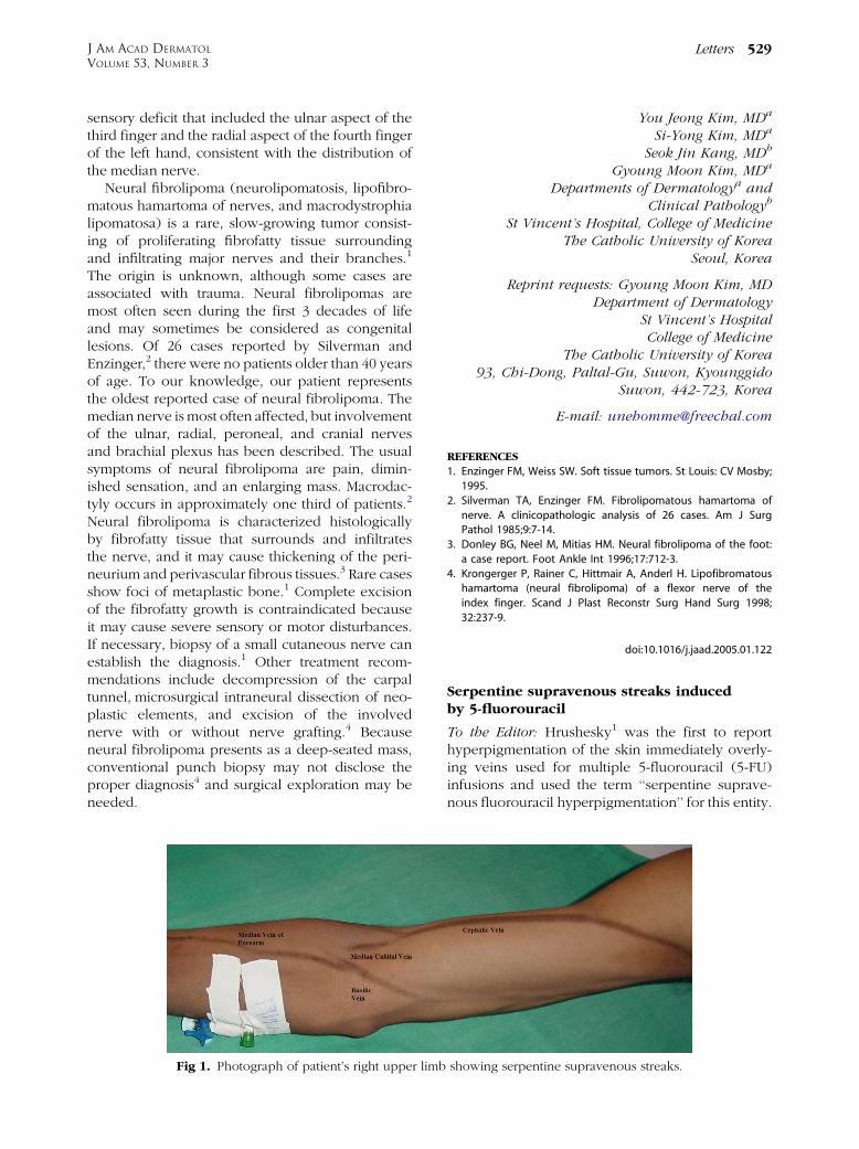
A 30-year-old male developed asymptomatic ser-piginous hyperpigmentation characteristically over-lying the right cephalic, basilic, median cubital veins,and median vein of forearm (Fig 1) after receiving2 cycles of adjuvant 5-FU as continuous intravenousinfusion for rectal adenocarcinoma. There was noclinical evidence of thrombophlebitis, and theseveins were still patent. The histopathology of theinvolved skin showed an increase in the epidermalmelanin content and melanin incontinence into thedermis (Fig 2). The patient received a third cycle of5-FU that did not worsen the pigmentation.
The exact mechanism of this phenomenon isunknown, but it has been postulated that thesecytotoxic agents cause endothelial injury. The agentthen leaches from the vessel to the overlying epi-dermal melanocytes and interferes in melanosomepackaging with the keratinocytes,2 thereby resultingin hyperpigmentation of that skin.
This is an isolated occurrence of 5-FUeinducedserpentine supravenous streaks; in the absence ofother dermatologic, hematologic, or gastrointestinalside effects, this is an extremely rare phenomenon.There is no specific treatment3 for the pigmentarystreaks induced by 5-FU. In time, the pigmentationdisappears and may not warrant discontinuationof the drug.
Vikas Jain, MBBS, MSa
Siddharth Bhandary, MBBS, MSa
Guruswami Nagendra Prasad, MBBS, MSa
Shrutakirti D. Shenoi, MBBS, MDb
Departments of Surgerya and Dermatologyb
Kasturba Medical CollegeManipal, India
Correspondence to: Vikas Jain, MBBS, MSDepartment of Surgery
Kasturba Medical CollegeManipal, India
Fig 2. Photomicrograph showing increased epidermalmelanin content and melanin incontinence into the dermis.(Hematoxylin-eosin stain; original magnification: 3100.)
REFERENCES
1. Hrushesky WJ. Unusual pigmentary changes associated with
5-fluorouracil therapy. Cutis 1980;26:181-2.
2. O’Daugherty D. Hyperpigmentation after cancer chemother-
apy. Lancet 1975;2:365.
3. Perlin E, Ahlgern JD. Pigmentary effects from protracted infu-
sion of 5-fluorouracil. Int J Dermatol 1991;30:43-4.
doi:10.1016/j.jaad.2005.01.127