SERIE BLANCA NORMAL Y PATOLÓGICA - Amazon...
63
SERIE BLANCA NORMAL Y PATOLÓGICA Bqco. Gonzalo Ojeda Hematología Clínica Fa.C.E.N.A – U.N.N.E 2011
Transcript of SERIE BLANCA NORMAL Y PATOLÓGICA - Amazon...

SERIE BLANCA NORMAL Y PATOLÓGICABqco. Gonzalo OjedaHematología ClínicaFa.C.E.N.A – U.N.N.E2011
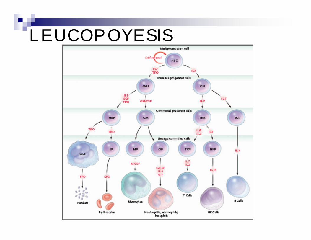
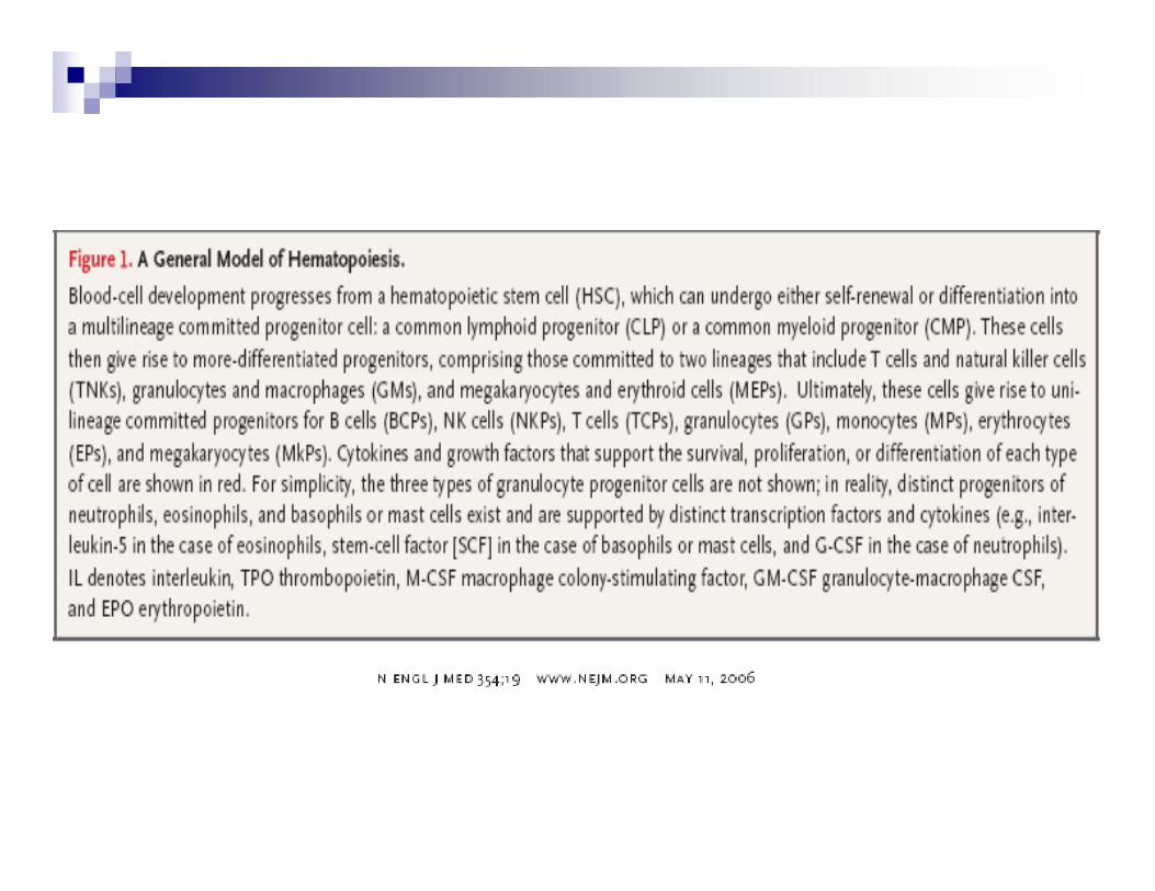
LEUCOPOYESIS

Neutrófilos( 50-70%) Basófilos(0-2%) Eosinófilos (0-5%)
Monocitos (1-9%) Linfocitos(20-40%)
Leucocitos granulares
Leucocitos agranulares


80 a 8002 a 8MONOCITOS
1500 a 400020 a 40LINFOCITOS
0 a 1000 a 1BASOFILOS
40 a 4001 a 4EOSINOFILOS
3000 a 600050 a 70SEGMENTADOS
0 a 3000 a 3CAYADOS
ABSOLUTA (mm3)
RELATIVA %

FUNCIONES DESTACADAS
GRANULOCITOS : inmunidad innata
MONOCITOS: inmunidad innata/adaptativa
LINFOCITOS: inmunidad adaptativa

GRANULOCITOS

GRANULOPOYESIS NEUTRÓFILO
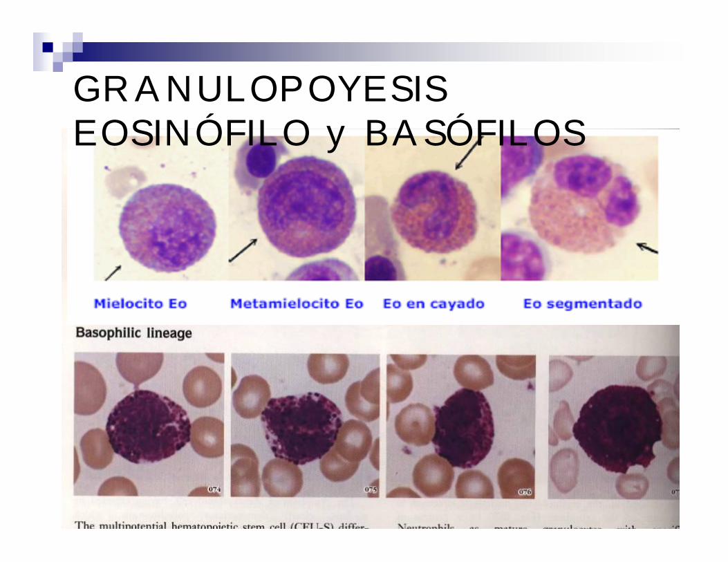
GRANULOPOYESIS EOSINÓFILO y BASÓFILOS
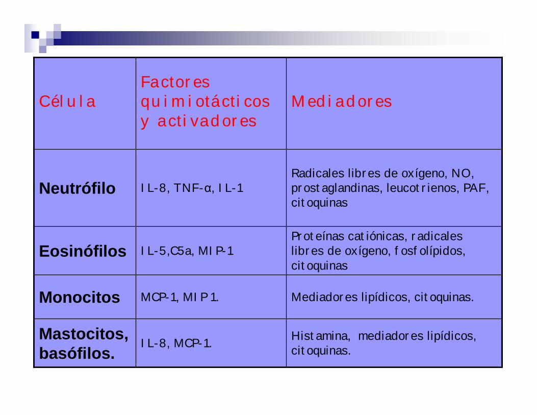
Histamina, mediadores lipídicos, citoquinas.IL-8, MCP-1.Mastocitos,
basófilos.
Proteínas catiónicas, radicales libres de oxígeno, fosfolípidos, citoquinas
IL-5,C5a, MIP-1Eosinófilos
Mediadores lipídicos, citoquinas.MCP-1, MIP 1.Monocitos
IL-8, TNF-α, IL-1
Factores quimiotácticosy activadores
Radicales libres de oxígeno, NO, prostaglandinas, leucotrienos, PAF, citoquinas
Neutrófilo
MediadoresCélula

NEUTRÓFILO
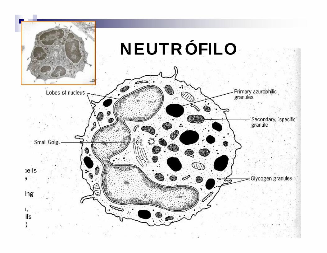
NEUTRÓFILO

GRANULOCITO NEUTRÓFILOSP:3000 A 6000 cel/mm3Tamaño: 12-20 µmGRANULOS 1°- AZUROFILOS
INESPECIFICOSLisosomas 1°,10 a 20% del contenido granularMieloperoxidasa (MPO)Fosfatasa acida (FAC)EsterasasBeta glucuronidasa y beta galactosidasaLisozimaOtras proteinas básicas catiónicas

*VESICULAS SECRETORIAS:Contienen FAL
*GRANULOS GELATINOSOSGelatinasa, lisozima
*Contienen alto contenido de glucógeno citoplasmático
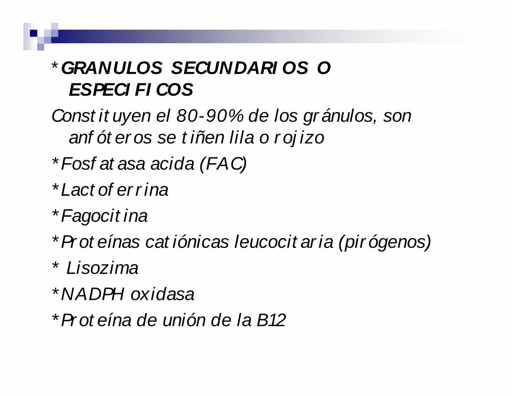
*GRANULOS SECUNDARIOS O ESPECIFICOS
Constituyen el 80-90% de los gránulos, son anfóteros se tiñen lila o rojizo
*Fosfatasa acida (FAC)*Lactoferrina*Fagocitina*Proteínas catiónicas leucocitaria (pirógenos)* Lisozima*NADPH oxidasa*Proteína de unión de la B12

FUNCIONES DEL GN
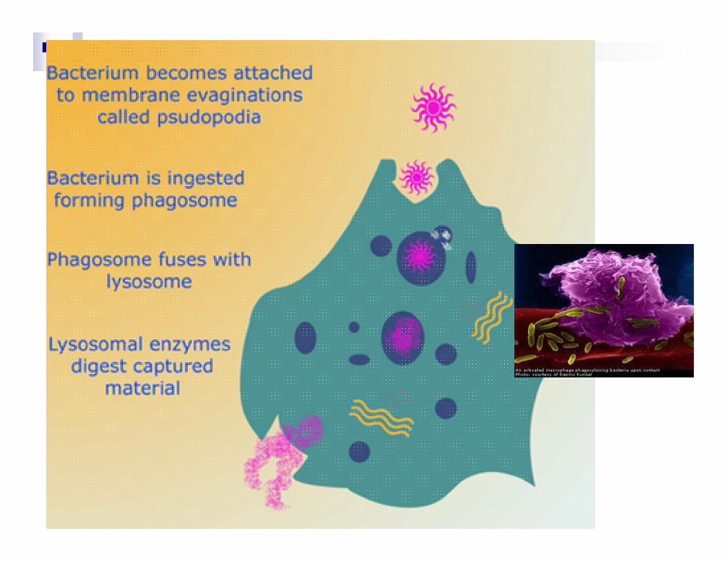
1-Defensa antimicrobiana: fagocitosis, bactericidia
2-Síntesis de la Proteína transportadora de B12
3- Síntesis de pirógeno leucocitario
4-Biosíntesis de nucleótidos

MECANISMOS DE LA FUNCION FAGOCITARIA
ADHERENCIA AL ENDOTELIO QUIMIOTAXIS OPSONIZACION Y RECONOCIMIENTO ENDOCITOSIS O INGESTION DEGRANULACION ACTIVACION DEL METABOLISMO
OXIDATIVO SISTEMAS BACTERICIDAS

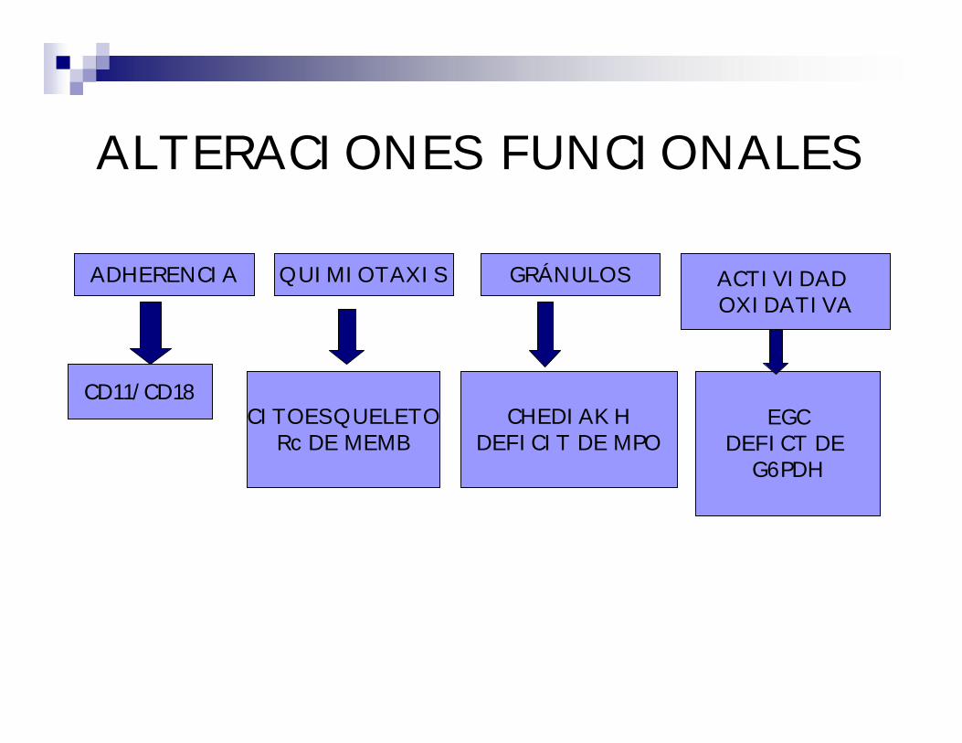
ALTERACIONES FUNCIONALES
ADHERENCIA ACTIVIDAD OXIDATIVA
QUIMIOTAXIS GRÁNULOS
CD11/CD18CITOESQUELETO
Rc DE MEMBCHEDIAK H
DEFICIT DE MPOEGC
DEFICT DE G6PDH

MIELOBLASTOS
1. myeloblast 2. neutrophil myelocyte 3. band neutrophil
Magnification: x 1000
Staining: MGGComment: One myeloblast and two more mature neutrophilic cells (myelocyte and band neutrophil leucocyte) are seen. Platelets with small number of granules.
Occurrence:blood: not presentmarrow: < 5%
Nucleoli: visible, medium or large size 1 to 4; brighter than chromatin
Nuclear/cytoplasmic ratio: high
Type of chromatin: fine, with reticular appearance
Nucleus' shape: usually oval, sometimes irregular, rarely round
Granularity: nongranular cytoplasm or a few thick azurophilic granules
Colour of cytoplasm: blue, without distinct perinuclear halo or with extended perinuclear halo
Shape of the cell: oval, sometimes round
Size of the cell: 15 - 25 m

1. myeloblast 2. promyelocyte 3. neutrophil myelocyte 4. neutrophil metamyelocyte 5. band neutrophil 6. segmented neutrophil7. pycnotic normoblast 8. polychromatic normoblast 9. basophilic normoblast 10. proerythroblast
Magnification: x 1000
Staining: MGGComment: Early myeloblast with very high cytoplasm - nucleus ratio, without granules. In the picture there are numerous other cells representing next stages of maturation of the series of granulopoiesis.
Occurrence:blood: not presentmarrow: < 5%
Nucleoli: visible, medium or large size 1 to 4; brighter than chromatin
Nuclear/cytoplasmic ratio: high or relatively high
Type of chromatin: fine, with reticular appearance
Nucleus' shape: usually oval, sometimes irregular, rarely round
Granularity: nongranular cytoplasm or a few thick azurophilic granules
Colour of cytoplasm: blue, without distinct perinuclear halo or with extended perinuclear halo
Shape of the cell: oval, sometimes round
Size of the cell: 15 - 25 m
MIELOBLASTOS

PROMIELOCITOS
1. neutrophil myelocyte 2. neutrophil metamyelocyte 3. band neutrophil 4. segmented neutrophil 5. plasmocyte 6. eosinophil7. megakaryoblast
Magnification: x 1000
Staining: MGGComment: The arrow indicates one promyelocyte, which is the only promyelocyte in the field. The nearby large cell of the granulopoiesis series is not a completely differentiated promyelocyte (lack of perinuclear zone, and not abundant granules).
Occurrence:blood: not presentmarrow: < 5 %
Nucleoli: visible, medium or large size, brighter than chromatin, 1-2. Sometimes not visible.
Nuclear/cytoplasmic ratio: moderate, low or very low
Type of chromatin: start of condensation
Nucleus' shape: oval
Granularity: thick, azurophilicabundant or very abundant
Colour of cytoplasm: light-blue, with distinct halo
Shape of the cell: oval or round
Size of the cell: 15 - 30 m

Magnification: × 1000
Staining: MGGComment: The promyelocyte contains very abundant primary granules and a distinct zone of perinuclear halo. Degranulated platelets and discrete anisocytosis of the erythrocytes are also seen.
Occurrence:blood: not presentmarrow: < 5 %
Nucleoli: visible, medium or large size, brighter than chromatin, 1-2. Sometimes not visible.
Nuclear/cytoplasmic ratio: moderate, low or very low
Type of chromatin: start of condensation
Nucleus' shape: oval
Granularity: thick, azurophilicabundant or very abundant
Colour of cytoplasm: light-blue, with distinct halo
Shape of the cell: oval or round
Size of the cell: 15 - 30 m
PROMIELOCITOS

Magnification: x 1000
Staining: MGGComment: The promyelocyte contains abundant primary granules and a distinct zone of perinuclear halo. Also distinct anisocytosis of the erythrocytes.
Occurrence:blood: not presentmarrow: < 5 %
Nucleoli: visible, medium or large size, brighter than chromatin, 1-2. Sometimes not visible.
Nuclear/cytoplasmic ratio: moderate, low or very low
Type of chromatin: start of condensation
Nucleus' shape: oval
Granularity: thick, azurophilicabundant or very abundant
Colour of cytoplasm: light-blue, with distinct halo
Shape of the cell: oval or round
Size of the cell: 15 - 30 mPROMIELOCITOS

1. neutrophil myelocyte 2. neutrophil metamyelocyte 3. band neutrophil 4. segmented neutrophil 5. lymphocyte 6. plasmocyte7. proerythroblast 8. polychromatic normoblast 9. pycnotic normoblast
Magnification: x 1000
Staining: MGGComment: The arrow indicates neutrophil myelocyte with pink cytoplasm and disappearing primary granules. In the field there are also four other cells at a similar stage of maturation and numerous other maturating neutrophil cells.
Occurrence:blood: not presentmarrow: 5 - 20 %
Nucleoli: not visible
Nuclear/cytoplasmic ratio: low or very low
Type of chromatin: partially condensed
Nucleus' shape: oval or kidney shaped
Granularity: abundant, thick azurophilic and neutrophilicgranulation
Colour of cytoplasm: light-blue or of pale pink colour undiscernible halo
Shape of the cell: oval or round
Size of the cell: 15 - 25 m
MIELOCITOS NEUTRÓFILOS

Magnification: x 1000
Staining: MGGComment: Early neutrophil myelocyte in the blood. Also two matured neutrophilic leucocytes, a lymphocyte and platelets.
Occurrence:blood: not presentmarrow: 5 - 20 %
Nucleoli: not visible
Nuclear/cytoplasmic ratio: low or very low
Type of chromatin: partially condensed
Nucleus' shape: oval or kidney shaped
Granularity: abundant, thick azurophilic and neutrophilicgranulation
Colour of cytoplasm: light-blue or of pale pink colour undiscernible halo
Shape of the cell: oval or round
Size of the cell: 15 - 25 m
MIELOCITOS NEUTRÓFILOS
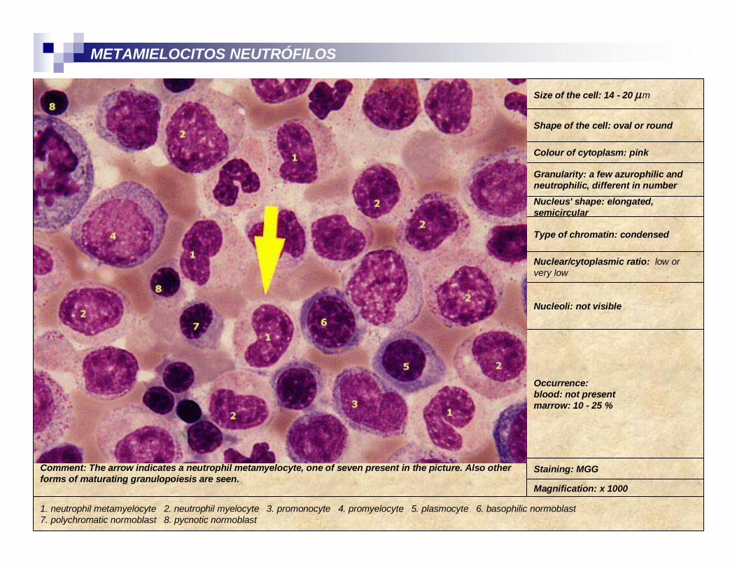
METAMIELOCITOS NEUTRÓFILOS
1. neutrophil metamyelocyte 2. neutrophil myelocyte 3. promonocyte 4. promyelocyte 5. plasmocyte 6. basophilic normoblast7. polychromatic normoblast 8. pycnotic normoblast
Magnification: x 1000
Staining: MGGComment: The arrow indicates a neutrophil metamyelocyte, one of seven present in the picture. Also other forms of maturating granulopoiesis are seen.
Occurrence:blood: not presentmarrow: 10 - 25 %
Nucleoli: not visible
Nuclear/cytoplasmic ratio: low or very low
Type of chromatin: condensed
Nucleus' shape: elongated, semicircular
Granularity: a few azurophilic and neutrophilic, different in number
Colour of cytoplasm: pink
Shape of the cell: oval or round
Size of the cell: 14 - 20 m

Magnification: x 1000
Staining: MGGComment: Neutrophil metamyelocyte indicated by the arrow is present in blood. Besides, neutrophilsegmented and band-forms leucocytes are seen. Platelets not rich in granules.
Occurrence:blood: not presentmarrow: 10 - 25 %
Nucleoli: not visible
Nuclear/cytoplasmic ratio: low or very low
Type of chromatin: condensed
Nucleus' shape: elongated, semicircular
Granularity: a few azurophilic and neutrophilic, different in number
Colour of cytoplasm: pink
Shape of the cell: oval or round
Size of the cell: 14 - 20 m
METAMIELOCITOS NEUTRÓFILOS

Magnification: x 1000
Staining: MGGComment: Two band forms and one segmented neutrophil leucocytes in the blood. Also crenated blood cells and platelets without granules are seen.
Occurrence:blood: < 5%marrow: 5 - 20 %
Nucleoli: not visible
Nuclear/cytoplasmic ratio: low or very low
Type of chromatin: condensed
Nucleus' shape: semicircular
Granularity: a few azurophilic and neutrophilic, different in number
Colour of cytoplasm: pink
Shape of the cell: oval or round
Size of the cell: 14 - 20 m
CAYADOS
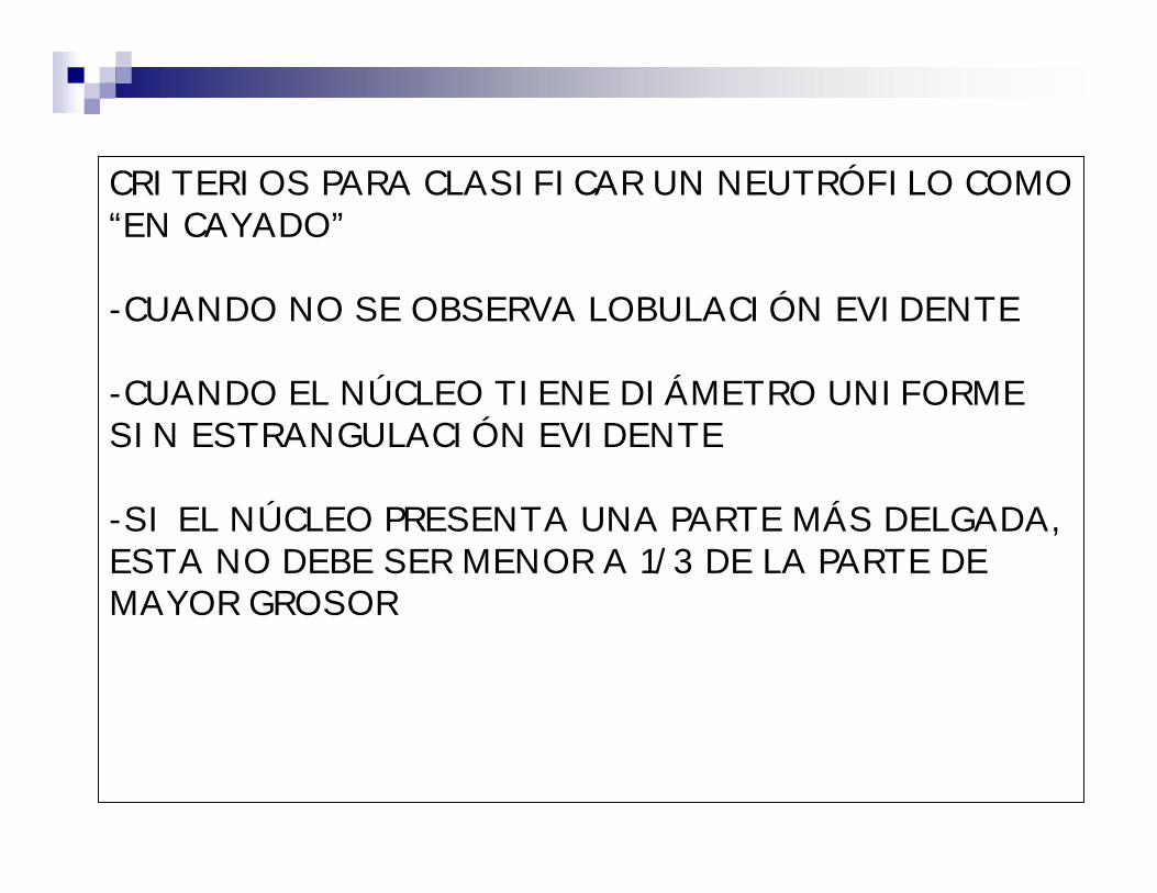
CRITERIOS PARA CLASIFICAR UN NEUTRÓFILO COMO “EN CAYADO”
-CUANDO NO SE OBSERVA LOBULACIÓN EVIDENTE
-CUANDO EL NÚCLEO TIENE DIÁMETRO UNIFORME SIN ESTRANGULACIÓN EVIDENTE
-SI EL NÚCLEO PRESENTA UNA PARTE MÁS DELGADA, ESTA NO DEBE SER MENOR A 1/3 DE LA PARTE DE MAYOR GROSOR


Magnification: x 1000
Staining: MGGComment: Three-lobulated segmented neutrophil leucocyte with fine neutrophil granularity.
Occurrence:blood: 40 - 75 %marrow: 5 - 20 %
Nucleoli: not visible
Nuclear/cytoplasmic ratio: low or very low
Type of chromatin: condensed
Nucleus' shape: lobulated (normally less than 5 lobes)
Granularity: a few azurophilic and neutrophilic, different in number granulation
Colour of cytoplasm: pink
Shape of the cell: oval or round
Size of the cell: 14 - 20 m
NEUTRÓFILO SEGMENTADO


ALTERACIONES LEUCOCITARIAS
NUCLEARES CITOPLASMÁTICAS
CUALITATIVAS
LEUCOCITOSISLEUCOPENIAS
CUANTITATIVAS
F(X)
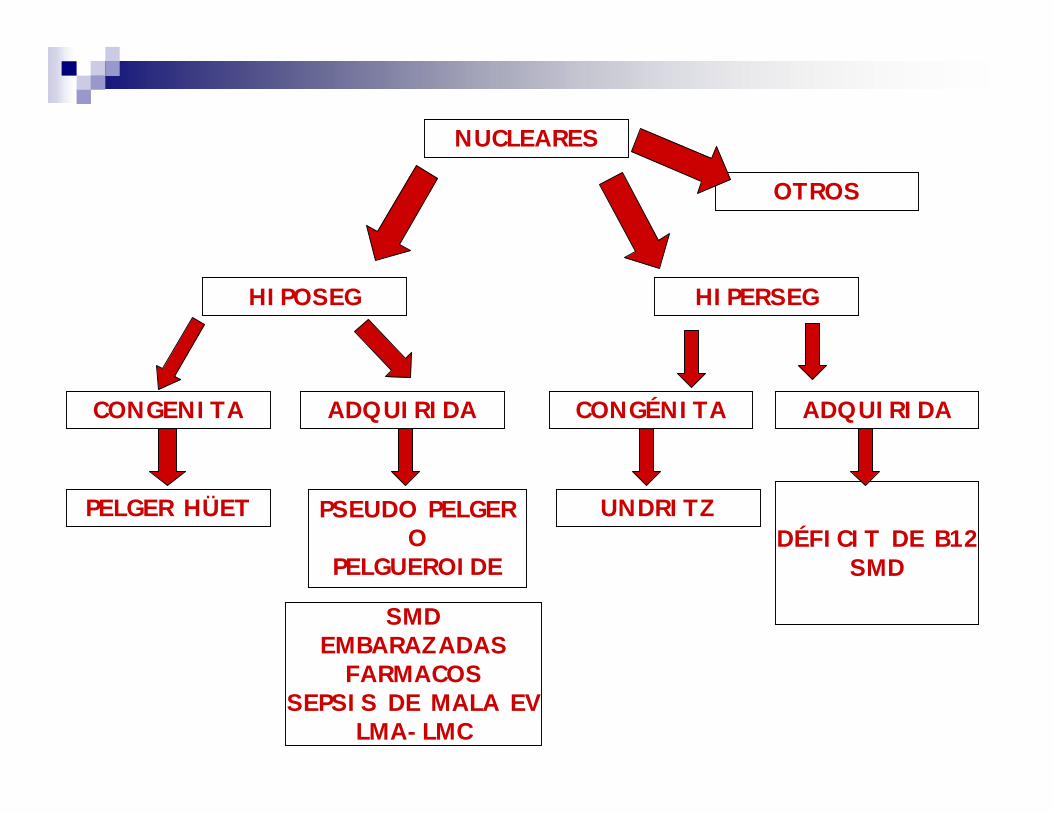
ADQUIRIDA
HIPERSEG
CONGÉNITA ADQUIRIDACONGENITA
HIPOSEG
NUCLEARES
PELGER HÜETDÉFICIT DE B12
SMD
UNDRITZPSEUDO PELGERO
PELGUEROIDE
SMDEMBARAZADAS
FARMACOSSEPSIS DE MALA EV
LMA-LMC
OTROS

CITOPLASMÁTICAS
ADQUIRIDASCONGÉNITAS
-A. DE CHEDIAK-HIGASHI-A. DE MAY HEGGLIN
-A. DE ALDER-DEGRANULADOS
-GRAN. TÓXICAS-VACUOLAS
-CUERPOS DE DöHLE-DEGRANULADOS

HIPOSEGMENTACIÓN NUCLEAR NO CONFUNDIR PELGER CON CAYADOS
CROMATINA MEDIANAMENTE CONDENSADACROMATINA HIPERCON-
DENSADALLEGA A MADURO SIN
SEGMENTAR
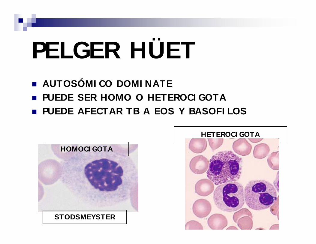
PELGER HÜET AUTOSÓMICO DOMINATE PUEDE SER HOMO O HETEROCIGOTA PUEDE AFECTAR TB A EOS Y BASOFILOS
HOMOCIGOTA
HETEROCIGOTA
STODSMEYSTER


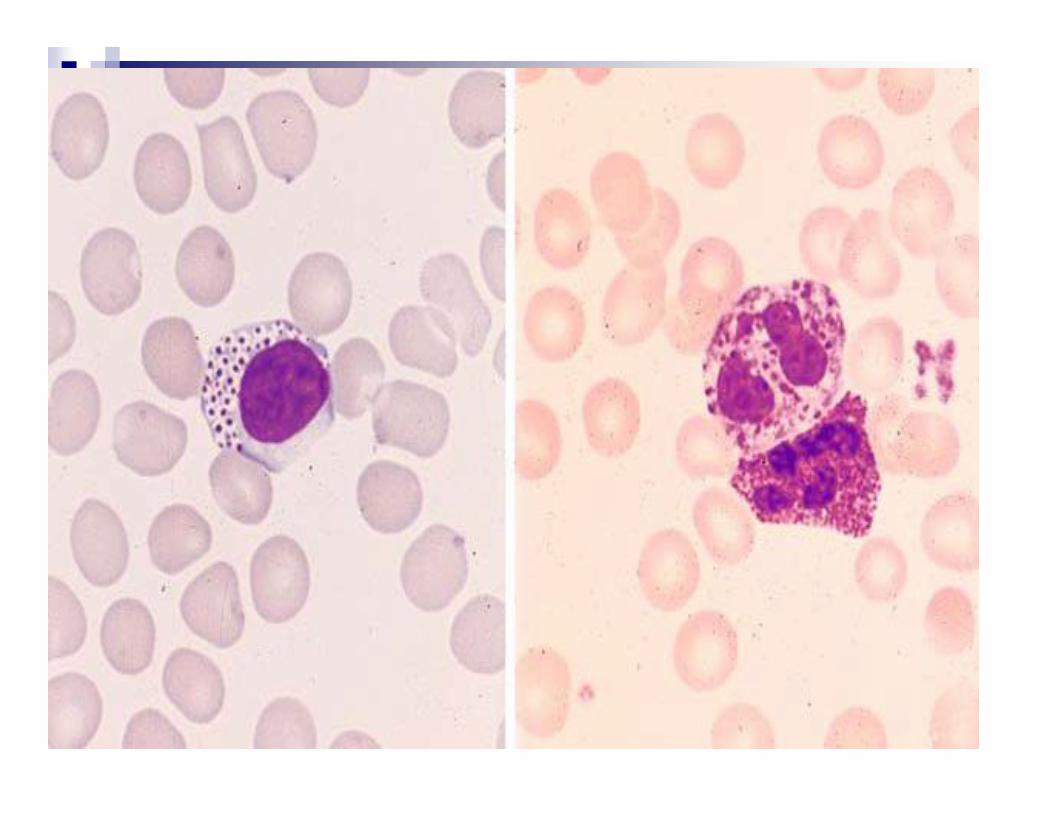
HIPERSEGMENTACIÓN
> A 5 LÓBULOS LIGERO AUMENTO DE TAMAÑO CELULAR CONGÉNITA (UNDRITZ) – AUTOSÓMICA
DOMINANTE- MUY RARA-80 A 90 % NEUTRÓFILOS CON MÁS DE 5 LÓBULOS
PUEDE AFECTAR A EOSINÓFILOS ADQUIRIDA- BASTANTE FRECUENTE-
SMD-ANEMIAS MEGALOBLÁSTICAS -ALCOHOLISMO

HIPERSEGMENTANCIÓN
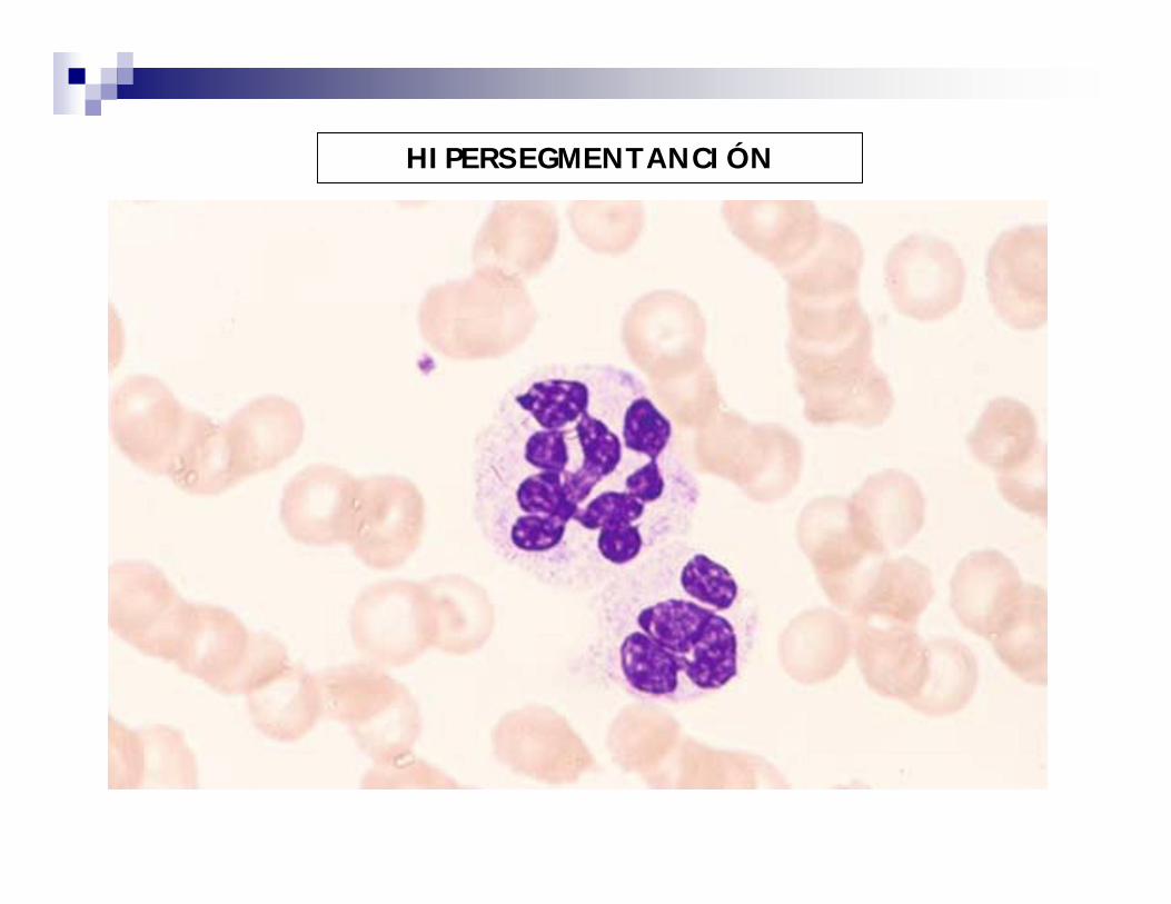
HIPERSEGMENTANCIÓN

ALTERACIONES CITOPLASMÁTICAS CHEDIAK HIGASHI


ALBINISMO PARCIAL

MAY HEGGLIN


ALDER REILLY

Magnification: x 1000
Staining: MGGComment: Granulesless segmented neutrophil.
Occurrence in blood: normally not present
NEUTROFILO AGRANULAR

Magnification: x 1000
Staining: MGGComment: Band neutrophil leucocyte with dark, very abundant toxic granulation. Also anisocytosis of erythrocytes. Numerous ovalocytes. Normal platelets.
Occurrence in blood: normally not present
Granularity: thick granules, more eosinophilic stained than typical neutrophilicgranularity. Single granules with a tendency to aggregate.
GRANULACIONES TOXICAS


Magnification: x 1000
Staining: MGGComment: The arrow points Döhle’s body in the granulocyte. Erythrocytes difficult to assess.
Occurrence in blood: normally not present.
Definition: Dotted inclusions of blue, non-granular cytoplasm in pink cytoplasm of mature neutrophil leucocyte.
CUERPOS DE DÖHLE

Magnification: x 1000
Staining: MGGComment: Döhle’s body pointed by the arrow. Also anisocytosis of erythrocytes.
Occurrence in blood: normally not present.
Definition: Dotted inclusions of blue, non-granular cytoplasm in pink cytoplasm of mature neutrophil leucocyte.

Magnification: x 1000
Staining: MGGComment: Distinct vacuoles changes in the neutrophilic leucocytes. Two target cells and one ovalocyte are present.
Occurrence: normal mature neutrophil leucocytes may contain single small vacuoles in cytoplasm
VACUOLAS

Magnification: x 1000
Staining: MGGComment: Small vacuoles in a neutrophil leucocyte with fine neutrophilic granulation. Also anizocytosis of erythrocytes. A single spherocyte and a polychromatophilic cell are seen.
Occurrence: normal mature neutrophil leucocytes may contain single small vacuoles in cytoplasm
VACUOLAS


NEUTRÓFILO APOPTÓTICO/MUESTRA VIEJA