
Sergei Nekhai, Ph.D. Objectives - Home | Howard University Immunology 201… · Immune Response to...
79
02/25/13 VIRAL IMMUNOLOGY Sergei Nekhai, Ph.D. Objectives: • Overview of immune system •Intrinsic antiviral response • Innate immune response •Adaptive immunity
Transcript of Sergei Nekhai, Ph.D. Objectives - Home | Howard University Immunology 201… · Immune Response to...

02/25/13 VIRAL IMMUNOLOGY
Sergei Nekhai, Ph.D.
Objectives:
• Overview of immune system
•Intrinsic antiviral response
• Innate immune response
•Adaptive immunity
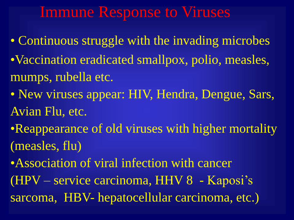
Immune Response to Viruses
• Continuous struggle with the invading microbes
•Vaccination eradicated smallpox, polio, measles,
mumps, rubella etc.
• New viruses appear: HIV, Hendra, Dengue, Sars,
Avian Flu, etc.
•Reappearance of old viruses with higher mortality
(measles, flu)
•Association of viral infection with cancer
(HPV – service carcinoma, HHV 8 - Kaposi’s
sarcoma, HBV- hepatocellular carcinoma, etc.)
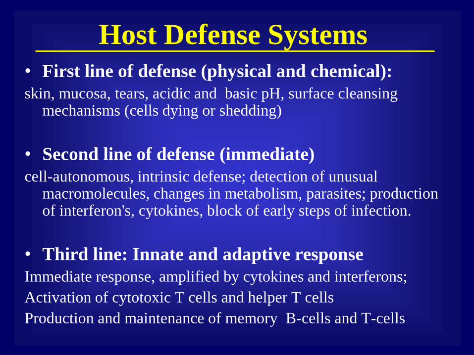
Host Defense Systems
• First line of defense (physical and chemical):
skin, mucosa, tears, acidic and basic pH, surface cleansing mechanisms (cells dying or shedding)
• Second line of defense (immediate)
cell-autonomous, intrinsic defense; detection of unusual macromolecules, changes in metabolism, parasites; production of interferon's, cytokines, block of early steps of infection.
• Third line: Innate and adaptive response
Immediate response, amplified by cytokines and interferons;
Activation of cytotoxic T cells and helper T cells
Production and maintenance of memory B-cells and T-cells
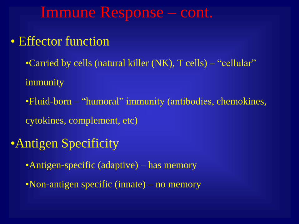
Immune Response – cont.
• Effector function
•Carried by cells (natural killer (NK), T cells) – “cellular”
immunity
•Fluid-born – “humoral” immunity (antibodies, chemokines,
cytokines, complement, etc)
•Antigen Specificity
•Antigen-specific (adaptive) – has memory
•Non-antigen specific (innate) – no memory
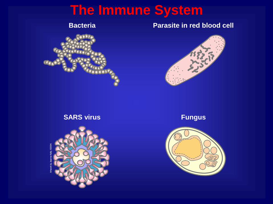
The Immune System
SARS virus
Parasite in red blood cell
Fungus
Bacteria

Markers of Self
Muscle cell
Nerve cell
Epithelial cell
Leukocyte
Class I MHC self-marker protein

Markers of Non-Self
Non-self leukocyte
Antibody
Epitope Class I MHC protein
Epitope
Antibody
Antigen
Antigen
Bacteria
Non-self nerve cell
SARS virus
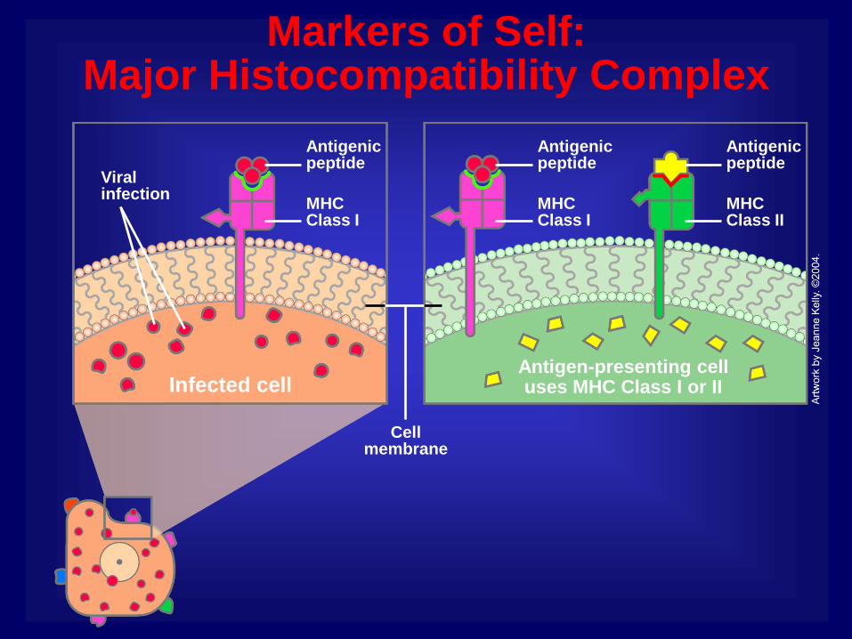
Markers of Self: Major Histocompatibility Complex
Antigenic peptide
Antigen-presenting cell uses MHC Class I or II
Cell membrane
MHC Class II
Antigenic peptide
Viral infection
Infected cell
MHC Class I
Antigenic peptide
MHC Class I
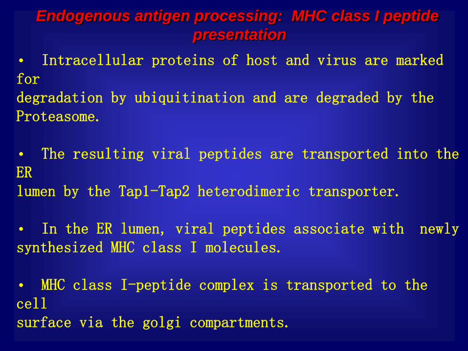
Endogenous antigen processing: MHC class I peptide
presentation
• Intracellular proteins of host and virus are marked for degradation by ubiquitination and are degraded by the Proteasome. • The resulting viral peptides are transported into the ER lumen by the Tap1-Tap2 heterodimeric transporter. • In the ER lumen, viral peptides associate with newly synthesized MHC class I molecules. • MHC class I-peptide complex is transported to the cell surface via the golgi compartments. • On the cell surface, the MHC class I-peptide complex

Endogenous antigen processing: MHC class I peptide presentation
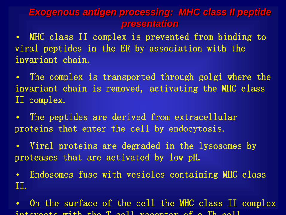
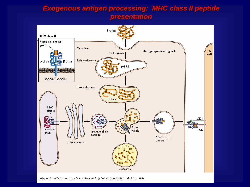
Exogenous antigen processing: MHC class II peptide
presentation
• MHC class II complex is prevented from binding to viral peptides in the ER by association with the invariant chain.
• The complex is transported through golgi where the invariant chain is removed, activating the MHC class II complex.
• The peptides are derived from extracellular proteins that enter the cell by endocytosis.
• Viral proteins are degraded in the lysosomes by proteases that are activated by low pH.
• Endosomes fuse with vesicles containing MHC class II.
• On the surface of the cell the MHC class II complex interacts with the T cell receptor of a Th cell
Exogenous antigen processing: MHC class II peptide
presentation
Organs of the Immune System
Tonsils and adenoids
Lymph nodes
Bone marrow
Appendix
Lymphatic vessels
Lymph nodes
Thymus
Peyer’s patches
Spleen
Lymphatic vessels
Lymph nodes
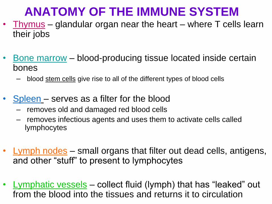
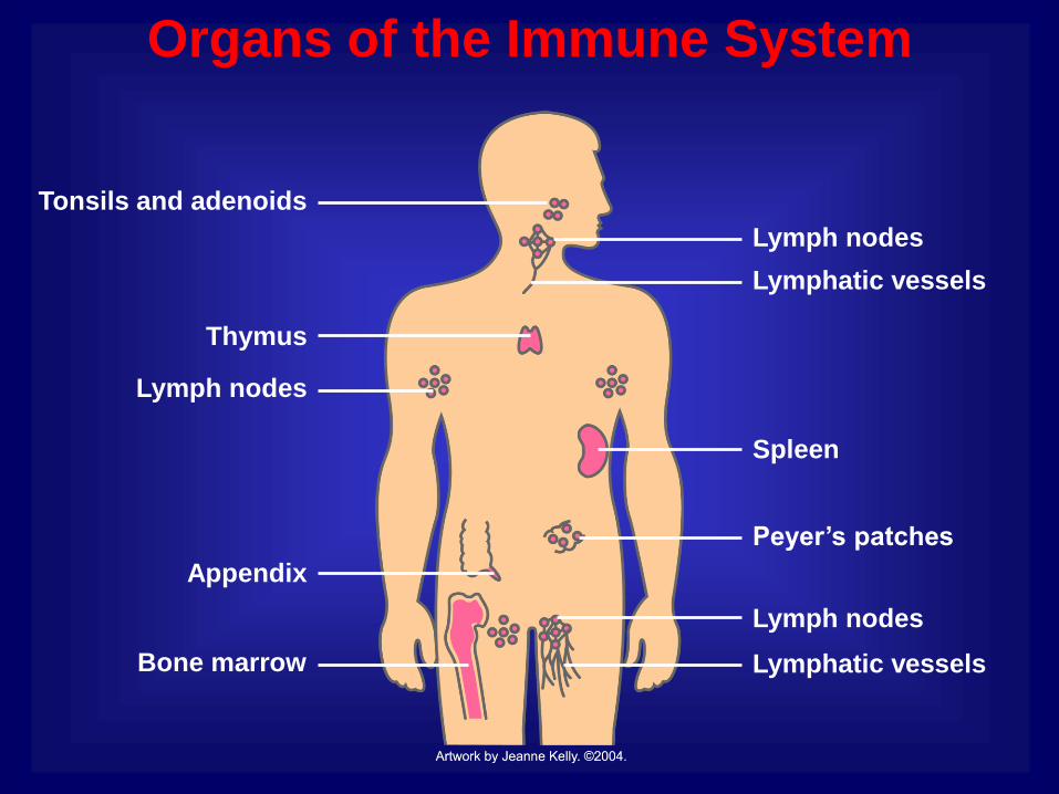
ANATOMY OF THE IMMUNE SYSTEM • Thymus – glandular organ near the heart – where T cells learn
their jobs
• Bone marrow – blood-producing tissue located inside certain bones – blood stem cells give rise to all of the different types of blood cells
• Spleen – serves as a filter for the blood – removes old and damaged red blood cells
– removes infectious agents and uses them to activate cells called lymphocytes
• Lymph nodes – small organs that filter out dead cells, antigens, and other “stuff” to present to lymphocytes
• Lymphatic vessels – collect fluid (lymph) that has “leaked” out from the blood into the tissues and returns it to circulation

Lymph Node
Germinal center
Vein
Cortex
Paracortex
Incoming lymphatic vessel
Outgoing lymphatic vessel
Artery
Medulla
Follicle
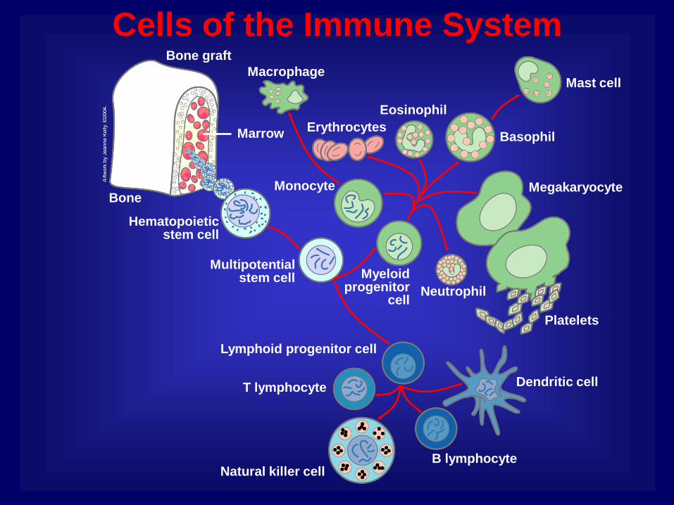
Cells of the Immune System Bone graft
Multipotential stem cell
Hematopoietic stem cell
Platelets
Macrophage
Erythrocytes
Eosinophil
Neutrophil
Megakaryocyte
Mast cell
Basophil
T lymphocyte
Natural killer cell
Dendritic cell
B lymphocyte
Lymphoid progenitor cell
Myeloid progenitor
cell
Monocyte
Marrow
Bone

B Cells
Plasma cell
Class II MHC and processed antigen are displayed
Antigen-presenting bacteria
Antigen
Antigen-specific B cell receptor
Antibodies B cell
Activated helper T cell
Lymphokines

Antibody
Assembled antibody molecule
Heavy chain
Antigen-binding region
Constant region
Light chain

Immunoglobulins
IgA
IgM
IgG, IgD, IgE, and IgA
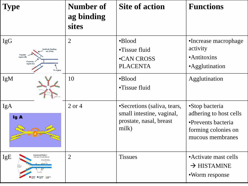
Type Number of
ag binding
sites
Site of action Functions
IgG 2 •Blood
•Tissue fluid
•CAN CROSS
PLACENTA
•Increase macrophage
activity
•Antitoxins
•Agglutination
IgM 10 •Blood
•Tissue fluid
Agglutination
IgA 2 or 4 •Secretions (saliva, tears,
small intestine, vaginal,
prostate, nasal, breast
milk)
•Stop bacteria
adhering to host cells
•Prevents bacteria
forming colonies on
mucous membranes
IgE 2 Tissues •Activate mast cells
HISTAMINE
•Worm response

T Cells
Activated killer cell Activated helper T cell
Resting cytotoxic T cell Resting helper T cell
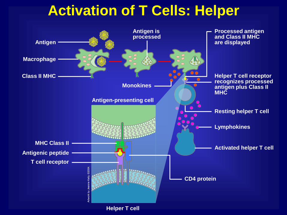
Activation of T Cells: Helper
Activated helper T cell
Monokines
MHC Class II
T cell receptor
Antigen-presenting cell
CD4 protein
Antigenic peptide
Antigen is processed
Resting helper T cell
Class II MHC
Lymphokines
Helper T cell receptor recognizes processed antigen plus Class II MHC
Macrophage
Processed antigen and Class II MHC are displayed Antigen
Helper T cell
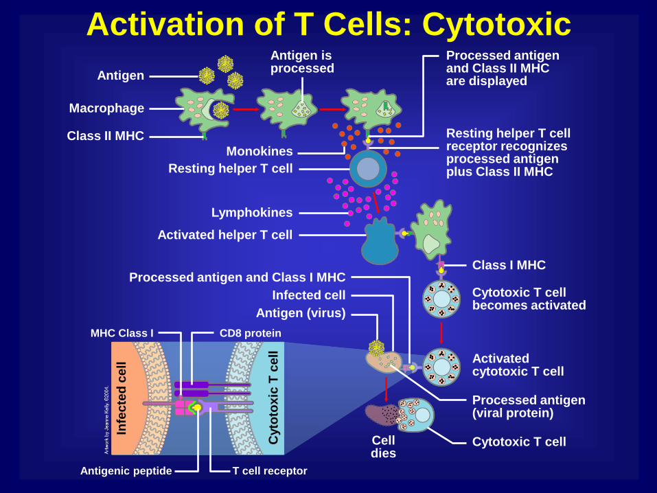
Activation of T Cells: Cytotoxic
Processed antigen and Class I MHC
Lymphokines
Class I MHC
Class II MHC
Processed antigen and Class II MHC are displayed Antigen
Resting helper T cell receptor recognizes processed antigen plus Class II MHC
Macrophage
Monokines
Cyto
tox
ic T
ce
ll
Infe
cte
d c
ell
MHC Class I
Antigenic peptide T cell receptor
CD8 protein
Resting helper T cell
Cytotoxic T cell
Cytotoxic T cell becomes activated
Antigen (virus)
Processed antigen (viral protein)
Cell dies
Infected cell
Activated cytotoxic T cell
Activated helper T cell
Antigen is processed

Cytokines
Mature helper T cell
Monokines Lymphokines
Macrophage

Killer Cells: Cytotoxic Ts and NKs
Killer cell
Target-oriented granules
Surface contact
Target cell

Phagocytes and Their Relatives
Monocyte
Dendritic cell
Eosinophil
Neutrophil
Basophil
Mast cell
Macrophage
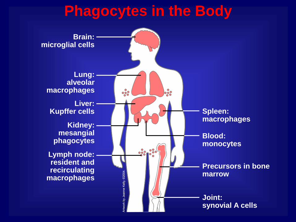
Phagocytes in the Body
Brain: microglial cells
Joint: synovial A cells
Precursors in bone marrow
Lymph node: resident and recirculating
macrophages
Blood: monocytes
Kidney: mesangial
phagocytes
Spleen: macrophages
Liver: Kupffer cells
Lung: alveolar
macrophages
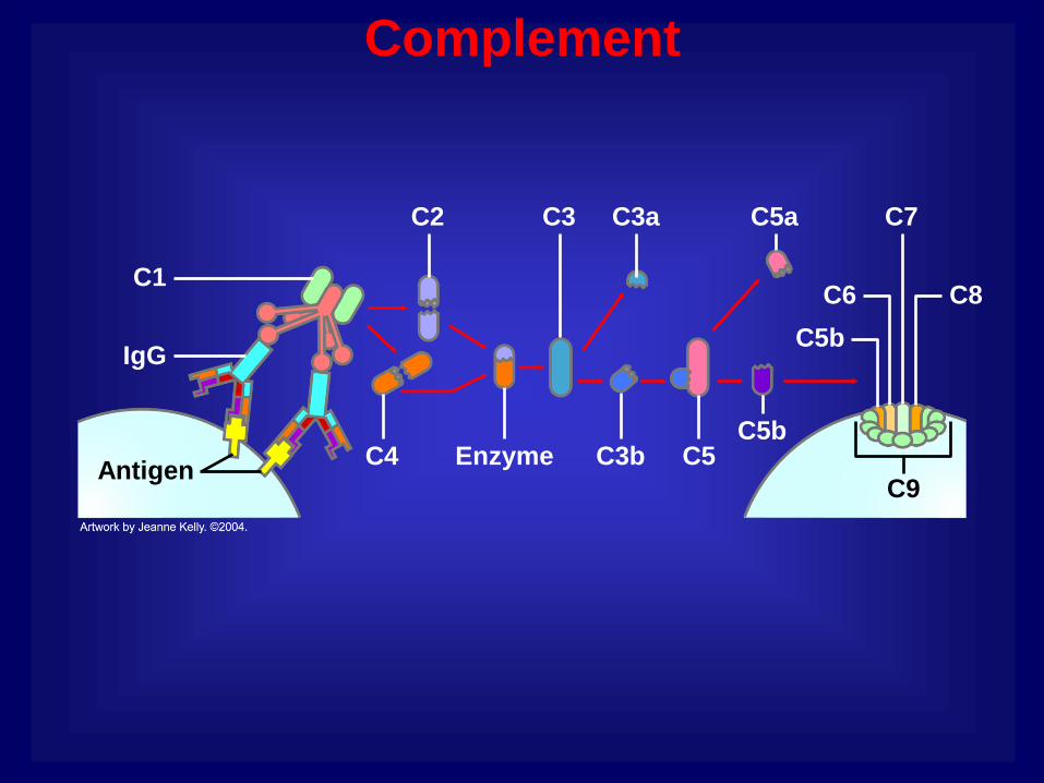
Complement
C9
Enzyme
C2
C5 C3b
C3a C3
C4 Antigen
IgG
C1 C8
C7
C6
C5b
C5b
C5a

Mounting an Immune Response
Complement
Virus
Killer cell
B cell
Antibodies
T cell
Lymphokines
Macrophage
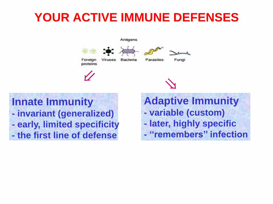
YOUR ACTIVE IMMUNE DEFENSES
INDUCTION OF AN IMMUNE RESPONSE
Foreign invaders - viruses, bacteria, allergens, toxins and parasites- constantly bombard our body.
Innate Immunity - invariant (generalized)
- early, limited specificity
- the first line of defense
Adaptive Immunity - variable (custom)
- later, highly specific
- ‘‘remembers’’ infection
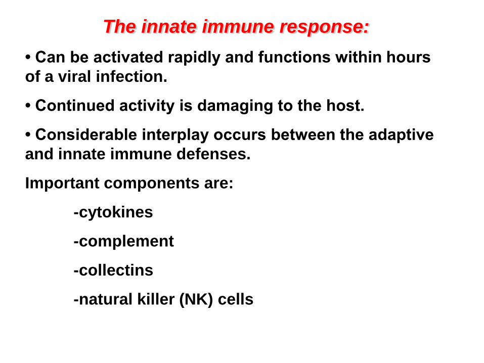
The innate immune response:
• Can be activated rapidly and functions within hours
of a viral infection.
• Continued activity is damaging to the host.
• Considerable interplay occurs between the adaptive
and innate immune defenses.
Important components are:
-cytokines
-complement
-collectins
-natural killer (NK) cells
Field’s Virology, Fifth Edition
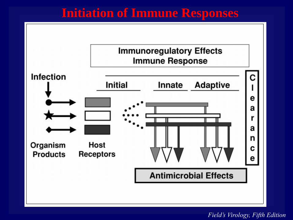
Initiation of Immune Responses
Field’s Virology, Fifth Edition
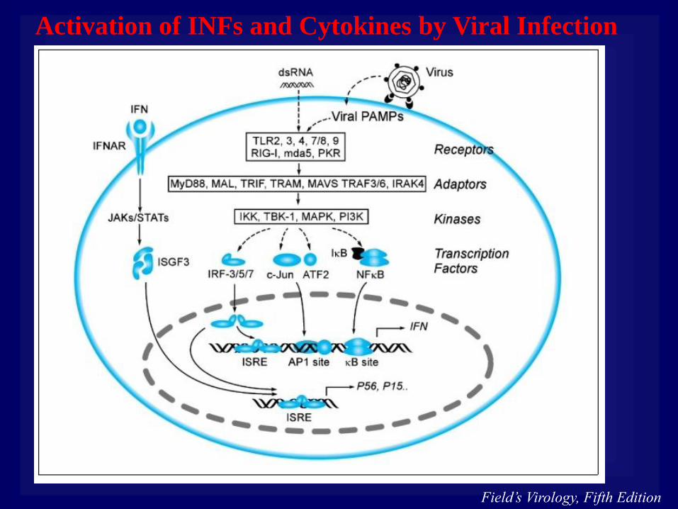
Activation of INFs and Cytokines by Viral Infection

Recognition of Foreign Nucleic Acids
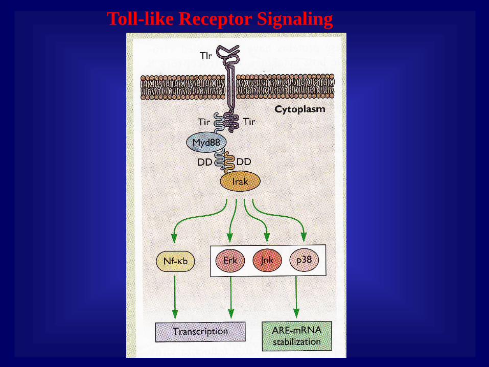
Toll-like Receptor Signaling
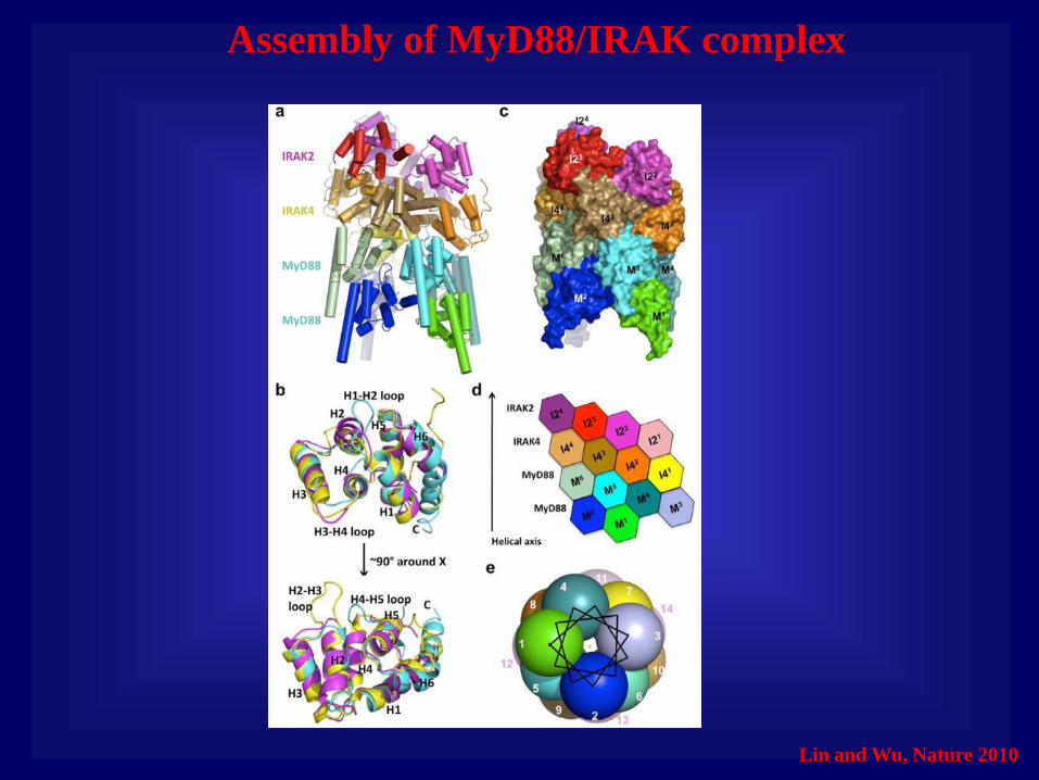
Assembly of MyD88/IRAK complex
Lin and Wu, Nature 2010

Toll-like Receptors Recognition Patterns
Toll-like Receptor Pattern Recognized
TLR1 Triacyl lipoproteins
TLR2 Lipoproteins, viral glycoproteins, gram-positive
peptidoglycans
TRL3 double-stranded RNA
TLR4 Lipopolysaccharids, viral glycoproteins
TLR5 Flagellin
TLR6 Diacyl lypoproteins
TLR7 Single-stranded RNA
TLR8 Single-stranded RNA (siRNA)
TLR9 CpG DNA, unmethylated CpG oligonucleotides
TLR10 Unknown
TLR11 profilin

Recognition of RNA by RigI and Mda5

Toll-like Receptors Recognition Patterns
Toll-like Receptor Pattern Recognized
TLR1 Triacyl lipoproteins
TLR2 Lipoproteins, viral glycoproteins, gram-positive
peptidoglycans
TRL3 double-stranded RNA
TLR4 Lipopolysaccharids, viral glycoproteins
TLR5 Flagellin
TLR6 Diacyl lypoproteins
TLR7 Single-stranded RNA
TLR8 Single-stranded RNA (siRNA)
TLR9 CpG DNA, unmethylated CpG oligonucleotides
TLR10 Unknown
TLR11 profilin

Three Primary Classes of Cytokines
Function Members Activity
Proinflammatory IL-1, TNF-α, IL-6, IL-12 Activation of leukocytes
Anti-inflammatory IL-10, IL-4, TGF-β Supress activity of pro-
inflammatory cytokines
Chemokines IL-8 Early stages rectrutiment
of immune cells

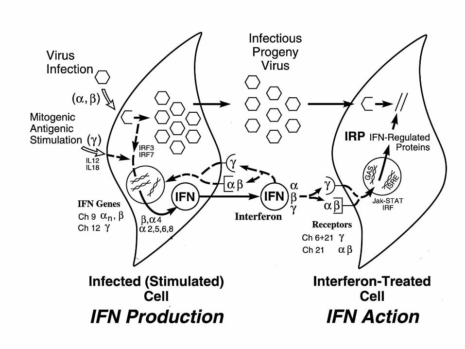
IFn-g is induced only when certain lymphocytes are
stimulated to replicate and divide after binding a foreign
antigen
IFn-a and IFn-b are induced by viral infection of any
cell type
Interferons
• IFN is induced by accumulation of double stranded
RNA (dsRNA).
• IFN induces gene expression at the transcriptional
level after binding to specific cell surface receptors.
• A cell that is bound to interferon and responds to it
is in an antiviral state.
• IFN induces expression of more that 100 genes,
products of many of these genes possess broad
spectrum antiviral activity.
• They lead to cell death by apoptosis or programmed
cells death, limiting cell to cell spread of virus.
• Production of large amounts if IFN causes common
symptoms such as fever, chills, nausea, etc.
Interferons

Interferon induced antiviral responses:
• Both viral and cellular protein synthesis stops in IFN
treated cells.
• This is due to two cellular proteins, ds-RNA activated
protein kinase (PKR) and ribonuclease L (RNase L).
• PKR is a serine/threonine kinase that has antiviral
properties, as well as antiproliferative and antitumor
functions.
• Activated PKR phosphorylates the alpha subunit of the
translation initiation factor eIF2, inhibiting translation.
• RNase L is a nuclease that can degrade cellular and viral
RNA; its concentration increases after IFN treatment.
• RNase L concentration increases 10-1,000 fold after
Ifn treatment, but is inactive unless 2’-5’-oligo(A)
synthetase is produced.
• 2’-5’-oligo(A) synthetase produces 2’, 5’ oligomers of
adenylic acid, only when activated by dsRNA.
• These poly(A) oligomers then activate RNase L,
which degrades all host and viral mRNA in the cell.
• RNase L participates not only in Ifn-mediated
antiviral defense, but also in apoptosis.
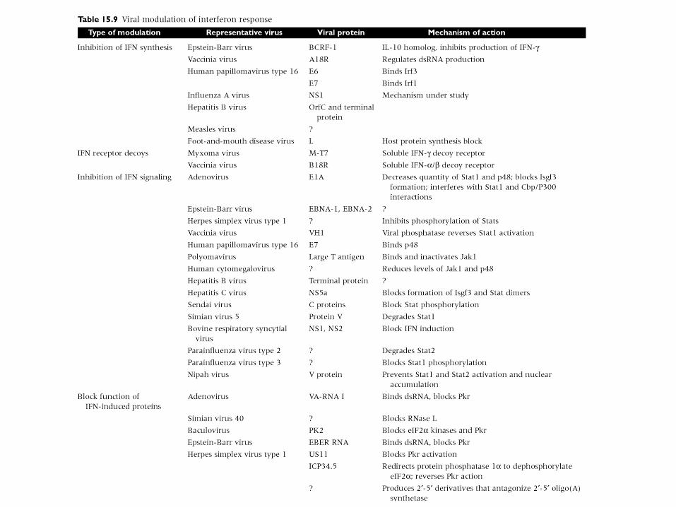
• IFN is a broad spectrum, highly effective antiviral
agent. However, viruses have developed numerous
mechanisms for inhibiting interferon action.
Interferon induced antiviral responses:

Interferon-induced Repression of Translation
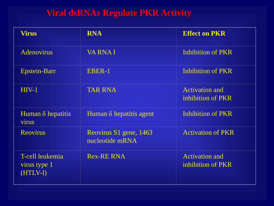
Viral dsRNAs Regulate PKR Activity
Virus
RNA
Effect on PKR
Adenovirus
VA RNA I
Inhibition of PKR
Epstein-Barr
EBER-1
Inhibition of PKR
HIV-1
TAR RNA
Activation and
inhibition of PKR
Human hepatitis
virus
Human hepatitis agent
Inhibition of PKR
Reovirus
Reovirus S1 gene, 1463
nucleotide mRNA
Activation of PKR
T-cell leukemia
virus type 1
(HTLV-I)
Rex-RE RNA
Activation and
inhibition of PKR
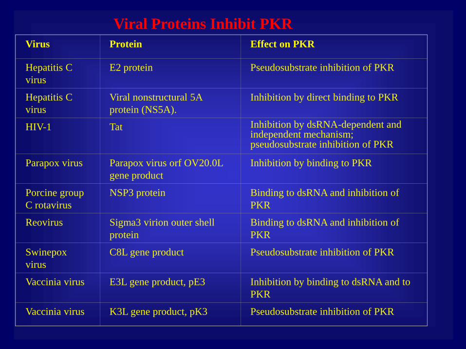
Viral Proteins Inhibit PKR
Virus
Protein
Effect on PKR
Hepatitis C
virus
E2 protein
Pseudosubstrate inhibition of PKR
Hepatitis C
virus
Viral nonstructural 5A
protein (NS5A).
Inhibition by direct binding to PKR
HIV-1
Tat
Inhibition by dsRNA-dependent and independent mechanism; pseudosubstrate inhibition of PKR
Parapox virus
Parapox virus orf OV20.0L
gene product
Inhibition by binding to PKR
Porcine group
C rotavirus
NSP3 protein
Binding to dsRNA and inhibition of
PKR
Reovirus
Sigma3 virion outer shell
protein
Binding to dsRNA and inhibition of
PKR
Swinepox
virus
C8L gene product
Pseudosubstrate inhibition of PKR
Vaccinia virus
E3L gene product, pE3
Inhibition by binding to dsRNA and to
PKR
Vaccinia virus
K3L gene product, pK3
Pseudosubstrate inhibition of PKR
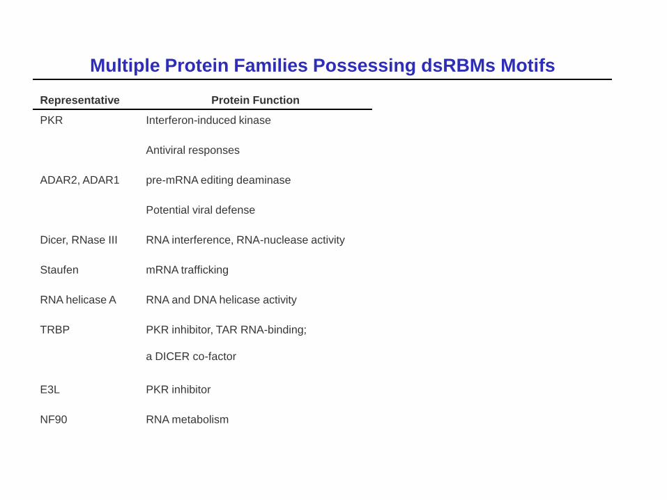
Multiple Protein Families Possessing dsRBMs Motifs
Representative Protein Function
PKR Interferon-induced kinase
Antiviral responses
ADAR2, ADAR1 pre-mRNA editing deaminase
Potential viral defense
Dicer, RNase III RNA interference, RNA-nuclease activity
Staufen mRNA trafficking
RNA helicase A RNA and DNA helicase activity
TRBP PKR inhibitor, TAR RNA-binding;
a DICER co-factor
E3L PKR inhibitor
NF90 RNA metabolism
Drakesmith and Prentice, 2008
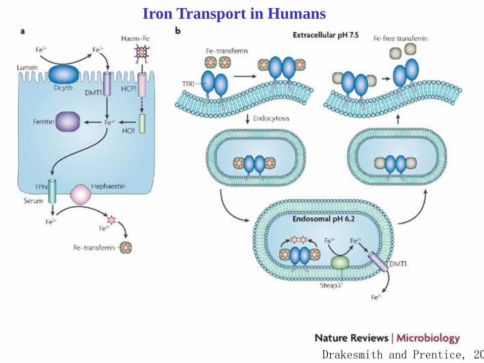
Iron Transport in Humans
Drakesmith and Prentice, 2008
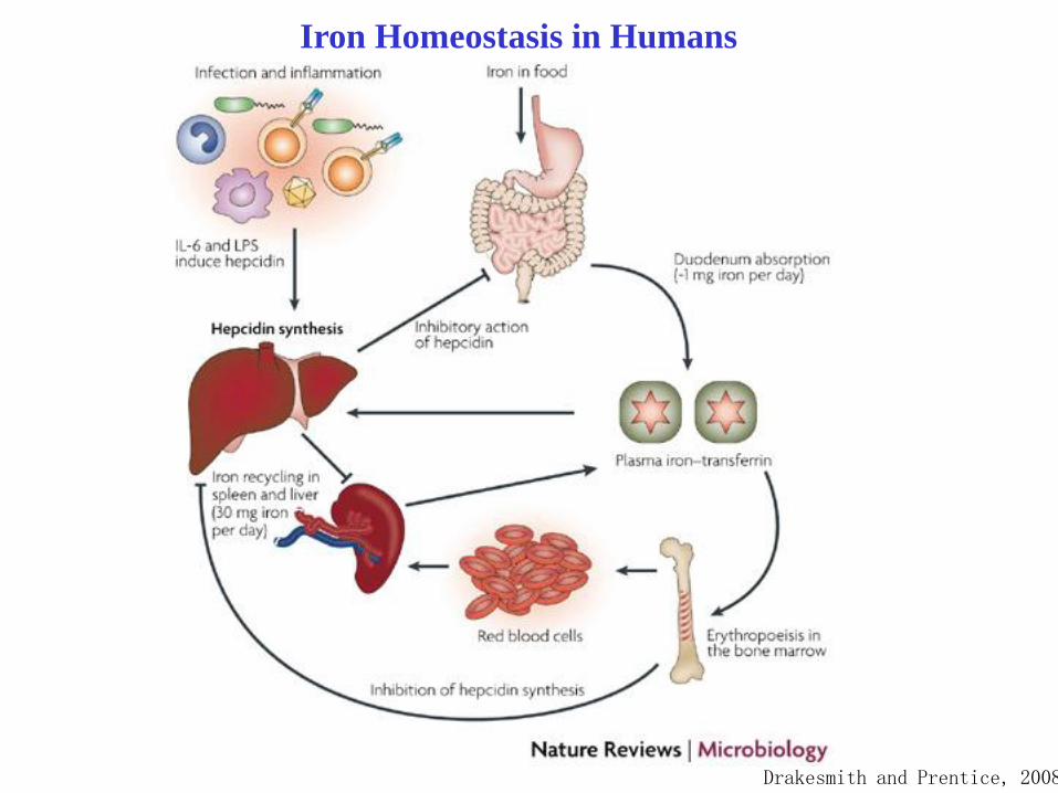
Iron Homeostasis in Humans
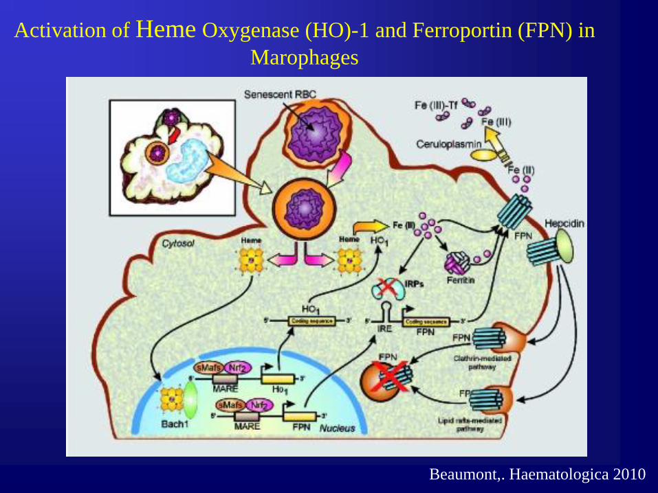
Activation of Heme Oxygenase (HO)-1 and Ferroportin (FPN) in
Marophages
Beaumont,. Haematologica 2010
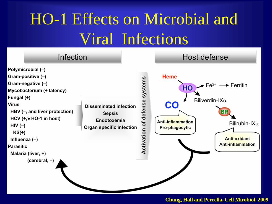
HO-1 Effects on Microbial and
Viral Infections
Chung, Hall and Perrella, Cell Mirobiol. 2009

Apoptosis
• Controlled cell death
• Uses regulators to ensure cells die off – Regulators include various
proteins that either inhibit or promote certain parts of the caspase cascade.
– Caspases (which originally exist as procaspases) which act as proteases and initiate the caspase cascade which causes cell death.
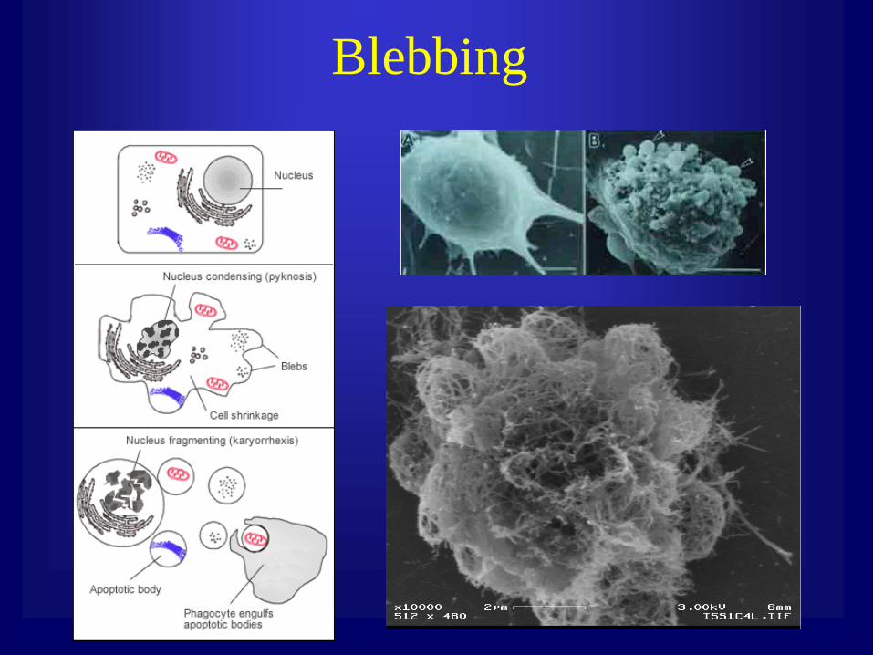
Blebbing

Mechanisms of Apoptosis Activation

Viral inhibition of apoptosis

Other Intrinsic Antivirail Responces
•Autophagy Formation of specialized membrane
compartments related to lysosomes
•Epigenetic silencing Defense against DNA
containing viruses, formation of chromatin structure
•RNA silencing Sequence-specific RNA
degradation
•Cytosine Deamination (APOBEC) C’s to U’s
conversion
•TRIM Proteins Targeting capsid protein by
TRM5a protein
•Tetherin Inability of the virions to bud
Nitric oxide synthase
Directs synthesis of NO in NK cells
Cytotoxic
Inhibits poxvirus and herpesvirus replication
Ubiquitin-proteosome pathway components
Proteins tagged with ubiquitin are targeted to the proteosome for
degradation
Other Intrinsic Antivirail Responces-cont.

Retrovirus Capsid Sensing by TRIM5
Pertel et al., Nature 2011

Micro RNA •Founding members of miRNAs, 22 nt and 61 nt RNAs coded by C.elegans
Lin-1 gene
complementary to 3’UTR of Lin-14 gene that blocked translation of Lin-14
• Control of cell proliferaton, cell death and fat metabolism in flies
•Modulation of hematopoietic lineage differentiation in mammals
•Leaf and flower development in plants
•Majority of miRNAs are transcribed independently
•Some (quarter) miRNA are derived from intrones
•miRNA are conserved
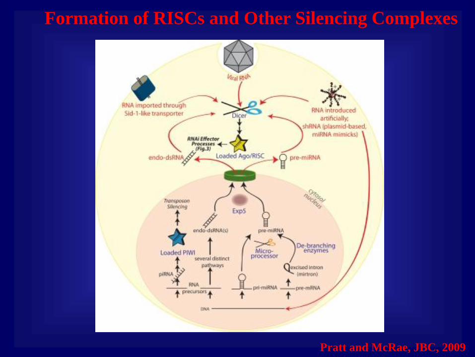
Formation of RISCs and Other Silencing Complexes
Pratt and McRae, JBC, 2009

Examples of Metazoan miRNAs

Maturation of miRNA
Plants Metazoa Animals
Cleavege
with Drosha
Additional
Cleavage
with Dicer
Complex with
RISC (RNA-induced
Silencing complex)
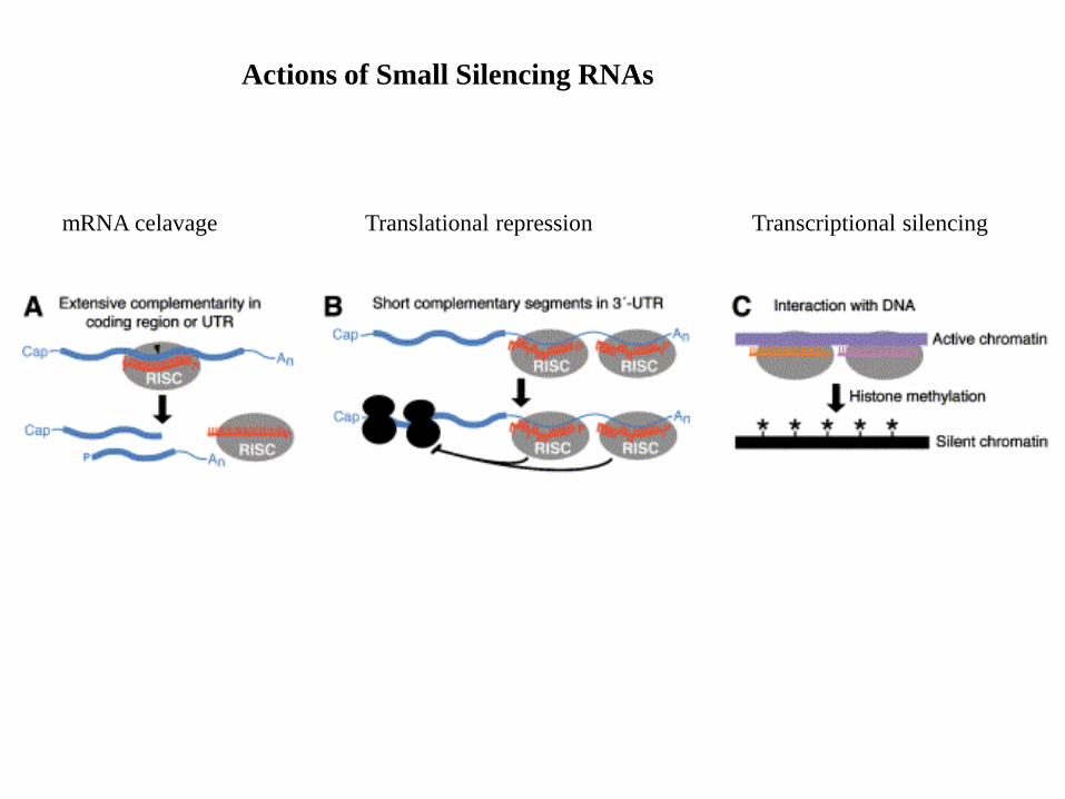
Actions of Small Silencing RNAs
mRNA celavage Translational repression Transcriptional silencing
Field’s Virology, Fifth Edition
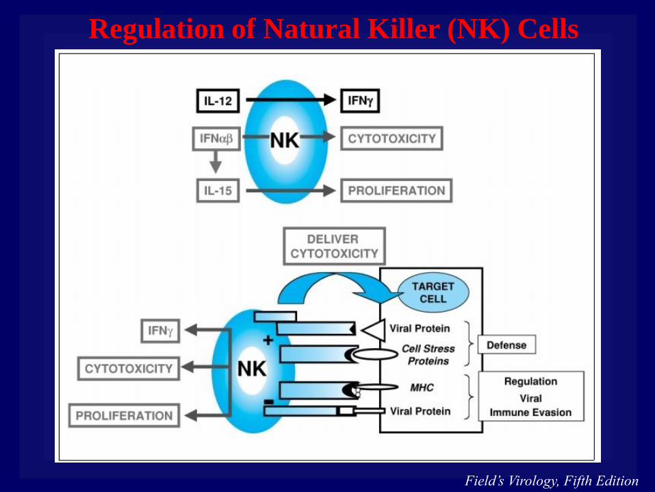
Regulation of Natural Killer (NK) Cells

Field’s Virology, Fifth Edition
Activation of Adaptive by Innate Immune Responces
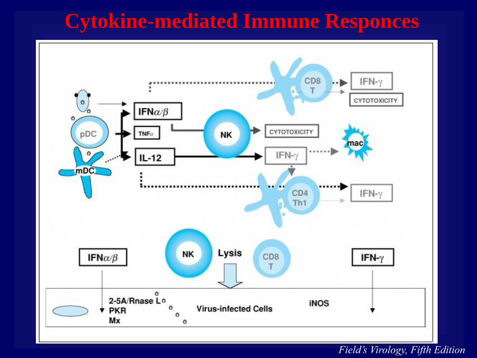
Field’s Virology, Fifth Edition
Cytokine-mediated Immune Responces

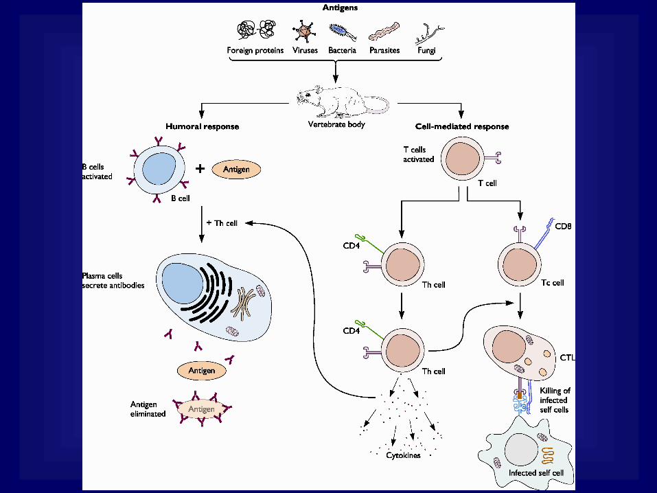
• Humoral response
Consists of lymphocytes of the B-cell lineage
Interaction of a specific receptor on precursor B
lymphocytes with antigens promotes differentiation
into antibody secreting cells (plasma cells).
• Cell-mediated response
Consists of lymphocytes of the T-cell lineage
Cytotoxic T cells (Tc cells) and T-helper cells (Th
cells) are the key effectors of this response.
The adaptive immune response:

The antigen receptors on the surface of B and T cells
B cells have about 100,000 molecules of a single antibody receptor per cell, which has specificity for one antigen epitope. T cells bearing the surface membrane protein CD4 always recognize peptides bound to MHC class II proteins and function as Th cells. T cells bearing the surface membrane protein CD8 always recognize peptide antigens bound to MHC class I proteins and function as cytotoxic T cells.

Field’s Virology, Fifth Edition
Antibody Activities in Viral Infection

Field’s Virology, Fifth Edition
Maturation of CD4+ and CD8+ T cells

Lymphocyte Subsets
TCR
a:b
Dominant T-cells Responding to Viral Infection
CD8+ recognize MHC I viral pepide complex
CD4+ recognize MHC II viral peptide complex,
regulate B- cell differentiation and inflammation
TH1 produce antiviral cytokines (IFNg)
TH2 produce cytokines for allergic response (IL-4, IL-5)
TCR g:
“Innate-like” effectors cells
express CD3 but not CD4 or CD8
recognize products of stressed cells
NKT
“innate-like” immune effects early in the immune response
express CD4, but not CD8
Treg
Control T- and B-responses
Express CD4 and CD25
Usually suppress T-and B-cell responces

• T lymphocytes recognize antigens on the surface of
self cells.
• The antigens on self cells can be recognized only by a
receptor on the surface of T cells when they are bound
to the MHC family of membrane proteins.
• The Th cells recognize antigens bound to MHC class II
molecules and produce powerful cytokines that affect
other lymphocytes (B and T cells) by promoting or
inhibiting cell division and gene expression.
• Once activated by Th cells, Tc cells differentiate into
CTLs that can kill virus infected cells.
Cell-mediated Response
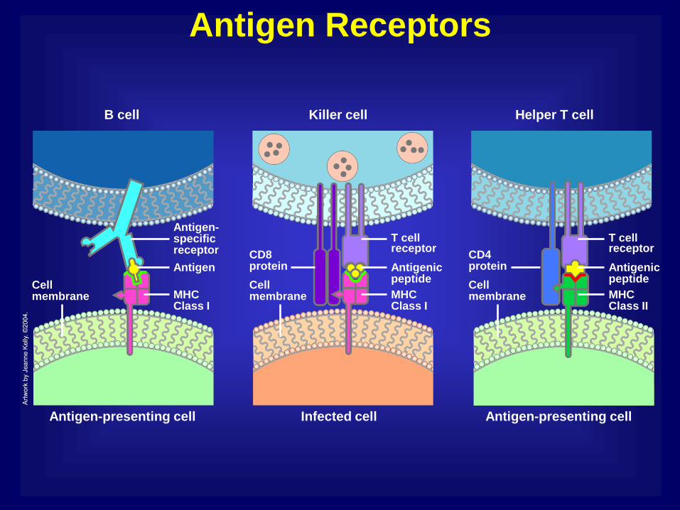
Antigen Receptors
Killer cell
Infected cell Antigen-presenting cell Antigen-presenting cell
CD8 protein
Cell membrane MHC
Class I MHC Class I
Antigenic peptide
T cell receptor
CD4 protein
Cell membrane MHC
Class II
Antigenic peptide
T cell receptor
Cell membrane
Antigen
Antigen-specific receptor
Helper T cell B cell

Regulatory T Cells
Regulatory T cells
Mature dendritic
cell
Regulatory T cell
Proliferation
T cells compete for cytokine signals
T cells compete for same antigen
Cytotoxic T cell