SEQUESTERING OF RAC BY THE YERSINIA EFFECTOR YOPO … · 1 SEQUESTERING OF RAC BY THE YERSINIA...
23
1 SEQUESTERING OF RAC BY THE YERSINIA EFFECTOR YOPO BLOCKS FCγ RECEPTOR-MEDIATED PHAGOCYTOSIS Eleanor Groves 1 , Katrin Rittinger 2 , Marlise Amstutz 3 , Sara Berry 1 , David W. Holden 1 , Guy R. Cornelis 3 , Emmanuelle Caron 1 † 1 Centre for Molecular Microbiology and Infection, Imperial College London, SW7 2AZ, United Kingdom 2 Division of Molecular Structure, National Institute for Medical Research, The Ridgeway, London, NW7 1AA, United Kingdom 3 Infection Biology, Biozentrum, University of Basel, Klingenbergstraße 50/70, CH-4056 Basel, Switzerland † Emmanuelle Caron passed away on July 8, 2009. Running head: YopO, a Rac-targeting membrane-bound GDI Address correspondence to: Eleanor Groves, CMMI2, Flowers Building, Imperial College, London, UK, SW15 3SH. Phone: + 44 207 594 3074, Fax: +44 207 594 3076, email: [email protected] Pathogenic Yersinia species neutralize innate immune mechanisms by injecting type three secretion effectors into immune cells, altering cell signalling. Our study elucidates how one of these effectors, YopO, blocks phagocytosis. We demonstrate using different phagocytic models that YopO specifically blocks Rac- dependent FcγR-internalisation pathway but not CR3-dependent uptake, which is controlled by Rho activity. We show that YopO prevents Rac activation, but does not affect Rac accumulation at the phagocytic cup. In addition, we show that plasma membrane localisation and the GDI-like domain of YopO cooperate for maximal anti-phagocytosis. While YopO has the same affinity for Rac1, Rac2 and RhoA in vitro, it selectively interacts with Rac isoforms in cells. This is due to the differential localisation of the Rho-family G proteins in resting cells: Rac isoforms partially exist as a GDI-free pool at the membrane of resting cells, whereas RhoA is trapped in the cytosol by RhoGDIα. We propose that YopO exploits this basic difference in localisation and availability to selectively inhibit Rac-dependent phagocytosis. INTRODUCTION Phagocytosis is a multi-step process displayed by a variety of cell types but particularly efficient in professional phagocytes (e.g. macrophages). It involves recognition, internalisation and degradation of particulate material over 0.5μm in diameter in a membrane-bound compartment called the phagosome. The first critical step during classical, zipper-like phagocytosis is the ligation of cell surface receptors (1). The best studied receptors are the opsono- receptors, Fcγ receptor (FcγR) and complement receptor 3 (CR3), which bind immobilised immunoglobulin and surface- deposited C3bi, respectively. Following ligation, receptors are thought to cluster around the phagocytic target and thereby initiate downstream signalling. This involves recruitment and activation of mediators that ultimately result in actin nucleation and polymerisation, through activation of Arp2/3, an event crucial to particle engulfment (2). Receptor-induced actin polymerisation drives the wrapping of membrane around the particle in a “zipper- like” manner, generally through the advancement of pseudopodia (3). Membrane delivery from intracellular membrane compartments is also required for completion of internalisation (4). Phagocytosis, like most processes reliant upon actin dynamics, is controlled through the action of Rho-family small G proteins. Indeed, Rho proteins link receptor ligation and actin polymerisation in all phagocytic events studied so far (5). However, different receptors employ distinct Rho proteins: RhoA activity is essential for internalisation through CR3, whilst Rac1 and Cdc42 act downstream of FcγR (6). Rho-family members act as molecular switches in eukaryotic cells, cycling between http://www.jbc.org/cgi/doi/10.1074/jbc.M109.071035 The latest version is at JBC Papers in Press. Published on November 19, 2009 as Manuscript M109.071035 Copyright 2009 by The American Society for Biochemistry and Molecular Biology, Inc.
Transcript of SEQUESTERING OF RAC BY THE YERSINIA EFFECTOR YOPO … · 1 SEQUESTERING OF RAC BY THE YERSINIA...

1
SEQUESTERING OF RAC BY THE YERSINIA EFFECTOR YOPO BLOCKS FCγ RECEPTOR-MEDIATED PHAGOCYTOSIS
Eleanor Groves1, Katrin Rittinger2, Marlise Amstutz3, Sara Berry1, David W. Holden1, Guy R. Cornelis3, Emmanuelle Caron1†
1Centre for Molecular Microbiology and Infection, Imperial College London, SW7 2AZ, United Kingdom 2Division of Molecular Structure, National Institute for Medical Research,
The Ridgeway, London, NW7 1AA, United Kingdom 3Infection Biology, Biozentrum, University of Basel, Klingenbergstraße 50/70, CH-4056 Basel, Switzerland
† Emmanuelle Caron passed away on July 8, 2009.
Running head: YopO, a Rac-targeting membrane-bound GDI
Address correspondence to: Eleanor Groves, CMMI2, Flowers Building, Imperial College, London, UK, SW15 3SH. Phone: + 44 207 594 3074, Fax: +44 207 594 3076, email:
Pathogenic Yersinia species neutralize innate immune mechanisms by injecting type three secretion effectors into immune cells, altering cell signalling. Our study elucidates how one of these effectors, YopO, blocks phagocytosis. We demonstrate using different phagocytic models that YopO specifically blocks Rac-dependent FcγR-internalisation pathway but not CR3-dependent uptake, which is controlled by Rho activity. We show that YopO prevents Rac activation, but does not affect Rac accumulation at the phagocytic cup. In addition, we show that plasma membrane localisation and the GDI-like domain of YopO cooperate for maximal anti-phagocytosis. While YopO has the same affinity for Rac1, Rac2 and RhoA in vitro, it selectively interacts with Rac isoforms in cells. This is due to the differential localisation of the Rho-family G proteins in resting cells: Rac isoforms partially exist as a GDI-free pool at the membrane of resting cells, whereas RhoA is trapped in the cytosol by RhoGDIα. We propose that YopO exploits this basic difference in localisation and availability to selectively inhibit Rac-dependent phagocytosis.
INTRODUCTION
Phagocytosis is a multi-step process displayed by a variety of cell types but particularly efficient in professional phagocytes (e.g. macrophages). It involves recognition, internalisation and degradation
of particulate material over 0.5µm in diameter in a membrane-bound compartment called the phagosome. The first critical step during classical, zipper-like phagocytosis is the ligation of cell surface receptors (1). The best studied receptors are the opsono-receptors, Fcγ receptor (FcγR) and complement receptor 3 (CR3), which bind immobilised immunoglobulin and surface-deposited C3bi, respectively. Following ligation, receptors are thought to cluster around the phagocytic target and thereby initiate downstream signalling. This involves recruitment and activation of mediators that ultimately result in actin nucleation and polymerisation, through activation of Arp2/3, an event crucial to particle engulfment (2). Receptor-induced actin polymerisation drives the wrapping of membrane around the particle in a “zipper-like” manner, generally through the advancement of pseudopodia (3). Membrane delivery from intracellular membrane compartments is also required for completion of internalisation (4). Phagocytosis, like most processes reliant upon actin dynamics, is controlled through the action of Rho-family small G proteins. Indeed, Rho proteins link receptor ligation and actin polymerisation in all phagocytic events studied so far (5). However, different receptors employ distinct Rho proteins: RhoA activity is essential for internalisation through CR3, whilst Rac1 and Cdc42 act downstream of FcγR (6).
Rho-family members act as molecular switches in eukaryotic cells, cycling between
http://www.jbc.org/cgi/doi/10.1074/jbc.M109.071035The latest version is at JBC Papers in Press. Published on November 19, 2009 as Manuscript M109.071035
Copyright 2009 by The American Society for Biochemistry and Molecular Biology, Inc.

2
inactive GDP-bound and active GTP-bound states (5). Guanine nucleotide exchange factors (GEFs) facilitate exchange of GDP for GTP, thereby activating small G proteins and allowing their GTP-dependent interaction with downstream effectors. The intrinsically low catalytic ability of Rho GTPases is greatly increased by interaction with GTPase-activating proteins (GAPs), leading to the recycling of the G protein to its GDP-bound, inactive form. A further level of regulation comes from the trafficking of the Rho proteins between different cellular compartments. The current models assign the Rho guanine dissociation inhibitors (RhoGDIs) a key function in keeping the Rho proteins in an inactive, cytosolic pool. Upon activation the G proteins are released from RhoGDI and recruited to membranes through their prenylated C-termini (7). All Rho-family members are thought to be regulated in a similar manner. Unsurprisingly, many bacterial effectors that manipulate phagocytic signalling do so by deregulating the activity of Rho GTPases (8,9).
Gram negative bacteria commonly subvert phagocytic uptake through the action of protein effectors that are directly injected into the host cell via a needle-like multi-protein complex called Type III secretion system (T3SS) (10). Yersinia species (Y. pestis, Y. pseudotuberculosis and Y. enterocolitica) are important extracellular human pathogens that utilise the T3SS injectosome to translocate a number of effector proteins into phagocytic cells (macrophages, dendritic cells and neutrophils), which in turn block bacterial uptake (11,12). The effector YopO, a 82 kDa multi-domain protein, was reported to trigger apoptosis, prevent cytokine secretion and nitric oxide production in epithelial cells and yeasts, modulate the actin cytoskeleton (typically with loss of actin stress fibres), and importantly inhibit phogocytosis (13-16). Indeed, unopsonised and IgG-coated yopO Yersinia mutants are internalised at a higher level than wild-type bacteria by mouse macrophages (11,15).
At a molecular level, YopO can be sub-divided into four functional domains. The N-terminal region of YopO functions as the chaperone-binding site prior to translocation
through the T3SS and mediates plasma membrane localisation after injection into host cells (17,18). It is flanked by a serine/threonine kinase domain recently shown to phosphorylate the heterotrimeric G protein subunit Gαq (19). The C-terminal region of YopO contains a GDI-like domain able to bind Rac and Rho in vitro, a property supported by the recently solved crystal structure of this domain (20,21). Finally a short coronin-homology domain is proposed to bind G-actin and promote kinase activity (13,22).
Despite the extensive analysis of YopO, its mechanism of action and the role of its individual domains remain poorly understood. Herein, we use an in vitro model of phagocytosis to study YopO-mediated anti-phagocytosis and the mechanism thereof. We found that the N-terminal membrane-addressing region in conjunction with the GDI-like domain specifically targets the Rac1-mediated phagocytic pathway. Importantly, we found that YopO executes it anti-phagocytic activity by targeting pre-existing GDI-free Rac1 at the plasma membrane.
EXPERIMENTAL PROCEDURES
Expression vectors
Eukaryotic plasmids encoding wild-type (wt) YopO (pEGFP::yopO, encoding YopOwt) and an N-terminal truncation (pEGFP::yopOΔ20-77, encoding YopO Δ20-77) mutants were described previously (18). pEGFP::yopO435-729 (encoding YopORBD) was constructed from pEGFP::yopO using Expand Long Template PCR System (Roche Applied Science) and the following primers: 5’-GGGGGGCTCGAGAGGCGGATAACACCCAAG-3’ and 5’-GGGGGGGAATTCTTATCACCATTCCCGCTCCAACCGGTTCAG-3’, and cloned into pEGFP-C3 (Clontech). Similarly, pEGFP::yopOΔ435-720 (encoding YopOΔRBD) and pEGFP::yopOD267AΔ435-720 (encoding YopOKDΔRBD) were constructed from pEGFP::yopO and pEGFP::yopOD267A respectively, using the following primers: 5’- GGGGGGACGCGTCGACTGAACCGGTT

3
CGAGCGGGAATCG-3' and 5’- GGGGGGACGCGTCCTTACATCAGTAGATAATGGGCTCATTTC-3’. pGEX::yopO435-729 was constructed using the following primers: forward 5’- GGGGGGGGATCCAGGCGGATAACACCCAAG-3’, reverse 5’- GGGGGGGAATTCTTATCACCATTCCCGCTCCAACCGGTTCAG-3’. pEGFP::yopOΔ83-434 (encoding YopO Δkinase) constructed by inverse PCR from pEGFP::yopOwt using the following primers: 5’- GGGGGGACGCGTAGGCGGATAACACCCAAGAAGCTTCG-3’ and 5’- GGGGGGACGCGTGAGTGTCTCAGTAAGGTTACGAGC -3', and the Expand Long Template PCR kit according to manufacturers instructions. All constructs were checked by sequencing (MWG) and prepared for transfection using the Qiagen Endo-free maxi-prep kit. Eukaryotic expression vectors encoding FcγRIIA and CR3 (αM and β 2 integrin chains) have been described previously (6). pGEX, plasmids encoding GST-tagged Rac1, Rac2 and RhoA were a kind gift of K.Rittinger. pGEX plasmids encoding GST-tagged PAK-CRIB and Rhotekin have been previously described (23). UD-GFP was a kind gift of MJ Bijlmakers (Division of Immunology, Infection and Inflammatory Disease, King's College London, London, UK) (24).
Cell culture and transfection
African green monkey COS-7 fibroblast, J774A.1 and RAW264.7 mouse macrophage cell lines were obtained from the ATCC and maintained in Dulbecco’s Modified Eagles Medium (DMEM, Gibco) supplemented with 10% heat-inactivated foetal calf serum (FCS, Sigma), penicillin (Sigma, 100 Units/ml) and streptomycin (Sigma, 0.1 mg/ml) at 37ºC, 5% CO2. COS-7 cells were transiently transfected using Lipofectamine2000 (Invitrogen), with a 0.8:2 DNA:Lipofectamine2000 ratio, according to manufacturer’s instructions. RAW264.7 were transiently transfected using a 2:3 DNA:Lipofectamine2000 (Invitrogen) ratio, or by electroporation (AMAXA, NucleofactorV, program D-32) according to the manufacturer’s instructions. For pull-down experiments 9µg DNA was used to transfect 3x10⁵ COS-7 cells using
the SuperFect (Qiagen) reagent according to manufacturer’s instructions.
For siRNA transfection COS-7 cells were seeded at 5x10⁵ cells per 10cm-dish. The following day cells were transfected with 5nM of luciferase or RhoGDIα duplexes (SMARTpools, Dharmacon) using Interferin (Polyplus transfection) according to manufacturers instructions. Cells were left for 48 hours before use.
Phagocytosis assays
Transfected COS-7 or RAW264.7 cells were first pre-incubated for at least 1 hour in Serum-free DMEM with 10mM Hepes (Sigma). In order to activate the endogenous CR3 receptors, RAW264.7 were pre-treated with phorbol-12-myristate-13-acetate (PMA, 150ng/ml, Sigma) for 15 minutes at 37ºC. Cells were challenged with 0.5µl of opsonised sheep red blood cells (sRBC, TCS Biosciences) per 13mm coverslip for 30 minutes, washed with PBS to remove unbound sRBC and fixed with 4% paraformaldehyde (PFA) for 15 minutes at 4ºC. Opsonisation with IgG or C3bi was performed as previously described (6). Coverslips were then washed and fixed as described above.
Immunofluorescence and scoring
Binding and phagocytosis were distinguished by differential staining of sRBC before or after permeabilisation with 0.2% Triton X-100, using combinations of Cy5-, rhodamine- and AMCA- conjugated donkey anti-rabbit IgG (Jackson Immunoresearch Laboratories). Coverslips were inverted onto glass slides on 5µl Mowiol 4-88 (Calbiochem), and observed using Olympus BX50 epifluorescence or LSM510 Zeiss Axiovert 100M confocal microscopes. In total 100 COS-7 cells (50 RAW264.7 cells) or phagosomes (representing 10-20 cells) were counted for each experiment. Association and phagocytic indices (AI/PI) reflect the total number of sRBC or bacteria associated to or internalised by 100 transfected cells, respectively. % internalisation corresponds to (AI/PI)x100. At least three experiments were performed per condition. Student’s t tests for significance were performed as indicated and p values <0.05 are shown.

4
Protein expression and GST-pulldown assay
Expression of YopO435-729, Rac1, Rac2 and RhoA in fusion with GST was induced in subcultures of Escherichia coli BL21 (DE3) using 0.5mM isopropyl-beta-D-thiogalactopyranoside (IPTG). After 4 hours at 30ºC, bacteria were resuspended in 50mM Tris pH8, 40mM EDTA, 25% sucrose, 100mM MgCl2, 0.2% Triton X-100, 1mM PMSF and Complete Protease inhibitor cocktail (Roche). To purify GST-Rac1, -Rac2 and -RhoA, bacteria were resuspended in lysis buffer [50mM Tris pH7.5, 300mM NaCl, 4mM DTT and EDTA-free protease inhibitors (Roche)] and sonicated. After lysate clearing by ultra-centrifugation, fusion proteins were affinity-purified on Glutathione Sepharose 4B beads (Amersham Biosciences) according to manufacturer’s instructions.
J774A.1 or transfected COS-7 cells were lysed in ice-cold lysis buffer (10% glycerol, 1% NP40, 50mM Tris pH7.6, 200mM NaCl, 10mM MgCl2, 1mM PMSF, Roche complete tablet). Lysates were incubated with glutathione sepharose beads coupled to 25µg of either GST or GST-RBD for 1 hour at 4ºC. Beads were washed three times in cold lysis buffer before analysis by SDS-PAGE and Western blotting. The following primary antibodies were used: anti-Rac (23A8, Upstate), anti-RhoA (119, Santa Cruz Biotechnology), anti-myc (4A6, Upstate), β-tubulin (TUB2.1, Sigma). HRP-conjugated anti-mouse or anti-rabbit (Amersham) antibodies were used as appropriate, and bands detected using the Enhanced chemiluminescence kit (ECL, Amersham). G protein loading was achieved by addition of GDP or GTPγS (100µM) and EDTA (5mM) followed by incubation at 30ºC for 10 minutes, and addition of MgCl2 (50mM). For mass spectrometry, bead eluates were separated by SDS-PAGE and stained using a compatible silver nitrate protocol as previously described (25).
Cell fractionation
J774A.1 macrophages were seeded the day prior to fractionation, washed once with cold PBS, resuspended in lysis buffer (10mM Tris pH7.5, 0.5mM DTT, 5mM NaCl, Roche complete inhibitor) and sonicated on ice for
30 seconds. Post-nuclear supernatants were subjected to ultra-centrifugation at 100 000g, for 45 minutes at 4ºC. The supernatant (cytosol fraction) and the pellet (membrane fraction) were resuspended in equal volumes of lysis buffer supplemented with 1% Triton X-100.
Mass spectrometry (MS) analysis
In preparation for mass spectrometry the desired protein band was excised, lyophilized and digested with trypsin (E.C.3.4.21.4, Promega) overnight. Peptides were extracted from gel pieces and nano-liquid chromatography was performed on an Ultimate 3000 using a PepMap 100 75µm × 15-cm fused silica C18 analytical column (LC Packings, Dionex, Sunnyvale, CA), coupled to a Probot for fraction collection and matrix addition with 2,5-dihydrobenzoic acid as the matrix. Matrix-assisted laser desorption/ionisation-time of flight (MALDI TOF/TOF) MS was performed using a Applied Biosystems 4800 mass spectrometer (Foster City, CA.) in the positive reflectron mode with Delayed Extraction. MS precursor acquisition was followed by interpretation and data-dependent MS/MS acquisition with the CID on. Data interpretation was configured to select a maximum of 10 precursor ions per fraction with a minimum signal-to-noise ratio of 50. The data were processed using GPS Explorer (Applied Biosystems, CA) against the Swis-Prot database. Search parameters were: enzyme = trypsin: fixed modifications = carboxymethyl (C); variable modifications = oxidation (M); mass tolerance ± 100 ppm: fragment mass tolerance = 0.3 Da: maximum missed cleavages = 1; mass values = monoisotopic.
Isothermal titration calorimetry (ITC) analysis
Purified recombinant proteins produced in E. coli from pGEX expression vectors were cleaved from GST by incubation with human thrombin (Calbiochem) then further purified for ITC by gel filtration on an equilibrated (50mM Tris ph7.5, 100mM NaCl) Superdex S75 column. Fractions containing pure protein were pooled and concentrated before use. Purified proteins were dialyzed into ITC buffer (50mM Tris pH 7.5, 50mM NaCl, 2mM MgCl2, 2mM
5
DTT) overnight prior to use. Isothermal calorimetric titrations were performed with a Microcal omega VP-ITC (MicroCal Inc., Northampton, MA). Typically 400-700µM of G protein was titrated into 40-70µM of YopORBD. Data were fitted by the least-squares method using the evaluation software, Microcal Origin version 5.0. All measurements were repeated at least two times and the estimated error is the standard deviation between measurements.
RESULTS
YopO is present at phagocytic cups and specifically blocks FcγR-phagocytosis
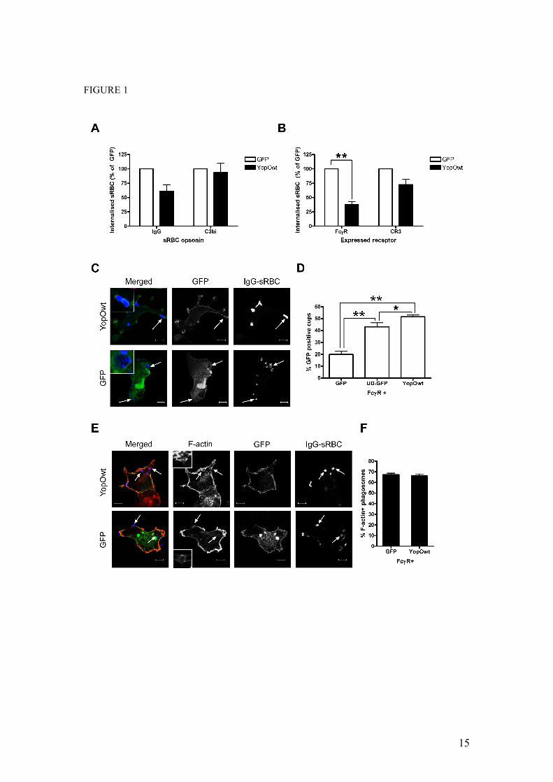
It is established that YopO contributes to anti-phagocytosis mediated by Yersinia type three secretion effectors (11). However, the molecular mechanism involved is unknown. To address this question we took advantage of the existence of two well-defined phagocytic pathways, triggered by ligation of the FcγR and CR3 receptors, which are governed by distinct signalling cascades in macrophages and receptor-transfected fibroblasts (6). We expressed GFP or GFP-tagged YopO in RAW264.7 macrophages (Figure 1A) and COS-7 fibroblasts co-transfected with constructs encoding either FcγRIIA or CR3 (Figure 1B). Cells were then challenged with IgG- or C3bi-opsonised red blood cells (RBC) to selectively target one phagocytic receptor or the other. In both cell systems, YopO had a greater inhibitory effect on the internalisation of IgG- than of C3bi-RBC. In macrophages, FcγR-mediated phagocytosis was reduced by 40%, with no reduction seen for CR3-dependent uptake. In transfected COS-7 cells, FcγR internalisation was inhibited by 60%, whilst only a small reduction (ca. 25%) was observed for CR3 phagocytosis. Importantly, expression of YopO had no effect on the level of RBC attachment (data not shown), suggesting YopO acts on phagocytic signalling rather than on receptor expression or availability. Of note, the selectivity of YopO towards FcγR-dependent uptake parallels the behaviour of yopO-deleted Y. enterocolitica, where the presence of YopO was important for phagocytosis of IgG-opsonised bacteria,
but loss of YopO had no affect on uptake of complement-opsonised bacteria (11).
Regardless of the receptor involved, phagocytic uptake is subject to tight spatio-temporal control. Most of its regulators and effectors (e.g. Rho proteins, Arp2/3 complex, actin filaments) are recruited and functional at the site of particle binding (9). Since YopO preferentially blocks FcγR-mediated phagocytosis, we tested whether YopO could be found underneath bound IgG-RBC. In COS-7 cells co-expressing FcγRIIA and either GFP or GFP-tagged YopO, then challenged with RBC, YopO was generally membrane associated and clearly enriched as a ring around 50% of the bound RBC, unlike GFP (Figure 1C and D) that showed 20% localisation to phagocytic cups. YopO also showed significantly higher enrichment than a membrane-targeted GFP construct, where the unique domain (UD) of the Src-family kinase Lck mediates association of GFP to the membrane (Figure 1D, p=0.0339) implying that YopO enrichment at phagocytic cups is not solely a result of its membrane localisation domain. Overall, these data suggest that YopO might interfere with phagocytic pathways downstream of receptor ligation to prevent internalisation. As it has been reported that YopO over-expression disrupts the actin cytoskeleton (13,17), we investigated the possibility that YopO would prevent F-actin polymerisation at sites of sRBC binding. YopO-expressing COS-7 cells had no visible defect in actin cup formation (Figure 1E and F), indicating that YopO does not drastically affect the de novo actin polymerisation initiated during phagocytosis. Interestingly, expression of either dominant negative Rac or Cdc42 has no effect on actin cup formation but reduces phagocytosis; only when both Cdc42 and Rac are simultaneously inactivated is actin cup formation abolished (26). This suggests that Rac and Cdc42 both contribute to actin cup formation at sites of FcγR uptake, and that YopO targets one of these Rho proteins, since YopO expression phenocopies inactivation of one of the two Rho proteins in this respect.
YopO prevents Rac activation not its localisation to the phagocytic cup

6
Since YopO does not interact with Cdc42 (21,27) (Supplementary Figure 2), Rac is the most likely Rho protein target downstream of FcγR signalling. We tested whether YopO blocks FcγR-phagocytosis by inhibiting Rac recruitment to the phagocytic cup. COS-7 cells were co-transfected with FcγRIIA and GFP or YopO-GFP and challenged for 10 minutes after synchronisation with IgG-RBC. In parallel a FcγR-mutant (FcγR2YF), which is defective in Rac recruitment (26), was included as a negative control. Endogenous Rac was visualised by immunofluorescence microscopy using a specific antibody and levels of recruitment to RBC were scored (Figure 2A). Cells expressing GFP or YopO showed similar levels of endogenous Rac recruitment to cups, whereas as expected the signalling mutant FcγRY2F showed a much lower level (Figure 2B). This suggests that the presence of YopO does not exclude Rac from the phagocytic cup.
We next investigated if YopO blocks Rac activation. To this end COS-7 cells co-expressing FcγR and GFP or YopO were subjected to a synchronised IgG-RBC challenge. Cells were lysed at 0 and 10 min after incubation at 37ºC and cell lysates were incubated with the Rac/Cdc42-binding domain of p21activated kinase (GST-PAK CRIB) immobilised on beads. Bead eluates and whole cell lysates were analysed by SDS-PAGE and western blot, probing for Rac (Figure 3A) and results were quantified (Figure 3B). In cells expressing GFP there was a consistent activation, approximately 4-fold, of Rac at 10 min compared to 0 min. Significantly, this activation was not detectable in cells expressing YopO, suggesting that YopO expression blocks activation of Rac that is required for successful phagocytosis downstream of FcγR ligation.
The GDI-like region of YopO is the preponderant domain mediating anti-phagocytosis
Next, we investigated the role of YopO domains in the inhibition of FcγR-mediated uptake. To this end several YopO mutants were constructed, co-transfected with a vector encoding FcγRIIA into COS-7 cells and analysed for their ability to interfere
with phagocytosis of IgG-RBC. A mutant lacking the kinase-domain (YopOΔkinase), a deletion mutant lacking the GDI-like region (YopOΔRBD for Rho-family G protein Binding Domain), a kinase-dead and GDI-like region-deleted double mutant (YopOkinase-deadΔRBD) and the isolated GDI-like domain (YopORBD) were studied in parallel (Figure 4). No significant difference in protein expression from the different constructs was observed by western blot or during microscopic analysis (Supplementary Figure 1) and none of the YopO constructs significantly reduced the ability of RBC to bind transfected COS-7 cells (Figure 4A). The mutant lacking the kinase domain reduced RBC uptake to a similar extent as YopOwt, indicating that this region is not required for activity, (Figure 4B). In contrast, the mutant lacking the GDI-like region showed significantly reduced anti-phagocytic activity compared to YopOwt. The kinase-dead, RBD-deleted mutant showed total abrogation of anti-phagocytosis. Consistently, expression of the isolated RBD alone significantly reduced uptake.
The GDI-like domain of YopO binds Rac1 and RhoA with equal affinity in vitro
The C-terminal half of YopO was reported to interact with Rac1 and RhoA in vitro (20,21,27). Furthermore, recent evidence indicates that YopO GDI-like domain prevents guanine nucleotide exchange on both Rac1 and RhoA in vitro (20). This contrasts with the selectivity (Rac over Rho) we observed in our phagocytosis assays. To further investigate the in vitro interactions of YopO GDI-like region with small G proteins we performed pulldown assays, incubating purified recombinant Rac1 (Figure 5A) or RhoA (Figure 5B), with purified GST-YopO RBD. As the nucleotide dependency of the YopO-small G protein interaction is unclear, we pre-loaded the purified G proteins with either GDP or GTPγS, to mimic the inactive and active state of RhoA and Rac1. Nucleotide loading was confirmed by a parallel pulldown assay using beads coated with GST-PAK-CRIB or GST-Rhotekin, which interact specifically with the active, GTP-bound forms of Rac1 and RhoA, respectively (28,29). Bound proteins were analysed by SDS-PAGE, followed by

7
immunoblotting. When compared to the input, the levels of bound G proteins were similar, suggesting that YopO RBD binds Rac1 and RhoA equally well in vitro. Furthermore, in both cases the inactive forms of the G proteins appeared to interact more strongly than the active forms. Additionally, we performed pulldown experiments from lysates of COS-7 cells over-expressing myc-tagged, Cdc42, RhoA or Rac1 using GST-YopO RBD or GST alone as a control (Supplementary Figure 2). Overexpressed Rac1 and RhoA, but not Cdc42, interacted specifically and equally with the YopO RBD beads. These results suggest that prenylated Rac1 and RhoA behave similarly to the bacterially expressed proteins and that YopO does not bind Cdc42.
We next quantified the YopO RBD-G protein interaction using isothermal titration calorimetry (ITC) (Figure 5C,D). Increasing concentrations of the small G proteins were titrated into YopO RBD, and the change in heat evoked upon complex formation was measured. Representative titrations of GDP-bound Rac1 and RhoA are shown. Rac1.GDP bound to YopO RBD with an affinity of 1.14 µM, and a 1:1 stoichiometry. Similarly, RhoA.GDP formed a 1:1 complex with YopO RBD with an affinity of 0.91 µM. There was no detectable difference between the respective affinities of YopO RBD for Rac1 and RhoA in vitro. ITC experiments using GTP loaded G proteins were attempted but there was not sufficient interaction to measure by ITC (data not shown), supporting the idea that YopO RBD has a preference for inactive-GDP bound G proteins. The isolated YopO RBD behaves like RhoGDI
The paradoxical difference between the in vitro and in cellulo results, where Rac-dependent FcγR phagocytosis is most sensitive to YopO, prompted us to explore further the biochemical and functional properties of isolated YopO RBD and compare them with those of eukaryotic RhoGDIα. Firstly, the ability of RhoGDIα to interact with Rac1 and RhoA was confirmed (Figure 6A). Macrophage lysates were incubated with immobilised GST-RhoGDIα, and its interaction with RhoA and Rac1 was
tested. As expected (30), RhoGDIα interacted to a similar extent with both G protein isoforms. To directly compare the anti-phagocytic capacities of YopO RBD and RhoGDIα we co-transfected COS-7 cells with vectors expressing GFP-tagged YopO RBD, RhoGDIα or GFP alone together with either FcγR or CR3 and measured phagocytosis of RBC (Figure 6B). Overexpression of RhoGDIα reduced phagocytosis through both receptors by approximately 50%. YopO RBD also reduced phagocytosis through both receptors, but to a noticeably lesser extent (by ca 25%) than RhoGDIα. This difference cannot be attributed to different expression levels of YopO RBD and RhoGDIα, as shown by western blotting (data not shown). Our results suggest a fundamental functional difference between full length YopO and its isolated RBD domain, since the isolated RBD shows no anti-phagocytic preference for a particular pathway, similar to the effect of overexpressing RhoGDIα.
YopO RBD binds preferentially to cellular Rac
Since full length YopO has a selective anti-phagocytic activity on the FcγR-mediated pathway, we decided to further investigate the interaction of YopO RBD with Rho-family proteins in a cellular context. Lysates from J774A.1 macrophages (Figure 7A) and COS-7 fibroblasts (Figure 7B) were incubated with GST-YopO RBD; bound proteins were analysed by SDS-PAGE and immunoblotting to reveal endogenous Rac1 and RhoA. In both cell types, relative to input, YopO RBD repeatedly showed a stronger interaction with Rac1 compared to RhoA. Further evidence that YopO RBD preferentially interacts with Rac in a cellular context comes from mass spectrometry analysis of proteins pulled-down with the isolated RBD domain. J774A.1 lysates were incubated with beads coated with GST-YopO RBD, and bead-associated proteins were separated by SDS-PAGE and visualised by silver nitrate staining. A specific YopO-associated band of approximately 20-25 kDa (the apparent molecular mass of Rho-family proteins) was excised and prepared for nano-liquid chromatography and MS-MS analysis (Figure 7C). When analyzed using the

8
SwissProt database, the only two significant peptide hits corresponded to mouse Rac2 and Rac1, with scores of 284 and 146 respectively. The sequence coverage of the matched peptides was 31% of the Rac2 protein (Figure 7D). Importantly, no other Rho-family protein, in particular RhoA or Cdc42-related peptides, was identified. Since binding of Rac2 to YopO has never been reported, we quantified their interaction by ITC. The Kd measured was 1µM, similar to that determined for Rac1 and RhoA, (data not shown).
YopO targets a GDI-free pool of Rac at the plasma membrane
One possible explanation for the observed interaction of YopO for Rac over RhoA is that, within cells, the former is more available to YopO than the latter, which could be linked with the intracellular locale of the proteins involved and/or their interaction with binding partners. It is generally assumed that inactive Rho-family proteins are confined to the cytosol, and recruited and/or activated locally at membranes, but this has not been systematically analysed for all Rho proteins. Therefore we investigated the intracellular localisation of RhoA and Rac by separating cells into membrane and cytosolic fractions. Fractions from J774A.1 macrophages (Figure 8A) and COS-7 fibroblasts (which do not express Rac2 (31), Figure 8B) were first tested for the presence of TfnR and β -tubulin. As shown in Figure 8A and B, there was no significant contamination of these proteins into the cytosolic and membrane fractions, respectively. When these fractions were probed for Rac1, Rac2 and RhoA, the Rac isoforms distributed to the membrane and cytosol fractions, whereas RhoA was only detected in the cytosolic fraction. In macrophages Rac2 was found almost exclusively at the membrane, whereas Rac1 was separated into a membrane pool and a larger cytosolic fraction. In COS-7 cells the majority of Rac1 was found at the membrane, suggesting differences in the localisation of Rac1 between different cell types. Interestingly, endogenous RhoGDI was, like Rho, exclusively cytosolic. This suggests that there is a pool of RhoGDI-free Rac at the membrane, and that RhoA is retained in the cytosol, possibly through an
interaction with RhoGDI. In such a scenario YopO RBD would interact better with Rac than RhoA because it cannot efficiently out-compete RhoGDI binding to RhoA. To test this hypothesis, we silenced expression of RhoGDIα in COS-7 cells and compared the ability of YopO RBD to interact with Rac1 and RhoA in pull-down assays (Figure 8C). Upon loss of RhoGDIα there was an increased interaction of YopO RBD with both Rac1 and RhoA, compared to control cells (Figures 8C and 7A). Strikingly, where previously no interaction of YopO RBD and RhoA could be detected, knockdown of RhoGDIα allowed an interaction suggesting that in control cells RhoA is trapped by RhoGDIα, and that loss of this interaction by knockdown allows interaction with YopO RBD.
YopO localises to the plasma membrane of transfected or infected cells, through its N-terminal region (17,18). To test the hypothesis that YopO localisation at the plasma membrane is required for anti-phagocytic activity, a GFP-tagged mutant of YopO (YopOΔ20-77), lacking the region required for plasma membrane localisation was used. This deletion mutant was almost exclusively cytosolic, like GFP, but unlike full-length YopO. This was observed both in RAW264.7 macrophages (Figure 9A) and in COS-7 cells (Figure 7B) and was confirmed independently by cell fractionation followed by western blotting. There was no obvious contamination of the fractions, as judged by the separation profile of tubulin (cytosol fraction) and transferrin receptor (TfnR, membrane fraction) (data not shown). GFP alone was primarily found in the cytosolic fraction, as expected. Conversely, the vast majority of GFP-tagged YopO was in the membrane fraction. Importantly, overexpressed YopOΔ20-77 was found in the cytosol fraction, with only a small proportion still detectable within the membrane fraction. Next, we co-expressed the YopO localisation mutant (YopOΔ20-77) with FcγRIIA in COS-7 cells. Upon phagocytic challenge with IgG-sRBC, this mutant (which still harbours an intact RBD) had significantly reduced anti-phagocytic ability (Figure 9D, p=0.0489). The remaining anti-phagocytic activity of this mutant is similar to that observed against

9
CR3-mediated phagocytosis (28% for FcγR, 15% for CR3) suggesting it could be a non-specific effect of overexpression of a GDI-like molecule. Altogether, we conclude that YopO mediates specific anti-phagocytosis towards FcγR, where activity is primarily mediated by its Rho-family binding GDI-like domain, through co-compartmentalisation with a pool of RhoGDI-free Rac at the plasma membrane of target cells.
DISCUSSION
Herein, we demonstrate that YopO is able to specifically block FcγR-mediated phagocytosis whilst having no significant effect on CR3-dependent internalisation. This is in sharp contrast to the T3SS effector EspJ of enteropathogenic E. coli which blocks both FcγR-mediated and CR3-dependent phagocytosis, the underlining mechanism of which is not yet known (32). Furthermore, we show that the N-terminal localisation domain and the GDI-like Rho-family binding domain cooperate in mediating inhibition of Rac-dependent uptake, whilst the kinase domain of YopO is dispensable. Furthermore, this study has revealed differential localisation of RhoA, which is only detectable in the cytosol and Rac isoforms that are present, at least in part, as a GDI-free pool at the plasma membrane. We propose that this membrane-associated Rac is the cellular target for YopO anti-phagocytic activity.
Our data establish cooperation between the N-terminal localisation domain and the G-protein binding regions (RBD) of YopO for anti-phagocytic function. First, we show that YopO deletion mutants lacking only the RBD domain are significantly reduced in their anti-phagocytic potential; this region alone is also sufficient for activity. Second, the N-terminal mutant (Δ20-77) is severely affected in anti-phagocytosis. Third, the selective activity of the RBD towards Rac-dependent phagocytosis is only observed when the N-terminal region, responsible for plasma membrane localisation, is present. In contrast, the kinase domain of YopO plays no role in anti-phagocytosis, although our data could suggest a regulatory role for the full kinase domain, since its presence but
inactivity seems to totally abrogate YopO activity.
The N-terminal region of YopO has no independent anti-phagocytic activity of its own, since a double mutant of YopO -lacking kinase activity and RBD domain- had no effect on FcγR-dependent phagocytosis. The YopO N-terminal region has a dual function as a chaperone-binding site within bacteria and, once translocated, is responsible for plasma membrane localisation in host cells (18). As yet, the mechanism by which it mediates this localisation is unknown; in particular, it does not harbour any obvious post-translational modification sites, e.g. for prenylation or myristoylation. Interestingly, the N-terminal domain of YopO has a net positive charge, hinting at a possible charge-dependent mechanism by which YopO N-terminus may be interacting with the negatively-charged inner leaflet of the plasma membrane, as proposed for several Ras-family proteins (33).
We show that YopO selectively blocks Rac-dependent uptake pathways. YopO consistently blocked FcγR-mediated uptake both in macrophages and in FcγR-transfected COS-7 cells. We would predict that, since Yersinia uptake via invasin is Rac-dependent, bacterial uptake would also be blocked by YopO. This would confirm that YopO is able to act on phagocytic particles of different sizes, a possibility which we have not addressed in this study. Interestingly, the functional selectivity of YopO for Rac-dependent pathways in cells is not paralleled by an increased affinity of its RBD for Rac over Rho in vitro. As was seen previously, we did not detect any interaction with Cdc42. Importantly, using pull-down assays followed by mass spectrometry we have identified Rac1 and Rac2 as the predominant Rho-family members able to interact with YopO RBD in macrophages. This suggests that cellular Rho is not available for interaction with YopO RBD in pull-down experiments, most likely because of the tight interaction of RhoA and RhoGDI in the cytosol (7).
A recent report convincingly showed that YopO RBD has a GDI-like structure and activity, although unlike GDI, YopO RBD

10
does not interact with the C-terminal prenyl moiety of RhoA and Rac1 (20). In accordance with the idea that YopO RBD is a GDI mimic, overexpression of either RhoGDI or YopO RBD decreased FcγR- mediated phagocytosis. However, our work strongly suggests that the biochemical and structural similarities between YopO RBD and host cell RhoGDIs mask some important functional differences. Indeed, unlike full length YopO, RhoGDI also blocks CR3-mediated uptake. The main difference between RhoGDI and YopO is their sub-cellular localisation: RhoGDI resides mainly in the cytosol, while YopO associates with the plasma membrane when overexpressed (this study, (18)) or when translocated following infection (17). All known T3SS effectors that impact on the regulation on Rho-family G proteins (e.g. SopE from Salmonella typhimurium, ExoS from Pseudomonas aeruginosa) are thought to act purely as mimics (34). Remarkably, our work has revealed that the mechanism of action of YopO depends both on GDI-mimicry and a specific sub-cellular localisation.
Our data show that YopO RBD can bind Rac1 and Rac2 in vitro; it targets membrane-associated Rac1 and Rac2 in macrophages but only Rac1 in COS-7 cells. Rac2 expression is restricted to haematopoietic cells, and our results therefore suggest that FcγR-phagocytosis is reliant upon Rac1 in COS-7 cells, as suggested before (6). However, our data cannot differentiate which is the predominant isoform required during FcγR-phagocytosis in macrophages. Nevertheless they provide an explanation for why YopO has an effect on NADPH oxidase activation, a process which is Rac2 dependent (35). Furthermore, our work supports the data that suggest Yersinia target β1-integrin mediated uptake by phagocytes, a process shown to require Rac and not Rho (36).
Interestingly, our data provide strong experimental evidence for a differential
localisation of endogenous Rac and RhoA in cells. Overexpressed RhoA was found to localise to the cytosol even when highly overexpressed, whereas overexpressed Rac1 and Rac2 were localised to membrane compartments, in agreement with our results (37,38). The localisation of Rac isoforms to the plasma membrane is thought to be due to the strong polybasic domain of Rac. This charge-dependent membrane association is further utilised by the cell to regulate Rac distribution: when the surface potential is altered, e.g. following activation of phagocytic receptors, Rac is released from the membrane of the forming phagosome (33). By contrast, RhoA has a weak polybasic domain that does not support membrane association. In addition, we show that RhoGDIα helps to retain RhoA in the cytosol and free from binding partners. The functional implications of these fundamental differences are many. The classical GTPase cycle is thought to involve activation and localisation to the membrane, followed by inactivation and cytosolic GDI-trapping. Our data show that this is not a true representation of the activation cycle of all G proteins. Rac is clearly already present at the membrane in resting cells, and presumably it would be locally activated upon initiation of phagocytosis or other Rac-activating stimuli. Our data show that binding of YopO to membrane Rac interferes with the activation process. By contrast, Rho-dependent phagocytosis and other Rho-dependent processes require trafficking of the RhoGDI-RhoA complex, or RhoA alone, to the membrane. The link between function and sub-cellular localisation of G proteins is only just starting to be understood, and the subtle differences in primary sequence between the different isoforms will have important implications for their regulation and function. The fact that a bacterial virulence factor has evolved to exploit this highlights its importance in cell signalling. Our data also suggest that YopO could be used as a tool to specifically target Rac-dependent pathways.

11
REFERENCES 1. Griffin, F. M., Jr., Griffin, J. A., Leider, J. E., and Silverstein, S. C. (1975) J Exp Med
142(5), 1263-1282 2. May, R. C., Caron, E., Hall, A., and Machesky, L. M. (2000) Nat Cell Biol 2(4), 246-
248 3. Swanson, J. A. (2008) Nat Rev Mol Cell Biol 9(8), 639-649 4. Booth, J. W., Trimble, W. S., and Grinstein, S. (2001) Semin Immunol 13(6), 357-364 5. Patel, J. C., Hall, A., and Caron, E. (2000) Methods Enzymol 325, 462-473 6. Caron, E., and Hall, A. (1998) Science 282(5394), 1717-1721 7. DerMardirossian, C., and Bokoch, G. M. (2005) Trends Cell Biol 15(7), 356-363 8. Finlay, B. B. (2005) Curr Top Microbiol Immunol 291, 1-10 9. Groves, E., Dart, A. E., Covarelli, V., and Caron, E. (2008) Cell Mol Life Sci 10. Cornelis, G. R. (2006) Nat Rev Microbiol 4(11), 811-825 11. Grosdent, N., Maridonneau-Parini, I., Sory, M. P., and Cornelis, G. R. (2002) Infect
Immun 70(8), 4165-4176 12. Marketon, M. M., DePaolo, R. W., DeBord, K. L., Jabri, B., and Schneewind, O.
(2005) Science 309(5741), 1739-1741 13. Juris, S. J., Rudolph, A. E., Huddler, D., Orth, K., and Dixon, J. E. (2000) Proc Natl
Acad Sci U S A 97(17), 9431-9436 14. Nejedlik, L., Pierfelice, T., and Geiser, J. R. (2004) Yeast 21(9), 759-768 15. Wiley, D. J., Nordfeldth, R., Rosenzweig, J., DaFonseca, C. J., Gustin, R., Wolf-Watz,
H., and Schesser, K. (2006) Microb Pathog 40(5), 234-243 16. Park, H., Teja, K., O'Shea, J. J., and Siegel, R. M. (2007) J Immunol 178(10), 6426-
6434 17. Hakansson, S., Galyov, E. E., Rosqvist, R., and Wolf-Watz, H. (1996) Mol Microbiol
20(3), 593-603 18. Letzelter, M., Sorg, I., Mota, L. J., Meyer, S., Stalder, J., Feldman, M., Kuhn, M.,
Callebaut, I., and Cornelis, G. R. (2006) Embo J 25(13), 3223-3233 19. Navarro, L., Koller, A., Nordfelth, R., Wolf-Watz, H., Taylor, S., and Dixon, J. E.
(2007) Mol Cell 26(4), 465-477 20. Prehna, G., Ivanov, M. I., Bliska, J. B., and Stebbins, C. E. (2006) Cell 126(5), 869-
880 21. Barz, C., Abahji, T. N., Trulzsch, K., and Heesemann, J. (2000) FEBS Lett 482(1-2),
139-143 22. Trasak, C., Zenner, G., Vogel, A., Yuksekdag, G., Rost, R., Haase, I., Fischer, M.,
Israel, L., Imhof, A., Linder, S., Schleicher, M., and Aepfelbacher, M. (2007) J Biol Chem 282(4), 2268-2277
23. Wiedemann, A., Patel, J. C., Lim, J., Tsun, A., van Kooyk, Y., and Caron, E. (2006) J Cell Biol 172(7), 1069-1079
24. Bijlmakers, M. J., Isobe-Nakamura, M., Ruddock, L. J., and Marsh, M. (1997) J Cell Biol 137(5), 1029-1040
25. Shevchenko, A., Wilm, M., Vorm, O., and Mann, M. (1996) Anal Chem 68(5), 850-858 26. Cougoule, C., Hoshino, S., Dart, A., Lim, J., and Caron, E. (2006) J Biol Chem
281(13), 8756-8764 27. Dukuzumuremyi, J. M., Rosqvist, R., Hallberg, B., Akerstrom, B., Wolf-Watz, H., and
Schesser, K. (2000) J Biol Chem 275(45), 35281-35290 28. Reid, T., Furuyashiki, T., Ishizaki, T., Watanabe, G., Watanabe, N., Fujisawa, K.,
Morii, N., Madaule, P., and Narumiya, S. (1996) J Biol Chem 271(23), 13556-13560 29. Thompson, G., Owen, D., Chalk, P. A., and Lowe, P. N. (1998) Biochemistry 37(21),
7885-7891 30. Gorvel, J. P., Chang, T. C., Boretto, J., Azuma, T., and Chavrier, P. (1998) FEBS Lett
422(2), 269-273 31. Wheeler, A. P., Wells, C. M., Smith, S. D., Vega, F. M., Henderson, R. B.,
Tybulewicz, V. L., and Ridley, A. J. (2006) J Cell Sci 119(Pt 13), 2749-2757 32. Marches, O., Covarelli, V., Dahan, S., Cougoule, C., Bhatta, P., Frankel, G., and
Caron, E. (2008) Cell Microbiol 10(5), 1104-1115 33. Yeung, T., Terebiznik, M., Yu, L., Silvius, J., Abidi, W. M., Philips, M., Levine, T.,
Kapus, A., and Grinstein, S. (2006) Science 313(5785), 347-351 34. Alto, N. M., Shao, F., Lazar, C. S., Brost, R. L., Chua, G., Mattoo, S., McMahon, S.
A., Ghosh, P., Hughes, T. R., Boone, C., and Dixon, J. E. (2006) Cell 124(1), 133-145

12
35. Yamauchi, A., Kim, C., Li, S., Marchal, C. C., Towe, J., Atkinson, S. J., and Dinauer, M. C. (2004) J Immunol 173(10), 5971-5979
36. Alrutz, M. A., Srivastava, A., Wong, K. W., D'Souza-Schorey, C., Tang, M., Ch'Ng, L. E., Snapper, S. B., and Isberg, R. R. (2001) Mol Microbiol 42(3), 689-703
37. Michaelson, D., Silletti, J., Murphy, G., D'Eustachio, P., Rush, M., and Philips, M. R. (2001) J Cell Biol 152(1), 111-126
38. Adamson, P., Paterson, H. F., and Hall, A. (1992) J Cell Biol 119(3), 617-627
ACKNOWLEDGEMENTS
We would like to gratefully acknowledge Professor Gadi Frankel, Drs J Mota, M Bright and D Lees for critical reading of this manuscript and Dr MJ Bijlmakers and B. Stieglitz for providing reagents. The Medical Research Council (MRC, UK) funded this work through a studentship to EG and a core grant to KR. Work in the Caron lab is funded by grants from the MRC and the Biotechnology and Biological Sciences Council (BBSRC, UK).
ABBREVIATIONS LIST
CR3: complement receptor 3, FcγR: Fcγ receptor, GDI: Guanine-nucleotide dissociation inhibitor, MS: mass spectrometry, RBD: Rho-family binding domain, T3SS: Type III secretion, wt: wild-type
DEDICATION We would like to dedicate this manuscript to the memory of Emmanuelle Caron, who tragically passed away after suffering from cancer during the preparation of this manuscript. She was an incredibly talented young scientist who brightened the lives of all who knew her, and was an inspirational, warm and caring mentor to her “phago” group, who will always miss her.

13
FIGURE LEGENDS FIGURE 1 Preferential inhibition of FcγR-mediated phagocytosis by YopO
(A) RAW264.7 mouse macrophages were transiently transfected with vectors expressing GFP or GFP-tagged YopOwt (YopOwt), then challenged with IgG- or C3bi-opsonised sRBC for 30 minutes. (B) COS-7 cells were co-transfected with FcγRIIA or CR3 vectors and either GFP or YopOwt, and challenged with appropriately opsonised sRBC for 30 minutes. Phagocytosis assays were performed and scored as described in materials and methods. At least 50 GFP-expressing RAW264.7 macrophages or 100 COS-7 cells were scored per experiment. Data represent mean ± SEM from at least 3 independent experiments. (C) COS-7 cells were transiently transfected with FcγRIIA and either GFP or GFP-YopOwt, then challenged with IgG-opsonised sRBC for 15 minutes after synchronisation at 4ºC. sRBC and F-actin were labelled and observed by confocal microscopy. GFP enrichment at site of sRBC binding was examined. Note the localisation of YopO, but not GFP around sRBC (arrows). (D) Quantification of GFP localisation to phagocytic cups in cells co-expressing FcγRIIA and GFP, YopO-GFP or UD-GFP (membrane targeted GFP). (E) Following sRBC challenge, cells expressing GFP or YopOwt were analysed for F-actin enrichment at sites of sRBC binding as described in materials and methods. Representative images are shown, with typical F-actin rings enlarged. (F) Quantification of actin cup formation. At least 100 sRBC were scored for the presence of F-actin-rich cups per experiment. Data represent mean + SEM from at least 3 independent experiments. Scale bars, 10µm. * indicates p<0.05, ** indicates p<0.01
FIGURE 2 YopO does not affect Rac recruitment to FcγR-phagocytic cups
(A) COS-7 cells were transiently transfected with FcγRIIA wildtype (wt) or mutant FcγRIIA (Y2F) and either GFP or GFP-YopOwt, then challenged with IgG-opsonised sRBC for 10 minutes after synchronisation at 4ºC. sRBC and endogenous Rac were labelled and observed by confocal microscopy. Rac enrichment at site of sRBC binding was examined. Scale bars, 10µm. (B) Quantification of Rac localisation to phagocytic cups in cells co-expressing FcγRIIA or FcγRIIA2YF and GFP or YopO-GFP. At least 100 sRBC were scored for the presence of Rac at sRBC cups per experiment. Data are mean + SEM of 3 independent experiments.
FIGURE 3 YopO blocks Rac activation during FcγR-phagocytosis
(A) COS-7 cells were transiently transfected with FcγRIIA and either GFP or GFP-YopOwt, then challenged with IgG-opsonised sRBC for 10 minutes at 37ºC after synchronisation at 4ºC. Cells were lysed at 0 min and 10 min and incubated with GST-PAK CRIB immobilised on glutathione sepharose beads. Bead eluates and lysates were analysed by SDS-PAGE and western blot probing for Rac. A representative exoeriment is shown. (B) The fold activation of Rac was quantified and shown relative to activation levels at 0 minutes (GFP-expressing cells). Data are mean + SEM of 3 independent experiments. * indicates p<0.05
FIGURE 4 Domain analysis of YopO: The GDI-like domain (RBD) is the main contributor to anti-phagocytosis
(A) COS-7 cells were co-transfected with FcγRIIA and GFP-tagged constructs as indicated. After 24 hours, cells were challenged with IgG-opsonised sRBC for 30 minutes and processed for immuno-fluorescence as described in materials and methods. Total associated sRBC for each condition were scored in at least 100 GFP-positive cells per experiment. (B) COS-7 cells were transfected as above and phagocytosed sRBC were scored for each condition. Data are expressed relative to GFP control, arbitrarily set at 100%. Data represent mean + SEM of at least 3 independent experiments. (C) Domain organisation of the constructs used above. * indicates p<0.05
FIGURE 5 YopO RBD shows no preference for Rac1 or RhoA in vitro
Purified recombinant Rac1 (A) and RhoA (B) were loaded with GDP or GTPγS, then incubated at 1µM or 0.1µM with 20µg of GST-YopO RBD immobilised on glutathione

14
sepharose beads. Following 1 hour incubation, eluates were analysed by SDS-PAGE and western blotting using antibodies to Rac and Rho. As a nucleotide loading control, GDP or GTPγS- bound Rac1 and RhoA were incubated with the Rac/Cdc42-binding domain of p21 activated kinase (PAK-CRIB) and the Rho binding domain of Rhotekin (Rhotekin) respectively, immobilised on sepharose beads and analysed as above. Interactions between purified G proteins and YopO RBD were further analysed by isothermal titration calorimetry. Representative titration of purified Rac1 (C) and RhoA (D). Experiments were repeated on at least 2 independent occasions with the same results.
FIGURE 6 RhoGDI and YopO RBD block both FcγR- and CR3-mediated phagocytosis
(A) J774A.1 lysates were incubated with GST-RhoGDI immobilised on glutathione sepharose beads and resolved by SDS-PAGE and western blot, probing for Rac, RhoA and actin. A sample of RhoGDI beads and bead eluates were run in parallel. At least 100 GFP-expressing COS-7 cells were scored per experiment. Results shown are representative of 3 experiments. (B) COS-7 cells were co-transfected with FcγRIIA or CR3 and a GFP-tagged construct: either RhoGDIα, YopO RBD or GFP alone, challenged with appropriately opsonised sRBC for 30 minutes and scored for sRBC phagocytosis. Data are expressed relative to the GFP control, arbitrarily set at 100%. Data are mean + SEM of at least 2 independent experiments. * indicates p<0.05
FIGURE 7 Unlike RhoGDI, YopO RBD prefentially binds Rac from cell lysates
J774A.1 (A) or COS-7 (B) lysates were incubated with GST-YopO RBD immobilised on glutathione sepharose beads and bead eluates were resolved by SDS-PAGE and western blotting, probing for Rac, RhoA and β -tubulin. Results shown are representative of 3 experiments. (C) After pulldown and SDS-PAGE, as described for (A), bead eluates were stained by a MS-compatible silver nitrate stain. The 25kDa band was analysed by MALDI-TOF MS. (D) Mouse Rac2 amino acid sequence, showing the overall sequence coverage of Rac1 and Rac2 peptides identified by MS (bold red). Amino acid differences between Rac1 and 2 are underlined.
FIGURE 8 A GDI-free pool of Rac molecules exists at the plasma membrane of resting cells
J774A.1 (A) and COS-7 (B) lysates were separated into membrane (M) and cytosol (C) fractions as described in the methods. After SDS-PAGE, blots were probed for Transferrin receptor (TfnR), β -tubulin, Rac, Rho and RhoGDI. (C) COS-7 cells were treated with luciferase- or RhoGDIα- specific siRNA duplexes, as described in materials and methods. After 48 hours cells were lysed and incubated with GST-YopO RBD or GST immobilised on glutathione sepharose beads. Bead eluates and samples of lysates were resolved by SDS-PAGE and western blot, probing for RhoGDI (confirming knockdown), Rac, Rho and actin (loading control). Data are representative of at least 3 independent experiments.
FIGURE 9 Membrane localisation of YopO is necessary for anti-phagocytosis
(A) RAW264.7 macrophages were transiently transfected with GFP-tagged versions of YopO (wt or Δ20-77) or GFP alone. After 24 hours, cells were stained for F-actin and observed using a Zeiss Axiovert 100M confocal microscope. Representative examples are shown. Scale bars, 10µm. (B) COS-7 cells were transfected and processed as described in (A). Scale bars, 20µm. (C) Lysates of COS-7 cells transfected with GFP-tagged constructs as indicated were separated into cytosol (C) and membrane (M) fractions, as described in the methods. After SDS-PAGE, fractions were probed using anti-GFP antibodies. (D) COS-7 cells were co-transfected with FcγRIIA and either GFP-tagged YopO (wt or Δ20-77), challenged with IgG-sRBC and at least 100 GFP-expressing COS-7 cells were scored for phagocytosis per experiment. Data are given as mean + SEM of at least 3 independent experiments. * indicates p<0.05
15
FIGURE 1

16
FIGURE 2

17
FIGURE 3

18
FIGURE 4

19
FIGURE 5

20
FIGURE 6

21
FIGURE 7

22
FIGURE 8

23
FIGURE 9