Sequential treatment with 5aza2deoxycytidine and ... 2015.pdf · and deacetylase inhibitors...
22
Research Article Sequential treatment with 5-aza-2 0 -deoxycytidine and deacetylase inhibitors reactivates HIV-1 Sophie Bouchat 1 , Nadège Delacourt 1 , Anna Kula 1 , Gilles Darcis 1,2 , Benoit Van Driessche 1 , Francis Corazza 3 , Jean-Stéphane Gatot 1,† , Adeline Melard 4 , Caroline Vanhulle 1 , Kabamba Kabeya 5 , Marion Pardons 1 , Véronique Avettand-Fenoel 4 , Nathan Clumeck 5 , Stéphane De Wit 5 , Olivier Rohr 6,7 , Christine Rouzioux 4 & Carine Van Lint 1,* Abstract Reactivation of HIV gene expression in latently infected cells together with an efficient cART has been proposed as an adjuvant therapy aimed at eliminating/decreasing the reservoir size. Results from HIV clinical trials using deacetylase inhibitors (HDACIs) ques- tion the efficiency of these latency-reversing agents (LRAs) used alone and underline the need to evaluate other LRAs in combina- tion with HDACIs. Here, we evaluated the therapeutic potential of a demethylating agent (5-AzadC) in combination with clinically tolerable HDACIs in reactivating HIV-1 from latency first in vitro and next ex vivo. We showed that a sequential treatment with 5-AzadC and HDACIs was more effective than the corresponding simultaneous treatment both in vitro and ex vivo. Interestingly, only two of the sequential LRA combinatory treatments tested induced HIV-1 particle recovery in a higher manner than the drugs alone ex vivo and at concentrations lower than the human tolera- ble plasmatic concentrations. Taken together, our data reveal the benefit of using combinations of 5-AzadC with an HDACI and, for the first time, the importance of treatment time schedule for LRA combinations in order to reactivate HIV. Keywords demethylating agent; epigenetics; HDACIs; HIV latency; HIV reservoir Subject Categories Microbiology, Virology & Host Pathogen Interaction; Pharmacology & Drug Discovery DOI 10.15252/emmm.201505557 | Received 19 June 2015 | Revised 6 November 2015 | Accepted 9 November 2015 Introduction Thirty years after its discovery, human immunodeficiency virus type 1 (HIV-1) remains a major problem of public health. Combination antiretroviral therapy (cART) is potent but not curative. cART requires lifelong adherence and does not fully restore health or a normal immune status in HIV-1-infected individuals. Although multiple reservoirs may exist, the HIV-1 reservoirs containing stably integrated, transcriptionally silent but replication-competent proviruses are recognized to predominate among infected CD4 + T cells (Eisele & Siliciano, 2012). They are therefore a permanent source for virus reactivation and could be responsible for the rebound of plasma viremia observed after cART interruption (Tyagi & Bukrinsky, 2012). Persistence of truly latent (i.e. non-defective) HIV-1 proviruses represents a major obstacle to eradication, as suggested by the failure of cART intensification strategies at clearing the viral reservoirs (Dinoso et al, 2009; Gandhi et al, 2010; Yukl et al, 2010). Indeed, the levels of HIV-1 reservoirs appear as one of the critical factors influencing the duration of a remission after cART cessation (Saez-Cirion et al, 2013). Consequently, a decline of the HIV-1 latent reservoirs to a level sufficient to permit an efficient control of the infection by the host immune system might allow interruptions in therapy (“treatment-free windows”). Reactivation of HIV gene expression in latently infected cells together with an efficient or intensified cART could serve as an adjuvant therapy aimed at eliminating/decreasing the pool of latent viral reservoirs. The chromatin organization and the epigenetic control of the HIV-1 promoter are key elements in transcriptional silencing (Van Lint et al, 2013). The repressive nucleosome nuc-1, located immedi- ately downstream of the transcription start site, is maintained hypoacetylated by histone deacetylases (HDACs) in latent condi- tions (Verdin et al, 1993; Van Lint et al, 1996). The use of HDAC inhibitors (HDACIs) as latency-reversing agents (LRAs) has been 1 Service of Molecular Virology, Department of Molecular Biology (DBM), Université Libre de Bruxelles (ULB), Gosselies, Belgium 2 Service des Maladies Infectieuses, Centre Hospitalier Universitaire (CHU) de Liège, Domaine Universitaire du Sart-Tilman, Université de Liège, Liège, Belgium 3 Laboratory of Immunology, IRISLab, CHU-Brugmann, Université Libre de Bruxelles (ULB), Brussels, Belgium 4 Service de Virologie, EA7327, AP-HP, Hôpital Necker-Enfants-Malades, Université Paris-Descartes, Paris, France 5 Service des Maladies Infectieuses, CHU St-Pierre, Université Libre de Bruxelles (ULB), Brussels, Belgium 6 IUT Louis Pasteur de Schiltigheim, University of Strasbourg, Schiltigheim, France 7 Institut Universitaire de France (IUF), Paris, France *Corresponding author. Tel: +32 2 650 98 07; Fax: +32 2 650 98 00; E-mail: [email protected] † Present address: Service de Génétique, Centre Hospitalier Universitaire (CHU) de Liège, Domaine Universitaire du Sart-Tilman, Liège, Belgium ª 2015 The Authors. Published under the terms of the CC BY 4.0 license EMBO Molecular Medicine 1 Published online: December 17, 2015
Transcript of Sequential treatment with 5aza2deoxycytidine and ... 2015.pdf · and deacetylase inhibitors...

Research Article
Sequential treatment with 5-aza-20-deoxycytidineand deacetylase inhibitors reactivates HIV-1Sophie Bouchat1, Nadège Delacourt1, Anna Kula1, Gilles Darcis1,2, Benoit Van Driessche1,
Francis Corazza3, Jean-Stéphane Gatot1,†, Adeline Melard4, Caroline Vanhulle1, Kabamba Kabeya5,
Marion Pardons1, Véronique Avettand-Fenoel4, Nathan Clumeck5, Stéphane De Wit5, Olivier Rohr6,7,
Christine Rouzioux4 & Carine Van Lint1,*
Abstract
Reactivation of HIV gene expression in latently infected cellstogether with an efficient cART has been proposed as an adjuvanttherapy aimed at eliminating/decreasing the reservoir size. Resultsfrom HIV clinical trials using deacetylase inhibitors (HDACIs) ques-tion the efficiency of these latency-reversing agents (LRAs) usedalone and underline the need to evaluate other LRAs in combina-tion with HDACIs. Here, we evaluated the therapeutic potential ofa demethylating agent (5-AzadC) in combination with clinicallytolerable HDACIs in reactivating HIV-1 from latency first in vitroand next ex vivo. We showed that a sequential treatment with5-AzadC and HDACIs was more effective than the correspondingsimultaneous treatment both in vitro and ex vivo. Interestingly,only two of the sequential LRA combinatory treatments testedinduced HIV-1 particle recovery in a higher manner than the drugsalone ex vivo and at concentrations lower than the human tolera-ble plasmatic concentrations. Taken together, our data reveal thebenefit of using combinations of 5-AzadC with an HDACI and, forthe first time, the importance of treatment time schedule for LRAcombinations in order to reactivate HIV.
Keywords demethylating agent; epigenetics; HDACIs; HIV latency;
HIV reservoir
Subject Categories Microbiology, Virology & Host Pathogen Interaction;
Pharmacology & Drug Discovery
DOI 10.15252/emmm.201505557 | Received 19 June 2015 | Revised 6 November
2015 | Accepted 9 November 2015
Introduction
Thirty years after its discovery, human immunodeficiency virus type
1 (HIV-1) remains a major problem of public health. Combination
antiretroviral therapy (cART) is potent but not curative. cART
requires lifelong adherence and does not fully restore health or a
normal immune status in HIV-1-infected individuals. Although
multiple reservoirs may exist, the HIV-1 reservoirs containing stably
integrated, transcriptionally silent but replication-competent
proviruses are recognized to predominate among infected CD4+
T cells (Eisele & Siliciano, 2012). They are therefore a permanent
source for virus reactivation and could be responsible for the
rebound of plasma viremia observed after cART interruption (Tyagi
& Bukrinsky, 2012). Persistence of truly latent (i.e. non-defective)
HIV-1 proviruses represents a major obstacle to eradication, as
suggested by the failure of cART intensification strategies at clearing
the viral reservoirs (Dinoso et al, 2009; Gandhi et al, 2010; Yukl
et al, 2010). Indeed, the levels of HIV-1 reservoirs appear as one of
the critical factors influencing the duration of a remission after cART
cessation (Saez-Cirion et al, 2013). Consequently, a decline of the
HIV-1 latent reservoirs to a level sufficient to permit an efficient
control of the infection by the host immune system might allow
interruptions in therapy (“treatment-free windows”). Reactivation
of HIV gene expression in latently infected cells together with an
efficient or intensified cART could serve as an adjuvant therapy
aimed at eliminating/decreasing the pool of latent viral reservoirs.
The chromatin organization and the epigenetic control of the
HIV-1 promoter are key elements in transcriptional silencing (Van
Lint et al, 2013). The repressive nucleosome nuc-1, located immedi-
ately downstream of the transcription start site, is maintained
hypoacetylated by histone deacetylases (HDACs) in latent condi-
tions (Verdin et al, 1993; Van Lint et al, 1996). The use of HDAC
inhibitors (HDACIs) as latency-reversing agents (LRAs) has been
1 Service of Molecular Virology, Department of Molecular Biology (DBM), Université Libre de Bruxelles (ULB), Gosselies, Belgium2 Service des Maladies Infectieuses, Centre Hospitalier Universitaire (CHU) de Liège, Domaine Universitaire du Sart-Tilman, Université de Liège, Liège, Belgium3 Laboratory of Immunology, IRISLab, CHU-Brugmann, Université Libre de Bruxelles (ULB), Brussels, Belgium4 Service de Virologie, EA7327, AP-HP, Hôpital Necker-Enfants-Malades, Université Paris-Descartes, Paris, France5 Service des Maladies Infectieuses, CHU St-Pierre, Université Libre de Bruxelles (ULB), Brussels, Belgium6 IUT Louis Pasteur de Schiltigheim, University of Strasbourg, Schiltigheim, France7 Institut Universitaire de France (IUF), Paris, France
*Corresponding author. Tel: +32 2 650 98 07; Fax: +32 2 650 98 00; E-mail: [email protected]†Present address: Service de Génétique, Centre Hospitalier Universitaire (CHU) de Liège, Domaine Universitaire du Sart-Tilman, Liège, Belgium
ª 2015 The Authors. Published under the terms of the CC BY 4.0 license EMBO Molecular Medicine 1
Published online: December 17, 2015

well characterized in several latency models and in ex vivo
cART-treated HIV-1+ patient cell cultures (Quivy et al, 2002; Archin
et al, 2009; Contreras et al, 2009; Reuse et al, 2009; Matalon et al,
2010). Several anti-HIV latency clinical studies and trials using
HDACIs have been reported in the HIV field [VPA (Lehrman et al,
2005; Siliciano et al, 2007; Archin et al, 2008, 2010; Sagot-Lerolle
et al, 2008; Routy et al, 2012a,b), SAHA (Archin et al, 2012, 2014;
Elliott et al, 2014), panobinostat (Rasmussen et al, 2015), and romi-
depsin (Sogaard et al, 2015)]. Altogether, these studies are encour-
aging but question the efficiency of HDACIs used alone to reduce
the size of the HIV-1 reservoirs and underline the need to evaluate
other classes of LRAs, alone or in combination with HDACIs.
Targeting simultaneously different mechanisms of latency should
be more efficient when viral eradication/remission is the objective
since the combination of different classes of compounds could
synergize (i.e. result in a higher reactivation level than the sum of
the reactivations produced by each compound individually) to reac-
tivate HIV expression in latently infected cells. In this regard, we
have previously demonstrated proof-of-concepts for the coadminis-
tration of two different classes of promising LRAs [an NF-jBinducer + an HDACI (Quivy et al, 2002; Adam et al, 2003; Reuse
et al, 2009), an NF-jB inducer + a P-TEFb-releasing agent (Darcis
et al, 2015), a histone methyltransferase inhibitor (HMTI) + an
HDACI (Bouchat et al, 2012), an HMTI + an NF-jB inducer
(Bouchat et al, 2012)] as a therapeutic perspective to decrease the
pool of latent HIV-1 reservoirs in the presence of efficient cART.
Epigenetically, it is known that DNA methylation and histone
deacetylation cooperate to establish and maintain a heterochromatin
environment. In the case of HIV, the HIV-1 promoter has been previ-
ously shown to be hypermethylated ex vivo and resistant to reactiva-
tion in the latent reservoirs from cART-treated aviremic HIV-1
infected individuals, as opposed to the hypomethylated 50 LTR of
integrated proviruses present in viremic patients (Blazkova et al,
2009). Although controversy remains about the level of DNA methy-
lation in patient cells in vivo (Blazkova et al, 2009, 2012; Palacios
et al, 2012; Ho et al, 2013), the DNA methylation status of the HIV-1
promoter could contribute to “lock” the silent state of the provirus
in cooperation with histone repressive post-translational modifi-
cations such as histone deacetylation, thereby making the return of
the provirus to an active state more difficult (Blazkova et al, 2009).
In this view, demethylating agents could represent promising candi-
date drugs in combination with HDACIs for reducing the pool of
latent HIV reservoirs. Two well-characterized nucleoside analog
DNA methylation inhibitors, 5-azacytidine (5-AzaC, marketed as
Vidaza) and 5-aza-20-deoxycytidine (5-AzadC, marketed as Daco-
gen), are currently FDA-approved to treat myelodysplastic
syndrome and used in cancer therapies (Kantarjian et al, 2006).
Few studies have already tested the HIV-1 reactivation potential of
5-AzadC + HDACI combinatory treatments using latently infected
cell lines but have failed to show any synergistic effect in vitro
(Blazkova et al, 2009; Kauder et al, 2009; Fernandez & Zeichner,
2010).
In this report, we thoroughly studied the sequential aspect of
cellular treatments combining demethylating agents with clinically
tolerable HDACIs in latently infected T-cell lines and in ex vivo
cultures of CD8+-depleted PBMCs or resting CD4+ T cells from
cART-treated aviremic HIV-1+ patients. We demonstrated that these
two classes of LRAs synergistically reactivated HIV in the context of
sequential treatments. Moreover, we determined their metabolic
activity profiles and their impact on global T-cell activation. Taken
together, our data reveal the benefit of using combinations of a
demethylating agent and an HDACI and, for the first time, the
importance of treatment time schedule for LRA combinations in
order to reactivate HIV.
Results
The DNA methylation inhibitor 5-AzadC induces HIV-1transcription and production in a latently infected T-cell line
Several postintegration latency models exist to study the mecha-
nisms of transcriptional reactivation and the pathogenesis of HIV-1.
In order to test the HIV-1 reactivation potential of 5-AzaC and
5-AzadC DNA methylation inhibitors, we used the HIV-1 latently
infected J-Lat 8.4 cell line since the Verdin’s laboratory has previ-
ously reported that two CpG islands flanking the transcription start
site are hypermethylated in several latently infected J-Lat cell lines
(Kauder et al, 2009).
As these drugs are nucleoside analogs and are incorporated into
DNA or RNA, we performed stimulation kinetics (24, 48 and 72 h)
and only obtained viral production in culture supernatants after
72 h of treatment. Therefore, Fig 1A shows only the data for the
72-h time point. At 72 h post-treatment, we observed that 5-AzadC,
but not 5-AzaC at the same doses, induced viral production in a
dose-dependent manner from 400 nM to 6.25 lM (Fig 1A). WST-1
assays revealed that metabolic activities decreased in a dose-depen-
dent manner from 400 nM ranging from 66.3% to 39.3% when
using increasing 5-AzadC concentrations (Fig 1B). We confirmed
the potency of 5-AzadC in comparison with 5-AzaC (observed by
quantification of p24 viral production in Fig 1A) by showing that
5-AzadC increased the number of GFP-positive cells (assessed by
FACS in Fig 1C) and increased viral mRNA expression (assessed by
RT–qPCR in Fig 1D). Of note, relative levels of initiated (TAR)
transcripts were lower than those of elongated (tat) transcripts for
all conditions as compared to mock-treated condition. This
phenomenon can be explained by the fact that more TAR tran-
scripts are detected in mock-treated condition due to RNA poly-
merase II pausing present in latency condition. We also analyzed
the mean fluorescence intensities (MFI) of the GFP-positive cell
populations following increasing concentrations of 5-AzadC
(Appendix Fig S1), and we showed that the amount of GFP
produced per cell was also increased, indicating an enhanced HIV-1
gene expression.
As 5-AzadC reactivated HIV-1 from latency at a concentration of
400 nM (Fig 1A) that is lower than the tolerable peak of plasmatic
concentration (Cmax) after usual dosage (20 mg/m²) in human anti-
cancer therapy [around 650 nM (Inc E (2014) Dacogen (decitabine)
for injection, full prescribing information)], we decided to use
400 nM of 5-AzadC as working concentration in our next experi-
ments (indicated by an arrow, Fig 1A). Study of the 5-AzaC reacti-
vation potential at higher doses than the ones we used had no
interest because the Cmax after usual dosage (75 mg/m²) in human
anticancer therapy is around 3 lM for 5-AzaC (Laille et al, 2014).
The higher reactivation potential of 5-AzadC compared to 5-AzaC
can be explained by the different intracellular metabolisms of these
EMBO Molecular Medicine ª 2015 The Authors
EMBO Molecular Medicine Sequential treatment with 5-AzadC + HDACI induces HIV-1 Sophie Bouchat et al
2
Published online: December 17, 2015

A
B
C
D
E
Figure 1. The DNA methylation inhibitor 5-AzadC induces HIV-1 expression in latently infected T cells.
A–D J-Lat 8.4 cells were mock-treated or treated with increasing concentrations of 5-AzadC or 5-AzaC. At 72 h post-treatment, viral production was measured byquantifying p24 antigen production in culture supernatants (A); metabolic activity was assessed by a WST-1 assay (B); viral protein expression was analyzed byFACS (C); and initiated (primers TAR) or elongated (primers tat) transcripts were quantified by RT–qPCR (D). The selected dose was indicated by an arrow.
E J-Lat 8.4 cells were mock-treated or treated with 5-AzadC (400 nM) or TNF-a (10 ng/ml) as a positive control. At 24, 48 or 72 h post-treatment, initiated (primersTAR) or elongated (primers tat) transcripts were quantified by RT–qPCR.
Data information: For (D, E), results were normalized using b-actin gene primers and are presented as histograms indicating the fold inductions compared to mock-treated condition for each time period. For (A–E), means and standard errors of the means from three independent biological duplicates (n = 6) are indicated. The resultobtained with mock-treated cells was arbitrarily set at a value of 1 (D, E) and 100% (B).
ª 2015 The Authors EMBO Molecular Medicine
Sophie Bouchat et al Sequential treatment with 5-AzadC + HDACI induces HIV-1 EMBO Molecular Medicine
3
Published online: December 17, 2015
two drugs (Li et al, 1970). Indeed, in contrast to its reduced analog
5-AzadC, 5-AzaC is a ribonucleoside and has to be first reduced in a
deoxynucleoside via a limiting enzymatic step before being incorpo-
rated into DNA. Moreover, while 5-AzadC incorporates exclusively
into DNA, only a small percentage (10–20%) of 5-AzaC is incorpo-
rated into DNA, the remainder being incorporated into RNA (Li
et al, 1970).
Since a 72-h treatment with 5-AzadC was required to observe an
increase in HIV-1 expression (see here above), we performed kinet-
ics studies to follow the viral mRNA level increase in response to
5-AzadC treatment. As shown in Fig 1E, 5-AzadC caused an increased
expression of both initiated (TAR) and elongated (tat) HIV-1 tran-
scripts, which was the highest at 72 h post-treatment, whereas a
24-h treatment was sufficient to observe the effect of the NF-jBinducer TNF-a on viral transcriptional activity (Fig 1E).
In conclusion, we demonstrated for the first time that 5-AzadC,
in contrast to 5-AzaC, reactivated HIV-1 expression from latency
in latently infected J-Lat 8.4 cells treated for 72 h, time needed to
obtain an effective removal of the viral transcription block, proba-
bly due, at least partially, to DNA demethylation as previously
shown by Verdin and colleagues (Kauder et al, 2009). Conse-
quently, Dacogen (5-AzadC), but not Vidaza (5-AzaC), used at
concentrations lower than that generally achieved in human
cancers could be a promising LRA and be used in combination
with other LRAs in reactivation strategies aimed at reducing the
HIV-1 reservoirs.
Determination of an optimal concentration of several clinicallytolerable HDACIs used in human therapy to induce HIV-1production in a latently infected T-cell line
In order to highlight new therapeutic approaches to purge latent
HIV reservoirs, we selected some clinically tolerable HDACIs that
could be administrated in future HIV clinical trials. We compared
the reactivation potentials of HDACIs previously extensively tested
in several HIV-1 reactivation studies (VPA, NaBut, MS-275, and
SAHA) (reviewed in Van Lint et al, 2013) to those of three promis-
ing and more recently tested HDACIs (belinostat, panobinostat, and
romidepsin) (Rasmussen et al, 2013; Wei et al, 2014). Table 1
describes the characteristics of these HDACIs.
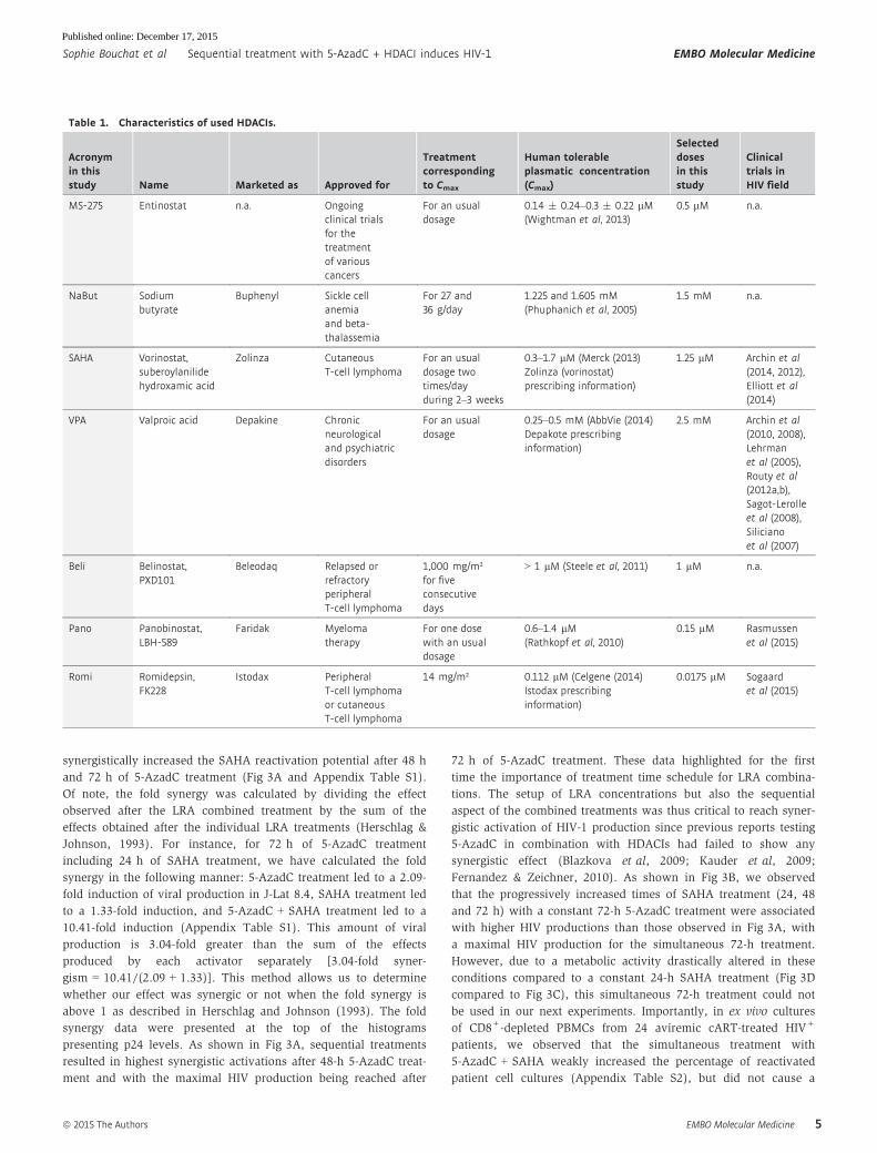
As shown in Fig 2, all selected HDACIs, except MS-275, induced
viral production after 24 h in a dose-dependent manner in the
latently infected J-Lat 8.4 cell line (Fig 2A and B). This measure-
ment is commonly performed 24 h post-treatment for HDACIs in
in vitro HIV reactivation experiments (Reuse et al, 2009). The
absence of reactivation with MS-275 was in agreement with results
previously reported (Reuse et al, 2009; Wightman et al, 2013). Of
note, in the Lewin’s study (Wightman et al, 2013), a significant viral
production has been detected following MS-275 treatment after 48 h
of stimulation in the ACH2 cell line and after 3 days of stimulation
in a primary CD4+ T-cell model for HIV-1 latency.
As shown in Fig 2A, NaBut presented a higher reactivation
potential than those observed with VPA, MS-275, and SAHA.
However, the HDACIs belinostat, panobinostat, and romidepsin
induced higher HIV-1 production than VPA, NaBut, MS-275, and
SAHA (Fig 2B, compared to Fig 2A). Moreover, if we only consid-
ered the HDACI concentrations close to their Cmax (indicated by a
box in Fig 2A and B), panobinostat and romidepsin were more
potent than the other HDACIs (including belinostat). Taken
together, we confirmed and extended previous works (Rasmussen
et al, 2013; Wei et al, 2014) by comparing the HIV-1 reactivation
potentials of several HDACIs. This allowed us to determine for
each HDACI an optimal concentration (outlined by an arrow in
Fig 2A and B) in terms of both their HIV-1 reactivation potential
and their Cmax (mentioned at the bottom of Fig 2A and B). In our
next experiments, we selected the following HDACI concentrations
for combinatory treatments with 5-AzadC: (i) MS-275 at a concen-
tration of 0.5 lM in agreement with Cmax; (ii) NaBut at 1.5 mM
consistent with Cmax; (iii) SAHA at 1.25 lM in agreement with
Cmax; (iv) VPA at 2.5 mM, which is higher than Cmax but allowed
HIV reactivation as opposed to VPA reactivation incapacity at Cmax
used in clinical trials (Lehrman et al, 2005; Siliciano et al, 2007;
Archin et al, 2008, 2010; Sagot-Lerolle et al, 2008; Routy et al,
2012a,b); (v) belinostat at 1 lM consistent with Cmax; (vi) panobi-
nostat at 0.15 lM corresponding to the dose initiating the plateau
phase; and (vii) romidepsin at 0.0175 lM corresponding to an
intermediate dose between the concentration initiating the plateau
phase and Cmax.
In parallel, we assessed cellular metabolic activities after treat-
ment with increasing HDACI concentrations and observed dose-
dependent decreases (Fig 2C and D). Those decreases were drastic
for belinostat, panobinostat, and romidepsin after the second tested
dose despite their approval in human therapy (Fig 2D). Metabolic
activity decrease did not seem to be due to viral cytopathic effects
but only to intrinsic toxicity of the drugs since the same profiles
were observed after LRA treatment of the uninfected Jurkat cell line
[the parental cell line of the J-Lat cell clones (Jordan et al, 2003)]
(Appendix Fig S2).
In conclusion, we determined for each HDACI an optimal
concentration based on its HIV-1 reactivation potential in a latently
infected T-cell line and on its Cmax. We demonstrated that panobi-
nostat and romidepsin were more potent than the other HDACIs
tested at a dose 4- to 9.3-fold and 6.4-fold inferior to their corre-
sponding Cmax, respectively.
Sequential treatment with the DNA methylation inhibitor5-AzadC and several HDACIs synergistically activates HIV-1 geneexpression and production in latently infected T-cell lines
DNA methylation and histone deacetylation contribute to gene
silencing. Consequently, both mechanisms could also cooperate to
establish a heterochromatin environment.
To evaluate the reactivation potential of treatments combining
an HDACI and 5-AzadC in latently infected J-Lat 8.4 cells, we
measured HIV-1 production after sequential or simultaneous
treatment in order to determine whether 5-AzadC treatment could
have a favorable effect on viral reactivation induced by HDACIs.
To this end, we progressively increased the time of 5-AzadC treat-
ment (24, 48 and 72 h) while maintaining a constant 24-h SAHA
treatment before harvesting the cells. More precisely, treatments
were as follows: (i) a simultaneous 24-h treatment with both
5-AzadC and SAHA; (ii) a 24-h 5-AzadC pretreatment followed by a
24-h SAHA induction, corresponding to a total 48-h 5-AzadC treat-
ment; and (iii) a 48-h 5-AzadC pretreatment followed by a 24-h
SAHA induction, corresponding to a total 72-h 5-AzadC treatment.
By extracellular p24 ELISA assays, we observed that 5-AzadC
EMBO Molecular Medicine ª 2015 The Authors
EMBO Molecular Medicine Sequential treatment with 5-AzadC + HDACI induces HIV-1 Sophie Bouchat et al
4
Published online: December 17, 2015
synergistically increased the SAHA reactivation potential after 48 h
and 72 h of 5-AzadC treatment (Fig 3A and Appendix Table S1).
Of note, the fold synergy was calculated by dividing the effect
observed after the LRA combined treatment by the sum of the
effects obtained after the individual LRA treatments (Herschlag &
Johnson, 1993). For instance, for 72 h of 5-AzadC treatment
including 24 h of SAHA treatment, we have calculated the fold
synergy in the following manner: 5-AzadC treatment led to a 2.09-
fold induction of viral production in J-Lat 8.4, SAHA treatment led
to a 1.33-fold induction, and 5-AzadC + SAHA treatment led to a
10.41-fold induction (Appendix Table S1). This amount of viral
production is 3.04-fold greater than the sum of the effects
produced by each activator separately [3.04-fold syner-
gism = 10.41/(2.09 + 1.33)]. This method allows us to determine
whether our effect was synergic or not when the fold synergy is
above 1 as described in Herschlag and Johnson (1993). The fold
synergy data were presented at the top of the histograms
presenting p24 levels. As shown in Fig 3A, sequential treatments
resulted in highest synergistic activations after 48-h 5-AzadC treat-
ment and with the maximal HIV production being reached after
72 h of 5-AzadC treatment. These data highlighted for the first
time the importance of treatment time schedule for LRA combina-
tions. The setup of LRA concentrations but also the sequential
aspect of the combined treatments was thus critical to reach syner-
gistic activation of HIV-1 production since previous reports testing
5-AzadC in combination with HDACIs had failed to show any
synergistic effect (Blazkova et al, 2009; Kauder et al, 2009;
Fernandez & Zeichner, 2010). As shown in Fig 3B, we observed
that the progressively increased times of SAHA treatment (24, 48
and 72 h) with a constant 72-h 5-AzadC treatment were associated
with higher HIV productions than those observed in Fig 3A, with
a maximal HIV production for the simultaneous 72-h treatment.
However, due to a metabolic activity drastically altered in these
conditions compared to a constant 24-h SAHA treatment (Fig 3D
compared to Fig 3C), this simultaneous 72-h treatment could not
be used in our next experiments. Importantly, in ex vivo cultures
of CD8+-depleted PBMCs from 24 aviremic cART-treated HIV+
patients, we observed that the simultaneous treatment with
5-AzadC + SAHA weakly increased the percentage of reactivated
patient cell cultures (Appendix Table S2), but did not cause a
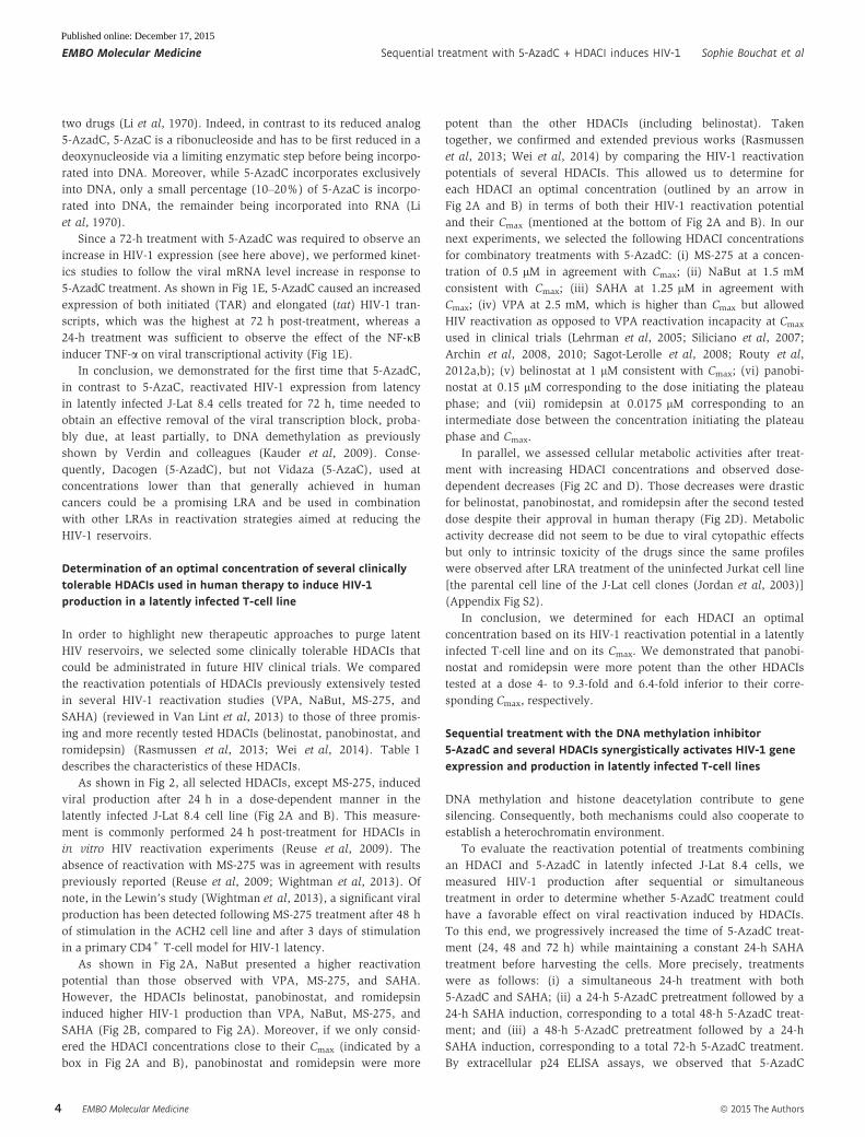
Table 1. Characteristics of used HDACIs.
Acronymin thisstudy Name Marketed as Approved for
Treatmentcorrespondingto Cmax
Human tolerableplasmatic concentration(Cmax)
Selecteddosesin thisstudy
Clinicaltrials inHIV field
MS-275 Entinostat n.a. Ongoingclinical trialsfor thetreatmentof variouscancers
For an usualdosage
0.14 � 0.24–0.3 � 0.22 lM(Wightman et al, 2013)
0.5 lM n.a.
NaBut Sodiumbutyrate
Buphenyl Sickle cellanemiaand beta-thalassemia
For 27 and36 g/day
1.225 and 1.605 mM(Phuphanich et al, 2005)
1.5 mM n.a.
SAHA Vorinostat,suberoylanilidehydroxamic acid
Zolinza CutaneousT-cell lymphoma
For an usualdosage twotimes/dayduring 2–3 weeks
0.3–1.7 lM (Merck (2013)Zolinza (vorinostat)prescribing information)
1.25 lM Archin et al(2014, 2012),Elliott et al(2014)
VPA Valproic acid Depakine Chronicneurologicaland psychiatricdisorders
For an usualdosage
0.25–0.5 mM (AbbVie (2014)Depakote prescribinginformation)
2.5 mM Archin et al(2010, 2008),Lehrmanet al (2005),Routy et al(2012a,b),Sagot-Lerolleet al (2008),Silicianoet al (2007)
Beli Belinostat,PXD101
Beleodaq Relapsed orrefractoryperipheralT-cell lymphoma
1,000 mg/m²for fiveconsecutivedays
> 1 lM (Steele et al, 2011) 1 lM n.a.
Pano Panobinostat,LBH-589
Faridak Myelomatherapy
For one dosewith an usualdosage
0.6–1.4 lM(Rathkopf et al, 2010)
0.15 lM Rasmussenet al (2015)
Romi Romidepsin,FK228
Istodax PeripheralT-cell lymphomaor cutaneousT-cell lymphoma
14 mg/m² 0.112 lM (Celgene (2014)Istodax prescribinginformation)
0.0175 lM Sogaardet al (2015)
ª 2015 The Authors EMBO Molecular Medicine
Sophie Bouchat et al Sequential treatment with 5-AzadC + HDACI induces HIV-1 EMBO Molecular Medicine
5
Published online: December 17, 2015
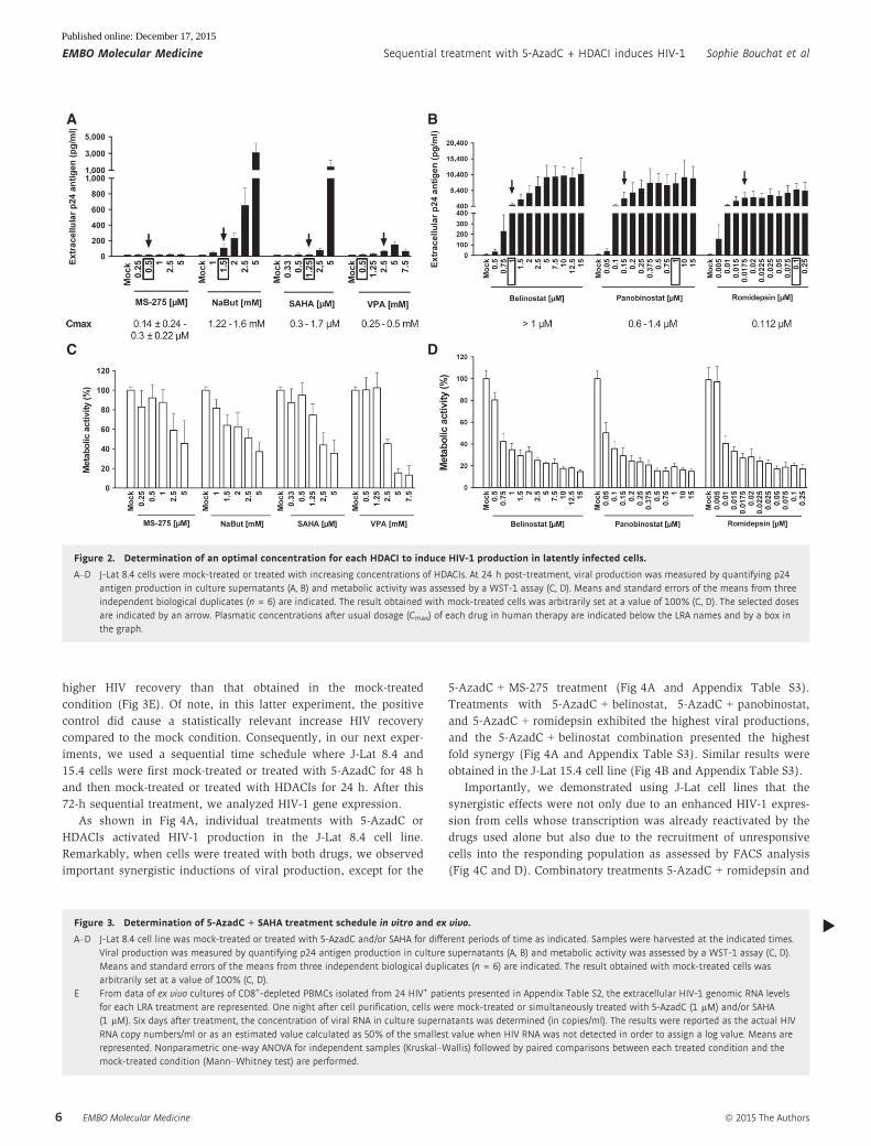
higher HIV recovery than that obtained in the mock-treated
condition (Fig 3E). Of note, in this latter experiment, the positive
control did cause a statistically relevant increase HIV recovery
compared to the mock condition. Consequently, in our next exper-
iments, we used a sequential time schedule where J-Lat 8.4 and
15.4 cells were first mock-treated or treated with 5-AzadC for 48 h
and then mock-treated or treated with HDACIs for 24 h. After this
72-h sequential treatment, we analyzed HIV-1 gene expression.
As shown in Fig 4A, individual treatments with 5-AzadC or
HDACIs activated HIV-1 production in the J-Lat 8.4 cell line.
Remarkably, when cells were treated with both drugs, we observed
important synergistic inductions of viral production, except for the
5-AzadC + MS-275 treatment (Fig 4A and Appendix Table S3).
Treatments with 5-AzadC + belinostat, 5-AzadC + panobinostat,
and 5-AzadC + romidepsin exhibited the highest viral productions,
and the 5-AzadC + belinostat combination presented the highest
fold synergy (Fig 4A and Appendix Table S3). Similar results were
obtained in the J-Lat 15.4 cell line (Fig 4B and Appendix Table S3).
Importantly, we demonstrated using J-Lat cell lines that the
synergistic effects were not only due to an enhanced HIV-1 expres-
sion from cells whose transcription was already reactivated by the
drugs used alone but also due to the recruitment of unresponsive
cells into the responding population as assessed by FACS analysis
(Fig 4C and D). Combinatory treatments 5-AzadC + romidepsin and
A B
C D
Figure 2. Determination of an optimal concentration for each HDACI to induce HIV-1 production in latently infected cells.
A–D J-Lat 8.4 cells were mock-treated or treated with increasing concentrations of HDACIs. At 24 h post-treatment, viral production was measured by quantifying p24antigen production in culture supernatants (A, B) and metabolic activity was assessed by a WST-1 assay (C, D). Means and standard errors of the means from threeindependent biological duplicates (n = 6) are indicated. The result obtained with mock-treated cells was arbitrarily set at a value of 100% (C, D). The selected dosesare indicated by an arrow. Plasmatic concentrations after usual dosage (Cmax) of each drug in human therapy are indicated below the LRA names and by a box inthe graph.
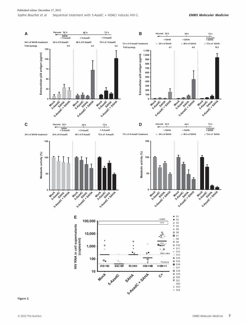
▸Figure 3. Determination of 5-AzadC + SAHA treatment schedule in vitro and ex vivo.
A–D J-Lat 8.4 cell line was mock-treated or treated with 5-AzadC and/or SAHA for different periods of time as indicated. Samples were harvested at the indicated times.Viral production was measured by quantifying p24 antigen production in culture supernatants (A, B) and metabolic activity was assessed by a WST-1 assay (C, D).Means and standard errors of the means from three independent biological duplicates (n = 6) are indicated. The result obtained with mock-treated cells wasarbitrarily set at a value of 100% (C, D).
E From data of ex vivo cultures of CD8+-depleted PBMCs isolated from 24 HIV+ patients presented in Appendix Table S2, the extracellular HIV-1 genomic RNA levelsfor each LRA treatment are represented. One night after cell purification, cells were mock-treated or simultaneously treated with 5-AzadC (1 lM) and/or SAHA(1 lM). Six days after treatment, the concentration of viral RNA in culture supernatants was determined (in copies/ml). The results were reported as the actual HIVRNA copy numbers/ml or as an estimated value calculated as 50% of the smallest value when HIV RNA was not detected in order to assign a log value. Means arerepresented. Nonparametric one-way ANOVA for independent samples (Kruskal–Wallis) followed by paired comparisons between each treated condition and themock-treated condition (Mann–Whitney test) are performed.
EMBO Molecular Medicine ª 2015 The Authors
EMBO Molecular Medicine Sequential treatment with 5-AzadC + HDACI induces HIV-1 Sophie Bouchat et al
6
Published online: December 17, 2015
A B
C D
E
Figure 3.
ª 2015 The Authors EMBO Molecular Medicine
Sophie Bouchat et al Sequential treatment with 5-AzadC + HDACI induces HIV-1 EMBO Molecular Medicine
7
Published online: December 17, 2015
BA
DC
FE
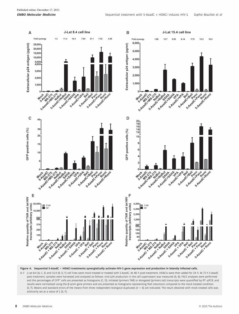
Figure 4. Sequential 5-AzadC + HDACI treatments synergistically activate HIV-1 gene expression and production in latently infected cells.
A–F J-Lat 8.4 (A, C, E) and 15.4 (B, D, F) cell lines were mock-treated or treated with 5-AzadC. At 48 h post-treatment, HDACIs were then added for 24 h. At 72 h 5-AzadCpost-treatment, samples were harvested and analyzed as follows: viral p24 production in the cell supernatant was measured (A, B); FACS analyses were performedand the percentages of GFP+ cells are presented as histograms (C, D); initiated (primers TAR) or elongated (primers tat) transcripts were quantified by RT–qPCR, andresults were normalized using the b-actin gene primers and are presented as histograms representing fold inductions compared to the mock-treated condition(E, F). Means and standard errors of the means from three independent biological duplicates (n = 6) are indicated. The result obtained with mock-treated cells wasarbitrarily set at a value of 1 (E, F).
EMBO Molecular Medicine ª 2015 The Authors
EMBO Molecular Medicine Sequential treatment with 5-AzadC + HDACI induces HIV-1 Sophie Bouchat et al
8
Published online: December 17, 2015
5-AzadC + panobinostat induced HIV-1 expression in a higher
proportion of cells than the drugs alone and than the other combina-
tions we tested, with percentages of J-Lat 8.4 GFP-positive cells of
28.9 and 30.8% and of J-Lat 15.4 GFP-positive cells of 13.8 and
16.0%, respectively (Fig 4C and D). We also analyzed the mean flu-
orescence intensities (MFI) of the GFP-positive cell populations
following the 5-AzadC + HDACIs treatments (Appendix Fig S3A
and B), and we showed that the amount of GFP produced per cell
was also more potently increased as compared to individual LRA
treatments. These data showed that synergy was due to both an
increase in the number of cells expressing virus and an enhanced
HIV-1 gene expression.
In order to test the effect of combined 5-AzadC + HDACIs treat-
ments on HIV-1 promoter transcriptional activity, initiated (TAR)
versus elongated (tat) HIV-1 transcripts were measured by RT–
qPCRs. As shown in Fig 4E, treatments with 5-AzadC alone or with
HDACIs alone (except for panobinostat and romidepsin) increased
the relative amount of both initiated and elongated viral transcripts.
Importantly, 5-AzadC + HDACIs combined treatments (especially
5-AzadC + panobinostat and 5-AzadC + romidepsin) caused higher
accumulations of initiated and elongated transcripts than those
caused by the drugs alone and by the other combinations tested
(Fig 4E). Similar results were obtained in the J-Lat 15.4 cell line
(Fig 4F).
In conclusion, sequentially combined 5-AzadC + HDACIs treat-
ments synergistically activated HIV-1 transcription and production
in two latently infected T-cell lines. Moreover, these combinations
removed the block to viral transcription observed in latently
infected J-Lat cells in a superior manner than the drugs alone. These
combinations also induced HIV-1 expression in a higher proportion
of cells than the drugs alone. Altogether, our in vitro results in
latently infected cell lines suggested that the 5-AzadC + panobinostat
and 5-AzadC + romidepsin combinations were promising in order
to reactivate HIV and prompted us to test these types of combina-
tions in ex vivo cultures of patient cells.
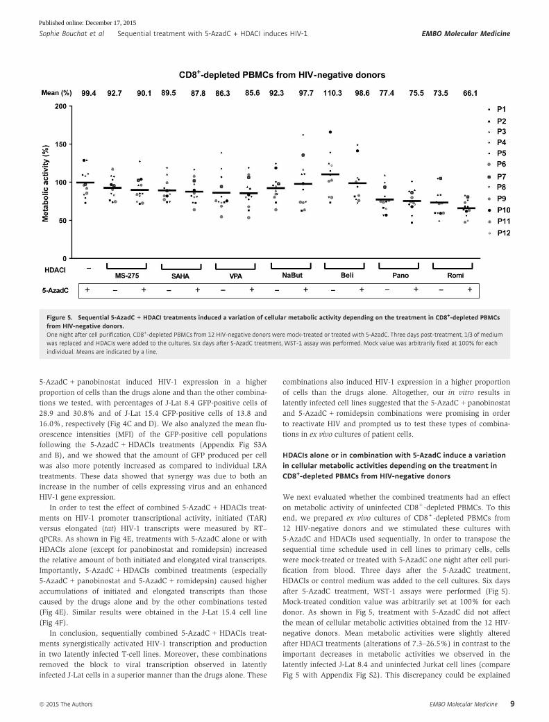
HDACIs alone or in combination with 5-AzadC induce a variationin cellular metabolic activities depending on the treatment inCD8+-depleted PBMCs from HIV-negative donors
We next evaluated whether the combined treatments had an effect
on metabolic activity of uninfected CD8+-depleted PBMCs. To this
end, we prepared ex vivo cultures of CD8+-depleted PBMCs from
12 HIV-negative donors and we stimulated these cultures with
5-AzadC and HDACIs used sequentially. In order to transpose the
sequential time schedule used in cell lines to primary cells, cells
were mock-treated or treated with 5-AzadC one night after cell puri-
fication from blood. Three days after the 5-AzadC treatment,
HDACIs or control medium was added to the cell cultures. Six days
after 5-AzadC treatment, WST-1 assays were performed (Fig 5).
Mock-treated condition value was arbitrarily set at 100% for each
donor. As shown in Fig 5, treatment with 5-AzadC did not affect
the mean of cellular metabolic activities obtained from the 12 HIV-
negative donors. Mean metabolic activities were slightly altered
after HDACI treatments (alterations of 7.3–26.5%) in contrast to the
important decreases in metabolic activities we observed in the
latently infected J-Lat 8.4 and uninfected Jurkat cell lines (compare
Fig 5 with Appendix Fig S2). This discrepancy could be explained
Figure 5. Sequential 5-AzadC + HDACI treatments induced a variation of cellular metabolic activity depending on the treatment in CD8+-depleted PBMCsfrom HIV-negative donors.One night after cell purification, CD8+-depleted PBMCs from 12 HIV-negative donors were mock-treated or treated with 5-AzadC. Three days post-treatment, 1/3 of mediumwas replaced and HDACIs were added to the cultures. Six days after 5-AzadC treatment, WST-1 assay was performed. Mock value was arbitrarily fixed at 100% for eachindividual. Means are indicated by a line.
ª 2015 The Authors EMBO Molecular Medicine
Sophie Bouchat et al Sequential treatment with 5-AzadC + HDACI induces HIV-1 EMBO Molecular Medicine
9
Published online: December 17, 2015

by the fact that HDACIs induce a cancer cell-specific cytotoxicity.
Indeed, a previous study has shown that panobinostat presents
toxicity in transformed cell lines but is relatively sparring in
primary cells (Prince et al, 2009). Importantly, we observed no
additional decrease in the mean of metabolic activities following
the combined 5-AzadC + HDACI treatments as compared to individ-
ual treatments, except for the 5-AzadC + romidepsin treatment for
which we observed a decrease in metabolic activity as compared to
individual romidepsin treatment (66.1% compared to 73.5%).
In conclusion, we highlighted that 5-AzadC alone and all
5-AzadC + HDACI treatments induced weak decreases in mean
metabolic activities (up to a 33.9% maximal decrease) depending
on the treatment as compared to the mean metabolic activities
observed in mock condition.
5-AzadC + panobinostat and 5-AzadC + romidepsin sequentialtreatments induce HIV recovery in CD8+-depleted PBMCs isolatedfrom cART-treated aviremic HIV+ patients
To address the physiological relevance of our in vitro reactivation
results, we investigated the reactivation potential of the
5-AzadC + HDACI combinations in ex vivo cultures of cells isolated
from cART-treated aviremic HIV-1+ patients. We purified CD8+-
depleted PBMCs from the blood of 19 selected volunteer patients
(Table 2). In order to evaluate the frequency of infected cells
during plating, we quantified cell-associated total HIV-1 DNA. One
night after purification, cells were either mock-treated or treated
with 5-AzadC and/or HDACIs (according to the sequential time
schedule described above) or with anti-CD3 + anti-CD28 antibodies
as positive control (Pierres et al, 1988; Costello et al, 1993).
Importantly, in all our experiments, purified cells were cultured in
the absence of both IL-2 and allogenic stimulation in order to
avoid non-specific global T-cell activation and proliferation, which
might cause an increase in genomic viral RNA level. Six days after
treatment, we measured HIV-1 genomic RNA concentrations in
culture supernatants.
We detected genomic viral RNA in the mock-treated culture
supernatant from 4 out of 19 patient cell cultures (X16-X19,
Table 2). This observation could be explained by the activation of
HIV-infected cells during the purification procedure. Global analysis
of all 19 patient cell cultures showed that treatment with 5-AzadC
alone led to the reactivation of 21.1% of the patient cell cultures, a
percentage similar to that obtained in the mock-treated condition.
In contrast, HDACIs alone increased the percentage of reactivated
Table 2. Representation of reactivation status of ex vivo cultures of CD8+-depleted PBMCs isolated from HIV+ patients.
Total HIV DNA (copies/106
CD8+-depleted PBMCs)
mock 5-AzadC Pano 5-AzadC + Pano
Beli 5-AzadC + Beli
Romi 5-AzadC + RomI
SAHA 5-AzadC + SAHA
NaBut 5-AzadC + NaBut
VPA 5-AzadC + VPA
MS-275 5-AzadC + MS-275
Positive control
αCD3+αCD28
X1 11 ND ND ND ND ND ND ND ND ND ND ND ND ND ND ND ND 391X2 670 ND ND ND 293 48 ND 285 537 ND ND ND ND NA NA NA NA 829X3 464 ND ND ND ND 1,209 ND 317 2,050 ND ND 598 ND ND ND NA NA 11,450X4 587 ND ND 3,977 4,403 ND ND 733 ND ND 1,072 536 ND 316 ND 80 918 19,630X5 724 ND ND ND 673 829 1,206 ND ND 1,079 619 747 ND ND ND 483 453 314X6 2,552 ND ND 147 2,121 ND 216 ND 446 243 278 275 ND 487 115 24 230 6,785X7 # 548 ND ND ND ND ND ND ND 273 ND 203 ND ND ND ND ND ND 6,601X8 1,089 ND 97 317 ND ND ND 458 ND 198 ND ND ND NA NA NA NA 21,850X9 125 ND ND 716 ND 219 236 ND ND 114 ND ND ND NA NA NA NA 3,367X10 744 ND 203 ND 1,269 230 ND ND ND ND ND ND ND NA NA NA NA NDX11 # <23 ND ND ND 161 ND ND ND ND ND ND ND ND ND ND NA NA NDX12 876 ND ND 20 ND 189 201 22 321 400 ND 65 ND 437 184 ND ND 3,877X13 863 ND ND 615 447 428 191 ND 581 578 ND ND ND 200 403 396 ND 205X14 # 57 ND ND ND 423 ND ND ND ND ND ND ND ND 8 ND NA NA 414X15 257 ND ND ND 205 ND ND ND ND ND 889 ND ND ND ND 407 ND 14,533X16 1,567 588 ND ND ND 822 ND 223 ND ND 305 ND ND ND ND ND ND 133X17 909 248 229 398 ND 210 ND ND ND 254 ND 502 235 NA NA NA NA 3,852X18 4,486 3,722 241 749 ND 922 551 1,511 605 264 465 233 ND 972 225 513 373 22,060X19 1,083 397 504 917 295 216 395 932 1,003 NA NA NA NA NA NA NA NA 2,880
Number of reactivated
patient's cell cultures with extracellular
HIV RNA > 150 copies/ml 4 4 8 10 10 7 7 8 7 7 6 1 5 3 4 4 16Number of
tested patient samples 19 19 19 19 19 19 19 19 18 18 18 18 13 13 10 10 19
% of reactivated
cultures 21.1 21.1 42.1 52.6 52.6 36.8 36.8 42.1 38.9 38.9 33.3 5.6 38.5 23.1 40.0 40.0 84.2
CD8+-depleted PBMCs Concentration of HIV RNA in culture supernatants after six days of treatment (copies/ml)
One night after cell purification, cells were mock-treated or treated with 5-AzadC. Three days post-treatment, 1/3 of medium was removed and HDACIs wereadded in the cultures. Six days after 5-AzadC treatment, the concentration of viral RNA in culture supernatants was determined (in copies/ml; ND meansundetectable, and NA indicates an untested condition). Total HIV-1 DNA is expressed as total HIV-1 DNA copies/106 CD8+-depleted PBMCs. The cultureshighlighted in gray showed a higher viral production with the LRA combination than with the corresponding LRAs alone, while the cultures highlighted in blackwere reactivated only by the combinatory treatment and not by the LRAs individually. Patients indicated by the # symbol are only reactivated by one or morecombinations.
EMBO Molecular Medicine ª 2015 The Authors
EMBO Molecular Medicine Sequential treatment with 5-AzadC + HDACI induces HIV-1 Sophie Bouchat et al
10
Published online: December 17, 2015

patient cell cultures (33.3–52.6% depending on the HDACI tested)
(Table 2). Sequential combinatory treatments, except for the
5-AzadC + NaBut and 5-AzadC + VPA combinations, were more
potent than the mock-treated condition and led to the reactivation
of 36.8–52.6% of cell cultures. Interestingly, only 5-AzadC + pano-
binostat and 5-AzadC + romidepsin combinations produced
increases in percentage of reactivated patient cell cultures as
compared to individual drug treatments (Table 2). These percent-
ages, 52.6 and 42.1%, respectively, indicated ~2-fold increases
compared to the percentage obtained with the mock-treated condi-
tion. Importantly, we also observed that, in some ex vivo patient
cell cultures, some combined treatments caused higher levels of
viral production than the levels observed after the individual treat-
ments (Table 2, values highlighted in gray). Moreover, HIV-1 recovery
was only observed with some LRA combinations, but not with the
corresponding individual LRAs (Table 2, values highlighted in
black). Among these patient cell cultures presenting values in black,
three cultures (X7, X11, and X14 indicated by a # symbol in
Table 2) presented the reactivation of viral production exclusively
after the sequential combined treatments: 5-AzadC + romidepsin
and 5-AzadC + SAHA combinations for X7, 5-AzadC + panobinostat
combination for X11, and 5-AzadC + panobinostat for X14. In terms
of extracellular HIV RNA (Fig 6A), we observed that only the
5-AzadC + panobinostat and 5-AzadC + romidepsin combinations
presented mean levels of extracellular viral RNA higher than the
mean levels observed after the individual LRA treatments and
higher than the mean level obtained in mock-treated condition.
However, the results including all patient cell cultures were not
statistically relevant, except for the positive control condition
(Fig 6A). Importantly, we observed that the 15 patient cell cultures
presenting no viral activation in mock condition (patients X1–X15)
clearly exhibited a more potent viral production following
5-AzadC + panobinostat and 5-AzadC + romidepsin treatments (see
Table 2, gray and black boxes) in comparison with the last 4
patient cell cultures presenting viral reactivation in mock condition
(patients X16–X19). Therefore, we decided to analyze separately the
reactivation data from the two groups: (i) the four patient cell
cultures exhibiting viral activation in mock-treated condition
(Fig 6B); and (ii) the 15 patient cell cultures presenting no viral
reactivation in mock condition (Fig 6C).
When performing analysis for the first group including the four
patient cell cultures presenting HIV-1 recovery in mock-treated
condition (Fig 6B), we observed that all means of HIV extracellular
RNA levels after the different treatments, except after the positive
control, were lower than the mean observed in mock-treated
condition (Fig 6B).
For the second group, the reactivation profiles we obtained were
similar to the reactivation profiles observed after global analysis of
the 19 patient cell cultures (compare Fig 6C to Fig 6A). Interestingly,
not only combinations 5-AzadC + panobinostat and 5-AzadC +
romidepsin but also 5-AzadC + SAHA and 5-AzadC + MS-275
produced higher mean levels of viral RNA than the mean levels
A
B
C
Figure 6. Representation of reactivation status of ex vivo cultures ofCD8+-depleted PBMCs isolated from HIV+ patients.
A–C From data of ex vivo cultures of CD8+-depleted PBMCs isolated from HIV+
patients (Table 2), the extracellular HIV-1 genomic RNA levels for each LRAtreatment are represented from all patient cell cultures (A), from patient cellcultures presenting no viral reactivation in mock condition (C), and frompatient cell cultures exhibiting reactivation in mock condition (B). One nightafter cell purification, cells were mock-treated or treated with 5-AzadC.Three days post-treatment, 1/3 of medium was replaced and HDACIs wereadded to the cultures. Six days after 5-AzadC treatment, the concentrationof viral RNA in culture supernatants was determined (in copies/ml). Theresults were reported as the actual HIV RNA copy numbers/ml or as anestimated value calculated as 50% of the smallest value when HIV RNA wasnot detected in order to assign a log value. Means are represented.Nonparametric one-way ANOVA for independent samples (Kruskal–Wallis)followed by paired comparisons between each treated condition and themock-treated condition (Mann–Whitney test) are performed.
ª 2015 The Authors EMBO Molecular Medicine
Sophie Bouchat et al Sequential treatment with 5-AzadC + HDACI induces HIV-1 EMBO Molecular Medicine
11
Published online: December 17, 2015
obtained with the individual treatments (Fig 6C). Moreover, in
contrast to what we observed for the 19 patient cell culture global
analysis, we observed statistically relevant viral recoveries for the
second group of 15 patient cell cultures (Fig 6C). Indeed, in this
group, all individual treatments, except 5-AzadC, produced statisti-
cally relevant increases in the mean levels of extracellular HIV-1
RNA (Fig 6C). Moreover, all 5-AzadC + HDACI combinations,
except 5-AzadC + NaBut and 5-AzadC + VPA, produced statistically
relevant increases in HIV recovery as compared to mock-treated
condition. Notably, the 5-AzadC + panobinostat combined treat-
ment was the most potent and the only one to produce highly
statistically relevant increase (P = 0.0007) as compared to mock
condition.
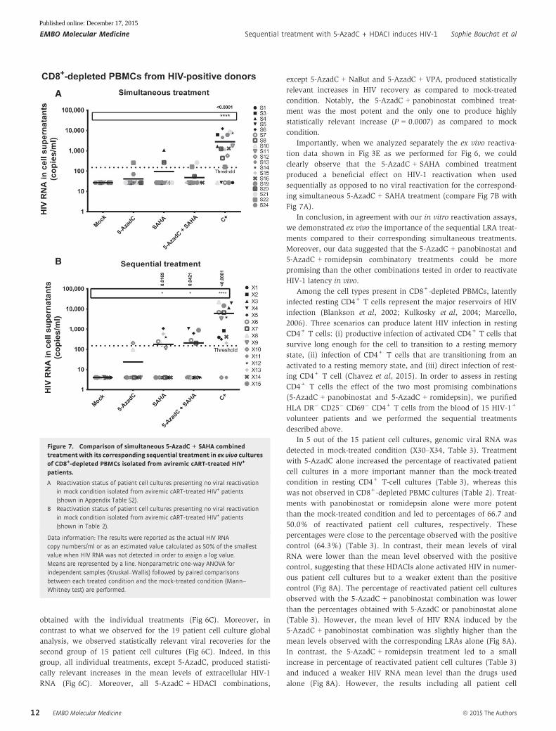
Importantly, when we analyzed separately the ex vivo reactiva-
tion data shown in Fig 3E as we performed for Fig 6, we could
clearly observe that the 5-AzadC + SAHA combined treatment
produced a beneficial effect on HIV-1 reactivation when used
sequentially as opposed to no viral reactivation for the correspond-
ing simultaneous 5-AzadC + SAHA treatment (compare Fig 7B with
Fig 7A).
In conclusion, in agreement with our in vitro reactivation assays,
we demonstrated ex vivo the importance of the sequential LRA treat-
ments compared to their corresponding simultaneous treatments.
Moreover, our data suggested that the 5-AzadC + panobinostat and
5-AzadC + romidepsin combinatory treatments could be more
promising than the other combinations tested in order to reactivate
HIV-1 latency in vivo.
Among the cell types present in CD8+-depleted PBMCs, latently
infected resting CD4+ T cells represent the major reservoirs of HIV
infection (Blankson et al, 2002; Kulkosky et al, 2004; Marcello,
2006). Three scenarios can produce latent HIV infection in resting
CD4+ T cells: (i) productive infection of activated CD4+ T cells that
survive long enough for the cell to transition to a resting memory
state, (ii) infection of CD4+ T cells that are transitioning from an
activated to a resting memory state, and (iii) direct infection of rest-
ing CD4+ T cell (Chavez et al, 2015). In order to assess in resting
CD4+ T cells the effect of the two most promising combinations
(5-AzadC + panobinostat and 5-AzadC + romidepsin), we purified
HLA DR� CD25� CD69� CD4+ T cells from the blood of 15 HIV-1+
volunteer patients and we performed the sequential treatments
described above.
In 5 out of the 15 patient cell cultures, genomic viral RNA was
detected in mock-treated condition (X30–X34, Table 3). Treatment
with 5-AzadC alone increased the percentage of reactivated patient
cell cultures in a more important manner than the mock-treated
condition in resting CD4+ T-cell cultures (Table 3), whereas this
was not observed in CD8+-depleted PBMC cultures (Table 2). Treat-
ments with panobinostat or romidepsin alone were more potent
than the mock-treated condition and led to percentages of 66.7 and
50.0% of reactivated patient cell cultures, respectively. These
percentages were close to the percentage observed with the positive
control (64.3%) (Table 3). In contrast, their mean levels of viral
RNA were lower than the mean level observed with the positive
control, suggesting that these HDACIs alone activated HIV in numer-
ous patient cell cultures but to a weaker extent than the positive
control (Fig 8A). The percentage of reactivated patient cell cultures
observed with the 5-AzadC + panobinostat combination was lower
than the percentages obtained with 5-AzadC or panobinostat alone
(Table 3). However, the mean level of HIV RNA induced by the
5-AzadC + panobinostat combination was slightly higher than the
mean levels observed with the corresponding LRAs alone (Fig 8A).
In contrast, the 5-AzadC + romidepsin treatment led to a small
increase in percentage of reactivated patient cell cultures (Table 3)
and induced a weaker HIV RNA mean level than the drugs used
alone (Fig 8A). However, the results including all patient cell
A
B
Figure 7. Comparison of simultaneous 5-AzadC + SAHA combinedtreatment with its corresponding sequential treatment in ex vivo culturesof CD8+-depleted PBMCs isolated from aviremic cART-treated HIV+
patients.
A Reactivation status of patient cell cultures presenting no viral reactivationin mock condition isolated from aviremic cART-treated HIV+ patients(shown in Appendix Table S2).
B Reactivation status of patient cell cultures presenting no viral reactivationin mock condition isolated from aviremic cART-treated HIV+ patients(shown in Table 2).
Data information: The results were reported as the actual HIV RNAcopy numbers/ml or as an estimated value calculated as 50% of the smallestvalue when HIV RNA was not detected in order to assign a log value.Means are represented by a line. Nonparametric one-way ANOVA forindependent samples (Kruskal–Wallis) followed by paired comparisonsbetween each treated condition and the mock-treated condition (Mann–Whitney test) are performed.
EMBO Molecular Medicine ª 2015 The Authors
EMBO Molecular Medicine Sequential treatment with 5-AzadC + HDACI induces HIV-1 Sophie Bouchat et al
12
Published online: December 17, 2015
cultures were not statistically relevant. Importantly, we also
observed that, in some ex vivo patient cell cultures, both combina-
tory treatments allowed higher levels of viral production than the
levels observed with the individual treatments (Table 3, values
highlighted in gray). As performed for the CD8+-depleted PBMCs,
we decided to analyze separately the reactivation data for (i) the
group of 5 patient cell cultures exhibiting viral activation in mock
condition (Fig 8B) and (ii) the group of 10 patient cell cultures
presenting no viral reactivation in mock condition (Fig 8C). Interest-
ingly, as shown in Fig 8B, the first group of patient cell cultures
exhibited higher viral production following 5-AzadC + panobinostat
treatment as compared to the individual panobinostat treatment,
suggesting that cells from these patients might have been weakly
reactivated during purification, but remained responsive to some of
the LRAs we used. In the second group of patient cell cultures, we
observed that panobinostat and romidepsin alone presented higher
and statistically relevant means of extracellular HIV RNA level
than the means observed with the 5-AzadC + panobinostat and
5-AzadC + romidepsin combinations (Fig 8C). The differences in
the effect of the combinations observed between the patient
cell cultures which presented viral reactivation in mock condition
(1st group) and the patient cell cultures presenting no viral recovery
in mock condition (2nd group) could result from differences in
cellular division status of reactivated cells and consequently
differences in 5-AzadC DNA incorporation.
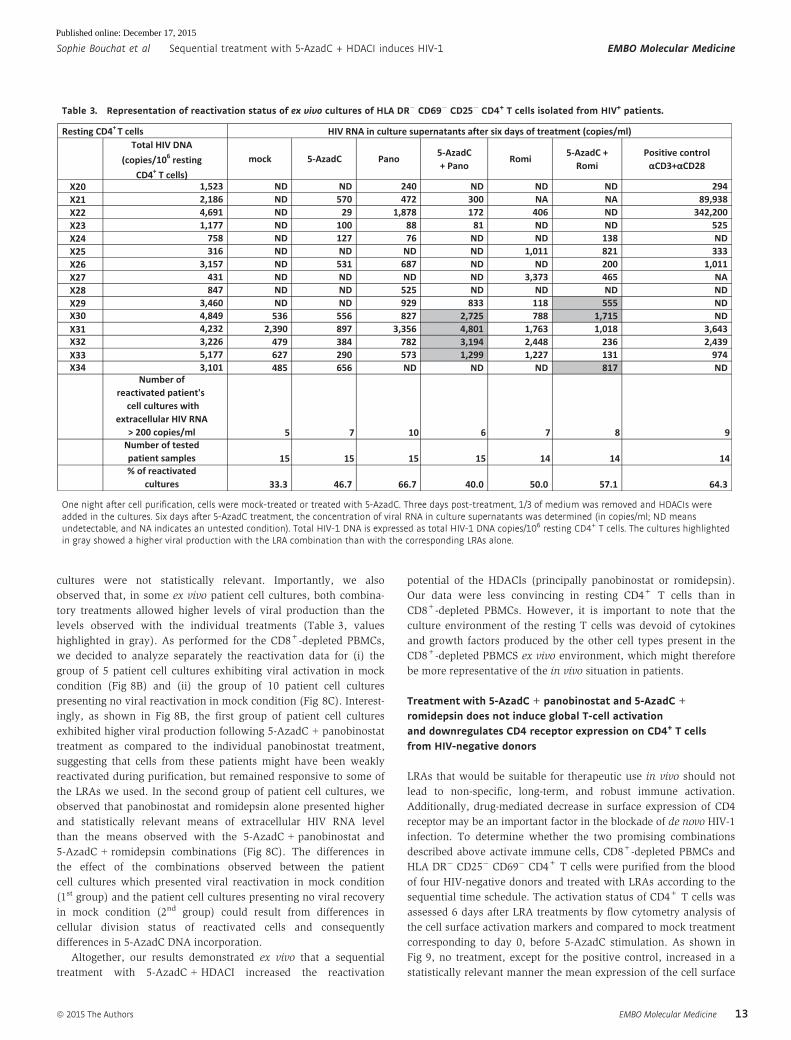
Altogether, our results demonstrated ex vivo that a sequential
treatment with 5-AzadC + HDACI increased the reactivation
potential of the HDACIs (principally panobinostat or romidepsin).
Our data were less convincing in resting CD4+ T cells than in
CD8+-depleted PBMCs. However, it is important to note that the
culture environment of the resting T cells was devoid of cytokines
and growth factors produced by the other cell types present in the
CD8+-depleted PBMCS ex vivo environment, which might therefore
be more representative of the in vivo situation in patients.
Treatment with 5-AzadC + panobinostat and 5-AzadC +
romidepsin does not induce global T-cell activationand downregulates CD4 receptor expression on CD4+ T cellsfrom HIV-negative donors
LRAs that would be suitable for therapeutic use in vivo should not
lead to non-specific, long-term, and robust immune activation.
Additionally, drug-mediated decrease in surface expression of CD4
receptor may be an important factor in the blockade of de novo HIV-1
infection. To determine whether the two promising combinations
described above activate immune cells, CD8+-depleted PBMCs and
HLA DR� CD25� CD69� CD4+ T cells were purified from the blood
of four HIV-negative donors and treated with LRAs according to the
sequential time schedule. The activation status of CD4+ T cells was
assessed 6 days after LRA treatments by flow cytometry analysis of
the cell surface activation markers and compared to mock treatment
corresponding to day 0, before 5-AzadC stimulation. As shown in
Fig 9, no treatment, except for the positive control, increased in a
statistically relevant manner the mean expression of the cell surface
Table 3. Representation of reactivation status of ex vivo cultures of HLA DR� CD69� CD25� CD4+ T cells isolated from HIV+ patients.
Total HIV DNA (copies/106 resting
CD4+ T cells)mock 5-AzadC Pano 5-AzadC
+ PanoRomi 5-AzadC +
RomiPositive control
αCD3+αCD28
X20 1,523 ND ND 240 ND ND ND 294X21 2,186 ND 570 472 300 NA NA 89,938X22 4,691 ND 29 1,878 172 406 ND 342,200X23 1,177 ND 100 88 81 ND ND 525X24 758 ND 127 76 ND ND 138 NDX25 316 ND ND ND ND 1,011 821 333X26 3,157 ND 531 687 ND ND 200 1,011X27 431 ND ND ND ND 3,373 465 NAX28 847 ND ND 525 ND ND ND NDX29 3,460 ND ND 929 833 118 555 NDX30 4,849 536 556 827 2,725 788 1,715 NDX31 4,232 2,390 897 3,356 4,801 1,763 1,018 3,643X32 3,226 479 384 782 3,194 2,448 236 2,439X33 5,177 627 290 573 1,299 1,227 131 974X34 3,101 485 656 ND ND ND 817 ND
Number of reactivated patient's
cell cultures with extracellular HIV RNA
> 200 copies/ml 5 7 10 6 7 8 9Number of tested patient samples 15 15 15 15 14 14 14% of reactivated
cultures 33.3 46.7 66.7 40.0 50.0 57.1 64.3
Resting CD4+ T cells HIV RNA in culture supernatants after six days of treatment (copies/ml)
One night after cell purification, cells were mock-treated or treated with 5-AzadC. Three days post-treatment, 1/3 of medium was removed and HDACIs wereadded in the cultures. Six days after 5-AzadC treatment, the concentration of viral RNA in culture supernatants was determined (in copies/ml; ND meansundetectable, and NA indicates an untested condition). Total HIV-1 DNA is expressed as total HIV-1 DNA copies/106 resting CD4+ T cells. The cultures highlightedin gray showed a higher viral production with the LRA combination than with the corresponding LRAs alone.
ª 2015 The Authors EMBO Molecular Medicine
Sophie Bouchat et al Sequential treatment with 5-AzadC + HDACI induces HIV-1 EMBO Molecular Medicine
13
Published online: December 17, 2015

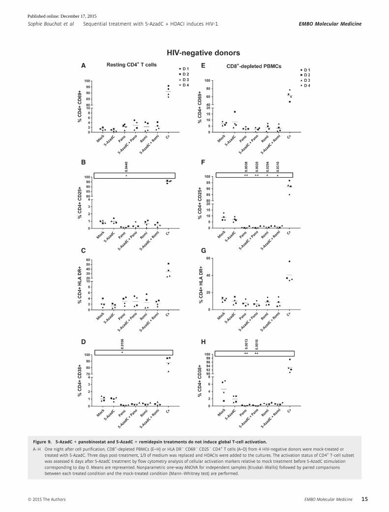
activation markers such as CD69 (early activation marker), CD25
(intermediate activation marker), HLA DR (late activation marker),
and CD38 (late activation marker and predictive marker of HIV-1
progression) in the two cell populations. Interestingly, in CD8+-
depleted PBMCs (cellular population including activated CD4+ T cells),
we observed that panobinostat and romidepsin alone or in combination
with 5-AzadC induced a statistically relevant decrease in CD25+ CD4+
cells. Panobinostat alone or in combination with 5-AzadC also induced
a statistically relevant decrease in CD38+ CD4+ cells. These results
were observed only with the two promising combinations (Fig 9) and
not with the other combinations (Appendix Fig S4).
In parallel, we evaluated whether the different treatments led to
downregulation of the CD4 receptor. As shown in Fig 10, panobi-
nostat and romidepsin alone or in combination with 5-AzadC
induced statistically relevant decreases in CD4 receptor expression
on the cell surface. This would limit the number of HIV-1 target
cells. These results were observed only with the two promising
combinations (Fig 10) and not with the other combinations
(Appendix Fig S5).
Altogether, we concluded that 5-AzadC + panobinostat and
5-AzadC + romidepsin treatments did not induce global T-cell
activation and were able to decrease the activated status of CD4+ T cells.
Moreover, we observed a negative regulation of the cell surface CD4
receptor expression after treatment with these combinations,
suggesting an ability to limit HIV-1 dissemination.
Discussion
In this report, we assessed in vitro and ex vivo the HIV-1 reactiva-
tion potential of combinations including demethylating agents and
HDACIs at clinically tolerable concentrations. We showed that the
DNA methylation inhibitor 5-AzadC alone, but not 5-AzaC,
induced HIV-1 expression after 72 h of treatment in a latently
infected T-cell line. Consequently, Dacogen (5-AzadC), but not
Vidaza (5-AzaC), used at concentrations below human usual
dosage, could be promising in combinatory treatments with other
LRAs in strategies aimed at reducing the HIV-1 reservoir size. After
determination for each selected HDACI (VPA, NaBut, MS-275,
SAHA, belinostat, panobinostat, and romidepsin) of an optimal
concentration in terms of its HIV-1 reactivation potential in a
latently infected T-cell line and of its human tolerable Cmax, we
demonstrated that a sequential 5-AzadC + HDACI (except MS-275)
treatment synergistically induced HIV-1 expression at both viral
RNA and protein levels in two latently infected T-cell lines. Our
data highlighted for the first time that, in addition to the setup of
LRA concentrations, the sequential time schedule of LRA combined
treatments was very important to reach these synergistic activa-
tions of HIV production. Our results showed that the
5-AzadC + panobinostat and 5-AzadC + romidepsin combinations
were more potent than the other combinations we tested, high-
lighting potential therapeutic implications for strategies aimed at
A
B
C
Figure 8. Representation of reactivation status of ex vivo cultures ofresting CD4+ T cells.
A–C From data of ex vivo cultures of resting CD4+ T cells isolated from HIV+
patients (Table 3), the extracellular HIV-1 genomic RNA levels for each LRAtreatment are represented from all patient cell cultures (A), from patient cellcultures presenting no viral reactivation in mock condition (C), and frompatient cell cultures exhibiting reactivation in mock condition (B). One nightafter cell purification, cells were mock-treated or treated with 5-AzadC.Three days post-treatment, 1/3 of medium was replaced and HDACIs wereadded in the cultures. Six days after 5-AzadC treatment, the concentrationof viral RNA in culture supernatants was determined (in copies/ml). Theresults were reported as the actual HIV RNA copy numbers/ml or as anestimated value calculated as 50% of the smallest value when HIV RNA wasnot detected in order to assign a log value. Means are represented.Nonparametric one-way ANOVA for independent samples (Kruskal–Wallis)followed by paired comparisons between each treated condition and themock-treated condition (Mann–Whitney test) are performed.
EMBO Molecular Medicine ª 2015 The Authors
EMBO Molecular Medicine Sequential treatment with 5-AzadC + HDACI induces HIV-1 Sophie Bouchat et al
14
Published online: December 17, 2015
A
B
C
D
E
F
G
H
Figure 9. 5-AzadC + panobinostat and 5-AzadC + romidepsin treatments do not induce global T-cell activation.
A–H One night after cell purification, CD8+-depleted PBMCs (E–H) or HLA DR� CD69� CD25� CD4+ T cells (A–D) from 4 HIV-negative donors were mock-treated ortreated with 5-AzadC. Three days post-treatment, 1/3 of medium was replaced and HDACIs were added to the cultures. The activation status of CD4+ T-cell subsetwas assessed 6 days after 5-AzadC treatment by flow cytometry analysis of cellular activation markers relative to mock treatment before 5-AzadC stimulationcorresponding to day 0. Means are represented. Nonparametric one-way ANOVA for independent samples (Kruskal–Wallis) followed by paired comparisonsbetween each treated condition and the mock-treated condition (Mann–Whitney test) are performed.
ª 2015 The Authors EMBO Molecular Medicine
Sophie Bouchat et al Sequential treatment with 5-AzadC + HDACI induces HIV-1 EMBO Molecular Medicine
15
Published online: December 17, 2015

reducing the pool of HIV-1 latent reservoirs in cART-treated
patients. Next, we addressed the physiological relevance of our
results using ex vivo cultures of CD8+-depleted PBMCs and of rest-
ing CD4+ T cells isolated from cART-treated aviremic HIV-1+
patients. In both types of cell cultures, we observed that HDACIs
alone were more potent in inducing HIV-1 recovery than 5-AzadC
alone. Recent studies have failed to observe increased HIV particle
production following ex vivo treatment with SAHA or romidepsin
(Bullen et al, 2014; Cillo et al, 2014; Mohammadi et al, 2014). This
discrepancy with the present study might result from the fact that
we analyzed viral production using a very sensitive technique, the
quantification of genomic HIV RNA in cell culture supernatants, as
performed by Wei and colleagues who, in agreement with our
data, have also observed a romidepsin- and SAHA-induced HIV
RNA release in resting CD4+ T-cell cultures (Wei et al, 2014).
Importantly, in agreement with our in vitro reactivation assays, we
demonstrated ex vivo the importance of the treatment sequen-
tial time schedule for LRA combinations compared to the
corresponding simultaneous combinatory treatments. This
phenomenon could be explained by the fact that, in eukaryotic cell
gene regulation, DNA methylation may function as a first control
level that locks gene transcriptional activity (reviewed in Jones,
1985). In some systems, such as myogenesis or thymidine kinase
induction, no stimulus other than demethylation is necessary for
gene expression. However, in other systems, gene activation occurs
in response to a cascade of events. For several genes whose activa-
tion requires 5-AzaC as a primary stimulus and another inducer as
a secondary stimulus, no significant response is observed with
either stimulus alone (Jones, 1985). In the present study, our data
suggest that the 5-AzadC + panobinostat and 5-AzadC + romidepsin
sequential combinatory treatments could be more promising than
the other combinations we tested in order to reactivate HIV-1
latency in vivo.
Nevertheless, we observed that ex vivo viral production levels
were weaker than the levels observed in our previous studies using
HMTIs, HDACIs, P-TEFb-releasing agents, or NF-jB inducers (Reuse
et al, 2009; Bouchat et al, 2012; Darcis et al, 2015). Of note, on
the one hand, in these previous studies, we used doses inducing
maximal HIV-1 viral production, whereas in contrast, in the present
report, we used LRA concentrations corresponding to compromises
between potent HIV-1 production and human tolerable Cmax. For
example, panobinostat and romidepsin were used at concentrations
4- to 9.3-fold and 6.4-fold lower than human Cmax, respectively. On
the other hand, 5-AzadC was reported as a compound exhibiting
HIV-1 antiretroviral effects. Indeed, Bouchard et al have reported
that 5-AzadC at the dose we used is able to completely inhibit HIV-1
replication when the drug is added to the cell medium at least 2 h
before infection (Bouchard et al, 1990). More recently, a study has
validated these latter results in a mouse model (murine acquired
immunodeficiency syndrome; MAIDS), where 5-AzadC alone or in
combination with gemcitabine decreases HIV-1 replication through
the introduction of lethal mutations during viral reverse transcrip-
tion, leading to a decrease in latent reservoir pool and in disease
progression (Clouser et al, 2012). Targeting reverse transcription,
5-AzadC has been compared to AZT and presents a higher inhibition
effect than AZT at equimolar amounts (Bouchard et al, 1990).
Importantly, the antiviral activity of 5-AzadC is also comparable to
that of tenofovir and raltegravir in the case of feline leukemia virus
(Greggs et al, 2012). Of note, in our ex vivo patient cell cultures, we
chose to work in the absence of antiretroviral compounds (i) to
amplify viral production, thereby facilitating measurement of cell
culture reactivation following LRA treatments, and (ii) to allow plat-
ing less cells per well, thereby allowing testing a larger number of
different LRA treatments with the blood sample of a single patient.
Therefore, viral productions we observed here could likely result
from either reactivation of HIV-1 gene expression in latent cells or
viral production in cells newly infected by the neosynthesized
viruses. Nevertheless, by treating cells with 5-AzadC, which has
been reported to exhibit an antiretroviral activity (Bouchard et al,
1990), we probably only measured HIV production from reactivated
latent cells. Indeed, the Bouchard et al’s study has shown that, in
patient cells, 5-AzadC, at the same dose than the one we used here,
limits the infection of the cells to one replication cycle (Bouchard
et al, 1990). This phenomenon could explain, at least in part, the
fact that in this report some patient cell cultures presented a level of
viral production following treatment with HDACIs alone higher than
the level observed following the corresponding 5-AzadC + HDACI
combinatory treatment.
The beneficial effect of the two combinations selected here
(5-AzadC + panobinostat and 5-AzadC + romidepsin) could be
explained by the cooperation between the two targeted epigenetic
mechanisms, but also by intrinsic properties of the HDACIs we
used. Firstly, panobinostat is likely the most potent pan-HDAC
inhibitor in clinical development (Prince et al, 2009). The synergis-
tic effect of 5-AzadC in combination with panobinostat could result
from the inhibition of DNA methyltransferase (DNMT) activity,
Figure 10. 5-AzadC + panobinostat and 5-AzadC + romidepsintreatments induce a significant decrease in the cell surface CD4 receptorexpression.One night after cell purification, HLA DR� CD69� CD25� CD4+ T cells from 4 HIV-negative donors were mock-treated or treated with 5-AzadC. Three days post-treatment, 1/3 of medium was replaced and HDACIs were added to the cultures.The median fluorescence intensity of CD4 receptor of viable CD4+ T-cell subsetwas assessed 6 days after 5-AzadC treatment by flow cytometry analysis relativeto mock treatment before 5-AzadC stimulation corresponding to day 0. Meansare represented. Nonparametric one-way ANOVA for independent samples(Kruskal–Wallis) followed by paired comparisons between each treated conditionand the mock-treated condition (Mann–Whitney test) are performed.
EMBO Molecular Medicine ª 2015 The Authors
EMBO Molecular Medicine Sequential treatment with 5-AzadC + HDACI induces HIV-1 Sophie Bouchat et al
16
Published online: December 17, 2015

since a downregulation of DNMT mRNAs and protein levels
after panobinostat treatment has been reported (Zopf et al, 2012).
Moreover, another study has shown that panobinostat treatment
depletes DNMT1 and the HMT EZH2 protein levels and disrupts the
interaction of DNMT1 with EZH2 (Fiskus et al, 2009), an HMT
implicated in HIV-1 transcriptional repression (Friedman et al,
2011). Secondly, romidepsin presents a unique intracellular pharma-
cology (Furumai et al, 2002). Moreover, synergistic activation
observed with 5-AzadC + romidepsin could be due to the fact that
romidepsin belongs to the depsipeptide class of HDACIs. Indeed, a
previous study has shown that depsipeptide exhibits a significant
demethylating activity on the promoters of several genes (Wu et al,
2008). Depsipeptide also suppresses the expression of the HMTs
G9A and SUV39H1, which in turn results in a decrease in di- and
trimethylated H3K9 around these gene promoters. Therefore, romi-
depsin not only interferes with histone acetylation but also with two
other epigenetic marks (di- and trimethylation of H3K9) previously
demonstrated as involved in HIV-1 latency (du Chene et al, 2007;
Marban et al, 2007; Imai et al, 2010).
In a therapeutic goal, the ideal compounds should not lead
to non-specific, long-term, and robust immune activation. In
this context, we observed that 5-AzadC + panobinostat and
5-AzadC + romidepsin treatment did not induce global T-cell activa-
tion and were able to decrease the activation level of activated
CD4+ T cells. Interestingly, we reported a negative regulation of the
cell surface CD4 receptor expression after treatment by these combi-
nations, suggesting a limitation of HIV-1 target cells after LRA treat-
ment and an obstacle for virus dissemination.
Despite promising aspects of these combinations, one concern
might be that 5-AzadC needs to be incorporated into DNA.
However, the pool of latently infected memory CD4+ T cells is
maintained throughout patient life by homeostatic proliferation of
memory T cells and/or intermittent antigen-driven clonal expansion
(Chomont et al, 2009). Moreover, two recent studies suggest that
HIV integrates preferentially into cancer-associated genes and other
genes that promote cell proliferation in a manner distinct from
homeostatic proliferation (Maldarelli et al, 2014; Wagner et al,
2014). Consequently, in these integration cases, HIV-1-infected
T cells would divide faster than uninfected T cells. Finally, it should
be noted that 5-AzadC could exhibit activities independent of its
DNA methylation inhibitory effect. Indeed, during the writing of this
manuscript, Peterlin and colleagues have elegantly demonstrated
that 5-AzaC (at 10 lM) releases P-TEFb from its inactive complex
similar to known P-TEFb-releasing agents (Fujinaga et al, 2015).
Consequently, 5-AzadC could also have an HIV-1 anti-latency effect
independently of its DNA incorporation.
In the race for a HIV cure, the levels of HIV-1 reservoirs appear
as one of the major factors influencing the duration of a remission
after cART cessation. Reactivation of HIV gene expression in
latently infected cells together with an efficient or intensive cART
could serve as an inducible adjuvant therapy aimed at eliminating/
decreasing the pool of latent viral reservoirs. Several clinical trials
have started with HDACIs alone. However, these studies question
the efficiency of these drugs used alone to induce a sufficient HIV-1
production level and to reduce the viral reservoir size. Our results
suggest that the use of 5-AzadC in combination with HDACI
could improve HIV-1 reactivation in such anti-latency clinical
trials. However, it has been reported that some HDACIs (SAHA,
panobinostat, and romidepsin) decrease the antiviral immune
system functions (Jones et al, 2014). Notably though, these side
effects were observed at higher doses than those reported in the
literature in anti-latency clinical trials using these HDACIs. Indeed,
immunomodulatory effects of HDACI have been widely studied in
the context of anticancer therapies. Undoubtedly, these effects will
vary depending on the specific pathological state since the effects of
an HDACI can be cell-, tissue-, or context-dependent and can
involve the modulation of specific inflammatory signaling pathways
as well as epigenetic mechanisms (Akimova et al, 2012; Fairlie &
Sweet, 2012). Overall, anti-inflammatory effects of HDACI in vivo
tend to target pathologic inflammatory responses but preserve
normal immune cell functions (Akimova et al, 2012). For example,
in the particular context of neoplasia, HDACIs prevent the ability of
tumors to evade the immune system by enhancing host immuno-
surveillance and inducing appropriate local immune effector
responses (Licciardi & Karagiannis, 2012). In the context of HIV
infection, Olesen et al (2015) have reported using data from two
recent separate clinical trials that the treatment of HIV-1-infected
patients with panobinostat and romidepsin did not decrease the
levels of HIV-1-specific effector memory CD8+ T cells. However,
even if HDACIs seem to preserve immune function, it is likely that
anti-HIV latency purging strategies will have to be complemented
with immunomodulatory approaches in order to clear reactivated
infected cells. Therefore, our results are promising in the view of
increasing viral production but if these combinations are tested in
clinical trials, a stimulation of the immune response should
probably be included in order to offset the effect of HDACIs.
Nevertheless, studies in cell lines revealed that cells expressing HIV
upon HDACI induction are more likely to undergo apoptosis than
cells not expressing HIV (Shehu-Xhilaga et al, 2009), suggesting
that HDACIs could display features of selective killing of HIV
expressing cells.
Altogether, our data showed that a sequential treatment with
5-AzadC + HDACI may increase the reactivation potential of some
HDACIs (principally panobinostat and romidepsin) and emphasized
the importance of considering the use of demethylating agents in
strategies to purge latent HIV reservoirs in some patients for whom
other LRAs would be inefficient. Interestingly, three clinical trials
for anticancer therapy in humans are ongoing with 5-AzadC in
combination with panobinostat (ClinicalTrial.gov NCT00691938,
NCT01194908, and NCT00925132) and two trials with
5-AzadC + romidepsin (ClinicalTrial.gov NCT00037817 and
NCT00114257). Some of these clinical trials apply a sequential
treatment. Consequently, the determination of the maximal
tolerated dose and dose limiting toxicity, the safety, and the toler-
ability of administration schedule of these combinations in
patients have already been studied in the context of anticancer
therapies. Nevertheless, we and others have observed an impor-
tant diversity between patients in terms of pattern of response to
different LRAs. This phenomenon reflects the heterogeneity of the
reservoirs and the multiplicity of the mechanisms which underlie
latency and likely vary from one patient to the other. These obser-
vations highlight the necessity to test a great number of HIV-1
patients in order to understand this heterogeneity. Our data also
underline the importance to demonstrate LRA efficacy ex vivo
before potential administration of these LRAs in vivo in clinical
trials.
ª 2015 The Authors EMBO Molecular Medicine
Sophie Bouchat et al Sequential treatment with 5-AzadC + HDACI induces HIV-1 EMBO Molecular Medicine
17
Published online: December 17, 2015
Materials and Methods
Reagents
5-AzadC, NaBut, VPA, and SAHA (vorinostat) were purchased from
Sigma Aldrich. MS-275 was purchased from Enzo Life Sciences.
Belinostat, panobinostat, and romidepsin were purchased from
Selleckchem.
Cell lines and cell culture
The T-lymphoid cell lines Jurkat and J-Lat (Jordan et al, 2003) were
obtained and used as described in Quivy et al (2002) and Reuse
et al (2009). These cell lines were grown in RPMI 1640 medium
(Gibco-BRL) supplemented with 10% fetal bovine serum, 50 U/ml
of penicillin, 50 lg of streptomycin at 37°C in a humidified 95% air/
5% CO2 atmosphere.
Virus production assays
HIV-1 production was measured in supernatants of the J-Lat 8.4 and
J-Lat 15.4 cell cultures by the INNOTEST� HIV Antigen mAb kit accord-
ing to the manufacturer’s instructions (Innogenetics). INNOTEST� HIV
Antigen mAb is an enzyme immunoassay for the detection of HIV p24
antigen in human serum, plasma, and cell culture supernatant.
Metabolic activity assays
Metabolic activity was evaluated by the colorimetric test WST-1
according to the manufacturer’s instructions (Roche). The Cell
Proliferation Reagent WST-1, based on its cleavage to a soluble
formazan depending on the glycolytic production of NAD(P)H in
viable cells, is used for the spectrophotometric quantification of cell
proliferation and viability in cell populations. Therefore, the amount
of formazan dye formed directly correlates with the number of
metabolically active cells in the culture.
Flow cytometry
In the J-Lat cell lines, transcriptional activation of the latent provirus
can be detected in individual cells by flow cytometry since these
cells harbor full-length latent HIV-1 provirus containing gfp gene in
place of nef. These J-Lat cell lines were mock-treated or treated as
indicated. FACS experiments were directly performed after cell fixa-
tion as previously described in Reuse et al (2009).
RNA extraction and analysis of HIV-1 transcripts
Total RNA samples were isolated using the RNeasy Plus kit (Qiagen)
and digested with TURBO DNase (TURBO DNA-freeTM kit,
Ambion). Retrotranscription was performed with Superscript III
(Invitrogen). Initiated and elongated transcripts were detected with
primers amplifying the TAR region (50-GTTAGACCAGATCTGAGCCT-30 and 50-GTGGGTTCCCTAGTTAGCCA-30) and primers
amplifying tat (50-ACTCGACAGAGGAGAGCAAG-30 and 50-GAGATCTGACTGTTCTGATGA-30), respectively. cDNAs were quantified
and normalized to the b-actin mRNA level (50-GTCGACAACGGCTCCGGC-30 and 50-GGTGTGGTGCCAGATTTTCT-30).
Study subjects
We selected 58 HIV+ volunteer treated patients at the St-Pierre
Hospital (Belgium) with undetectable viral load (< 50 HIV-1 RNA
copies/ml of plasma since at least 1 year), under cART (for at least
1 year), and with a CD4+ T-cell level superior to 300 cells/mm3 of
blood. Characteristics of these patients were well documented
and presented in Appendix Table S4. Ethical approval was
granted by the Human Subject Ethics Committees of hospital. All
patients enrolled in the study provided written informed
consent for donating blood. The blood samples from HIV-negative
donors were obtained from the transfusion center of Charleroi
(Belgium). The experiments concerning patient cells are
conformed to the principles set out in the WMA Declaration of
Helsinki and the Department of Health and Human Services
Belmont report.
Isolation of CD8+-depleted PBMCs and resting CD4+ T cells
CD8+-depleted PBMCs used in reactivation assays were isolated
from the fresh whole blood of HIV+ patients as previously
described (Reuse et al, 2009). Resting CD4+ T cells (HLA
DR� CD69� CD25� CD4+) were isolated using a negative selection
by an automate RoboSep (StemCell Technologies). PBMCs isolated
by density centrifugation on a Ficoll–Hypaque gradient (Pharmacia)
were washed with NaCl 0.9%, counted, and placed on RoboSep
according to the manufacturer’s instructions. CD8+-depleted PBMCs
or HLA DR� CD69� CD25� CD4+ T cells were seeded in LGM-3
growth medium (Lonza) at 2 × 10⁶ cells/ml (6 × 10⁶ cells/well) and
0.8 × 10⁶ cells/ml (2.5 × 10⁵ cells/well), respectively. One night after
isolation, cells were mock-treated or treated with anti-CD2 + anti-
CD28 (experiments performed in 2009) or anti-CD3 + anti-CD28
(experiments performed in 2014) antibodies as a positive control for
global T-cell activation (Pierres et al, 1988; Costello et al, 1993) or by
5-AzadC or by the appropriate HDACI for simultaneous treatment.
For sequential treatment, three days after 5-AzadC treatment, 1/3 of
medium was removed and replaced by medium containing the
appropriate HDACI.
Quantitative assessment of HIV-1 RNA
Six days after treatment, culture supernatants were collected
and HIV-1 RNAs were extracted by QIAamp Viral RNA Mini Kit
(Qiagen) according to the manufacturer’s instructions. Samples
were next tested for quantitative HIV-1 RNA levels using
the Generic HIV RNA cell kit based on RT–qPCR (Biocentric,
Bandol, France) (detection limits of HIV-1 RNA copies/ml
depended on supernatant volumes collected for RNA quan-
tification).
Quantification of total HIV-1 DNA
Total cellular DNA was extracted from patient cells after cell purifi-
cation using either the DNA blood and tissue kit (Qiagen) or the
QIAampDNA Micro kit (Qiagen) or QIAampDNA blood mini kit
(Qiagen). Total HIV-1 DNA was then quantified by the Generic HIV
DNA cell kit based on qPCR [Biocentric, Bandol, France (Avettand-
Fenoel et al, 2009)].
EMBO Molecular Medicine ª 2015 The Authors
EMBO Molecular Medicine Sequential treatment with 5-AzadC + HDACI induces HIV-1 Sophie Bouchat et al
18
Published online: December 17, 2015

Analysis of T-cell activation by flow cytometry
For T-cell activation analysis, CD8+-depleted PBMCs or resting
CD4+ T cells isolated from the blood of uninfected donors were
used to establish ex vivo cell cultures. Cells corresponding to day 0
and day 6 were collected and were stained with relevant antibodies.
All antibodies were purchased from BD Biosciences and were
included in two antibody cocktails. The first cocktail included anti-
CD4 (dilution 1:40, 557852), anti-CD8 (dilution 1:10, 345774), and
anti-CD38 (dilution 1:10, 345806). The second cocktail included
anti-CD4 (dilution 1:10, 345770), anti-CD8 (dilution 1:40, 348813),
anti-HLA DR (dilution 1:10, 347401), anti-CD69 (dilution 1:10,
347823), and anti-CD25 (dilution 1:40, 340907). Studies assessing
CD4 receptor downregulation were performed in resting CD4+
T cells which were incubated with anti-CD4 (dilution 1:10, 345770)
and LIVE/DEAD Fixable Near-IR Dead Cell Stain (Molecular Probes)
in order to discriminate between viable and non-viable cells. Median
fluorescent intensity (MFI) of CD4 was then analyzed on viable
cells. All experiments were assessed by flow cytometry analysis on
a FACS Canto II (Becton-Dickinson) and analyzed using the FACS
Diva Software (Becton-Dickinson). An arbitrary threshold was set
using mock-treated cells analyzed at day 0 for each HIV-negative
donor.
Statistical analysis
The HIV-positive donor selection was based on the three criteria
described above. The HIV-negative donor selection was based on
unique criteria where no HIV infection is detected. The donor
selection was randomized to avoid any personal or subjective bias.
T-cell activation data, ex vivo reactivation studies using patient cell
cultures of CD8+-depleted PBMCs or of resting CD4+ T cells are
shown as means. Data sets were analyzed using a nonparametric
one-way ANOVA for independent samples (Kruskal–Wallis)
followed by paired comparisons between each treated condition
and the mock-treated condition (Mann–Whitney test). The thresh-
old of statistical significance a was set at 0.05. P-value < 0.05
(*: P adjusted < 0.05, **: P adjusted < 0.01, ***: P adjusted
< 0.001, ****: P adjusted < 0.0001) was considered statistically rele-
vant. Analyses were performed using Prism version 6.0 (GraphPad
software).
Expanded view for this article is available online.
AcknowledgementsWe thank the HIV-1+ patients for their willingness to participate in this
study and Elodie Goudeseune, Joëlle Cailleau, and Annick Caestecker who
cared for the patients. We thank Christelle Cardona, Ludivine David, Hilde
Vereertbrugghen, and Sylvain Fauquenoy for excellent assistance in the
experiments. We thank the transfusion center from Charleroi (Hainaut,
Belgium) and principally Jacqueline Pineau for providing the blood from HIV-
negative donors. The CD4+ T-lymphoid cell lines Jurkat and J-Lat 8.4 and
15.4 were obtained from the AIDS Research and Reference Reagent Program
(National Institute of Allergy and Infectious Disease [NIAID], National
Institute of Health [NIH]).
We acknowledge grant support from the Belgian Fund for Scientific
Research (FRS-FNRS, Belgium), the ANRS (France Recherche Nord&Sud
Sida-HIV Hépatites), the “Fondation Roi Baudouin”, the NEAT Program, the
Walloon Region (the Excellence Program “Cibles” and the “Fond de matura-
tion” program), and the “Institut Universitaire de France (IUF)”. S.B. is a fellow
of the Belgian “Fonds pour la Recherche dans l’Industrie et l’Agriculture” (FRIA).
A.K. is a postdoctoral fellow of “Les Amis des Instituts Pasteur à Bruxelles, asbl”.
J.S.G. was a fellow of the “Cibles” Excellence program. B.V.D. is a postdoctoral
fellow of the ANRS. G.D. and C.V.L. are “Aspirant” and “Directeur de Recherches”
of the FRS-FNRS (Belgium), respectively.
Author contributionsCVL, CR, OR, and SB conceived and designed the experiments. SB, ND, AK, GD,
BVD, J-SG, CV, and MP performed the experiments. AM, VA-F, ND, SB, and CR
performed measurements of HIV-1 genomic RNA concentrations. AM, VA-F,
and CR performed measurements of total HIV-1 DNA concentrations. KK, SDW,
and NC performed patient selection. AK and FC performed analyses of global
T-cell activation. SB and CVL wrote the paper.
The paper explained
ProblemMore than 30 years after its discovery, HIV-1 remains a major prob-lem of public health. Persistence of truly latent (i.e. non-defective)HIV-1 proviruses represents a major obstacle to eradication. Indeed,the levels of HIV-1 reservoirs appear as one of the critical factorsinfluencing the duration of a remission after cART cessation. Conse-quently, a decline of the HIV-1 latent reservoirs to a level sufficient topermit an efficient control of the infection by the host immunesystem might allow interruptions in therapy (“treatment-freewindows”). Reactivation of HIV gene expression in latently infectedcells together with an efficient or intensified cART could serve as anadjuvant therapy aimed at eliminating/decreasing the pool of latentviral reservoirs. In this regard, results from HIV clinical trials usinghistone deacetylase inhibitors (HDACIs) question the efficiency ofthese drugs used alone and underline the need to evaluate otherlatency-reversing agents (LRAs) in combination with HDACIs.
ResultsIn this report, we thoroughly studied the sequential administration ofa DNA-demethylating agent and clinically tolerable HDACIs firstin vitro in latently infected T-cell lines and next ex vivo in a largenumber (n = 58) of cART-treated aviremic HIV-1+ patient cell cultures.Interestingly, we showed that a sequential treatment with 5-AzadCand HDACIs was more effective than the corresponding simultaneoustreatment to observe a synergistic activation of HIV productionin vitro and ex vivo. Moreover, only two of the sequential combinatorytreatments tested induced HIV-1 particle recovery in a higher mannerthan the LRAs alone in ex vivo patient cell cultures of CD8+-depletedPBMCs. In conclusion, we reported for the first time that, in additionto concentration setup and combinatory aspect, the treatmentsequential time schedule of LRA combination might be critical todesign purging strategies aimed at decreasing the HIV-1 reservoir size.
ImpactOur results based on a molecular virology view clearly illustrate a clini-cally focused translational research. We think that our study could inter-est and inspire HIV clinicians in order to elaborate clinical trial protocols.Indeed, this study clearly showed that the two promising combinations(5-AzadC + panobinostat and 5-AzadC + romidepsin) presented a reac-tivation potential at concentrations lower than the human tolerableplasmatic concentrations without presenting decreased cellular meta-bolic activity and global T-cell activation. Consequently, our findingsshould be beneficial for the design of future anti-latency therapeuticstrategies and therefore constitute a step forward for HIV remission.
ª 2015 The Authors EMBO Molecular Medicine
Sophie Bouchat et al Sequential treatment with 5-AzadC + HDACI induces HIV-1 EMBO Molecular Medicine
19
Published online: December 17, 2015
Conflict of interestThe authors declare that they have no conflict of interest.
References
Adam E, Quivy V, Bex F, Chariot A, Collette Y, Vanhulle C, Schoonbroodt S,
Goffin V, Nguyen TL, Gloire G et al (2003) Potentiation of tumor necrosis
factor-induced NF-kappa B activation by deacetylase inhibitors is
associated with a delayed cytoplasmic reappearance of I kappa B alpha.
Mol Cell Biol 23: 6200 – 6209
Akimova T, Beier UH, Liu Y, Wang L, Hancock WW (2012) Histone/protein
deacetylases and T-cell immune responses. Blood 119: 2443 – 2451
Archin NM, Eron JJ, Palmer S, Hartmann-Duff A, Martinson JA, Wiegand A,
Bandarenko N, Schmitz JL, Bosch RJ, Landay AL et al (2008) Valproic acid
without intensified antiviral therapy has limited impact on persistent HIV
infection of resting CD4+ T cells. Aids 22: 1131 – 1135
Archin NM, Espeseth A, Parker D, Cheema M, Hazuda D, Margolis DM
(2009) Expression of latent HIV induced by the potent HDAC inhibitor
suberoylanilide hydroxamic acid. AIDS Res Hum Retroviruses 25:
207 – 212
Archin NM, Cheema M, Parker D, Wiegand A, Bosch RJ, Coffin JM, Eron J,
Cohen M, Margolis DM (2010) Antiretroviral intensification and valproic
acid lack sustained effect on residual HIV-1 viremia or resting CD4+ cell
infection. PLoS ONE 5: e9390
Archin NM, Liberty AL, Kashuba AD, Choudhary SK, Kuruc JD, Crooks AM,
Parker DC, Anderson EM, Kearney MF, Strain MC et al (2012)
Administration of vorinostat disrupts HIV-1 latency in patients on
antiretroviral therapy. Nature 487: 482 – 485
Archin NM, Bateson R, Tripathy MK, Crooks AM, Yang KH, Dahl NP, Kearney
MF, Anderson EM, Coffin JM, Strain MC et al (2014) HIV-1 expression
within resting CD4+ T cells after multiple doses of vorinostat. J Infect Dis
210: 728 – 735
Avettand-Fenoel V, Chaix ML, Blanche S, Burgard M, Floch C, Toure K,
Allemon MC, Warszawski J, Rouzioux C (2009) LTR real-time PCR for HIV-1
DNA quantitation in blood cells for early diagnosis in infants born to
seropositive mothers treated in HAART area (ANRS CO 01). J Med Virol 81:
217 – 223
Blankson JN, Persaud D, Siliciano RF (2002) The challenge of viral reservoirs
in HIV-1 infection. Annu Rev Med 53: 557 – 593
Blazkova J, Trejbalova K, Gondois-Rey F, Halfon P, Philibert P, Guiguen A,
Verdin E, Olive D, Van Lint C, Hejnar J et al (2009) CpG methylation
controls reactivation of HIV from latency. PLoS Pathog 5: e1000554
Blazkova J, Murray D, Justement JS, Funk EK, Nelson AK, Moir S, Chun TW,
Fauci AS (2012) Paucity of HIV DNA methylation in latently infected,
resting CD4+ T cells from infected individuals receiving antiretroviral
therapy. J Virol 86: 5390 – 5392
Bouchard J, Walker MC, Leclerc JM, Lapointe N, Beaulieu R, Thibodeau L
(1990) 5-azacytidine and 5-azadeoxycytidine inhibit human
immunodeficiency virus type 1 replication in vitro. Antimicrob Agents
Chemother 34: 206 – 209
Bouchat S, Gatot JS, Kabeya K, Cardona C, Colin L, Herbein G, de Wit S,
Clumeck N, Lambotte O, Rouzioux C et al (2012) Histone
methyltransferase inhibitors induce HIV-1 recovery in resting CD4+ T cells
from HIV-1+ HAART-treated patients. Aids 26: 1473 – 1482
Bullen CK, Laird GM, Durand CM, Siliciano JD, Siliciano RF (2014) New ex vivo
approaches distinguish effective and ineffective single agents for reversing
HIV-1 latency in vivo. Nat Med 20: 425 – 429
Chavez L, Calvanese V, Verdin E (2015) HIV latency is established directly and
early in both resting and activated primary CD4 T cells. PLoS Pathog 11:
e1004955
du Chene I, Basyuk E, Lin YL, Triboulet R, Knezevich A, Chable-Bessia C,
Mettling C, Baillat V, Reynes J, Corbeau P et al (2007) Suv39H1 and
HP1gamma are responsible for chromatin-mediated HIV-1 transcriptional
silencing and post-integration latency. EMBO J 26: 424 –435
Chomont N, El-Far M, Ancuta P, Trautmann L, Procopio FA, Yassine-Diab B,
Boucher G, Boulassel MR, Ghattas G, Brenchley JM et al (2009) HIV
reservoir size and persistence are driven by T cell survival and
homeostatic proliferation. Nat Med 15: 893 – 900
Cillo AR, Sobolewski MD, Bosch RJ, Fyne E, Piatak M Jr, Coffin JM, Mellors JW
(2014) Quantification of HIV-1 latency reversal in resting CD4+ T cells
from patients on suppressive antiretroviral therapy. Proc Natl Acad Sci USA
111: 7078 – 7083
Clouser CL, Holtz CM, Mullett M, Crankshaw DL, Briggs JE, O’Sullivan MG,
Patterson SE, Mansky LM (2012) Activity of a novel combined
antiretroviral therapy of gemcitabine and decitabine in a mouse model for
HIV-1. Antimicrob Agents Chemother 56: 1942 – 1948
Contreras X, Schweneker M, Chen CS, McCune JM, Deeks SG, Martin J,
Peterlin BM (2009) Suberoylanilide hydroxamic acid reactivates HIV from
latently infected cells. J Biol Chem 284: 6782 – 6789
Costello R, Lipcey C, Algarte M, Cerdan C, Baeuerle PA, Olive D, Imbert J
(1993) Activation of primary human T-lymphocytes through CD2 plus
CD28 adhesion molecules induces long-term nuclear expression of NF-
kappa B. Cell Growth Differ 4: 329 – 339
Darcis G, Kula A, Bouchat S, Fujinaga K, Corazza F, Ait-Ammar A,
Delacourt N, Melard A, Kabeya K, Vanhulle C et al (2015) An in-depth
comparison of latency-reversing agent combinations in various in vitro
and ex vivo HIV-1 latency models identified bryostatin-1 + JQ1 and
ingenol-B+JQ1 to potently reactivate viral gene expression. PLoS Pathog
11: e1005063
Dinoso JB, Kim SY, Wiegand AM, Palmer SE, Gange SJ, Cranmer L, O’Shea
A, Callender M, Spivak A, Brennan T et al (2009) Treatment
intensification does not reduce residual HIV-1 viremia in patients on
highly active antiretroviral therapy. Proc Natl Acad Sci USA 106:
9403 – 9408
Eisele E, Siliciano RF (2012) Redefining the viral reservoirs that prevent HIV-1
eradication. Immunity 37: 377 – 388
Elliott JH, Wightman F, Solomon A, Ghneim K, Ahlers J, Cameron MJ, Smith
MZ, Spelman T, McMahon J, Velayudham P et al (2014) Activation of HIV
transcription with short-course vorinostat in HIV-infected patients on
suppressive antiretroviral therapy. PLoS Pathog 10: e1004473
Fairlie DP, Sweet MJ (2012) HDACs and their inhibitors in immunology:
teaching anticancer drugs new tricks. Immunol Cell Biol 90: 3 – 5
Fernandez G, Zeichner SL (2010) Cell line-dependent variability in HIV
activation employing DNMT inhibitors. Virol J 7: 266
Fiskus W, Buckley K, Rao R, Mandawat A, Yang Y, Joshi R, Wang Y, Balusu R,
Chen J, Koul S et al (2009) Panobinostat treatment depletes EZH2 and
DNMT1 levels and enhances decitabine mediated de-repression of JunB
and loss of survival of human acute leukemia cells. Cancer Biol Ther 8:
939 – 950
Friedman J, Cho WK, Chu CK, Keedy KS, Archin NM, Margolis DM, Karn J
(2011) Epigenetic silencing of HIV-1 by the histone H3 lysine 27
methyltransferase enhancer of Zeste 2. J Virol 85: 9078 – 9089
Fujinaga K, Luo Z, Schaufele F, Peterlin BM (2015) Visualization of positive
transcription elongation factor b (P-TEFb) activation in living cells. J Biol
Chem 290: 1829 – 1836
EMBO Molecular Medicine ª 2015 The Authors
EMBO Molecular Medicine Sequential treatment with 5-AzadC + HDACI induces HIV-1 Sophie Bouchat et al
20
Published online: December 17, 2015
Furumai R, Matsuyama A, Kobashi N, Lee KH, Nishiyama M, Nakajima H,
Tanaka A, Komatsu Y, Nishino N, Yoshida M et al (2002) FK228
(depsipeptide) as a natural prodrug that inhibits class I histone
deacetylases. Cancer Res 62: 4916 – 4921
Gandhi RT, Zheng L, Bosch RJ, Chan ES, Margolis DM, Read S, Kallungal B,
Palmer S, Medvik K, Lederman MM et al (2010) The effect of raltegravir
intensification on low-level residual viremia in HIV-infected patients on
antiretroviral therapy: a randomized controlled trial. PLoS Med 7.
Greggs WM III, Clouser CL, Patterson SE, Mansky LM (2012) Discovery of
drugs that possess activity against feline leukemia virus. J Gen Virol 93:
900 – 905
Herschlag D, Johnson FB (1993) Synergism in transcriptional activation: a
kinetic view. Genes Dev 7: 173 – 179
Ho YC, Shan L, Hosmane NN, Wang J, Laskey SB, Rosenbloom DI, Lai J,
Blankson JN, Siliciano JD, Siliciano RF (2013) Replication-competent
noninduced proviruses in the latent reservoir increase barrier to HIV-1
cure. Cell 155: 540 – 551
Imai K, Togami H, Okamoto T (2010) Involvement of histone H3 lysine 9
(H3K9) methyltransferase G9a in the maintenance of HIV-1 latency and its
reactivation by BIX01294. J Biol Chem 285: 16538 – 16545
Jones PA (1985) Altering gene expression with 5-azacytidine. Cell 40:
485 – 486
Jones RB, O’Connor R, Mueller S, Foley M, Szeto GL, Karel D, Lichterfeld M,
Kovacs C, Ostrowski MA, Trocha A et al (2014) Histone deacetylase
inhibitors impair the elimination of HIV-infected cells by cytotoxic T-
lymphocytes. PLoS Pathog 10: e1004287
Jordan A, Bisgrove D, Verdin E (2003) HIV reproducibly establishes a latent
infection after acute infection of T cells in vitro. EMBO J 22: 1868 – 1877
Kantarjian H, Issa JP, Rosenfeld CS, Bennett JM, Albitar M, DiPersio J, Klimek
V, Slack J, de Castro C, Ravandi F et al (2006) Decitabine improves patient
outcomes in myelodysplastic syndromes: results of a phase III randomized
study. Cancer 106: 1794 – 1803
Kauder SE, Bosque A, Lindqvist A, Planelles V, Verdin E (2009) Epigenetic
regulation of HIV-1 latency by cytosine methylation. PLoS Pathog 5:
e1000495
Kulkosky J, Sullivan J, Xu Y, Souder E, Hamer DH, Pomerantz RJ (2004)
Expression of latent HAART-persistent HIV type 1 induced by novel cellular
activating agents. AIDS Res Hum Retroviruses 20: 497 – 505
Laille E, Goel S, Mita AC, Gabrail NY, Kelly K, Liu L, Songer S, Beach CL
(2014) A phase I study in patients with solid or hematologic
malignancies of the dose proportionality of subcutaneous Azacitidine
and its pharmacokinetics in patients with severe renal impairment.
Pharmacotherapy 34: 440 – 451
Lehrman G, Hogue IB, Palmer S, Jennings C, Spina CA, Wiegand A, Landay
AL, Coombs RW, Richman DD, Mellors JW et al (2005) Depletion of
latent HIV-1 infection in vivo: a proof-of-concept study. Lancet 366:
549 – 555
Li LH, Olin EJ, Buskirk HH, Reineke LM (1970) Cytotoxicity and mode of action
of 5-azacytidine on L1210 leukemia. Cancer Res 30: 2760 – 2769
Licciardi PV, Karagiannis TC (2012) Regulation of immune responses by
histone deacetylase inhibitors. ISRN Hematol 2012: 690901
Maldarelli F, Wu X, Su L, Simonetti FR, Shao W, Hill S, Spindler J, Ferris AL,
Mellors JW, Kearney MF et al (2014) HIV latency. Specific HIV integration
sites are linked to clonal expansion and persistence of infected cells.
Science 345: 179 – 183
Marban C, Suzanne S, Dequiedt F, de Walque S, Redel L, Van Lint C, Aunis D,
Rohr O (2007) Recruitment of chromatin-modifying enzymes by CTIP2
promotes HIV-1 transcriptional silencing. EMBO J 26: 412 – 423
Marcello A (2006) Latency: the hidden HIV-1 challenge. Retrovirology 3: 7
Matalon S, Palmer BE, Nold MF, Furlan A, Kassu A, Fossati G, Mascagni P,
Dinarello CA (2010) The histone deacetylase inhibitor ITF2357 decreases
surface CXCR4 and CCR5 expression on CD4(+) T-cells and monocytes and
is superior to valproic acid for latent HIV-1 expression in vitro. J Acquir
Immune Defic Syndr 54: 1 – 9
Mohammadi P, di Iulio J, Munoz M, Martinez R, Bartha I, Cavassini M,
Thorball C, Fellay J, Beerenwinkel N, Ciuffi A et al (2014) Dynamics of HIV
latency and reactivation in a primary CD4 + T cell model. PLoS Pathog 10:
e1004156
Olesen R, Rasmussen TA, Lichterfeld M, Graversen ME, Leth S, Østergaard L,
Søgaard OS, Tolstrup M (2015) In vivo effects of panobinostat and
romidepsin on HIV-1-specific CD8+ T cell immunity. CROI; February 23-26;
Seattle, Washington, USA.
Palacios JA, Perez-Pinar T, Toro C, Sanz-Minguela B, Moreno V, Valencia E,
Gomez-Hernando C, Rodes B (2012) Long-term nonprogressor and elite
controller patients who control viremia have a higher percentage of
methylation in their HIV-1 proviral promoters than aviremic patients
receiving highly active antiretroviral therapy. J Virol 86: 13081 – 13084
Phuphanich S, Baker SD, Grossman SA, Carson KA, Gilbert MR, Fisher JD,
Carducci MA (2005) Oral sodium phenylbutyrate in patients with
recurrent malignant gliomas: a dose escalation and pharmacologic study.
Neuro Oncol 7: 177 – 182
Pierres A, Lopez M, Cerdan C, Nunes J, Olive D, Mawas C (1988) Triggering CD
28 molecules synergize with CD 2 (T 11.1 and T 11.2)-mediated T cell
activation. Eur J Immunol 18: 685 – 690
Prince HM, Bishton MJ, Johnstone RW (2009) Panobinostat (LBH589): a potent
pan-deacetylase inhibitor with promising activity against hematologic and
solid tumors. Fut Oncol 5: 601 – 612
Quivy V, Adam E, Collette Y, Demonte D, Chariot A, Vanhulle C, Berkhout B,
Castellano R, de Launoit Y, Burny A et al (2002) Synergistic activation of
human immunodeficiency virus type 1 promoter activity by NF-kappaB
and inhibitors of deacetylases: potential perspectives for the development
of therapeutic strategies. J Virol 76: 11091 – 11103
Rasmussen TA, Schmeltz Sogaard O, Brinkmann C, Wightman F, Lewin SR,
Melchjorsen J, Dinarello C, Ostergaard L, Tolstrup M (2013) Comparison of
HDAC inhibitors in clinical development: effect on HIV production in latently
infected cells and T-cell activation. Hum Vaccin Immunother 9: 993 – 1001
Rasmussen TA, Tolstrup M, Moller HJ, Brinkmann CR, Olesen R, Erikstrup C,
Laursen AL, Ostergaard L, Sogaard OS (2015) Activation of latent human
immunodeficiency virus by the histone deacetylase inhibitor panobinostat:
a pilot study to assess effects on the central nervous system. Open Forum
Infect Dis 2: ofv037
Rathkopf D, Wong BY, Ross RW, Anand A, Tanaka E, Woo MM, Hu J, Dzik-
Jurasz A, Yang W, Scher HI (2010) A phase I study of oral panobinostat
alone and in combination with docetaxel in patients with castration-
resistant prostate cancer. Cancer Chemother Pharmacol 66: 181 – 189
Reuse S, Calao M, Kabeya K, Guiguen A, Gatot JS, Quivy V, Vanhulle C, Lamine
A, Vaira D, Demonte D et al (2009) Synergistic activation of HIV-1
expression by deacetylase inhibitors and prostratin: implications for
treatment of latent infection. PLoS ONE 4: e6093
Routy JP, Angel JB, Spaans JN, Trottier B, Rouleau D, Baril JG, Harris M,
Trottier S, Singer J, Chomont N et al (2012a) Design and implementation
of a randomized crossover study of valproic acid and antiretroviral
therapy to reduce the HIV reservoir. HIV Clin Trials 13: 301 – 307
Routy JP, Tremblay CL, Angel JB, Trottier B, Rouleau D, Baril JG, Harris M,
Trottier S, Singer J, Chomont N et al (2012b) Valproic acid in association
with highly active antiretroviral therapy for reducing systemic HIV-1
ª 2015 The Authors EMBO Molecular Medicine
Sophie Bouchat et al Sequential treatment with 5-AzadC + HDACI induces HIV-1 EMBO Molecular Medicine
21
Published online: December 17, 2015

reservoirs: results from a multicentre randomized clinical study. HIV Med
13: 291 – 296
Saez-Cirion A, Bacchus C, Hocqueloux L, Avettand-Fenoel V, Girault I,
Lecuroux C, Potard V, Versmisse P, Melard A, Prazuck T et al (2013) Post-
treatment HIV-1 controllers with a long-term virological remission after
the interruption of early initiated antiretroviral therapy ANRS VISCONTI
Study. PLoS Pathog 9: e1003211
Sagot-Lerolle N, Lamine A, Chaix ML, Boufassa F, Aboulker JP, Costagliola D,
Goujard C, Pallier C, Delfraissy JF, Lambotte O (2008) Prolonged valproic
acid treatment does not reduce the size of latent HIV reservoir. Aids 22:
1125 – 1129
Shehu-Xhilaga M, Rhodes D, Wightman F, Liu HB, Solomon A, Saleh S, Dear
AE, Cameron PU, Lewin SR (2009) The novel histone deacetylase inhibitors
metacept-1 and metacept-3 potently increase HIV-1 transcription in
latently infected cells. Aids 23: 2047 – 2050
Siliciano JD, Lai J, Callender M, Pitt E, Zhang H, Margolick JB, Gallant JE,
Cofrancesco J Jr, Moore RD, Gange SJ et al (2007) Stability of the latent
reservoir for HIV-1 in patients receiving valproic acid. J Infect Dis 195:
833 – 836
Sogaard OS, Graversen ME, Leth S, Olesen R, Brinkmann CR, Nissen SK, Kjaer
AS, Schleimann MH, Denton PW, Hey-Cunningham WJ et al (2015) The
depsipeptide romidepsin reverses HIV-1 latency in vivo. PLoS Pathog 11:
e1005142
Steele NL, Plumb JA, Vidal L, Tjornelund J, Knoblauch P, Buhl-Jensen P, Molife
R, Brown R, de Bono JS, Evans TR (2011) Pharmacokinetic and
pharmacodynamic properties of an oral formulation of the histone
deacetylase inhibitor Belinostat (PXD101). Cancer Chemother Pharmacol 67:
1273 – 1279
Tyagi M, Bukrinsky M (2012) Human immunodeficiency virus (HIV) latency:
the major hurdle in HIV eradication. Mol Med 18: 1096 – 1108
Van Lint C, Bouchat S, Marcello A (2013) HIV-1 transcription and latency: an
update. Retrovirology 10: 67
Van Lint C, Emiliani S, Verdin E (1996) The expression of a small fraction of
cellular genes is changed in response to histone hyperacetylation. Gene
Expr 5: 245 – 253
Verdin E, Paras P Jr, Van Lint C (1993) Chromatin disruption in the promoter
of human immunodeficiency virus type 1 during transcriptional activation.
EMBO J 12: 3249 – 3259
Wagner TA, McLaughlin S, Garg K, Cheung CY, Larsen BB, Styrchak S, Huang
HC, Edlefsen PT, Mullins JI, Frenkel LM (2014) HIV latency. Proliferation of
cells with HIV integrated into cancer genes contributes to persistent
infection. Science 345: 570 – 573
Wei DG, Chiang V, Fyne E, Balakrishnan M, Barnes T, Graupe M, Hesselgesser
J, Irrinki A, Murry JP, Stepan G et al (2014) Histone deacetylase inhibitor
romidepsin induces HIV expression in CD4 T cells from patients on
suppressive antiretroviral therapy at concentrations achieved by clinical
dosing. PLoS Pathog 10: e1004071
Wightman F, Lu HK, Solomon AE, Saleh S, Harman AN, Cunningham AL, Gray L,
Churchill M, Cameron PU, Dear AE et al (2013) Entinostat is a histone
deacetylase inhibitor selective for class 1 histone deacetylases and activates
HIV production from latently infected primary T cells. Aids 27: 2853 – 2862
Wu LP, Wang X, Li L, Zhao Y, Lu S, Yu Y, Zhou W, Liu X, Yang J, Zheng Z et al
(2008) Histone deacetylase inhibitor depsipeptide activates silenced genes
through decreasing both CpG and H3K9 methylation on the promoter. Mol
Cell Biol 28: 3219 – 3235
Yukl SA, Shergill AK, McQuaid K, Gianella S, Lampiris H, Hare CB, Pandori M,
Sinclair E, Gunthard HF, Fischer M et al (2010) Effect of raltegravir-
containing intensification on HIV burden and T-cell activation in multiple
gut sites of HIV-positive adults on suppressive antiretroviral therapy. Aids
24: 2451 – 2460
Zopf S, Ocker M, Neureiter D, Alinger B, Gahr S, Neurath MF, Di Fazio P
(2012) Inhibition of DNA methyltransferase activity and expression
by treatment with the pan-deacetylase inhibitor panobinostat
in hepatocellular carcinoma cell lines. BMC Cancer 12: 386
License: This is an open access article under the
terms of the Creative Commons Attribution 4.0
License, which permits use, distribution and reproduc-
tion in any medium, provided the original work is
properly cited.
EMBO Molecular Medicine ª 2015 The Authors
EMBO Molecular Medicine Sequential treatment with 5-AzadC + HDACI induces HIV-1 Sophie Bouchat et al
22
Published online: December 17, 2015