Sequential pathology of experimental pasteurellosis in ... · injected intraperitoneally with 0.1...
10
DISEASES OF AQUATIC ORGANISMS Dis. aquat. Org. Published March 30 l Sequential pathology of experimental pasteurellosis in gilthead seabream Sparus aurata. A light- and electron-microscopic study 'Departamento de Biologia Fundamental and 'Departamento de Microbiologia y Parasitologia, Facultad de Biologia, Universidad de Santiago de Compostela, E-15706 Santiago de Compostela, Spain ABSTRACT The haematological and histopatholog~cal changes caused by Pasteurella pisacida or by its extracellular products (ECPs) are described for gllthead seabream Sparus aurata following expen- mental infection Results indicate that the ECPs weie haemolytic In vlvo, causing a significant decrease in the number of circulating red blood cells However, this decrease was not significant in fish Injected with bacteria The Inflammatory response Induced by bacteria and ECPs was simllar including lym- phopenia, granulocytosis, an Increase in the number of pentoneal exudate cells, and mobilization and degranulatlon of the eoslnoph~lic granular cells The study of per~toneal exudate cells showed that at 1 and 6 h post-injection numerous peritonea1 granulocytes had engulfed 1 or 2 bacteria per cell Gran- ule discharge occurred, and altered bactena were frequently observed in the phagocytic vacuoles of these granulocytes Macrophages containing phagocytosed bacteria were also noted After 1 d, P pls- acida occurred in large numbers within the pentoneal maclophages These bacteria were apparently intact The hlstopathological study showed that the bacterium was mainly phagocytosed by macro- phages and that the latter accumulated in several organs Maciophages with engulfed bacteria appeared in the ludney and spleen at 6 h post-inlection After 2 d, high numbers of macrophages, slngly or in aggregates, containing abundant phagocytosed bacteria were observed in these organs In later stages of the infection, the occurrence of degenerate macrophages full of intact-appearing bacter~a and of bacterial colonies of different sizes suggested that macrophages played an important role in dissem- inat~ng the pathogen throughout the flsh The lesions observed in the muscle adjacent to the site of injection and in the spleen ellipsoids of fish injected with ECPs were rare, possibly due to the low pro- teolytic activity of ECPs In contrast fish injected with ECPs developed severe lesions in the liver and gills, suggesting the presence of toxin(s) which may be Important in the pathogenesis of pasteurellosis KEY WORDS Gilthead seabream . Pasteurella plsaclda . Live cells . Extracellular products Haema- tology. H~stopathology INTRODUCTION The importance of Pasteurella piscicida as a pathogen responsible for extensive losses in different species of wild and farmed marine fish has been reported in numerous studies (see reviews of Toranzo et al. 1991, l t a o 1993, Kusuda & Salati 1993, Thune et al. 1993). External pathological signs of pasteurellosis are usually inconspicuous, surface lesions usually being absent in affected fish. Internally, infected fish show septicaemia and necrosis in most organs and can develop whitish areas in the spleen and kidney (Kub- ota et al. 1970a, Wolke 1975, Tung et al. 1985, Hawke et al. 1987, Toranzo et al. 1991). It has been reported that the extracellular products (ECPs) of Pasteurella piscicida are strongly toxlc for fish when injected intraperitoneally. In addition, these ECPs display in vitro haemolytic activity in turbot Scophthalmus maximus and rainbow trout Oncorhyn- chus mykiss and also contain a high phospholipase activity (Magarinos et al. 1992). These studies suggest that the ECPs are involved In the pathogenesis of P. 0 Inter-Research 1995
Transcript of Sequential pathology of experimental pasteurellosis in ... · injected intraperitoneally with 0.1...
DISEASES OF AQUATIC ORGANISMS Dis. aquat. Org.
Published March 30 l
Sequential pathology of experimental pasteurellosis in gilthead seabream Sparus aurata. A light- and
electron-microscopic study
'Departamento de Biologia Fundamental and 'Departamento de Microbiologia y Parasitologia, Facultad de Biologia, Universidad de Santiago de Compostela, E-15706 Santiago de Compostela, Spain
ABSTRACT The haematological and histopatholog~cal changes caused by Pasteurella pisacida or by its extracellular products (ECPs) are described for gllthead seabream Sparus aurata following expen- mental infection Results indicate that the ECPs weie haemolytic In vlvo, causing a significant decrease in the number of circulating red blood cells However, this decrease was not significant in fish Injected with bacteria The Inflammatory response Induced by bacteria and ECPs was simllar including lym- phopenia, granulocytosis, an Increase in the number of pentoneal exudate cells, and mobilization and degranulatlon of the eoslnoph~lic granular cells The study of per~toneal exudate cells showed that at 1 and 6 h post-injection numerous peritonea1 granulocytes had engulfed 1 or 2 bacteria per cell Gran- ule discharge occurred, and altered bactena were frequently observed in the phagocytic vacuoles of these granulocytes Macrophages containing phagocytosed bacteria were also noted After 1 d , P pls- ac ida occurred in large numbers within the pentoneal maclophages These bacteria were apparently intact The hlstopathological study showed that the bacterium was mainly phagocytosed by macro- phages and that the latter accumulated in several organs Maciophages with engulfed bacteria appeared in the ludney and spleen at 6 h post-inlection After 2 d , high numbers of macrophages, slngly or in aggregates, containing abundant phagocytosed bacteria were observed in these organs In later stages of the infection, the occurrence of degenerate macrophages full of intact-appearing bacter~a and of bacterial colonies of different sizes suggested that macrophages played an important role in dissem- ina t~ng the pathogen throughout the flsh The lesions observed in the muscle adjacent to the site of injection and in the spleen ellipsoids of fish injected with ECPs were rare, possibly due to the low pro- teolytic activity of ECPs In contrast fish injected with ECPs developed severe lesions in the liver and gills, suggesting the presence of toxin(s) which may be Important in the pathogenesis of pasteurellosis
KEY WORDS Gilthead seabream . Pasteurella plsaclda . Live cells . Extracellular products Haema- tology. H~stopathology
INTRODUCTION
The importance of Pasteurella piscicida as a pathogen responsible for extensive losses in different species of wild and farmed marine fish has been reported in numerous studies (see reviews of Toranzo et al. 1991, l t a o 1993, Kusuda & Salati 1993, Thune et al. 1993). External pathological signs of pasteurellosis are usually inconspicuous, surface lesions usually being absent in affected fish. Internally, infected fish show septicaemia and necrosis in most organs and can
develop whitish areas in the spleen and kidney (Kub- ota et al. 1970a, Wolke 1975, Tung et al. 1985, Hawke et al. 1987, Toranzo et al. 1991).
It has been reported that the extracellular products (ECPs) of Pasteurella piscicida are strongly toxlc for fish when injected intraperitoneally. In addition, these ECPs display in vitro haemolytic activity in turbot Scophthalmus maximus and rainbow trout Oncorhyn- chus mykiss and also contain a high phospholipase activity (Magarinos et al. 1992). These studies suggest that the ECPs are involved In the pathogenesis of P.
0 Inter-Research 1995
178 DIS aquat. Org.
piscicida, although their contribution to the pathology of pasteurellosis is still unknown.
In this study we compared the lesions caused by Pas- teurella pisc~cida and by its ECPs in juvenile gilthead seabream Sparus aurata. The results indicated that P. piscicida ECPs were haemolytic and caused severe morphological changes in some of the organs analysed when injected. In consequence, the toxins included in the ECPs may be important factors in the pathogenesis of the disease.
MATERIALS AND METHODS
Bacteria and extracellular products (ECPs). A viru- lent strain of Pasteurella piscicida (DI-21), originally isolated from seabream, was used in this study. The lethal dose (LDS,) of the strain for this fish species was 1.6 X cells fish-' (Toranzo et a!. 1991, Magarifios et al. 1992). Cultures were stored frozen at -70°C in Tryp- tic Soy Broth (TSB, Difco) with 15% (v/v) glycerol until use. For these experiments, bacteria were grown overnight at 22°C in Brain-Heart Infusion broth (Difco) supplemented with NaCl to a final concentration of 2 % (BHI-2). The bacterial suspension was centrifuged at 6000 X g for 30 rnin and resuspended in sterile phos- phate-buffered saline (PBS) to obtain an initial concen- tration of approximately log cells ml-l (MacFarland standard no. 3). For injection into the fish, serial 10-fold dilutions of the bacterial suspensions were made in the same buffer.
ECPs were prepared by the cellophane overlay method as described by Magarinos et al. (1992). Ster- ilised cellophane sheets were placed on the surface of Brain-Heart Infusion Agar plates containing a final concentration of 2% NaCl (BHIA-2). The sheets were inoculated with 0.5 m1 of bacterial suspension and incubated for 48 h at 22°C. The bacterial cells were scraped from the sheets with PBS and centrifuged at l0000 X g at 4°C for 30 min. The supernatant was then filtered through 0.45 pm (Millipore) filter and lyo- philised. Before inoculation, the ECPs were redis- solved in sterile PBS and the protein concentration determined by the method of Bradford (1976). The ECPs used in this study had a haemolytic activity for seabream erythrocytes of 150 U ml-l, a proteolytic activity of 0.5 U ml-l, and a phospholipase activity of 400 U ml-' at a concentration of 2.8 mg protein ml-l.
Fish. Juvenile gilthead seabream Sparus aurata weighing approximately 5 to 10 g were used. Fish were maintained in aerated recirculating seawater at 18°C during the course of the experiment (2 wk).
Bacteria and ECP injection. Groups of 30 fish were injected intraperitoneally with 0.1 m1 of Pasteurella piscicida strain DT-21 (105 to 106 cells ml-I of PBS) or
with 0.1 m1 of different concentrations of ECPs (2.8, 0.70, and 0.28 mg protein ml-l of PBS) derived from the same strain. The lethal dose (LDS, = 2.8 pg protein g - l fish) of the ECPs was previously determined following procedures described by Magarinos et al. (1992). Con- trols were injected with 0.1 m1 of PBS. At least 2 flsh of each group were randomly sampled at 1 and 6 h, and at 1, 2 , 3, 4 and 5 d after injection. In addition, the remaining fish were collected when they became mon- bund (i.e. those showing changes in colour, irregular swimming, and loss of equilibrium).
Blood and peritoneal exudate cells. Blood and peri- toneal exudate cells were obtained from controls and fish injected with bacteria or ECPs. Following anaes- thesia with 3-aminobenzoic acid ethyl ester (Sigma), blood was collected from the caudal vessel in a heparinized tube. Peritonea1 exudate cells were obtained from the abdominal cavity after a careful wash with saline. 8:00d and peritoneal exudate cells were counted microscopically with an haemocyto- meter.
Blood and intraperitoneal liquid smears were fixed in methanol and stained with May-Griinwald-Giemsa (M-G), peroxidase, and periodic acid-Schiff (PAS) methods. PAS smears were counterstained with haematoxylin. One hundred cells were examined microscopically in each smear and the percentage of granulocytes, lymphocytes, thrombocytes and mono- cytes determined.
Although counts were carried out in all fish, only the blood and peritoneal exudate cell counts of 10 fish injected with ECP (0.28 mg protein ml-') and 10 fish injected with bacteria, which became moribund between 3 and 4 d post-injection, were compared sta- tistically.
Presentation of quantitative data. Quantitative results were subjected to an analysis of variance (ANOVA) and are shown as the mean rt the standard deviation. A significance level of a = 0.05 was used.
Light and electron microscopy. Tissue samples (gill, intestine, heart, liver, kidney and spleen) from injected and control fish were fixed with Bouin's and processed for light microscopy. Sections 7 pm thick were stained with haematoxylin-eosin. Other tissue samples were fixed for 6 h with 2% glutaraldehyde and 1% formaldehyde in 0.067 M Sorensen buffer (pH 7.4) and then post-fixed for 2 h with 1% OsO, in the same buffer. After dehydration in a graded acetone series, the tissues were embedded in Spurr resin. Semithin sections (1 pm thick) were cut on a Reichert-Jung ultramicrotome and stained with 1 % toluidine blue solution. For transmission electron microscopy (TEM), ultrathin sections were double-stained with uranyl acetate and lead citrate and examined with a Philips CM 12 microscope.

Noya et al.. Sequential pathology of experimental pasteurellosis 179
Table 1. Sparus aurata. Changes in blood and peritoneal exudate cells, in gilthead seabream injected intraperitoneally with Pas- teurella piscicida or with extracellular products (ECPs). The fish serving as source of the samples had become moribund between
3 and 4 d post-injection. Results are expressed as the mean value for 10 fish. 'Significantly different from control values
I I I Parameters Controls Bacteria ECPs I I Red blood cell counts (X 10* 111-l) 1.7 * 0.12 1.5 + 0.26 1.4 i 0.06' I I White blood cell counts (X 103 111-l)
Differential cell counts from blood smears Lymphocytes (%) Granulocytes (%) Thrombocytes (%)
Peritonea1 exudate cell counts (X 103 ml-l)
RESULTS jected with bacteria, degenerate granulocytes usually showed phagocytosed bacteria. Monocytes with
The majority of fish inoculated with the ECPs con- phagocytosed bacteria were also noted. Septicaemia taining the highest dose of protein became moribund was only observed, however, in 8 of 28 (28%) mori- in less than 24 h. Fish injected with the lowest dose bund fish which were injected with bacteria. reached this state between Days 2 and 3. In compari- son, fish injected with bacteria became moribund between Days 3 and 5. Changes in the peritoneal exudate cells
Gross lesions
Both groups of fish injected with bacteria or ECPs showed darkening of the dorsal and lateral surface of the body when they became moribund, but no other external changes were observed in most fish. Fish showed abundant accumulation of ascitic fluid although haemorrhages in internal organs were not observed. Some fish injected with ECPs showed exophthalmia.
Haematological changes
The number of erythrocytes decreased significantly in fish injected with ECPs (Table 1). Although some fish injected with bacteria showed low erythrocyte numbers, mean erythrocyte numbers in challenged and control fish were not significantly different.
The number of leucocytes and granulocytes in- creased in both fish injected with bacteria and with ECPs. However, in both groups, the number of lym- phocytes decreased significantly (Table 1). Monocytes, scarce in the blood of control fish, were frequently observed in the blood of injected fish.
Fish injected with bacteria or with ECPs also showed degenerated erythrocytes and leucocytes as well as abundant circulating erythroblasts and irnnla- ture granulocytes. Granulocytes of these fish showed a marked increase in the PAS positivity. In fish in-
Regardless of the injected inoculum (bacteria or ECPs), a marked increase in the number of exudate cells was observed in the peritoneal fluid 3 d after injection (Table 1). These cells were mainly granulo- cytes, macrophages and eosinophilic granular cells.
Examination of peritonea1 exudate cells 1 h after in- jection of bacteria showed that many of the granulo- cytes contained 1 or 2 phagocytosed bacteria per cell. Bacteria were also occasionally observed in macro- phages. Study of the peritoneal exudate cells with TEM showed that granulocytes suffered an intense degranulation during which the granule contents were released into the phagosomes which contained bacteria in process of being degraded (Fig. 1). At 6 h after injection, numerous granulocytes appeared necrotic, some of them containing degraded bacteria. At this stage, peritoneal macrophages containing moderate amounts of phagocytosed bacteria and cell debris, probably mainly degenerated granulocytes, were observed. These phagocytosed bacteria were normal in appearance. From 1 to 5 d post-injection, large numbers of phagocytosed bacteria occurred in peritoneal macrophages (Fig. 2a). These bacteria did not suffer any apparent morphological changes. How- ever, some of the macrophages showed a necrotic ap- pearance. At this stage, occasional granulocytes con- taining phagocytosed bacteria were also observed (Fig. 2b).
At 1 h after injection, fish injected with ECPs had numerous degenerate cells, mainly granulocytes, in the peritoneal exudate. With time, macrophages con-


Noya et al.: Sequent~al pathology of experimental pasteurellosis 181
taining large amounts phagocytosed cell debris were observed. These macrophages were especially abun- dant at 6 h and 1 d after intraperitoneal injection.
Histopathology
Fish that became moribund in the first 6 h after injec- tion of ECPs had few, if any, histopathological changes in the organs studied. After 1 d, alterations became more evident, and after 3 d they were especially severe.
Kidney and spleen
At 6 h after bacterial injection, scattered macro- phages containing a few phagocytosed bacteria were observed in the kidney and spleen. These phagocy- tosed bacteria were normal in appearance (Fig. 3 ) . After 2 d , the kidney showed a high number of macro- phages which accumulated in sinusoids and, occasion- ally, in the haematopoietic tissue. In the spleen, macrophages accumulated in the red pulp, the peri- ellipsoidal areas, or adjacent to the capillary walls. The melanomacrophage centres, little developed in control fish, increased markedly in size. Although in the first stages of infection macrophages contained low num- bers of phagocytosed bacteria, with time, large num- bers of macrophages full of bacteria appeared and accumulated in aggregates. These phagocytosed bac- teria were apparently unaffected (Fig. 4). Necrotic macrophages full of phagocytosed bacteria (Fig. 5) and large bacterial colonies (Fig. 6) were also observed.
Fish injected with ECPs showed an increase in the number of macrophages 24 h post-injection. After 3 d, large numbers of hypertrophied macrophages oc- curred in the kidney and the spleen. The macrophages contained mainly degenerate erythrocytes and cell debris. In the kidney, macrophages occupied the sinu-
soids and most of interstitial haematopoietic tissue (Fig. 7). In the spleen, macrophages (Fig. 8) were located in the ellipsoidal or in the capillary walls and in the red pulp. The endothelial cells of the ellipsoids and capillaries were, however, unaffected.
All fish injected with either bacteria or ECPs showed dilatation of kidney sin.usoids. In the haematopoietic tissue there was a depletion of leucocytes (granulo- cytes) and erythrocytes, this decrease being clearly evident 3 d after injection. The spleen of both groups of fish, however, was characterized by a marked increase in the number of granulocytes and immature leuco- cytes and erythrocytes as well as by the presence of scattered eosinophilic granular cells.
Liver
Bacteria were seen in the liver at 1 to 2 d post- injection and were observed in macrophages located in the sinusoids. In later stages of infection, bacteria appeared either inside sub-endothelial macrophages (Fig. g), or free in the space of Disse, where they formed small colonies without apparent damage to the sinusoidal endothelium, and in some hepatocytes. Invaded hepatocytes showed different levels of injury, depending on the numbers of bacteria they contained. In early stages, hepatocytes showed margination of nuclear chromatin and autolysosomes of varying size in the cytoplasm. Vesiculation of the endoplasmic reticulum was also frequently observed (Fig. 10). In later stages, hepatocytes were necrotic and bacteria formed small colonies. Hepatocytes surrounding the colonies usually had a normal appearance.
Livers of fish injected with ECPs showed consistent alterations, being especially marked 3 d after injection. Structural analysis revealed multiple foci of necrotic hepatocytes showing large intracytoplasmic granules and vacuoles, and some with pyknosis and karyolysis.
Figs. 1 to 6. Sparus aurata infected with Pasteurella piscicida. Rg 1 h post-injection (pi.). Transmission electron micrograph showing a peritonea1 granulocyte containing 2 phagocytosed bacteria. Note the altered bacterium (arrow) inside a phagocytic vacuole and the marked degranulation (arrowheads). An apparently intact bacterium (B) is also observed. ~ 1 0 5 0 0 . 4 d p.i. (a) Peritoneal macrophages (M) containing large numbers of phagocytosed bacteria. Granulocyte (G). (b) Peritoneal granulocyte showing phagocytosed bacteria (arrow). M-G, X 1250. Kidney, 6 h p.i. Transmission electron micrograph showing a macro- phage containing phagocytosed apparently Intact bacteria. A bacterium (arrowheads), which appeared to be dividing, is also noted. Nucleus (N). x9800. Figs. 4 , 5 & 6. IGdney of fish injected with bacteria (4 d p.1.). Fig. 4. Transmission electron micrograph showing 2 macrophages containing phagocytosed bacteria (B). Although macrophages have a different electron density, the plasma membrane is well preserved (arrowheads). Note also, extracellular bacteria (arrow) x5200. Fig. Transmission electron micrograph showing an altered macrophage containing many phagocytosed bacteria. The cell shows disruption of the phago- cytic vacuole membrane (arrow), condensation of the cytoplasm, and vesiculat~on of the endoplasmic reticulum (arrowheads). Phagolysosome (star). x6200. Fig. 6. Bacteria were observed inside macrophages (arrows) or forming a colony (B). Cells sur-
rounding the colony were apparently normal. Toluidine blue, X 1200
Fig. 7. Sparus aurata. h d n e y of fish injected with extracellular products (ECPs) (3 d p i ) . Large numbers of macrophages (M) con- taining abundant cell debris are seen within the haematopoietic tissue. Sinusoid (S). Toluidine blue, x5OO


Noya et al.. Sequential pathology of experimental pasteurellosis 183
Figs. 8 to 13. Sparus aurata. Fig. Transmission electron micrograph of the spleen of fish injected with ECPs (3 d p.i.). Hyper- trophied macrophage located in the ellipsoidal wall containing numerous phagolysosomes (stars). x6850. Figs. 9 & 10. Transmis- sion electron micrographs of the liver of fish injected with bacteria (4 d pi.). Fig. 9. Sub-endothelial macrophage containing a large number of phagocytosed bacteria, some of them dividing (arrow). Microvilli (Mi). Sinusoid (S). x6000. Fig. 10. Hepatocyte invaded by bacteria (stars). Note chromatin margination of the nucleus (N), the presence of lysosomes (arrows) containing cyto- plasmic organelles, interpreted as early autolysosomes, and vesiculation and dilatation of the rough endoplasmic reticulum (arrowheads). ~ 6 7 5 0 . Fig. 11 Liver of fish injected with ECPs (2 d p.1 ) . Transmission electron micrograph of an altered hepato- cyte showing 2 enlarged lysosomes (stars) as well as folding of the nuclear membrane. Nucleus (NI. x9450 Fig 12 . Gills of fish injected with bacteria (4 d p.1.). (a) Lamellae showing macrophages containing many bacteria (arrows) ~n the capillary lumen. (b) Note the presence of dark granules (arrowheads) in the cytoplasm of the epithelial (E) and chloride (C) cells in the primary gill lamellae. Toluidine blue, ~ 1 1 0 0 . Flg. 13. Gills of fish injected with ECPs (3 d p.i.) showing abundant dark granules (arrowheads)
in the cytoplasm of epithelial cells (E). Toluidine blue, X 1200
At the subcellular level, most hepatocytes had chro- matin margination as well as large autophagic vac- uoles and residual bodies in the cytoplasm (Fig. 11). Reduction of the size of hepatocyte microvilli or, some- times, their loss was observed, and the space of Disse was often collapsed.
Gills
Although there were differences between individuals, fish injected with either bacteria or ECPs exhibited changes in the gills. These changes were usually evi- dent after 1 d in fish injected with ECPs and after 3 d in
Fig. 14. Sparus aurata infected with Pasteurella piscicida. Transmission electron micrograph of the gills of fish injected
fish injected with bacteria. In both cases, a progressive increase in the number of macrophages and granulo- cytes was found in blood spaces (Fig. 12a) and between the epithelial cells of the lamellae, filament, and gill arch. Epithelial and chloride cells suffered degenerative changes, being more severe in fish injected with ECP. Using light microscopy, both types of cells had dark in- tracytoplasmic granules of different size (Figs. 12b & 13). Using electron microscopy, we observed that these granules were dense osmiophilic mitochondria or auto- lysosomes of different size (mainly containing mito- chondria and endoplasmic reticulum) (Fig. 14a, b).
At 6 h after ECPs injection, degranulation of eosino- philic granular cells of gill filaments and arches was observed. After 1 d, these cells were frequently de- granulated and with an irregular morphology (Fig. 15).
with bacteria (4 d p i ) . (i) chloride cell showing auto- lysosomes containing cytoplasmic membranous tubules Fig. 15. Sparus aurata. Gill arch of a fish injected with ECPs (arrowheads) or disintegrating mitochondria (arrows). ~ 9 3 0 0 . (1 d p.i.). Eosinophilic granular cells located between the con- (b) Epithelial cell showing a large autolysosome (arrowheads] nective tissue have an irregular morphology (arrow) and containing mitochondria and other cytoplasmic organelles. some of them seem to be degranulated (arrowheads). Tolui-
X 17 500 dine blue, X l000
184 Dis. aquat. Org 21: 177-186, 1995
Eosinophilic granular cells containing granules of varying size and macrophages containing abundant phagocytosed cell debris were also observed in these areas at 3 d post-injection.
the lamina propria and in the submucosa. The number of these macrophages increased with time. Both bacte- ria and ECPs induced degranulation and mobilization of the eosinophilic granular cells to the muscle layers and to the serous membrane.
Skin and muscle DISCUSSION
Bacteria multiplied extensively in the muscle adja- cent to the site of injection, and necrosis of muscle fibres occurred at the injection sites. An intense inflammatory reaction was observed at this level. Alterations caused by the ECPs in the muscle adjacent to the site of injection were mild and only leucocyte infiltration and vasodilatation were observed. Muscle fibres were, however, usually unaffected. No lesions were observed in the skin in any case.
Heart
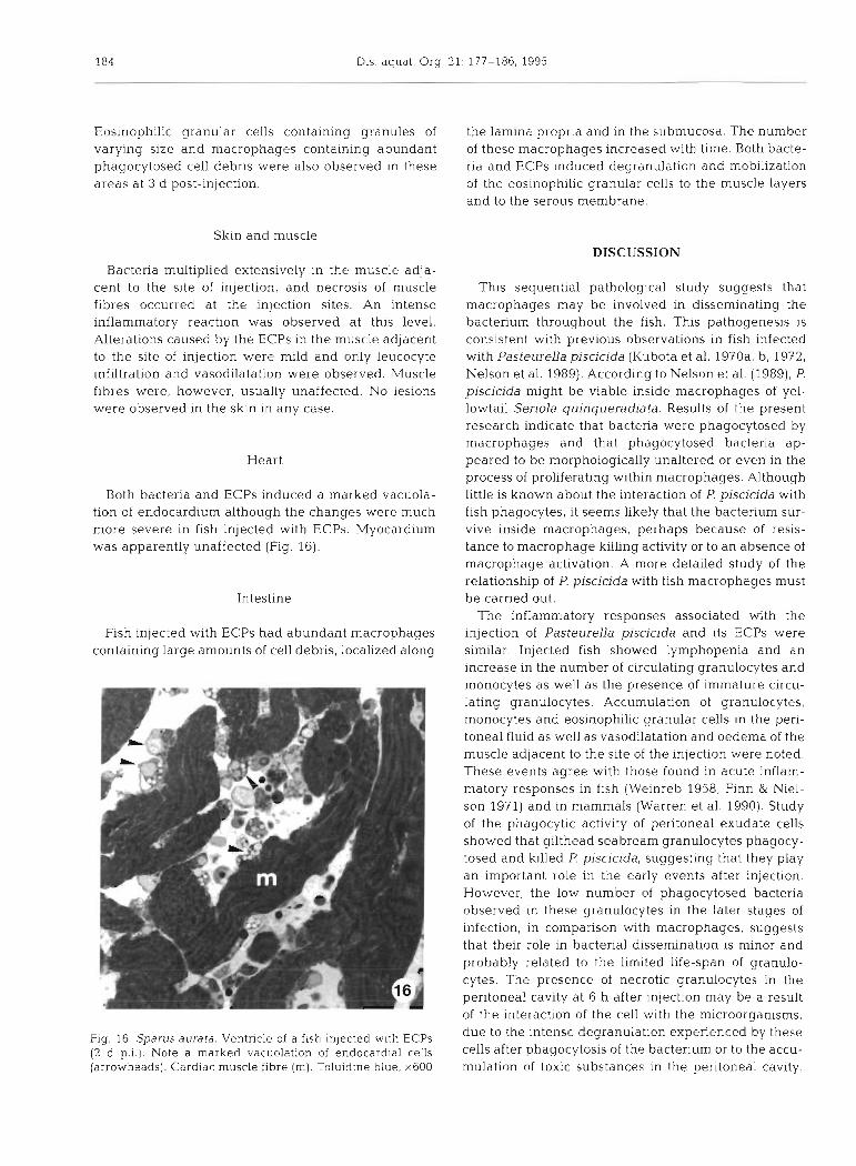
Both bacteria and ECPs induced a marked vacuola- tion of endocardium although the changes were much more severe in fish injected with ECPs. Myocardium was apparently unaffected (Fig. 16).
Intestine
Fish injected with ECPs had abundant macrophages containing large amounts of cell debris, localized along
This sequential pathological study suggests that macrophages may be involved in disseminating the bacterium throughout the fish. This pathogenesis is consistent with previous observations in fish infected with PasteureLla piscicida (Kubota et al. 1970a, b, 1972, Nelson et al. 1989). According to Nelson et al. (1989), P. piscicida might be viable inside macrophages of yel- lowtail Seriola quinqueradiata. Results of the present research indicate that bacteria were phagocytosed by macrophages and that phagocytosed bacteria ap- peared to be morphologically unaltered or even in the process of proliferating within macrophages. Although little is known about the interaction of P. piscicida with fish phagocytes, it seems likely that the bacterium sur- vive inside macrophages, perhaps because of resis- tance to macrophage killing activity or to an absence of macrophage activation. A more detailed study of the relationship of P. piscicida with fish macrophages must be carried out.
The inflammatory responses associated with the injection of Pasteurella piscicida and its ECPs were similar. Injected fish showed lymphopenia and an increase in the number of circulating granulocytes and monocytes as well as the presence of immature circu- lating granulocytes. Accumulation of granulocytes, monocytes and eosinophilic granular cells in the peri- toneal fluid as well as vasodilatation and oedema of the muscle adjacent to the site of the injection were noted. These events agree with those found in acute inflam- matory responses in fish (Weinreb 1958, Finn & Niel- son 1971) and In mammals (Warren et al. 1990). Study of the phagocytic activity of peritoneal exudate cells showed that gilthead seabream granulocytes phagocy- tosed and killed P. piscicida, suggesting that they play an important role in the early events after injection. However, the low number of phagocytosed bacteria observed in these granulocytes in the later stages of infection, in comparison with macrophages, suggests that their role in bacterial dissemination is minor and probably related to the limited life-span of granulo- cytes. The presence of necrotic granulocytes in the peritoneal cavity at 6 h after injection may be a result of the interaction of the cell with the microorganisms,
Fig. 16. Sparus aurata. Ventricle of a flsh inlected wlth ECPs due to the intense degranulation experienced by these
(2 d p.i.1. Note a marked vacuolation of endocardial cells cells after ~ h a g o c ~ t o s i s of the bacterium or to the accu- (arrowheads). Cardiac muscle f ~ b r e (m). Toluidine blue, x600 mulation of toxic substances in the peritoneal cavity.
Noya et al.. Sequential pathology of experimental pasteurellosis 185
The mobilization and changes in the morphology of the eosinophilic granular cells of the gill and intestine of gilthead seabream in response to the injection of P. pis- cicida or its ECPs were similar to the effects observed in the eosinophilic granular cells of rainbow trout injected with Aeromonas salmonicida and Vibrio anguillarum ECPs (Lamas et al. 1991, Powell et al. 1993). These changes suggest that eosinophilic granu- lar cells are involved in inflammatory responses in fish.
The lesions observed in Sparus aurata associated with Pasteurella piscicida may be a consequence of the invasion 'strategy' of the bacterium. In advanced stages of infection, aggregates of degenerate macro- phages full of apparently intact bacteria and bacterial colonies of varying size were observed in several organs, suggesting that these colonies may be a result of the destruction of microorganism-laden macro- phages. However, the histopathological changes sur- rounding the colonies were usually mild or unde- tectable, suggesting that the toxins released by the bacterium are not very destructive. These changes were consistent with those caused by the ECPs which also showed a tissue-destructive capacity that was very poor in comparison to the ECPs of other fish pathogens such as Aeromonas salmonicida (Ellis et al. 1981) or Vibrio anguillarum (Lamas 1992). The ab- sence of lesions in the muscle adjacent to the site of ECP injection or in the spleen ellipsoids following ECP injection may be related to the low proteolytic activity shown jn vitro by the ECPs of the strain used in this study (Magarilios et al. 1992) and may explain the low tissue-destructive capacity of the bacterium.
Fish injected with ECPs had a notable decrease in the number of circulating erythrocytes. The presence of numerous degenerate erythrocytes in the blood or phagocytosed by macrophages corroborates the pres- ence of the haemolytic activity demonstrated in vitro (Magarifios et al. 1992). However, the reduction in the number of circulating erythrocytes in fish injected with Pasteurella piscicida was not significant. This mild haemolysis due to the bacteria in vivo may reflect the fact that the bacteria spend most of the time inside macrophages during the development of the infection; it is only in the later stages of the infection, after destroying the macrophages, that they occur as free colonies. As a consequence, the release of haemolysins to the blood would be restricted to the last part of the infection. These results indicate that haemolysins may not be a major factor in the lethality of fish pasteur- ellosis.
The toxicity of the bacterium for hepatocytes and endothelial cells appeared to be restricted to the regions where the bacterium proliferated, although the epithelial and chloride cells of the gill lamellae showed acute degenerative changes even when they were not
lnvaded by the bacterium. These changes may be due to the toxic activities of its ECPs. The alterations caused by the ECPs to these organs were especially evident 3 d after injection and were more severe than those caused by bacteria. This difference in toxicity may be explained by the bacterium requiring time to produce the ECPs and the ECPs needing time to cause lesions. The severe lesions caused by the ECPs to these organs may be related to their physiological function. The liver is involved in detoxification and, in conse- quence, is especially liable to suffer injuries from tox- ins that it accumulates. The role of the epithelia1 and chloride cells in respiration and osmoregulation may also make them sensitive to the toxins circulating in the blood.
The cause of fish death by Pasteurella piscicida infection is still unknown. Previous reports have sug- gested that fish may die by bacterial embolism or by suffocation due to macrophage infiltration in the gill lamellae (Kubota et al. 1970a, Nelson et al. 1989). The toxic activities of the ECPs demonstrated in the present study, especially in certain critical organs, suggest that the toxins play a role in the pathogenesis of the dis- ease.
Acknowledgements. Thls study was supported by grants (MAR 91-1133-C02-01 and AGF94-1360-C03-01) from the Comision Interministerial de Ciencia y Tecnologia (CICYT). B.M. acknowledges the Xunta de Galicia for research fellow- ship. The authors thank Prof. Ramon Anadon for the critical reading of the manuscript and J. M. Leboreiro (Granja Cultivo Integral de Rodaballo, Quilmas, Carnota, Spain) for the sup- ply of fish needed for the various experiments.
LITERATURE CITED
Bradford MM (1976) A rapid and sensitive method for the quantitation of the principle of 248-254
microgram quantities of protein utilizing protein-dye bind~ng. Analyt Biochem 72:
Ellis AE, Hastings TS, Munro LS (1981) The role of Aeromonas salrnonicida extracellular products in the pathology of furunculosis. J Fish Dis 4:41-51
Finn JP, Nielson NO (1971) The inflammatory response of rainbow trout. J Fish Biol3:463-478
Hawke JP, Plakas SM, Minton RV, McPherson RM, Snider TG, Guarino AM (1987) Fish pasteurellosis of cultured striped bass, Morone saxatilis, in coastal Alabama. Aqua- culture 65:193-204
Kitao T (1993) Pasteurellosis. In: Inglis V, Roberts RJ, Bro- mage NR (eds) Bacterial diseases of fish. Blackwell Scien- tific Publ, Oxford, p 159-165
Kubota S, Krmura M, Egusa S (1970a) Studies of a bacterial tuberculoidosis of the yellowtail. I. Symptomatology and histopathology. Fish Pathol 4:111-118
Kubota S , Kimura M, Egusa S (1970b) Studies of a bacterial tuberculoidosis of the yellowtail. 11. Mechanism of nodule formation. Fish Pathol 5:31-34
Kubota S, Kimura M, Egusa S (1972) Studies of a bacterial tuberculoidosis of the yellowtail. 111. Findings on nodules and bacterial colonies in tissues. Fish Pathol 6:69-72
186 Dis. aquat. Org. 21: 177-186, 1995
Kusuda R, Salati F (1993) Major bacterial diseases affecting mariculture in Japan. In: Inglis V. Roberts RJ, Bromage NR (eds) Bacterial diseases of fish. Blackwell Scientif~c Publ, Oxford, p 69-85
Lamas J (1992) Alteraciones experimentales causadas por Vibno anguillarurn y sus productos extracelulares en trucha arcoiris. Aspectos citologicos e histopatologicos. PhD thesis, University of Santiago de Compostela
Lamas J , Bruno DW, Santos Y, Anadon R, Ellis AE (1991) Eosinophilic granular cells response to intraperitoneal injection with Vibno anguillarum and its extracellular products in rainbow trout, Oncorhynchus mykiss. Fish Shellfish Immunol 1:187-194
Magarifios B, Santos Y, Romalde JL, Barja JL, Toranzo AE (1992) Pathogenic activities of live cells and extracellular products of the fish pathogen Pasteurella piscicida. J gen Microbiol 138:2491-2498
Nelson JS, Kawahara E, Kawai K, Kusuda R (1989) Macro- phage infiltration in pseudotuberculosis of yellowtail, Seriola quinqueradiata. Bull mar Sci Fish, Kochi Univ 11:17-22
Powell MD, Briand HA, Wright GM, Burka JF (1993) Rainbow trout (Oncorhynchus rnykiss Walbaum) intestinal eosino- philic granule cells (EGC) response to Aeromonas sal- rnonicida and Vibrio anguillarum extracellular products.
Responsible Subject Editor: T EveIyn, Nanaimo, B.C., Canada
Fish Shellfish Immunol 3:279-289 Thune RL. Stanley LA, Cooper RK (1993) Pathogenesis of
gram-negative bacterial infections in warmwater fish. In: Falsal M, Hetnck FM (eds) Annual revlew of flsh diseases. Pergamon Press, New York, p 5-36
Toranzo AE, Barreiro S, Casal JF, Figueras A, Magarinos B, Barja JL (1991) Pasteurellosis in cultured gilthead sea- bream, Sparus aurata: first report in Spain. Aquaculture 99:l-15
Tung MC, Tsai SS, Ho LF, Huang ST, Chen SC (1985) An acute septicemic infection of Pasteurella organlsm in pond-cultured Formosa snake-head flsh (Channa rnacu- lata Lacepede) in Taiwan. Fish Path01 25.143-148
Warren JS, Ward PA, Johnson KJ (1990) The inflammatory response. In: Williams WJ, Beutler E. Erslev AJ, Llchtman MA (eds) Hematology, 4th edn. McGraw-Hill, Inc, New York, p 63-70
Weinreb EL (1958) Studies on the histology and histopathol- ogy of the rainbow trout, Salmo gairdneri irideus. 1. Hema- tology: under normal and experimental conditions of inflammation. Zoologica 46:145-152
Wolke RE (1975) Pathology of bacterial and fungal diseases affecting fish. In: Rubelin W, Migaki G (eds) The pathol- ogy of fishes. University of Wisconsin Press, Madison, p 33-116
manuscript first received: March 9, 1994 Rev~sed version accepted: November 6, 1994