Sequencing Site Booklet
28
For life science research only. Not for use in diagnostic procedures. FOR IN VITRO USE ONLY. DI G High Prime DNA Labeling and Detection Starter Kit I For color detection with NBT/BCIP. Random primed DNA labeling with digoxigenin-dUTP, alkali-labile and detection of hybrids by enzyme immunoassay Cat. No. 11 745 832 910 Store at 15 to 25° C Kit for 12 labeling reactions of 10 ng-3 g DNA and detection of 24 blots of 100 cm² Instruction Manual Version December 2005
-
Upload
biochemisthimself -
Category
Documents
-
view
217 -
download
0
Transcript of Sequencing Site Booklet

8/6/2019 Sequencing Site Booklet
http://slidepdf.com/reader/full/sequencing-site-booklet 1/28
For life science research only. Not for use in diagnostic procedures.FOR IN VITRO USE ONLY.
DIG High Prime DNA Labelingand Detection Starter Kit IFor color detection with NBT/BCIP.Random primed DNA labeling with digoxigenin-dUTP,alkali-labile and detection of hybrids by enzyme immunoassay
Cat. No. 11 745 832 910 Store at 15 to 25° CKit for 12 labeling reactions of 10 ng-3 g DNAand detection of 24 blots of 100 cm²
Instruction Manual
Version December 2005

8/6/2019 Sequencing Site Booklet
http://slidepdf.com/reader/full/sequencing-site-booklet 2/28
Roche Applied Science2
1. Preface
1.1 Table of Contents
1. Preface......................................................................................................................................................21.1 Table of Contents .................................................................................................................................................................. 21.2 Kit contents ............................................................................................................................................................................. 3
2. Introduction .............................................................................................................................................52.1 Product overview................................................................................................................................................................... 5
3. Procedures and required materials ....................................................................................................83.1 Before you begin................................................................................................................................................................... 83.2 DIG-DNA Labeling ............................................................................................................................................................... 93.3 Determination of labeling efficiency............................................................................................................................123.4 DNA transfer and fixation................................................................................................................................................153.5 Hybridization.........................................................................................................................................................................173.6 Immunological detection..................................................................................................................................................19
3.7 Stripping and reprobing of DNA blots........................................................................................................................214. Results ....................................................................................................................................................224.1 Typical results.......................................................................................................................................................................22
5. Appendix.................................................................................................................................................235.1 Trouble shooting .................................................................................................................................................................235.2 References.............................................................................................................................................................................245.3 Ordering Information.........................................................................................................................................................25
8/6/2019 Sequencing Site Booklet
http://slidepdf.com/reader/full/sequencing-site-booklet 3/28
Roche Applied Science3
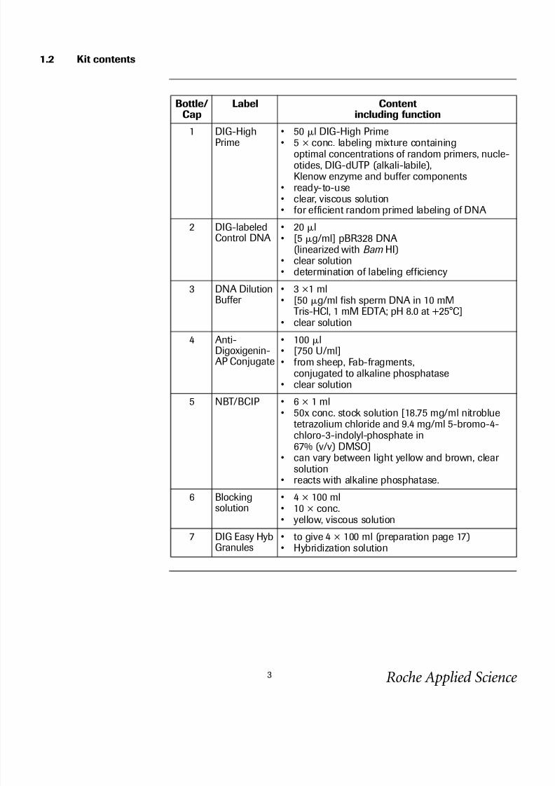
1.2 Kit contents
Bottle/Cap
Label Contentincluding function
1 DIG-HighPrime
• 50 l DIG-High Prime• 5 × conc. labeling mixture containing
optimal concentrations of random primers, nucle-otides, DIG-dUTP (alkali-labile),Klenow enzyme and buffer components
• ready-to-use• clear, viscous solution• for efficient random primed labeling of DNA
2 DIG-labeledControl DNA
• 20 l• [5 g/ml] pBR328 DNA
(linearized with Bam HI)• clear solution
• determination of labeling efficiency3 DNA Dilution
Buffer • 3 ×1 ml• [50 g/ml fish sperm DNA in 10 mM
Tris-HCl, 1 mM EDTA; pH 8.0 at +25°C]• clear solution
4 Anti-Digoxigenin-AP Conjugate
• 100 l• [750 U/ml]• from sheep, Fab-fragments,
conjugated to alkaline phosphatase• clear solution
5 NBT/BCIP • 6 × 1 ml
• 50x conc. stock solution [18.75 mg/ml nitrobluetetrazolium chloride and 9.4 mg/ml 5-bromo-4-chloro-3-indolyl-phosphate in67% (v/v) DMSO]
• can vary between light yellow and brown, clear solution
• reacts with alkaline phosphatase.
6 Blockingsolution
• 4 × 100 ml• 10 × conc.• yellow, viscous solution
7 DIG Easy Hyb
Granules
• to give 4 × 100 ml (preparation page 17)
• Hybridization solution
8/6/2019 Sequencing Site Booklet
http://slidepdf.com/reader/full/sequencing-site-booklet 4/28
Roche Applied Science4
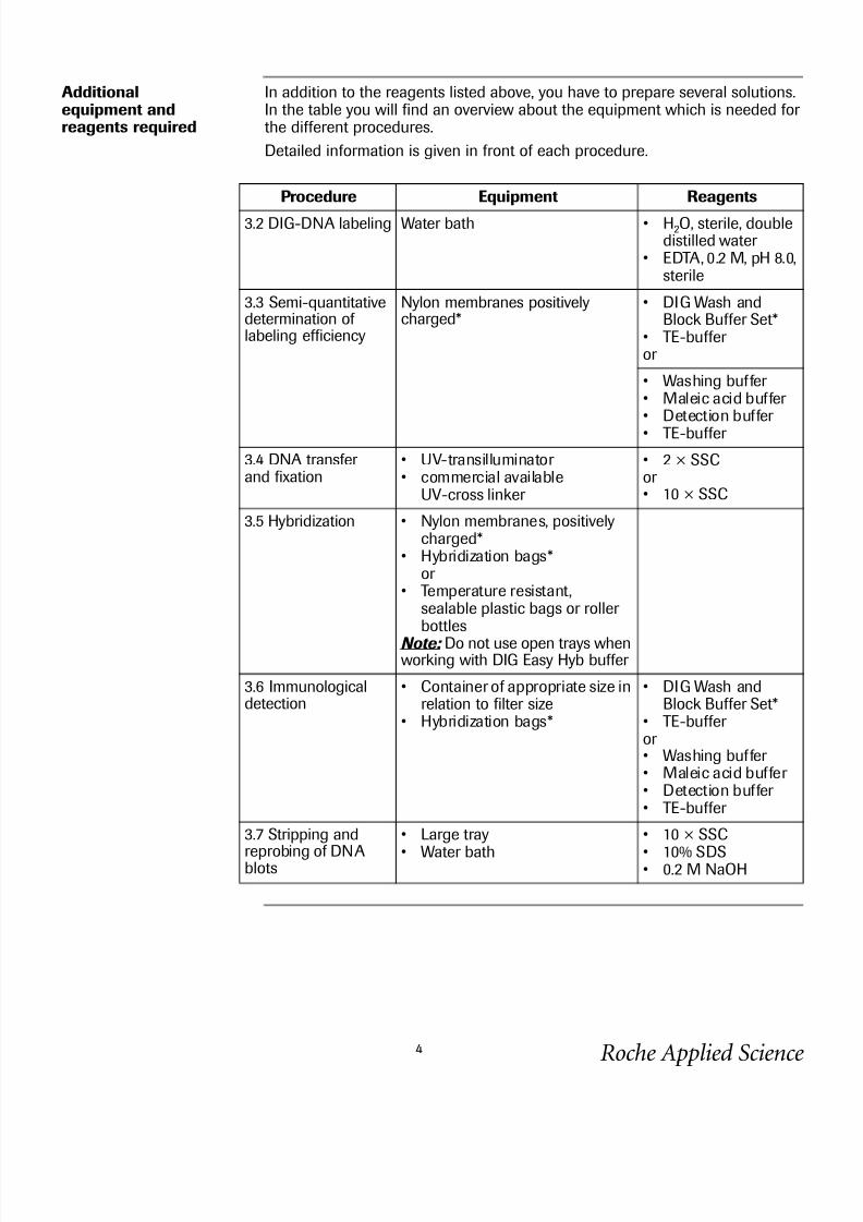
Additionalequipment andreagents required
In addition to the reagents listed above, you have to prepare several solutions.In the table you will find an overview about the equipment which is needed for the different procedures.
Detailed information is given in front of each procedure.
Procedure Equipment Reagents
3.2 DIG-DNA labeling Water bath • H2O, sterile, doubledistilled water
• EDTA, 0.2 M, pH 8.0,sterile
3.3 Semi-quantitativedetermination of labeling efficiency
Nylon membranes positivelycharged*
• DIG Wash andBlock Buffer Set*
• TE-buffer or
• Washing buffer • Maleic acid buffer • Detection buffer • TE-buffer
3.4 DNA transfer and fixation
• UV-transilluminator • commercial available
UV-cross linker
• 2 × SSCor • 10 × SSC
3.5 Hybridization • Nylon membranes, positivelycharged*
• Hybridization bags*or
• Temperature resistant,sealable plastic bags or roller bottles
Note: Do not use open trays whenworking with DIG Easy Hyb buffer
3.6 Immunologicaldetection
• Container of appropriate size inrelation to filter size
• Hybridization bags*
• DIG Wash andBlock Buffer Set*
• TE-buffer or • Washing buffer • Maleic acid buffer • Detection buffer • TE-buffer
3.7 Stripping and
reprobing of DNAblots
• Large tray
• Water bath
• 10 × SSC
• 10% SDS• 0.2 M NaOH

8/6/2019 Sequencing Site Booklet
http://slidepdf.com/reader/full/sequencing-site-booklet 5/28
Roche Applied Science5
2. Introduction
2.1 Product overview
Test principle The DIG High Prime DNA Labeling and Detection Starter Kit I uses digoxigenin(DIG), a steroid hapten, to label DNA probes for hybridization and subsequentcolor detection by enzyme immunoassay [1].
Stage Description
DNA labeling DIG-labeled DNA probes are generated with DIG-High Primeaccording to the random primed labeling technique. DIG-High Primeis a specially developed reaction mixture containing digoxigenin-dUTP, alkali-labile (Fig. 2) and all reagents, including enzymenecessary for random primed labeling, premixed in an optimized5 × concentrated reaction buffer.
Hybridization DIG-labeled probes are used for hybridization to membrane
blotted nucleic acids according to standard methods. The use of thealkali-labile form of DIG-11-dUTP enables easier and more efficientstripping of blots for rehybridization with a secondDIG-labeled probe.
Immunologicaldetection
The hybridized probes are immunodetected with anti-digoxigenin-AP,Fab fragments and are then visualized with the colorimetric substratesNBT/BCIP.

8/6/2019 Sequencing Site Booklet
http://slidepdf.com/reader/full/sequencing-site-booklet 6/28
Roche Applied Science6
Application DIG-labeled DNA probes can be used:
• for all types of filter hybridization
• for single copy gene detection in total genomic DNA, even from organismswith high complexity, e.g. human, barley, and wheat.
Sample material • DNA fragments of at least 100 bp
• linearized plasmid, cosmid or DNA• supercoiled DNA
Assay time This table lists the reaction time of the single steps
Number of tests 1 kit is sufficient for • 12 standard labeling reactions of up to 3 g template DNA
and detection of • 24 blots of 100 cm2.
Quality Control Using unlabeled control-DNA (pBR 328), labeled as described in the protocol,0.1 pg of homologous DNA is detected in a dot blot after 16 h color develop-ment (1 pg of homologous DNA can be detected after 1h color development).
Step Reaction time
DNA labeling 1 h-O/N
Hybridization 6 h or O/N
Immunological detection 1.5 h
Color development 0.5 - 16 h

8/6/2019 Sequencing Site Booklet
http://slidepdf.com/reader/full/sequencing-site-booklet 7/28
Roche Applied Science7
Kit storage/stability
The unopened kit is stable at -15 to -25° C until the expiration date printed onthe label. Shipping conditions on dry ice.
Once opened, please refer to the following table for proper storage.
Sensitivityand specificity
A single copy human gene is detected in a Southern blot of 1 g digested pla-centa DNA.
Note: Sensitivity depends both on the concentration of labeled DNA in thehybridization and on the time of color reaction.
Advantages This table describes benefits and features of the kit.
Kit component Storage
Anti-Digoxigenin-AP Conjugatecap 4
2-8° C, stable
Blocking solutionbottle 6
• unopened, stable at 15-25° C• once opened, it should be aliquoted and
stored at -15 to -25° C or at 2-8° C up to onemonth when keeping sterile
• working solution should always be preparedfresh
NBT/BCIPcap 6
• 2-8° C, stable• or at least 4 weeks at 15-25° CNote: During shipment of the kit on dry ice,a precipitate may occur which is easily dissolvedby briefly warming to 37°C
Benefit Feature
Accurate and fast The use of premixed DIG-High Prime mini-mizes the hands-on-time required to labelDNA probes and increases yields andreproducibility.
Sensitive Single-copy genes can be detected in totalhuman DNA complex and plant genomes.
Time-saving DIG-labeled probes can be stored for atleast one year. Hybridization solutions canbe reused 3 – 5 times, depending on theamount of labeled probe used for signalgeneration in each hybridization.

8/6/2019 Sequencing Site Booklet
http://slidepdf.com/reader/full/sequencing-site-booklet 8/28
Roche Applied Science8
3. Procedures and required materials
3.1 Before you begin
General handling rec-ommendations
This table describes general hints for DIG labeling and detection.
Overview
Recommendation Guideline
Work under clean conditions • Autoclave DIG System solutions• Filter-sterilize solutions containing SDS• Tween 20 should be added to previously
sterilized solutions
Use clean incubation trays Rigorously clean and rinse laboratory traysbefore each use.
Membrane handling requirements • Wear powder-free gloves• Handle membrane only on the edges and
with clean forceps
Section 3.2 DIG-DNA labeling
↓
Section 3.3 Quantification of labeling efficiency
↓Section 3.4 DNA fixation
↓
Section 3.5 Hybridization
↓
Section 3.6 Immunological detection
↓
Section 3.7 Stripping and reprobing of DNA blots

8/6/2019 Sequencing Site Booklet
http://slidepdf.com/reader/full/sequencing-site-booklet 9/28
Roche Applied Science9
3.2 DIG-DNA Labeling
Introduction DNA is random primed labeled with Digoxigenin-11-dUTP using DIG-HighPrime, a 5x concentrated labeling mixture of random hexamers, dNTP mix con-taining alkali-labile Digoxigenin-11-dUTP, labeling grade Klenow enzyme andan optimized reaction buffer.
Additionalequipment andreagents required
• water bath
• ice/water
This table lists composition, storage and use of the required reagents in addi-tion to kit components
Template DNA The following table lists the recommended features of the template DNA
Labeling of DNA isolated
from agarose
If you intend to perform genomic Southern blotting, you should separate thetemplate insert DNA from the vector by agarose gel electrophoresis.
Excise the DNA fragment from an agarose gel. For best results use either our High Pure PCR Product Purification Kit * or our Agarose Gel DNA ExtractionKit* to separate the DNA from the agarose.
Solution Composition Storage Use
Water Autoclaved, double distilled water 15-25° C,stable
Dilution of DNA
EDTA 0.2 M ethylenediamino- tetraacetic acid,pH 8.0
15-25° C,stable
Stopping thelabeling reaction
Feature Detail
Purity For plasmid DNA use the High Pure Plasmid Isolation Kit * for purification.When other commercially available purification kits are used, we recommendto do an additional phenol/chloroform extraction to remove residual protein.This step is also necessary when templates have been treated with restrictionor other modifying enzymes before labeling.
Size To obtain optimal results, template DNA should be linearized andshould have a size of 100 or larger. Template DNA >5 kb should be restric-
tion-digested using a 4 bp cutter (e.g. Hae III ) prior to labeling.Amount With the procedure described below principally 10 ng – 3 g of template
can be labeled, however, please check in the given table the necessaryamount of probe needed for your size of blot. By scaling up of all volumes andcomponents accordingly this procedure can be used for labeling of larger amounts. If single-copy gene detection in complex genomes is performed atleast 300 ng of template DNA (probe concentration: 25 ng/ml hybridizationsolution) should be labeled.

8/6/2019 Sequencing Site Booklet
http://slidepdf.com/reader/full/sequencing-site-booklet 10/28
Roche Applied Science10
Procedure This procedure is designed for 10 ng-3 g of DNA. Larger amounts (up to 10g) can be labeled by scaling up of all components and volumes.
Yield of labeling reac-tion
Table 1:
This table shows you the yield of DIG-High Prime labeling under optimal condi-tions.
In the standard reaction with 1 g DNA per assay approx. 15% of the nucle-otides are incorporated into about 0.8 g of newly synthesized DIG-labeledDNA within 1 h and approx. 38% of the nucleotides into about 2 g after 20 h.
Using DIG-High Prime solution, reactions were performed with increasingamountsof different template DNAs for 1 h and 20 h. The yield of DIG-labeled DNA wasdetermined by incorporation of a radioactive tracer and confirmed by a dot blot(Average of 10 independent labeling assays)
Step Action
1 Add 1g template DNA (linear or supercoiled) and autoclaved, doubledistilled water to a final volume of 16 l to a reaction vial.
2 Denature the DNA by heating in a boiling water bath for 10 min and quicklychilling in an ice/water bath.Note: Complete denaturation is essential for efficient labeling.
3 • Mix DIG-High Prime (vial 1) thoroughly and add 4 l to the denaturedDNA, mix and centrifuge briefly.
• Incubate for 1 h or O/N at 37° C.Note: Longer incubations (up to 20 h) will increase the yield of DIG-labeledDNA (see table below ).
4 Stop the reaction by adding 2l 0.2 M EDTA (pH 8.0) and/or by heating to 65°C for 10 min.Note: The length of the DIG-labeled fragments obtained with DIG-High Prime
range from 200 bp to 1000 bp or larger, depending on the length of theoriginal template.
Template DNA 1 h 20 h
10 ng 45 ng 600 ng
30 ng 130 ng 1050 ng
100 ng 270 ng 1500 ng
300 ng 450 ng 2000 ng
1000 ng 850 ng 2300 ng
3000 ng 1350 ng 2650 ng

8/6/2019 Sequencing Site Booklet
http://slidepdf.com/reader/full/sequencing-site-booklet 11/28
Roche Applied Science11
.
Fig. 2: Yield of DIG-labeled DNA from different amounts of template DNA after 1 and 20 h incu-bation of the DIG-High Prime reaction at 37°C.
3500
3000
2500
2000
1500
1000
500
0
10ng
30ng
100ng
300ng
1000ng
3000ng
amount of template DNA per labeling reaction
a m o u
n t o f s y n t h e s i z e d l a b e l e d
D N A ( n g )
1 h
20 h

8/6/2019 Sequencing Site Booklet
http://slidepdf.com/reader/full/sequencing-site-booklet 12/28
Roche Applied Science12
3.3 Determination of labeling efficiency
Introduction Determination of the yield of DIG-labeled DNA is most important for optimaland reproducible hybridization results. Too high of a probe concentration in thehybridization mix causes background, while too low of a concentration leads toweak signals.
Test principle The preferred method for quantification of labeled probes is the direct detec-tion method.
Preparation of additionalsolutions required
Please find in the following table composition and preparation of additionalreagents required. The following buffers are also available in the DIG Wash andBlock Buffer Set, DNase and RNase free.*Please note: These solutions are also used in the detection procedure of chapter 3.6. and can be prepared in larger quantities.
Stage Description
1 • A series of dilutions of DIG-labeled DNA is applied to a small strip of nylon membrane positively charged*.
• Part of the nylon membrane is preloaded with defined dilutions of DIG-labeled control DNA (vial 2) which are used as standards.
2 • The nylon membrane is subjected to immunological detection withanti-digoxigenin-AP conjugate (vial 4) and the premixed stock solutionof NBT/BCIP (vial 5).
• The intensities of the dilution series of DIG-labeled DNA and controlDNA are compared.
Solution Composition / Preparation Storage andstability
Use
Washing
buffer
0.1 M Maleic acid, 0.15 M NaCl;
pH 7.5 (20° C); 0.3% (v/v) Tween 20
15-25° C,
stable
Removal
of unboundantibody
Maleic acidbuffer
0.1 M Maleic acid, 0.15 M NaCl;adjust with NaOH (solid) to pH 7.5(20° C)
15-25° C,stable
Dilution of Blocking solution
Detectionbuffer
0.1 M Tris-HCl, 0.1 M NaCl,pH 9.5 (20° C)
15-25° C,stable
Adjustment of pH to 9.5
TE-buffer 10 mM Tris-HCl, 1 mM EDTA,pH 8.0
15-25° C,stable
Stopping color reaction
8/6/2019 Sequencing Site Booklet
http://slidepdf.com/reader/full/sequencing-site-booklet 13/28
Roche Applied Science13
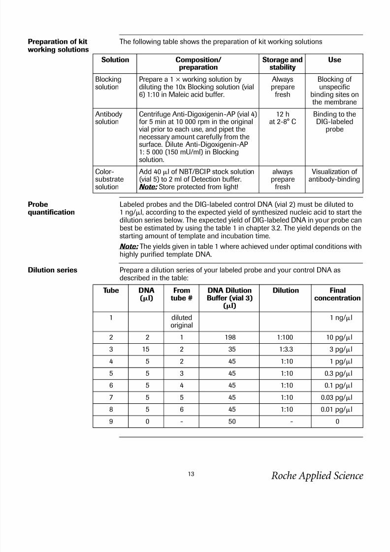
Preparation of kitworking solutions
The following table shows the preparation of kit working solutions
Probequantification
Labeled probes and the DIG-labeled control DNA (vial 2) must be diluted to1 ng/l, according to the expected yield of synthesized nucleic acid to start thedilution series below. The expected yield of DIG-labeled DNA in your probe canbest be estimated by using the table 1 in chapter 3.2. The yield depends on thestarting amount of template and incubation time.
Note: The yields given in table 1 where achieved under optimal conditions withhighly purified template DNA.
Dilution series Prepare a dilution series of your labeled probe and your control DNA asdescribed in the table:
Solution Composition/preparation
Storage andstability
Use
Blockingsolution
Prepare a 1 × working solution bydiluting the 10x Blocking solution (vial
6) 1:10 in Maleic acid buffer.
Alwaysprepare
fresh
Blocking of unspecific
binding sites onthe membrane
Antibodysolution
Centrifuge Anti-Digoxigenin-AP (vial 4)for 5 min at 10 000 rpm in the originalvial prior to each use, and pipet thenecessary amount carefully from thesurface. Dilute Anti-Digoxigenin-AP1: 5 000 (150 mU/ml) in Blockingsolution.
12 hat 2-8° C
Binding to theDIG-labeled
probe
Color-substratesolution
Add 40 l of NBT/BCIP stock solution(vial 5) to 2 ml of Detection buffer.Note: Store protected from light!
alwaysprepare
fresh
Visualization of antibody-binding
Tube DNA(l)
Fromtube #
DNA DilutionBuffer (vial 3)
(l)
Dilution Finalconcentration
1 dilutedoriginal
1 ng/l
2 2 1 198 1:100 10 pg/l
3 15 2 35 1:3.3 3 pg/l
4 5 2 45 1:10 1 pg/l
5 5 3 45 1:10 0.3 pg/l
6 5 4 45 1:10 0.1 pg/l7 5 5 45 1:10 0.03 pg/l
8 5 6 45 1:10 0.01 pg/l
9 0 - 50 - 0

8/6/2019 Sequencing Site Booklet
http://slidepdf.com/reader/full/sequencing-site-booklet 14/28
Roche Applied Science14
Procedure The following procedure describes the direct detection.
Note: Use sufficient buffer volumes to cover the membrane completely duringall steps.
Analyzingthe results
Compare the intensity of the spots out of your labeling reaction to the controland calculate the amount of DIG-labeled DNA. If the 0.1 pg dilution spots of
your probe and of the control are visible, then the labeled probe has reachedthe expected labeling efficiency (pls. see table 1 in 3.2.) and can be used in therecommended concentration in the hybridization.
The following spots should be visible
Step Action
1 Apply a 1 l spot of tubes 2-9 from your labeled probes and the labeled controlto the nylon membrane.
2 Fix the nucleic acid to the membrane by cross linking with UV-light or bakingfor 30 min at 120 ° C.
3 • Transfer the membrane into a plastic container with 20 mlMaleic acid buffer .
• Incubate under shaking for 2 min at 15-25° C.
4 Incubate for 30 min in 10 ml Blocking solution.
5 Incubate for 30 min in 10 ml Antibody solution.
6 Wash with 10 ml Washing buffer , 2 × 15 min.
7 Equilibrate 2-5 min in 10 ml Detection buffer .
8 Incubate membrane in 2 ml freshly prepared color substrate solution in aappropriate container in the dark. Do not shake during color development.Note: The color precipitate starts to form within a few minutes. The membranecan be exposed to light for short time periods to monitor color development.
9 Stop the reaction, when desired spot or band intensities are achieved,by washing the membrane for 5 min with 50 ml of sterile double dist. water or with TE-buffer.Results can be documented by photocopying the wet filter or by photography.
Incubation time Appearance
5-10 min 30 pg spot
30 min 3 pg spot

8/6/2019 Sequencing Site Booklet
http://slidepdf.com/reader/full/sequencing-site-booklet 15/28
Roche Applied Science15
3.4 DNA transfer and fixation
Transfer methods andmembranes
Standard protocols for gel electrophoresis, denaturation and neutralization of the gel are described in Sambrook et al. (2). Gels lacking ethidium bromide arepreferred, because ethidium can cause uneven background problems. All com-mon types of
DNA transfer methods are suitable for subsequent DIG hybridization (4,5).In our experience, best results are obtained when gels are blotted by capillarytransfer with 20 × SSC on nylon membranes*, positively charged.
Note: Alkali transfer (e.g., in 0.4 M NaOH) is not suitable for the transfer of DIG-labeled molecular weight markers*.

8/6/2019 Sequencing Site Booklet
http://slidepdf.com/reader/full/sequencing-site-booklet 16/28
Roche Applied Science16
Fixation procedure Fix the DNA to the membrane by any of the following procedures:
Storage of the membrane
Please refer to the following table.
IF you want to... THEN...
UV-crosslinking(nylon membrane)
• Place the membrane on Whatman 3MM-paper soakedwith 10 × SSC.
• UV-crosslink the wet membrane without prior washing.
• After the UV-crosslinking, rinse the membrane brieflyin double distilled water and allow to air-dry.
bake at 120° C(nylon membrane)
• Wash the membrane briefly in 2 × SSC.• Bake the nylon membrane at 120° C for 30 min or
according to the manufacturer`s instructions.
bake at 80° C(nylon membrane)
• Wash the membrane briefly in 2 × SSC.• Bake at 80° C for 2 h under vacuum.
IF... THEN...
you want to go ahead. Use the membrane immediately for prehybridization.
you want to work later on store the membrane dry at 2-8° C.

8/6/2019 Sequencing Site Booklet
http://slidepdf.com/reader/full/sequencing-site-booklet 17/28
Roche Applied Science17
3.5 Hybridization
Additionalequipment required
• ice/water • shaking water-bath• or hybridization oven• temperature resistant plastic or glass boxes, petri dishes, roller bottles or
sealable plastic bags.Note: Do not use open containers with DIG Easy Hyb buffer.
Preparation of DIG Easy Hyb workingsolution
Add carefully 64 ml sterile double distilled water in two portions to the DIG EasyHyb Granules (bottle 7), dissolve by stirring immediately for 5 min at 37° C.
Hybridizationtemperature
The appropriate hybridization temperature is calculated according to GC con-tent and percent homology of probe to target according to the following equa-tion:
Tm = 49.82 + 0.41 (% G + C) - (600/l) [l = length of hybrid in base pairs]
Topt
. = Tm
–20 to 25° C
(The given numbers of the equation were calculated according to a standardequation for hybridization solutions containing formamide, 50%.) The actualhybridization temperature Topt. for hybridization with DIG Easy Hyb is 20-25° Cbelow the calculated Tm value. Topt. can be regarded as a stringent hybridizationtemperature allows up to 18% mismatches between probe and target. Whenthe degree of homology of your probe to template is less than 80%, you shouldlower Topt. accordingly (approx. 1.4°C below Tm per 1 % mismatch) and alsoadjust the stringent washing steps accordingly (i.e. increase SSC concentrationand lower washing temperature).
8/6/2019 Sequencing Site Booklet
http://slidepdf.com/reader/full/sequencing-site-booklet 18/28
Roche Applied Science18
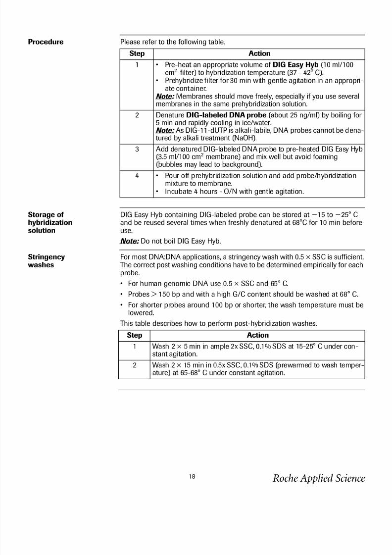
Procedure Please refer to the following table.
Storage of hybridizationsolution
DIG Easy Hyb containing DIG-labeled probe can be stored at 15 to 25° Cand be reused several times when freshly denatured at 68°C for 10 min beforeuse.
Note: Do not boil DIG Easy Hyb.
Stringencywashes
For most DNA:DNA applications, a stringency wash with 0.5 × SSC is sufficient.The correct post washing conditions have to be determined empirically for eachprobe.
• For human genomic DNA use 0.5 × SSC and 65° C.
• Probes > 150 bp and with a high G/C content should be washed at 68° C.
• For shorter probes around 100 bp or shorter, the wash temperature must belowered.
This table describes how to perform post-hybridization washes.
Step Action
1 • Pre-heat an appropriate volume of DIG Easy Hyb (10 ml/100cm2 filter) to hybridization temperature (37 - 42° C).
• Prehybridize filter for 30 min with gentle agitation in an appropri-ate container.
Note: Membranes should move freely, especially if you use severalmembranes in the same prehybridization solution.
2 Denature DIG-labeled DNA probe (about 25 ng/ml) by boiling for 5 min and rapidly cooling in ice/water.Note: As DIG-11-dUTP is alkali-labile, DNA probes cannot be dena-tured by alkali treatment (NaOH).
3 Add denatured DIG-labeled DNA probe to pre-heated DIG Easy Hyb(3.5 ml/100 cm2 membrane) and mix well but avoid foaming(bubbles may lead to background).
4 • Pour off prehybridization solution and add probe/hybridizationmixture to membrane.
• Incubate 4 hours - O/N with gentle agitation.
Step Action
1 Wash 2 × 5 min in ample 2x SSC, 0.1% SDS at 15-25° C under con-stant agitation.
2 Wash 2 × 15 min in 0.5x SSC, 0.1% SDS (prewarmed to wash temper-ature) at 65-68° C under constant agitation.

8/6/2019 Sequencing Site Booklet
http://slidepdf.com/reader/full/sequencing-site-booklet 19/28
Roche Applied Science19
3.6 Immunological detection
Additional reagentsrequired
Please find in the following table composition and preparation of additionalreagents required. The following buffers are also available in the DIG Wash andBlock Buffer Set, DNase and RNase free*.
Preparation of kitworking solutions
In the following table the preparation of kit working solutions is described.
Solution Composition / Preparation Storage and
stability
Use
Washingbuffer
0.1 M Maleic acid, 0.15 M NaCl;pH 7.5 (20° C); 0.3% (v/v) Tween20
15-25° C,stable
Washing of membrane
Maleic acidbuffer
0.1 M Maleic acid, 0.15 M NaCl;adjust with NaOH (solid) to pH7.5 (20° C)
15-25° C,stable
Dilution of Blockingsolution
Detectionbuffer
0.1 M Tris-HCl, 0.1 M NaCl,pH 9.5 (20° C)
15-25° C,stable
Alkalinephosphatasebuffer
TE-buffer 10 mM Tris-HCl, 1 mM EDTA, pH
8.0
15-25° C,
stable
Stopping
color reaction
Solution Composition / Preparation Storage andstability
Use
Blockingsolution
Prepare a 1x working solution bydiluting 10 × Blocking solution(vial 6) 1:10 with Maleic acidbuffer.
Alwaysprepare fresh
Blocking of unspecificbinding sites
Antibody
solution
Centrifuge Anti-Digoxigenin-AP
(vial 4) for 5 min at 10 000 rpm inthe original vial prior to each use,and pipet the necessary amountcarefully from the surface. DiluteAnti-Digoxigenin-AP 1:5000 (150mU/ml) in Blocking solution.
12 h at 2-8° C Binding to the
DIG-labeledprobe
Color-substratesolution
Add 200 l of NBT/BCIP stock solution (vial 5) to 10 ml of Detec-tion buffer.Note: Store protected from light!
Alwaysprepare fresh
Visualizationof antibody-binding

8/6/2019 Sequencing Site Booklet
http://slidepdf.com/reader/full/sequencing-site-booklet 20/28
Roche Applied Science20
Procedure This table describes how to perform the immunological detection ona 100 cm2 membrane.
Note: All incubations should be performed at 15-25° C with agitation. If themembrane is to be reprobed, do not allow the membrane to dry at any time.
Storage of membrane
Please refer to the following table.
Step Action
1 After hybridization and stringency washes, rinse membrane briefly
(1-5) min in Washing buffer .
2 Incubate for 30 min in 100 ml Blocking solution.
3 Incubate for 30 min in 20 ml Antibody solution.
4 Wash 2 x 15 min in 100 ml Washing buffer .
5 Equilibrate 2-5 min in 20 ml Detection buffer .
6 Incubate membrane in 10 ml freshly prepared color substrate solu-tion in a appropriate container in the dark. Do not shake duringcolor development.Note: The color precipitate starts to form within a few minutes andthe reaction is usually complete after 16 h. The membrane can be
exposed to light for short time periods to monitor color development.7 Stop the reaction, when desired spot or band intensities are
achieved, by washing the membrane for 5 min with 50 ml of steriledouble dist. water or with TE-buffer.Results can be documented by photocopying the wet filter or by pho-tography.
IF... THEN...
you want to reprobe the membrane the membrane should not dry off at any
time, store in sealed plastic bag.Note : If you want to maintain the color,store membranes in TE buffer do notallow the membrane to dry.
you don´t want to reprobe dry the membrane at 15 to 25°C for stor-age.Note: Color fades upon drying, to revita-lize the color, wet the membrane in TEbuffer.

8/6/2019 Sequencing Site Booklet
http://slidepdf.com/reader/full/sequencing-site-booklet 21/28
Roche Applied Science21
3.7 Stripping and reprobing of DNA blots
General The alkali-labile form of DIG-11-dUTP enables easier and more efficient strip-ping of blots for rehybridization experiment.
Additional reagents
required
• Dimethylformamid (DMF)
• 0.2 N NaOH, 0.1% SDS (w/v)• 2 × SSC
Protocol
Storage of strippedmembrane
Please refer to the following table.
Note : When stripping and rehybridization of blots is planned, the membraneshould not dry off at any time.
CAUTION: Work under a fume hood
Step Action
1 • Heat DMF in a large glass beaker in a water bath under a fumehood to 50-60° C.
• Incubate the membranes in the heated DMF until the blue color precipitate is removed from the filter.CAUTION: DMF is volatile and can be ignited above 67° C.
2 Rinse membrane briefly in double distilled water .
3 Wash for 2 × 15 min in 0.2 N NaOH, SDS, 0.1% (w/v) at 37° Cunder constant agitation.
4 Equilibrate briefly in 2 × SSC.
5 Prehybridize and hybridize with a second probe.
Once the membrane is stripped, it can be stored in Maleic acid buffer or 2 × SSC until used again.

8/6/2019 Sequencing Site Booklet
http://slidepdf.com/reader/full/sequencing-site-booklet 22/28
Roche Applied Science22
4. Results
4.1 Typical results
Genomic
Southern blot
Figure 3: This figure shows you the detection of a single-copy gene (ß-actin) in total human DNAusing the standard protocol.
Genomic Southern blot
Size (kb) 1 2 3 4
21
5.1/5.04.3
3.5
2.01.9
1.61.4
0.95
0.83
0.56
2.55 1.0 g
Probe: DIG label -actin DNA fragmentLane 1: 100 ng DNA molecular weight marker III, digoxigenin-labeled.
Lane 2: 5 g
Lane 3: 2.5 g
Lane 4: 1 g
human placenta DNA,
Eco R1

8/6/2019 Sequencing Site Booklet
http://slidepdf.com/reader/full/sequencing-site-booklet 23/28
Roche Applied Science23
5. Appendix
5.1 Trouble shooting
Trouble shooting table This table describes various troubleshooting parameters for DIG-labeling anddetection
Problem Possiblecause
Recommendation
Lowsensitivity
Inefficientprobe labeling
• Check labeling efficiency. The labeling reactioncan be upscaled. Prolong incubation time toovernight.
• Clean up template DNA by phenolization.• Use only fragments < 5 kb or predigest with a
restriction enzyme (e.g., four bp cutter)• Make sure that template is efficiently
denatured before labeling.
Low probeconcentrationin the hybrid-ization
• Increase probe concentration, but do not usemore than 25 ng/ml DNA probe. Check hybrid-ization and washing conditions.
• Prolong hybridization time.• Prolong color development to 16 h.
Highback-ground
Inefficienthybridization
• Recalculate hybridization temperature.• Do not the allow the membrane to dry between
prehybridization and hybridization.• If you use plastic bags, remove all air bubbles
prior to sealing.
Wrong type of nylon mem-
brane
Some types of nylon membrane may causehigh background: use nylon membrane*,
especially tested for the DIG-System fromRoche Applied Science.
Inefficientblockingbeforeimmuno-assay
Prolong blocking and washing steps.
Ineffectivestringencywashes
Check temperature of stringency washes,prewarm wash solution to correct temperature
Special hintsfor immuno-assay
When using laboratory trays for the detectionprocedure, they should be rigorously cleanedbefore use. Anti-DIG-AP binding and color development should be done in separate trays.

8/6/2019 Sequencing Site Booklet
http://slidepdf.com/reader/full/sequencing-site-booklet 24/28
Roche Applied Science24
5.2 References
1 Höltke, H.J., Ankenbauer, W., Mühlegger, K., Rein, R., Sagner, G., Seibl, R., & Walter, T. (1995) The Digoxigenin (DIG)System for non-radioactive labeling and detection of nucleic acids-an overview. Cell. Mol. Biol. 41 (7): 883-905.
2 Sambrook, J., Fritsch, E.M. and Maniatis,T. (1989) Molecular cloning: a laboratory manual, 2nd edition,Cold Spring Harbor Laboratory, Cold Spring Harbor Labor, New York.
3 Southern E.M. (1975) Detection of specific sequences among DNA fragments separated by gel electrophoresis. J. Mol. Biol. 98: 503.
4 Khandijan, E.W. (1987) Optimized hybridization of DNA blotted and fixed to nitrocellulose and nylon membranes.Bio/Technology 5: 165.
5.3 Ordering Information
Kits For a complete overview, please visit and bookmark our “DIG Reagents and Kits
for Non-Radioactive Nucleic Acid Labeling and Detection” Special Interest Siteat http://www.roche-applied-science.
Product Pack Size Cat. No.
Agarose Gel DNA Extraction Kit 100 reactions 11 696 505 001
DNA Isolation Kit for Cells and Tis-sue for the extraction of genomicDNA from cells and tissue ranging insize from 50 to 150 kb
10 isolationsfor 400 mg tissue or
5 × 10⁷ cells
11 814 770 001
DNA Isolation Kit for mammalianBlood for the isolation of intactgenomic DNA from mammalianwhole blood or lymphocyte prepara-tions
25 purifications 11 667 327 001
High Pure PCR Product PurificationKitfor the purification of PCR reac-tion pro-ducts
50 purifications250 purifications
11 732 668 00111 732 676 001
High Pure Plasmid Isolation Kit smallscale mini-preps for sequencing,PCR, and cloning
50 purifications250 purifications
11 754 777 00111 754 785 001
PCR Clean Up Kitfor post-PCR DNA fragment purifi-cation
up to100 purifications
11 696 513 001

8/6/2019 Sequencing Site Booklet
http://slidepdf.com/reader/full/sequencing-site-booklet 25/28
Roche Applied Science25
Single reagents
* available from Roche Applied Science1) Tween is a trademark of ICI Americas Inc., USA2) High Pure is a trademark of a Member of the Roche Group
DISCLAIMER OF
LICENSE
The labeling of nucleic acids with DIG is covered by EP patents 0 324 474 and0
371 262 as well as the following US patents 5.344.757, 5.354.657 and 5.702.888owned by Roche Diagnostics GmbH.
Product Pack Size Cat. No.
Blocking reagent 50 g 11 096 176 001
DIG Easy Hyb (ready-to-use hybrid-ization solution without formamide)
500 ml 11 603 558 001
DNA Molecular Weight Marker,
Digoxigenin-labeled:DNA Molecular Weight Marker IIDNA Molecular Weight Marker IIIDNA Molecular Weight Marker VDNA Molecular Weight Marker VIDNA Molecular Weight Marker VIIDNA Molecular Weight Marker VII I
5 g (500 l)5 g (500 l)5 g (500 l)5 g (500 l)5 g (500 l)5 g (500 l)
11 218 590 91011 218 603 91011 669 931 91011 218 611 91011 669 940 91011 449 451 910
DIG Wash and Block Buffer Set 30 blots(10 × 10 cm2)
11 585 762 001
Hybridization bags 50 bags 11 666 649 001
Nylon Membrane, positively
charged(20 x 30 cm)(10 x 15 cm)(0.3 x 3 m roll)
10 sheets20 sheets1 roll
11 209 272 00111 209 299 00111 417 240 001

8/6/2019 Sequencing Site Booklet
http://slidepdf.com/reader/full/sequencing-site-booklet 26/28
Roche Applied Science26

8/6/2019 Sequencing Site Booklet
http://slidepdf.com/reader/full/sequencing-site-booklet 27/28
Roche Applied Science27

8/6/2019 Sequencing Site Booklet
http://slidepdf.com/reader/full/sequencing-site-booklet 28/28
Roche Diagnostics GmbHRoche Applied Science68298 Mannheim
1 2 0 5 . 1
1 7 4
6 8 4 7 ➇
www.roche-applied-science.com
to order, solve technical queries, find product information,
or contact your local sales representative.
www.roche-applied-science.com/pack-insert/11745832910a.pdf
Please visit our new Online Technical Support Site under www.roche-applied-science.com/support