September 26, 2014 Tamara S. Syrek Jensen, J.D. Mail … · September 26, 2014 . Tamara S. Syrek...
43
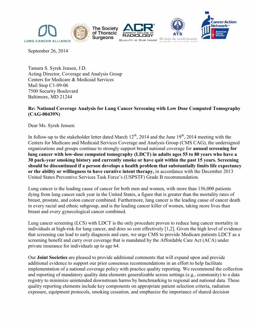
September 26, 2014 Tamara S. Syrek Jensen, J.D. Acting Director, Coverage and Analysis Group Centers for Medicare & Medicaid Services Mail Stop C1-09-06 7500 Security Boulevard Baltimore, MD 21244 Re: National Coverage Analysis for Lung Cancer Screening with Low Dose Computed Tomography (CAG-00439N) Dear Ms. Syrek Jensen: In follow-up to the stakeholder letter dated March 12 th , 2014 and the June 19 th , 2014 meeting with the Centers for Medicare and Medicaid Services Coverage and Analysis Group (CMS CAG), the undersigned organizations and groups continue to strongly support broad national coverage for annual screening for lung cancer with low-dose computed tomography (LDCT) in adults ages 55 to 80 years who have a 30 pack-year smoking history and currently smoke or have quit within the past 15 years. Screening should be discontinued if a person develops a health problem that substantially limits life expectancy or the ability or willingness to have curative intent therapy, in accordance with the December 2013 United States Preventive Services Task Force’s (USPSTF) Grade B recommendation. Lung cancer is the leading cause of cancer for both men and women, with more than 156,000 patients dying from lung cancer each year in the United States, a figure that is greater than the mortality rates of breast, prostate, and colon cancer combined. Furthermore, lung cancer is the leading cause of cancer death in every racial and ethnic subgroup, and is the leading cancer killer of women, taking more lives than breast and every gynecological cancer combined. Lung cancer screening (LCS) with LDCT is the only procedure proven to reduce lung cancer mortality in individuals at high-risk for lung cancer, and does so cost effectively [1,2]. Given the high level of evidence that screening can lead to early diagnosis and cure, we urge CMS to provide Medicare patients LDCT as a screening benefit and carry over coverage that is mandated by the Affordable Care Act (ACA) under private insurance for individuals up to age 64. Our Joint Societies are pleased to provide additional comments that will expand upon and provide additional evidence to support our prior consensus recommendations in an effort to help facilitate implementation of a national coverage policy with practice quality reporting. We recommend the collection and reporting of mandatory quality data elements generalizable across settings (e.g., community) to a data registry to minimize unintended downstream harms by benchmarking to regional and national data. These quality reporting elements include key components on appropriate patient selection criteria, radiation exposure, equipment protocols, smoking cessation, and emphasize the importance of shared decision
Transcript of September 26, 2014 Tamara S. Syrek Jensen, J.D. Mail … · September 26, 2014 . Tamara S. Syrek...
September 26, 2014 Tamara S. Syrek Jensen, J.D. Acting Director, Coverage and Analysis Group Centers for Medicare & Medicaid Services Mail Stop C1-09-06 7500 Security Boulevard Baltimore, MD 21244 Re: National Coverage Analysis for Lung Cancer Screening with Low Dose Computed Tomography (CAG-00439N) Dear Ms. Syrek Jensen: In follow-up to the stakeholder letter dated March 12th, 2014 and the June 19th, 2014 meeting with the Centers for Medicare and Medicaid Services Coverage and Analysis Group (CMS CAG), the undersigned organizations and groups continue to strongly support broad national coverage for annual screening for lung cancer with low-dose computed tomography (LDCT) in adults ages 55 to 80 years who have a 30 pack-year smoking history and currently smoke or have quit within the past 15 years. Screening should be discontinued if a person develops a health problem that substantially limits life expectancy or the ability or willingness to have curative intent therapy, in accordance with the December 2013 United States Preventive Services Task Force’s (USPSTF) Grade B recommendation. Lung cancer is the leading cause of cancer for both men and women, with more than 156,000 patients dying from lung cancer each year in the United States, a figure that is greater than the mortality rates of breast, prostate, and colon cancer combined. Furthermore, lung cancer is the leading cause of cancer death in every racial and ethnic subgroup, and is the leading cancer killer of women, taking more lives than breast and every gynecological cancer combined.
Lung cancer screening (LCS) with LDCT is the only procedure proven to reduce lung cancer mortality in individuals at high-risk for lung cancer, and does so cost effectively [1,2]. Given the high level of evidence that screening can lead to early diagnosis and cure, we urge CMS to provide Medicare patients LDCT as a screening benefit and carry over coverage that is mandated by the Affordable Care Act (ACA) under private insurance for individuals up to age 64. Our Joint Societies are pleased to provide additional comments that will expand upon and provide additional evidence to support our prior consensus recommendations in an effort to help facilitate implementation of a national coverage policy with practice quality reporting. We recommend the collection and reporting of mandatory quality data elements generalizable across settings (e.g., community) to a data registry to minimize unintended downstream harms by benchmarking to regional and national data. These quality reporting elements include key components on appropriate patient selection criteria, radiation exposure, equipment protocols, smoking cessation, and emphasize the importance of shared decision

Page 2 of 43
making, as detailed in the American College of Radiology (ACR) Lung Cancer Screening Center designation specifications under the ACR Computed Tomography (CT) accreditation program. The components of the ACR Lung Cancer Screening Center designation have been incorporated into the Lung Cancer Alliance (LCA) National Framework for Lung Cancer Screening Excellence. The Joint Societies support a coverage requirement that participating providers meet the specifications of the ACR’s Lung Screening Center designation. The Joint Societies also support a coverage requirement that ensures patient outcomes are effectively tracked. We are committed to tracking outcomes for patient populations other than those covered by Medicare, and are developing the Lung Cancer Screening Practice Registry as part of the ACR’s National Radiology Data Registry (NRDR) suite of registries. This year CMS approved the ACR National Radiology Data Registry as a Qualified Clinical Data Registry (QCDR) for the CMS Physician Quality Reporting System (PQRS) for the 2014 program year. The ACR has actively been leveraging our existing infrastructure to provide a clinical practice registry for lung cancer screening quality reporting. Other validated clinical practice registries that use the same recommended data elements discussed in this letter and map to ACR Lung-RADS™ for external reporting, such as International Early Lung Cancer Action Program (I-ELCAP), can provide through collaboration similar quality and outcomes reporting. Joint Society Consensus Recommendations:
I. The Joint Societies recommend unrestricted broad national coverage of LDCT lung cancer
screening for the patient population recommended by the USPSTF, with adherence to minimum quality elements, evidence based protocols and reporting to a clinical practice registry, including the following: [See next section for details]
a. Standards for centers performing lung cancer screening CT, as described in the ACR Lung
Cancer Screening Center Designation criteria under the CT accreditation program, the latter already required by CMS for outpatient imaging facilities under Medicare Improvements for Patients and Providers Act (MIPPA)
b. Structured reporting using ACR Lung Imaging Reporting and Data System (Lung-RADS™) for benchmarking clinical performance
c. Practice quality reporting through a clinical practice database, such as an ACR National Radiology Data Registry (NRDR)
II. The Joint Societies recommend Coverage with Evidence Development (CED) and data
registry consideration only for other high-risk individuals where evidence is promising [i.e., National Comprehensive Cancer Network (NCCN) Category 2 patient group] to inform future coverage decisions. This includes certain registry documentation requirements, including the following: [See next section for details]
a. Data registry protocol b. Standard data elements under a comprehensive CED registry c. Institutional agreements for participation d. Appropriate regulatory oversight as required for research data collection including institutional
review board approvals at participating sites

Page 3 of 43
III. Supplemental information is also provided in the following areas:
1) Access to CT scanner specific low dose chest CT protocols 2) Cost effectiveness of lung cancer screening with CT 3) Lung cancer screening and health equity for all individuals recommended by USPSTF 4) Management of individuals with a positive lung cancer screening CT 5) Quality improvements through a multidisciplinary coordination of efforts 6) Lung cancer screening and surgical outcomes
I. National Coverage for LDCT Lung Cancer Screening Recommendations
The Joint Societies fully support the USPSTF recommendations and agree the evidence is adequate to conclude that annual lung cancer screening with low dose CT is beneficial in high-risk individuals between ages 55 to 80 who are current smokers with at least 30 pack years of smoking history or former smokers who ceased tobacco use within the last 15 years, and that screening should be discontinued if a person develops a health problem that substantially limits life expectancy or the ability or willingness to have curative lung surgery.
For this patient group, we recommend national coverage of annual low dose CT with participating providers required to meet practice and quality reporting criteria outlined below and consistent with the ACR Lung-RADS™ reporting lexicon and the ACR Lung Cancer Screening Center designation under the CT accreditation program. This schema of structured reporting and adherence to practice quality criteria will minimize unintended downstream harms and minimize false positives. Provided below are the defined quality elements that we propose as the minimum data to be reported and maintained under broad national coverage. These minimum quality reporting and management operations are achievable across practice settings including the community setting, and are essential to establish practice benchmarks, and to report and compare practice quality. We recognize that there may be geographic or other variations that will be important to consider in setting benchmarks on some elements. For example, areas with endemic fungus in the soil such as the parts of the Midwest and Southwest may have higher screen positive rates.
As described in our March 12th stakeholder letter, the Joint Societies feel that continuous quality improvement should occur starting with adherence to best published practices. Our recommended criteria for eligible providers are outlined in our ACR Designated Lung Cancer Screening Center and provided below. LDCT LCS is already being performed safely and effectively in both academic and community settings and is generalizable to all practice settings if minimum quality criteria are in place.
a. The Following ACR Designated Lung Cancer Screening Center Criteria are Recommended

Page 4 of 43
Patient Criteria
1) The individual considering lung cancer screening with CT has undergone a clinical evaluation
including a medical history in a process of shared decision making with their primary healthcare provider and has met the following patient eligibility criteria.
The individual is 55 to 80 years of age Has a cigarette smoking history of 30 pack years and greater Has no underlying comorbidities which would preclude curative intent therapy (surgery or
stereotactic radiation) The referring and billing provider(s) have documented the appropriate evaluation and approved eligibility criteria of the Medicare beneficiary.
Greater than 90% adherence to appropriate patient selection is an appropriate benchmark.
Facility/Site Criteria
1) The facility/site implements follow-up systems, including a structured reporting protocol using ACR Lung-RADS™ (described in more detail below) and follows the ACR Practice Parameter for
Communication of Diagnostic Imaging Findings or the equivalent.
2) The facility/site offers or provides access to smoking cessation patient programs, education and counseling materials.
Have a mechanism in place to refer patients for smoking cessation counseling or provide smoking cessation materials either directly or as part of a lung cancer screening program.
Quality Control - Equipment Specifications and Radiation Exposure Protocols
1) The facility/site follows both state and federal requirements for equipment specifications and
performance, as well as the ACR Practice Parameters and Technical Standards or equivalent. In addition, the facility/site must have any accreditation needed to operate CT equipment.
Note: CT scanners used for the purpose of lung cancer screening should be multidetector helical (spiral) CT scanners and perform the exam in a single breath hold. Non-helical and single detector CT scanners are not appropriate for lung cancer screening CT.
2) The facility/site maintains compliance with a quality control (QC) program consistent with the ACR CT
Quality Control Manual or equivalent. 3) The facility/site implements and maintains an imaging protocol equivalent to the ACR Designated Lung
Cancer Screening Center requirements (see below specifications).
Radiation exposure levels consistent with lung screening protocols and not routine chest scans; the protocol shall have a CT Dose Index volume (CTDIvol) of < 3 mGy, for a standard size patient (5’7, 154lb) (using 32-cm diameter CTDI phantom)
Page 5 of 43
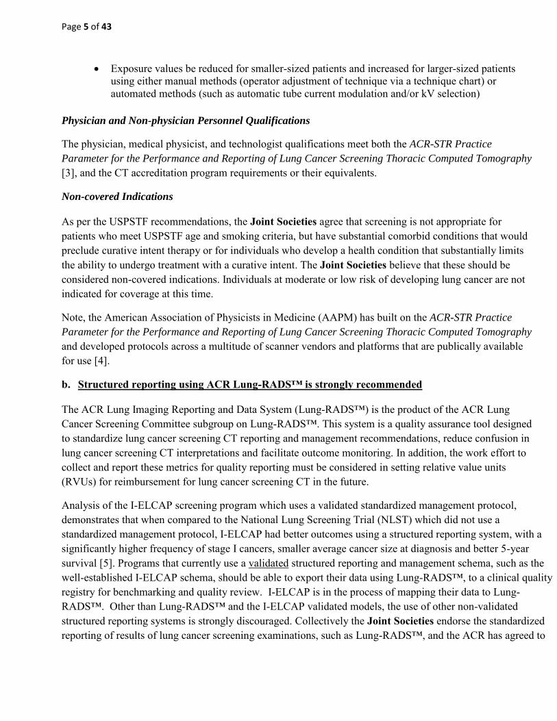
Exposure values be reduced for smaller-sized patients and increased for larger-sized patients using either manual methods (operator adjustment of technique via a technique chart) or automated methods (such as automatic tube current modulation and/or kV selection)
Physician and Non-physician Personnel Qualifications
The physician, medical physicist, and technologist qualifications meet both the ACR-STR Practice
Parameter for the Performance and Reporting of Lung Cancer Screening Thoracic Computed Tomography
[3], and the CT accreditation program requirements or their equivalents.
Non-covered Indications
As per the USPSTF recommendations, the Joint Societies agree that screening is not appropriate for patients who meet USPSTF age and smoking criteria, but have substantial comorbid conditions that would preclude curative intent therapy or for individuals who develop a health condition that substantially limits the ability to undergo treatment with a curative intent. The Joint Societies believe that these should be considered non-covered indications. Individuals at moderate or low risk of developing lung cancer are not indicated for coverage at this time.
Note, the American Association of Physicists in Medicine (AAPM) has built on the ACR-STR Practice
Parameter for the Performance and Reporting of Lung Cancer Screening Thoracic Computed Tomography and developed protocols across a multitude of scanner vendors and platforms that are publically available for use [4].
b. Structured reporting using ACR Lung-RADS™ is strongly recommended
The ACR Lung Imaging Reporting and Data System (Lung-RADS™) is the product of the ACR Lung Cancer Screening Committee subgroup on Lung-RADS™. This system is a quality assurance tool designed to standardize lung cancer screening CT reporting and management recommendations, reduce confusion in lung cancer screening CT interpretations and facilitate outcome monitoring. In addition, the work effort to collect and report these metrics for quality reporting must be considered in setting relative value units (RVUs) for reimbursement for lung cancer screening CT in the future.
Analysis of the I-ELCAP screening program which uses a validated standardized management protocol, demonstrates that when compared to the National Lung Screening Trial (NLST) which did not use a standardized management protocol, I-ELCAP had better outcomes using a structured reporting system, with a significantly higher frequency of stage I cancers, smaller average cancer size at diagnosis and better 5-year survival [5]. Programs that currently use a validated structured reporting and management schema, such as the well-established I-ELCAP schema, should be able to export their data using Lung-RADS™, to a clinical quality registry for benchmarking and quality review. I-ELCAP is in the process of mapping their data to Lung-RADS™. Other than Lung-RADS™ and the I-ELCAP validated models, the use of other non-validated structured reporting systems is strongly discouraged. Collectively the Joint Societies endorse the standardized reporting of results of lung cancer screening examinations, such as Lung-RADS™, and the ACR has agreed to

Page 6 of 43
include representatives from societies such as the American Thoracic Society (ATS), American College of Chest Physicians (ACCP), and The Society of Thoracic Surgeons (STS) for future iterations of Lung-RADS™, in particular with attention to management protocols for higher risk lesions.
The table below contains Version 1.0 of Lung-RADS™ and contains the assessment categories and accompanying management recommendations. A complete lexicon and atlas will be developed soon. The atlas will include a description of a medical audit and outcome monitoring process. The lexicon of lung cancer screening CT terms and the reporting format are meant to standardize the language used in lung cancer screening CT reports. In particular, the consistent use of the assessment categories will help clinicians understand disposition of their patients based on lung cancer screening CT and aid in auditing lung cancer screening CT practices and programs. Knowing how we perform will help to identify deficiencies, facilitate research and be of practical value in taking better care to avoid adverse medicolegal consequences. To minimize harms, it is essential that the evaluation and management of nodules highly suspicious for lung cancer should be guided by multidisciplinary protocols. Practices without lung cancer specialists or multidisciplinary programs should have a process in place for consultation with and/or referral for management of patients with screening results in higher categories (such as Lung-RADS™ 4B & 4X) to such specialists or multidisciplinary programs. The following specialties usually comprise a multidisciplinary program: Thoracic Radiology, Pulmonary Medicine, Thoracic Surgery, Medical Oncology and Radiation Oncology.
Category Descriptor
Category Descriptor Primary Category
Management
Incomplete - 0 Additional lung cancer screening CT images
and/or comparison to prior chest CT examinations is needed
Negative No nodules and
definitely benign nodules
1
Continue annual screening with LDCT in 12 months Benign
Appearance or Behavior
Nodules with a very low likelihood of
becoming a clinically active cancer due to
size or lack of growth
2
Probably Benign
Probably benign finding(s) - short term follow-up suggested;
includes nodules with a low likelihood of
becoming a clinically active cancer
3 6 month LDCT
Suspicious
Findings for which additional diagnostic testing and/or tissue
sampling is recommended
4A 3 month LDCT; PET/CT may be used when
there is a ≥ 8 mm solid component
4B Chest CT with or without contrast, PET/CT and/or tissue sampling depending on the
*probability of malignancy and comorbidities. PET/CT may be used when
there is a ≥ 8 mm solid component. 4X

Page 7 of 43
Significant - 0ther
S
Prior Lung Cancer
C
ACR Lung-RADS™: Safely improving the positive predictive value of CT lung screening
The NLST proved CT lung screening reduces lung cancer specific mortality in high-risk patients when the minimum size of a positive pulmonary nodule is set at 4-mm. 27.3% of prevalence exams in the NLST were positive for a nodule 4-mm or greater resulting in an overall positive predictive value (PPV) of 3.8%. More than half of these positive exams were positive for small nodules (4-6 mm), which had a much lower PPV around 0.5%. This suggests that increasing the threshold of a positive CT lung screening exam to 6-mm for solid and part solid nodules and 20-mm for non solid nodules could significantly reduce the false positive rate without impacting the sensitivity to detect malignancy. Applying Lung-RADS™ to the NLST prevalence screen decreases the positive screen rate to 13.7% on baseline screens and 5.9% on subsequent annual screens compared to the 27.3% and 27.9% seen on NLST baseline and year 1 screens, and 16.8% on the subsequent year 2 screens. The PPV increases to 6.9% at baseline screen and 10.9% on subsequent screens using Lung-RADS™ (Data in submission, September 2014, Annals of Internal
Medicine). Lung-RADS™ Classifications at Baseline Screen Applied to the NLST Study Data
Cancer No Cancer All Lung-RADS™ Score N (%) N (%) N (%)
1 or 2 (Negative Screen) 40 (13.9) 22672 (87.2) 22712 (86.3) 3 or 4 (Positive Screen) 248 (86.1) 3349 (12.8) 3597 (13.7) All 288 26021 26309
Lung-RADS™ Classifications at Post-Baseline Screens Applied to the NLST Study Data
Cancer No Cancer All Lung-RADS™ Score N (%) N (%) N (%)
1 or 2 (Negative Screen) 86 (21.4) 45701 (94.7) 45787 (94.1) 3 or 4 (Positive Screen) 315 (78.6) 2569 (5.3) 2974 (5.9) All 401 48270 48761
The I-ELCAP confirmed similar results in their CT lung screening experience. They reported a reduction in positive percentage to 10.2% using a 6-mm threshold vs 16% at a 5-mm threshold with the same number of lung cancers found within 12 months of baseline enrollment at both thresholds [6].

Page 8 of 43
Following publication of these research findings both the National Comprehensive Cancer Network (NCCN Guidelines®) and the American College of Radiology (Lung-RADS™) adopted 6-mm as the minimum nodule size threshold for a positive CT lung cancer screening examination. The CT lung cancer screening program at the Lahey Hospital & Medical Center (LHMC) demonstrated that the positive rates reported at 4-mm and 6-mm in the NLST and I-ELCAP are also observed in clinical practice. From January 2012 through May 2014, 2180 patients at high-risk for lung cancer according to NCCN Guidelines® underwent clinical CT lung screening exams at LHMC with all nodule sizes greater than 4-mm recorded. Results from LHMC at the NLST 4-mm threshold and at the ACR Lung-RADS™ 6-mm threshold are compared below. As detailed above, a 6-mm threshold (ACR Lung-RADS™) reduces the LHMC positive rate to levels reported in the NLST and I-ELCAP with a similar significant corresponding increase in PPV (17.3%) [7].

Page 9 of 43
c. Practice Quality Reporting through a Clinical Database is Recommended
i) Clinical Practice Quality Reporting Registry and the ACR National Radiology Data Registry (NRDR)
In order to evaluate the quality of lung cancer screening programs, participation in a clinical practice registry with standardized data elements is recommended. Sites would input data to a registry and receive back data both on the performance of the practice and individual radiologist performance, benchmarked to regional and national data.
The American College of Radiology launched the first of its registries, the National Oncologic PET Registry (NOPR) in 2005. NOPR was developed for Coverage with Evidence Development for PET scans. Building on that infrastructure, the ACR launched a collection of quality databases under the umbrella of the NRDR (www.nrdr.acr.org). These databases include the Dose Index Registry (DIR) established in 2011, National Mammography Database (NMD), CT Colonography registry (CTC) established in 2008, IV Contrast Extravasation Registry (ICE), and the General Radiology Improvement Database (GRID) established in 2008. Currently the ACR is in the process of adding functionality to collect data to support participation in the CMS Physician Quality Reporting System (PQRS) program. The PQRS database is under development and currently undergoing testing.
The guiding principle of the NRDR registries is to empower facilities and physicians to create a cyclical quality improvement process that involves transmitting data to NRDR, receiving semi-annual national benchmarking reports, comparing and analyzing institution’s results, and developing and implementing an improvement plan. Subsequent reports reveal which improvement activities had the desired effect. This quality improvement cycle can be easily implemented and conveniently monitored with each semi-annual benchmark report. The American Board of Radiology (ABR) has endorsed all five registry databases as approved professional quality improvement (PQI) projects for the maintenance of certification of radiologists. In 2014, NRDR was approved as a Qualified Clinical Data Registry (QCDR) for PQRS participation by CMS.
As of June 2014, there were over 1000 facilities in the registry and data on over 16 million exams. The number of data elements varies across databases, and lists of data elements for each database are available from the NRDR website. Participating facilities are spread all over the US and represent a broad variety of practice types.
For a CT lung cancer screening clinical practice registry, we strongly recommend that a longitudinal data collection process that tracks the entire patient experience be incorporated. Integration with The Society of Thoracic Surgeons’ database and other similar databases are examples being pursued. While the ACR is willing and able to be a single source registry for purposes of quality reporting and benchmarking, the ACR is also willing and able to collaborate with other registries that follow the report the minimum data elements, assuming data integrity standards are met, if deemed appropriate by CMS.

Page 10 of 43
ii) Lung Cancer Screening Practice Registry
Plans for a new ACR Lung Cancer Screening Clinical Practice Registry under NRDR are underway. The database will support clinical practice audits and the capability to collect longer-term outcome data to provide data for evidence development. Physician identifiers (IDs) will be collected as National Provider Identifiers (NPIs). CMS may use this ID to audit participation by providers billing Medicare for lung cancer screening exams.
Burden on participating physicians will vary based on mechanism of data collection and transmission used. In all cases, there will be some additional effort for the radiologist. Additional effort may include education about Lung-RADS™, using Lung-RADS™ in practice, developing and providing smoking cessation guidance, reviewing follow-up data on cancers, performing audits, and reviewing audit data. For a practice as a whole, there will be staff time required to obtain biopsy results and similar follow-up data from patients and referring physicians to maintain complete patient information for clinical audits. Automated reporting system with Lung-RADS™ templates and automated data submission to registry and/or automated audits will require fewer hours of physician effort but more up front investment in technology. There will be some cost tradeoffs based on whether data transmission is automatic behind-the-scenes or requires some staff time to run a query and upload data to a registry to obtain audit results.
The anticipated timeline is a launch in early 2015 with web-based data entry forms and data upload. Automated data upload from Radiology Information System (RIS) or Electronic Health Records (EHR) will be phased in over time.
Data element specification currently underway with clinical leadership Development of technical specifications started in August 2014 Building of software support planned to start in Fall 2014, to be tested by early 2015

Page 11 of 43
iii) Proposed Data Elements
The following elements are recommended for a clinical practice registry reporting for LCS LDCT. A discussion about reasonable compliance rates with quality metric submission achievable in practice will be important, as in many settings the screening event may occur in one practice site and the diagnostic evaluation may occur in another setting or settings. In addition, the work effort to collect and report these metrics for quality reporting must be considered in setting RVUs for reimbursement for lung cancer screening CT in the future.
Other comprehensive clinical practice registries that follow the same proposed minimum data elements and data quality standards for reporting outcomes for benchmarking and public reporting may be considered, and ideally would collaborate with the ACR LCS clinical practice registry (CPR) under NRDR.
At An Individual Patient Level At A Practice Aggregate Level By Interpreting Radiologist
1. Patient Identifiers: a. Name b. DOB
1. PPV 1 (abnormal screen): The percentage of all positive screening examinations (LungRADS™ Categories 0, 3 and 4) that result in a tissue diagnosis of lung cancer within 1 year. PPV 1= TP / (number of positive screening examinations)
2. Appropriateness of screening: a. Age b. Smoking status
i. Current smoker ii. Former smoker
iii. Non smoker c. If current or former smoker, # of pack-
years of smoking d. If former smoker, # of years since quit e.
2. PPV 2 (abnormal screen, interim CT recommended): The percentage of all lung cancer screening CT examinations recommended for 6 month LDCT, or 3 month LDCT with or without PET/CT, (LungRADS ™Categories 3 and 4A) that resulted in a tissue diagnosis of lung cancer within one year.
PPV 2 = TP / (number of screening examinations recommended for additional CT imaging)
3. Screening timing: a. Baseline screen (prevalence screen) b. Follow-up annual screen (incidence
screens)
3. PPV 3 (biopsy): The percentage of all known biopsies done as a result of positive screening with or without subsequent PET/CT or diagnostic chest CT examinations (LungRADS™ Category 4B & 4X) that resulted in a tissue diagnosis of lung cancer within 1 year. PPV3 = biopsies with lung cancer diagnosis / all biopsies
4. Smoking cessation addressed 4. PPV 4 (additional diagnostics up to and including biopsy): The percentage of all screening examinations for which additional diagnostics and/or biopsy is performed as a result of a positive screening examinations (LungRADS™ Category 4B & 4X) that resulted in a tissue diagnosis of lung cancer within 1 year. PPV4 = TP / (total # with additional diagnostics up to and
including biopsy)
5. Screening CT radiation exposure a. CTDIvol b. DLP
5. Cancer detection rate (CDR) for baseline (prevalence – CDRp) and follow-up (incidence – CDRi) screening examinations
a. For baseline (prevalence) screens: # of lung cancers per 1000 baseline screening examinations
b. For subsequent (incidence) screens: # of lung cancers per 1000 follow-up screening examinations
Page 12 of 43
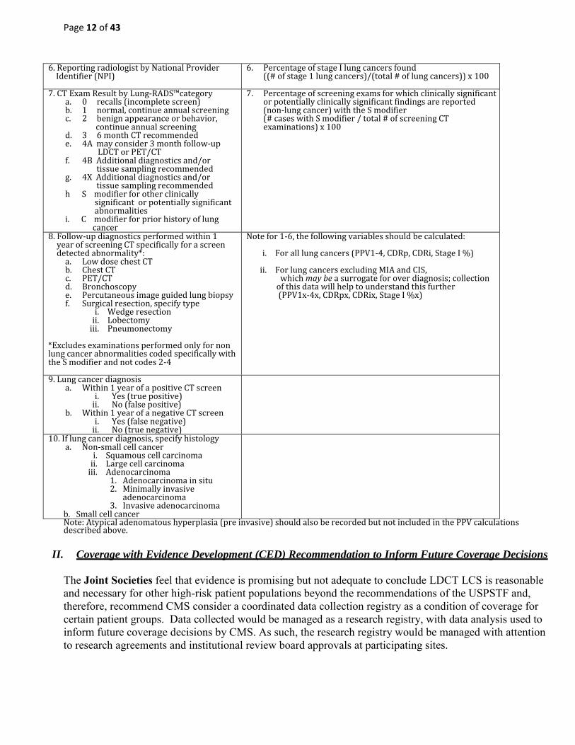
6. Reporting radiologist by National Provider Identifier (NPI)
6. Percentage of stage I lung cancers found ((# of stage 1 lung cancers)/(total # of lung cancers)) x 100
7. CT Exam Result by Lung-RADS™category a. 0 recalls (incomplete screen) b. 1 normal, continue annual screening c. 2 benign appearance or behavior,
continue annual screening d. 3 6 month CT recommended e. 4A may consider 3 month follow-up
LDCT or PET/CT f. 4B Additional diagnostics and/or tissue sampling recommended g. 4X Additional diagnostics and/or tissue sampling recommended h S modifier for other clinically significant or potentially significant abnormalities i. C modifier for prior history of lung cancer
7. Percentage of screening exams for which clinically significant or potentially clinically significant findings are reported (non-lung cancer) with the S modifier (# cases with S modifier / total # of screening CT examinations) x 100
8. Follow-up diagnostics performed within 1 year of screening CT specifically for a screen detected abnormality*:
a. Low dose chest CT b. Chest CT c. PET/CT d. Bronchoscopy e. Percutaneous image guided lung biopsy f. Surgical resection, specify type
i. Wedge resection ii. Lobectomy
iii. Pneumonectomy
*Excludes examinations performed only for non lung cancer abnormalities coded specifically with the S modifier and not codes 2-4
Note for 1-6, the following variables should be calculated:
i. For all lung cancers (PPV1-4, CDRp, CDRi, Stage I %)
ii. For lung cancers excluding MIA and CIS, which may be a surrogate for over diagnosis; collection of this data will help to understand this further (PPV1x-4x, CDRpx, CDRix, Stage I %x)
9. Lung cancer diagnosis a. Within 1 year of a positive CT screen
i. Yes (true positive) ii. No (false positive)
b. Within 1 year of a negative CT screen i. Yes (false negative)
ii. No (true negative)
10. If lung cancer diagnosis, specify histology a. Non-small cell cancer
i. Squamous cell carcinoma ii. Large cell carcinoma
iii. Adenocarcinoma 1. Adenocarcinoma in situ 2. Minimally invasive
adenocarcinoma 3. Invasive adenocarcinoma
b. Small cell cancer
Note: Atypical adenomatous hyperplasia (pre invasive) should also be recorded but not included in the PPV calculations described above.
II. Coverage with Evidence Development (CED) Recommendation to Inform Future Coverage Decisions
The Joint Societies feel that evidence is promising but not adequate to conclude LDCT LCS is reasonable and necessary for other high-risk patient populations beyond the recommendations of the USPSTF and, therefore, recommend CMS consider a coordinated data collection registry as a condition of coverage for certain patient groups. Data collected would be managed as a research registry, with data analysis used to inform future coverage decisions by CMS. As such, the research registry would be managed with attention to research agreements and institutional review board approvals at participating sites.

Page 13 of 43
Patient Criteria
1) The first category of individuals may be slightly younger or have a lower pack-year smoking history
than the USPSTF recommended population, but have additional risk factors for lung cancer. Although the NLST provided excellent randomized trial evidence of the benefit of LDCTs for a high-risk group of patients, as a clinical trial, the study limited its inclusion criteria to the risk factors of age and smoking history. A wealth of pre-existing data has demonstrated several other clinically important risk factors for lung cancer that have not been addressed in the USPSTF guidelines, yet should be strongly considered for CED. Since it is highly unlikely that there will be randomized clinical trials like NLST in the future to study other at risk populations for lung cancer and the use of CT screening, CED would serve the important role of systematically studying the performance of other high-risk individuals. In October 2011 the National Comprehensive Cancer Network (NCCN) recommended annual CT lung screening for an additional group of high-risk individuals, which extends beyond the USPSTF recommendation [8]. This population is often referred to as the “NCCN group 2”, and is described below, as taken from the attached NCCN guideline (page LCS-1). This is an example of the type of population that may be included in a CED decision.
NCCN High-Risk Group 2
Individuals ≥ 50 years of age, with a ≥ 20 pack year history of smoking who have at least one additional risk factor for lung cancer (other than second-hand smoke), such as: o Occupational exposure, specifically to agents that are identified as carcinogens
targeting the lungs, including silica, cadmium, asbestos, arsenic, beryllium, chromium, diesel fumes, nickel, coal smoke, and soot
o Cancer history, as there is an increased risk of developing new primary lung cancer among survivors of lung cancer, lymphomas, cancers of the head and neck, and smoking-related cancers
o Documented high radon exposure o Family history of lung cancer o Disease history of chronic obstructive pulmonary disease (COPD) or pulmonary fibrosis
Results of NCCN high-risk Group 2 in a clinical CT lung cancer screening program
In January 2012 the Lahey Hospital & Medical Center (LHMC) in Burlington Massachusetts began offering clinical CT lung screening as a community benefit to individuals aged 74 years and younger meeting either NCCN Group 1 or Group 2 high-risk criteria. Twenty-six percent of participants in the LHMC CT lung cancer screening program qualified for screening through Group 2. (Fig 1) Applied nationwide a Group 2 rate of 26% would equate to approximately 2 million Americans at high-risk for lung cancer outside the entry criteria of the NLST. Additionally, as nearly one third of their Group 2 population failed to meet Group 1 criteria solely because they quit smoking more than 15 years ago, 600,000 former smokers between 55 and 74 with 30 pack-year or more smoking history could lose access to screening with national eligibility limited to Group 1. Despite statistical significant differences in age, pack-years, and duration of smoking cessation LHMC found no significant difference in the rate of positive results between

Page 14 of 43
NCCN Group 2 and Group 1 (NLST population) with overall positive results equivalent to those reported in the prevalence screen of the NLST. The annualized cancer detection rate for NCCN Group 2 and Group 1 was also nearly identical at 1.8% and 1.6% respectively (Table 1, 2, and 3). Similar CT lung screening positive rates and malignancy detection rates between NCCN Group 2 and Group 1 (NLST population) (Table 4) offers the potential to save thousands of additional lives every year by expanding CT lung screening eligibility to include Group 2 high-risk individuals [9].
Table 1. Lahey Clinic Lung Cancer Screening Cohort of NCCN Group 1 and Group 2 Patients: Comparison of Patient Demographics, Smoking History & Follow-Up
Variable Total
NCCN
Group 2
NCCN
Group 1
p-value:
(Group 2 vs Group 1) NLST
Number qualified 2079 538 1541 NA NR
Number screened 1760 464 (26%) 1296 (74%) NA ~26,000
Average age (y) 64 61 65 p < 0.001 61
Male 52% 50% 53% p < 0.2 59%
Smoking history (pack-years) 47 40 51 p < 0.001 56
Current smoker 812 (46%) 167 (36%) 645 (50%) p < 0.001 48%
Former smoker duration (years) 10.3 18.5 6.7 p < 0.001 NR
Clinical follow-up available 1328 (75%) 331 (71%) 997 (77%) NR NA
Average follow-up (months) 12.5 12.1 12.7 NR 78
NLST = National Lung Screening Trial NR = Not Reported NA= Not Applicable

Page 15 of 43
Table 2: Lahey Clinic Lung Cancer Screening Cohort of NCCN Group 1 and Group 2 Patients
Screening Results
Result
Total Screened
n = 1760
NCCN Group 2
n = 464
NCCN Group 1
n = 1296
NLST
(T0)
Total Positive 481 27.3% 116 25.0% 365 28.2% 27.3%
Probably Benign 412 23.4% 103 22.2% 309 23.8% NR
Suspicious 69 3.9% 13 2.8% 56 4.3% NR
Probable Infection 114 6.5% 28 6.0% 86 6.6% NR
Significant Incidentals 108 6.1% 28 6.0% 80 6.2% 10.2%
NLST = National Lung Screening Trial NR = Not Reported
Table 3: Lahey Clinic Lung Cancer Screening Cohort of NCCN Group 1 and Group 2 Patients: Malignancy Rate and Average Follow-Up
Variable Overall Group 2 Group 1
Overall malignancy rate 23/1328 (1.7%) 6/331 (1.8%) 17/997 (1.7%)
Average follow-up (months) 12.5 12.1 12.7
Annualized malignancy rate 1.6% 1.8% 1.6%
Time to diagnosis (months) 4.1 5.6 3.7
Average follow-up from diagnosis (months) 7.8 5.3 8.6

Page 16 of 43
2) The second category of individuals that should be included for coverage beyond the
USPSTF guidelines, are 55 to 80 year olds who have a 30 pack-year or more history of
smoking and who may have stopped smoking for more than 15 years.
Although these individuals were not studied by the NLST, the risk of smoking-related cancers is predominantly related to total exposure and gradually decreases over time, meaning that these patients may remain at significant risk of lung cancer development. Further, an arbitrary cutoff of 15 years would result in an implementation dilemma for patients who are covered for initiation of lung cancer screening, and who are then no longer covered for continued follow-up and screening after they have succeeded in smoking cessation for more than 15 years. This exclusion could potentially lead to a paradox of incentives that “encourages” a patient to restart smoking in order to maintain eligibility for lung cancer screening coverage. With respect to the cost effectiveness of lung cancer screening in this population, analysis of NLST presented last June at the National Cancer Institute (NCI) demonstrated that the cost effectiveness of lung cancer screening is maintained, even if the relative risk of the population falls to 60% of the risk of the NLST enrolled population.
The Joint Societies’ goals through evidence collection are to monitor and evaluate the performance of lung cancer screening in these populations for which randomized controlled trial data does not exist and for whom the magnitude of the clinical outcomes is suspected to
0
10
20
30
40
50
60
70
80
90
100
0
200
400
600
800
1000
1200
1400
1600
1800
Pe
rce
nt
(%)
Nu
mb
er
of
pa
tie
nts
scr
ee
ne
d (
n)
Two year screening interval
Lahey Clinic Lung Cancer Screening Program
Volume and Percentages
Program Volume Group 2 Percentage Cumulative Self Referral Percentage
Page 17 of 43
be positive but remains uncertain. This robust registry would function as prospective clinical data collection and analysis using the same stringency and data validation used in clinical trials, to which sites would have to commit. Non-compliance with data submission and integrity would result in non-participation. It is expected that because of these requirements, only highly motivated centers committed to furthering the knowledge of lung cancer CT screening in this population would participate, which has been the ACR’s experience with other similar efforts.
We would expect that data collection for this purpose would meet the following basic criteria:
Contain required minimum data elements Written protocol on file Institutional Review Board review and approval Referral by qualified healthcare provider
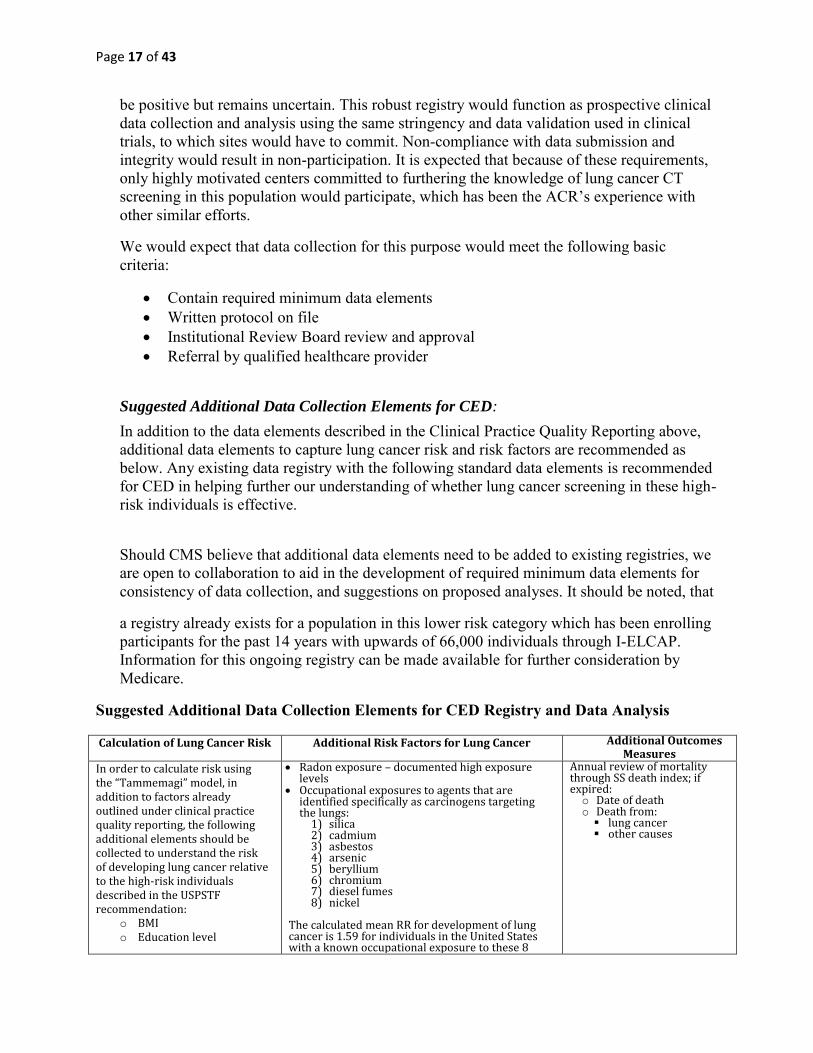
Suggested Additional Data Collection Elements for CED:
In addition to the data elements described in the Clinical Practice Quality Reporting above, additional data elements to capture lung cancer risk and risk factors are recommended as below. Any existing data registry with the following standard data elements is recommended for CED in helping further our understanding of whether lung cancer screening in these high-risk individuals is effective. Should CMS believe that additional data elements need to be added to existing registries, we are open to collaboration to aid in the development of required minimum data elements for consistency of data collection, and suggestions on proposed analyses. It should be noted, that
a registry already exists for a population in this lower risk category which has been enrolling participants for the past 14 years with upwards of 66,000 individuals through I-ELCAP. Information for this ongoing registry can be made available for further consideration by Medicare.
Suggested Additional Data Collection Elements for CED Registry and Data Analysis
Calculation of Lung Cancer Risk Additional Risk Factors for Lung Cancer Additional Outcomes Measures
In order to calculate risk using the “Tammemagi” model, in addition to factors already outlined under clinical practice quality reporting, the following additional elements should be collected to understand the risk of developing lung cancer relative to the high-risk individuals described in the USPSTF recommendation:
o BMI o Education level
Radon exposure – documented high exposure levels
Occupational exposures to agents that are identified specifically as carcinogens targeting the lungs:
1) silica 2) cadmium 3) asbestos 4) arsenic 5) beryllium 6) chromium 7) diesel fumes 8) nickel
The calculated mean RR for development of lung cancer is 1.59 for individuals in the United States with a known occupational exposure to these 8
Annual review of mortality through SS death index; if expired: o Date of death o Death from:
lung cancer other causes

Page 18 of 43
Reference McWilliams, A. et al. (2013). Probability of cancer in pulmonary nodules detected on first screening computed tomography. New England
Journal of Medicine, 369, 919;10. Note: several public sites exist for using this risk assessment tool, such as http://www.brocku.ca/lung-cancer-risk-calculator, and the tools are available for download on line directly as well.
agents.
References:
Driscoll, T. et al. (2005, December). The global burden of disease due to occupational carcinogens. American Journal of Industrial Medicine, 48(6), 419-431. Retrieved from http://www.ncbi.nlm.nih.gov/pubmed/16299703. Steeland K et al (1996) American Journal of Industrial medicine, 29(5), 474-490. Retrieved from http://www.ncbi.nlm.nih.gov/pubmed/8732921 For those who are exposed to these carcinogens, smokers have a greater risk for lung cancer than nonsmokers. Reference: Reid, A. (2006, August). The risk of lung cancer with increasing time since ceasing exposure to asbestos and quitting smoking. Occupational and Environmental Medicine, 63(8), 509-512. Retrieved from http://www.ncbi.nlm.nih.gov/pubmed/16849527
History of cancers that are associated with an
increased risk of developing a new primary lung cancer o prior lung cancer o lymphoma o head and neck cancer o other smoking-related cancers
Family history of lung cancer COPD Pulmonary fibrosis Second hand smoke exposure: Individuals
exposed to second-hand smoke have a highly variable exposure to the carcinogens, with varying evidence for increased risk after this variable exposure. Therefore, second-hand smoke is not independently considered a risk factor for lung cancer screening, unless there is documentation of excessive second hand smoking exposure
III. Supplemental Information
1) Access to CT Scanner Specific Low Dose Chest CT Protocols
The American Association of Physicists in Medicine (AAPM) recently posted version 2.0 of its’ low dose lung cancer screening protocols, extending the number of CT models for which protocols are available (see http://www.aapm.org/pubs/CTProtocols/?tab=5#CTabbedPanels). This update includes protocols for CT scanners for 31 CT scanner models from six different CT vendors, and covers the great majority of all CT scanners in use in the United States today, and is consistent with the ACR-STR Practice Parameter for the Performance and Reporting of Lung
Cancer Screening Thoracic Computed Tomography (CT). The ACR has made this material

Page 19 of 43
available on the ACR Lung Cancer Screening Resource Webpage. This material creates ready access to protocols for practices to use in their screening programs, optimized for the CT make and model in their practice and assembled by experts with attention to low radiation exposure and optimized for lung cancer screening CT.
2) CT Lung Cancer Screening is Cost Effective Extensive information regarding the cost effectiveness of CT lung cancer screening was included in the March 12, 2014 stakeholder letter. Importantly, this included the cost effectiveness analysis of NLST addition to the data presented by William Black and Paul Pinsky at the June 2013 NCI Advisory Board which is now in press with The New England Journal of Medicine. A preliminary application of Lung-RADS™ to the NLST being prepared for publication indicates that using Lung-RADS™ will reduce the incremental cost effectiveness ratio (ICER) about $3,000 per quality-adjusted life-year (QALY) gained. Additional data available since that letter includes the work of Pyenson, B.S. et. al. conducted by Milliman entitled “Offering Lung Cancer Screening to High-Risk Medicare Beneficiaries Saves Lives and Is Cost-Effective: An Actuarial Analysis” published on line in the American Health
and Drug Benefits in August 2014 [10]. This work is an actuarial analysis that demonstrates offering CT lung cancer screening to high-risk Medicare beneficiaries saves lives and is cost effective. The abstract of that work is presented below, with the full article included as an appendix.
Background: By a wide margin, lung cancer is the most significant cause of cancer death in the United States and worldwide. Because the incidence of lung cancer increases with age, Medicare beneficiaries are often at increased risk. Because of its demonstrated effectiveness in reducing mortality, lung cancer screening with low-dose computed tomography (LDCT) imaging will be covered without cost-sharing starting January 1, 2015, by non-grandfathered commercial plans. Medicare is considering coverage for lung cancer screening. Objective: To estimate the cost and cost effectiveness (i.e., cost per life-year saved) of LDCT lung cancer screening of the Medicare population at high-risk for lung cancer. Methods: Medicare costs, enrollment and demographics were used for this study; they were derived from the 2012 Centers for Medicare & Medicaid Services (CMS) beneficiary files and were forecast to 2014 based on CMS and US Census Bureau projections. Standard life and health actuarial techniques were used to calculate the cost and cost effectiveness of lung cancer screening. The cost, incidence rates, mortality rates, and other parameters chosen by the authors are taken from actual Medicare data and the modeled screenings are consistent with Medicare processes and procedures. Results: Approximately 4.9 million high-risk Medicare beneficiaries would be eligible for lung cancer screening in 2014. Without screening, Medicare patients
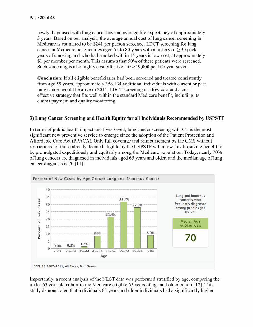
Page 20 of 43
newly diagnosed with lung cancer have an average life expectancy of approximately 3 years. Based on our analysis, the average annual cost of lung cancer screening in Medicare is estimated to be $241 per person screened. LDCT screening for lung cancer in Medicare beneficiaries aged 55 to 80 years with a history of ≥ 30 pack-years of smoking and who had smoked within 15 years is low cost, at approximately $1 per member per month. This assumes that 50% of these patients were screened. Such screening is also highly cost effective, at <$19,000 per life-year saved. Conclusion: If all eligible beneficiaries had been screened and treated consistently from age 55 years, approximately 358,134 additional individuals with current or past lung cancer would be alive in 2014. LDCT screening is a low cost and a cost effective strategy that fits well within the standard Medicare benefit, including its claims payment and quality monitoring.
3) Lung Cancer Screening and Health Equity for all Individuals Recommended by USPSTF In terms of public health impact and lives saved, lung cancer screening with CT is the most significant new preventive service to emerge since the adoption of the Patient Protection and Affordable Care Act (PPACA). Only full coverage and reimbursement by the CMS without restrictions for those already deemed eligible by the USPSTF will allow this lifesaving benefit to be promulgated expeditiously and equitably among the Medicare population. Today, nearly 70% of lung cancers are diagnosed in individuals aged 65 years and older, and the median age of lung cancer diagnosis is 70 [11].
Importantly, a recent analysis of the NLST data was performed stratified by age, comparing the under 65 year old cohort to the Medicare eligible 65 years of age and older cohort [12]. This study demonstrated that individuals 65 years and older individuals had a significantly higher
Page 21 of 43
prevalence of lung cancer and that CT had a significantly higher positive predictive value (4.9% vs. 3.0%) compared to individuals under 65 years of age. Despite a higher false positive rate in the older cohort, the resection rates for screen-detected cancer were similar (75.6% in the under-65 cohort vs. 73.2% in the 65 and older cohort) and the complications from invasive procedures were low and not significantly different between the two groups (9.8% in the under-65 cohort vs. 8.5% in the 65 years and older cohort). Screen-detected cancer was about twice as frequent in the 65 years and older cohort than in the under-65 cohort. Since PPACA does not require CMS to cover all Essential Health Benefits as it does commercial insurers, lack of coverage for the older aged individuals covered by the USPSTF recommended population would be a de facto denial of access to a significant percentage of this population, with disproportionate impact on those at highest need. It would be illogical and certainly inappropriate public health policy to deny previously eligible individuals coverage when they reach the age of 65, especially since that age approaches the peak age of 70 for lung cancer incidence and deaths. Equally inappropriate and illogical would be a CED for the 65 years of age and older individuals for which the USPSTF recommends screening. Limiting screening in a CED would effectively reduce access to patients able to participate through the academic and research centers that would participate in CED. This would restrict access for a significant portion of the Medicare population, including those financially or logistically unable to travel to those centers, and those, frequently inner city minorities, intimidated by the centers. For them, restricted access is tantamount to a denial of care. If lung cancer screening were to be entirely under a CED, it would disproportionately impact lower income and rural populations and establish an unprecedented government-mandated disparity of care. The fact is that no other cancer screening service has been as rigorously tested by a randomized clinical trial (RCT) and as rigorously reviewed and approved by the USPSTF prior to implementation. Indeed other studies and analyses on CT screening indicate that the actual mortality benefit of CT screening will be significantly higher than other cancer screening methods when carried out according to published best practices and protocols. To help ensure that CT screening would be implemented safely and responsibly and to educate the public on its risks and benefits and what to look for in a screening site, lung cancer specialists on Lung Cancer Alliance’s boards developed the Framework for Excellence in Screening and the Continuum of Care after the NLST results were published. Hundreds of sites around the country, from academic centers to community hospitals, have adopted the Framework and are screening. A Map of Lung Cancer Alliance Framework Centers of Screening Excellence can be found at: http://www.lungcanceralliance.org/get-information/am-i-at-risk/what-do-i-need-to-know-about-screening/where-should-i-be-screened/lung-cancer-screening-centers/ Their interests, and the interest of sound public health policy, will best be served by full
coverage and reimbursement by CMS for all those under the USPSTF recommendation.

Page 22 of 43
4) Management of Individuals with a Positive Lung Cancer Screening CT
The Society of Thoracic Surgeons (STS), American Thoracic Society (ATS) and the American College of Chest Physicians (ACCP) are committed to working collaboratively and as equal partners with the American College of Radiology (ACR) to advocate for CMS coverage of lung cancer screening for high-risk individuals. We are encouraged by CMS’ receptiveness to consider multidisciplinary, standardized, outcomes-driven approaches that will maximize benefits and minimize harms. STS, ATS and ACCP recognize that ACR has successfully overseen breast cancer screening programs and has established accreditation procedures and standardized reporting schemes that will be useful in lung cancer screening.
Collectively the Joint Societies endorse the standardized reporting of results of lung cancer screening examinations using Lung-RADS™ or that are mapped to Lung-RADS™, and the ACR has agreed to include representatives from societies such as the STS, ATS and ACCP for future iterations of Lung-RADS™.
See Appendices for the ATS/ACCP Lung Cancer Screening Policy Statement In Press (CHEST).
To minimize harms, it is essential that the evaluation and management of nodules highly suspicious for lung cancer should be guided by multidisciplinary protocols. Practices without lung cancer specialists or multidisciplinary programs should have a process in place for consultation with or referral for patients with screening results in higher categories (such as LungRADS 4B) to such specialists or multidisciplinary programs.
5) Multidisciplinary Coordination of Efforts and Support: The American Cancer Society and the American Medical Association
The American Cancer Society (ACS) has committed to convene representatives from the medical specialties important in the practice of lung cancer screening, and organize a meeting of key organizations and experts to identify challenges, solutions, and approaches to monitoring the increasing availability and uptake of lung cancer screening. A group like this could evolve into an organization like the National Colorectal Cancer Roundtable (NCCT), which is a national coalition of organizations working together to advance colorectal cancer control efforts. Some of the challenges and solutions as they pertain to lung cancer are addressed in the Policy Statement from the ACCP and ATS, which is appended.
The Roundtable, established by the American Cancer Society and the Centers for Disease Control and Prevention (CDC) in 1997, includes public organizations, private organizations, voluntary organizations, and invited individuals dedicated to reducing the incidence of and mortality from colorectal cancer in the U.S., through coordinated leadership, strategic planning, and advocacy.
The ultimate goal of the Roundtable is to increase the use of proven colorectal cancer screening tests among the entire population for whom screening is appropriate. The NCCRT is organized to magnify the impact that each organization can have in their efforts to improve colorectal cancer outcomes. Members learn about the latest news and events via the NCCRT website. NCCRT.org houses many tools and resources that are available for NCCRT member use.
Page 23 of 43
Members can attend the NCCRT Annual Meeting to share ideas, get up-to-date information about screening and wrestle with pressing issues. Lastly, the NCCRT offers its members a place to work together on various initiatives through multiple task groups, which have regular conference calls, and usually one annual meeting. The NCCRT and its Task Groups are designed to leverage each organization’s strengths in order to address pressing needs, in particular those areas of need that commonly are not the priority of any one organization, but are recognized as a priority by all organizations. It is fairly straightforward to see that the introduction of lung cancer screening could benefit from a similar organization. For, now, however, it will be useful to bring organizations together to begin collective thinking about how to best deliver high quality lung cancer screening and follow-up care.
We would also like to call your attention to the resolution approved at the American Medical Association annual meeting this this year, representing over 220,000 physicians across the spectrum of primary and specialty care practices, which states “That our American Medical Association recommend that coverage of lung cancer screening for high-risk patients by Medicare, Medicaid, and private insurance be a required covered benefit to ensure that everyone at risk has a fair and equitable opportunity to survive a lung cancer diagnosis” [13].
6) Lung Cancer Screening and Surgical Outcomes Major changes in practice over the past 10-15 years have resulted in continued improvements in patient outcomes following lung cancer resection. Although some critics have pointed to dated Surveillance, Epidemiology, and End Results (SEER)-Medicare outcomes to describe surgical morbidity and mortality, two substantive changes in recent practice are increasing specialization and the adoption of minimally invasive surgery. Multiple studies have now shown a benefit of surgical specialization in thoracic surgery in both short and long-term outcomes for lung cancer resection. Fewer and fewer general surgeons are continuing to practice thoracic surgery due to inadequate training, hospital credentialing limitations, as well as payor, consumer, and referring physician expectations. This is one factor resulting in improved modern outcomes. The second is an increasing adoption of minimally invasive surgical techniques for appropriate patients (smaller and earlier stage tumors). Further, although lobectomy is the standard operation for patients with lung cancer, surgeons have been testing whether early stage peripheral tumors may be able to be managed by lesser, sub-lobar resections that may result in even less morbidity. A key principle shown in current literature is the benefit of detecting early stage lung cancer, as is the most common detection in lung cancer screening programs. This allows surgery that is more often minimally invasive, with lower morbidity, and with lower cost than surgery or multi-disciplinary care (chemotherapy, radiation, and surgery) for more advanced lung cancer. Shown below is data from The STS General Thoracic Database and from current literature regarding outcomes for lung cancer surgery to be used in the context of developing lung cancer screening policy for Medicare beneficiaries. The STS National Database:
Raw data from The STS General Thoracic Database of 38,013 patients undergoing lobectomy for lung cancer from 2004-2013 demonstrates a mortality of < 1% in 55 to 64 year old patients and <2% in patients 65-80, while major morbidity, estimated by prolonged length of stay (>14 days)

Page 24 of 43
was approximately 5% in the younger aged group compared to approximately 6% in the Medicare age population (Source: The Society of Thoracic Surgeons General Thoracic Database, accessed June 19, 2014). The American College of Surgeons National Surgical Quality Improvement Program
Database:
In order to determine current operative mortality rates in people 65-80 years (Medicare population eligible for lung cancer screening), we analyzed American College of Surgeons National Surgical Quality Improvement Program (ACS-NSQIP) data files from 2005-2012. A total of 3,546 were available for analysis. Of the 3,546 performed, 2,126 were open lobectomy and 1,420 video-assisted thoracoscopic surgery lobectomy (VATS). A total of 78 postoperative deaths occurred for an overall mortality of 2.19%. The mortality rates between age groups were analyzed stratified into three groups: Group 1 65-70 years of age, Group 2 71-74 years, Group 3 75-80 years. No statistically significant difference was found across the three age groups. The similarity in mortality rates between age groups reflects a more judicious evaluation and selection of the elderly in the preoperative setting, along with a broad use of modern surgical techniques nationwide. Literature Review:
A review of the published literature on surgical resections is included as appendix #6.
Joint Societies
The signatories to this consensus document are a multi-society, multi-disciplinary stakeholders group including Lung Cancer Alliance, The Society of Thoracic Surgeons, American College of Radiology, American Thoracic Society, American Cancer Society Cancer Action Network, Academy of Radiology Research, American Association for Thoracic Surgery, American College of Surgeons’ Commission on Cancer, American Lung Association, American Roentgen Ray Society, American Society for Radiation Oncology, American Society of Clinical Oncology, Association of Community Cancer Centers, Association of University Radiologists, Blanchard Valley Hospital, Bluffton Hospital, Brigham and Women’s Health Care Lung Cancer Screening Program, Center for Cancer Prevention and Treatment, CHI Health Good Samaritan, Crozer Regional Cancer Center, Decesaris Cancer Institute at Anne Arundel Medical Center, Edward Cancer Center, Friends of Cancer Research, Henry Ford Medical Group, Hollings Cancer Center at the Medical University of South Carolina, Houston Methodist Hospital - Houston Methodist Research Institute Lung Cancer Screening Program, Inova Health System, International Early Lung Cancer Action Program, James Graham Brown Cancer Center, part of KentuckyOne Health, John T. Mather Memorial Hospital, Lahey Hospital and Medical Center, Lombardi Comprehensive Cancer Center, Mary Horrigan Connors Center for Women’s Health and Gender Biology at Brigham and Women’s Hospital, Massachusetts General Hospital Lung Cancer Screening Program, Methodist Lung Thoracic Oncology Clinic, Middlesex Hospital Total Lung Care Center, Moffitt Cancer Center, Montefiore Einstein Center for Cancer Care, National Comprehensive Cancer Network, National Council of Asian Pacific Islander Physicians, National Hispanic Medical Association, National Jewish Health Lung Cancer Screening CT Program, National Medical Association, NYU Lung Cancer Biomarker Center, Oakland

Page 25 of 43
University William Beaumont School of Medicine, PIH Health Hospital- Whittier, Premier Radiology, Prevent Cancer Foundation, Providence Health & Services, Quantitative Imaging Biomarkers Alliance, Radiological Society of North America, Roper St. Francis Cancer Care, Seattle Radiologists, Sharp Chula Vista Medical Center, Society of Chairs of Academic Radiology Departments, Society of Computed Body Tomography and Magnetic Resonance, Society of Thoracic Radiology, St. Elizabeth's Medical Center, St. Joseph Mercy Hospital Ann Arbor, St. Thomas Health, Swedish Cancer Institute, The American Association of Physicists in Medicine, The American Board of Radiology, The American Board of Radiology Foundation, The Fleischner Society, The University of Chicago, The University of Toledo Medical Center, Tuality Healthcare, UC Davis Comprehensive Lung Cancer Screening Program, UC Health University of Cincinnati Cancer Institute, UM Baltimore Washington Medical Center, University of Michigan Comprehensive Cancer Center, University of Minnesota Cancer Care, University of Virginia Health System Comprehensive Lung Cancer Screening Program, Upstate Medical University Cancer Center, and WellStar Health System. The Joint Societies ask that CMS move expeditiously in implementing broad national coverage so that individuals at high-risk across the country can have access to this lifesaving benefit. As mentioned, lung cancer is the leading cause of cancer death in the United States for both men and women, exceeding the number of deaths from cancers of the breast, colon, and prostate combined. For each of these three cancers, there are well established screening tests and programs. Approximately 85% of lung cancers are associated with cigarette smoking. Screening for current and former smokers with LDCT is the only method ever proven to reduce lung cancer mortality in this high-risk population and it has also been shown to be cost effective.
Conclusion The Joint Societies appreciate the opportunity to comment on the National Coverage Analysis, and to voice our overall support for national coverage for lung cancer screening of high-risk patients with LDCT per the USPSTF’s final recommendation (Grade B). We agree that asymptomatic smokers and former smokers between the ages of 55 and 80 who have at least 30-pack years of smoking and have used tobacco within the last 15 year and have no health problem that substantially limits life expectancy or the ability or willingness to have curative lung surgery, are the ideal candidates for annual low-dose CT lung cancer screens and should be the baseline for national screening coverage. We recommend that basic quality metrics detailed in this letter be included in a national coverage policy for LDCT lung cancer screening. Beyond the USPSTF recommended patient population, the Joint Societies recommend Coverage with Evidence Development in patient populations where evidence is clearly promising, as detailed in the NCCN guidelines group 2, and for individuals who meet the criteria for screening but who have stopped smoking for more than 15 years. The Joint Societies would be pleased to collaborate with CMS in developing protocols and basic quality elements in the development of a LDCT lung cancer screening program NCD. We look forward to working with CMS in establishing national coverage for a lifesaving screening service for those who will benefit the most from its use.

Page 26 of 43
Our Joint Societies urge CMS to save thousands of lives and implement broad national coverage for the Medicare population. We are committed to helping CMS facilitate implementation safely and effectively. If you have any questions or would like additional information, please contact Anita McGlothlin at 800-227-5463, ext. 4923 or via email at [email protected].
Sincerely,
Academy of Radiology Research American Association for Thoracic Surgery
American Cancer Society Cancer Action Network American College of Radiology
American College of Surgeons’ Commission on Cancer American Lung Association
American Roentgen Ray Society American Society for Radiation Oncology
American Society of Clinical Oncology American Thoracic Society
Association of Community Cancer Centers Association of University Radiologists
Blanchard Valley Hospital Bluffton Hospital
Brigham and Women’s Health Care Lung Cancer Screening Program Center for Cancer Prevention and Treatment
CHI Health Good Samaritan Crozer Regional Cancer Center
Decesaris Cancer Institute at Anne Arundel Medical Center Edward Cancer Center
Friends of Cancer Research Henry Ford Medical Group
Hollings Cancer Center at the Medical University of South Carolina Houston Methodist Hospital - Houston Methodist Research Institute
Lung Cancer Screening Program Inova Health System
International Early Lung Cancer Action Program James Graham Brown Cancer Center, part of KentuckyOne Health
John T. Mather Memorial Hospital Lahey Hospital and Medical Center
Page 27 of 43
Lombardi Comprehensive Cancer Center Lung Cancer Alliance
Mary Horrigan Connors Center for Women’s Health and Gender Biology at Brigham and Women’s Hospital
Massachusetts General Hospital Lung Cancer Screening Program Methodist Lung Thoracic Oncology Clinic
Middlesex Hospital Total Lung Care Center Moffitt Cancer Center
Montefiore Einstein Center for Cancer Care National Comprehensive Cancer Network
National Council of Asian Pacific Islander Physicians National Hispanic Medical Association
National Jewish Health Lung Cancer Screening CT Program National Medical Association
NYU Lung Cancer Biomarker Center Oakland University William Beaumont School of Medicine
PIH Health Hospital-Whittier Premier Radiology
Prevent Cancer Foundation Providence Health & Services
Quantitative Imaging Biomarkers Alliance Radiological Society of North America
Roper St. Francis Cancer Care Seattle Radiologists
Sharp Chula Vista Medical Center Society of Chairs of Academic Radiology Departments
Society of Computed Body Tomography and Magnetic Resonance Society of Thoracic Radiology St. Elizabeth's Medical Center
St. Joseph Mercy Hospital Ann Arbor St. Thomas Health
Swedish Cancer Institute The American Association of Physicists in Medicine
The American Board of Radiology The American Board of Radiology Foundation
The Fleischner Society The Society of Thoracic Surgeons
The University of Chicago The University of Toledo Medical Center
Tuality Healthcare UC Davis Comprehensive Lung Cancer Screening Program
UC Health University of Cincinnati Cancer Institute UM Baltimore Washington Medical Center

Page 28 of 43
University of Michigan Comprehensive Cancer Center University of Minnesota Cancer Care
University of Virginia Health System Comprehensive Lung Cancer Screening Program Upstate Medical University Cancer Center
WellStar Health System
Cc: Patrick Conway, MD, CMS Geraldine McGinty, MD, MBA, FACR Ella Kazerooni, MD, FACR Debra Monticciolo, MD, FACR Cindy Moran, ACR Chris Sherin, ACR Pam Wilcox, ACR Angela Kim, ACR Anita McGlothlin, ACR

Page 29 of 43
References
1. Pinsky P, Black WC. National Lung Screening Trial Subset Analysis. Board of Scientific Advisors & National Cancer Advisory Board, Department of Health and Human Services, National Institutes of Health, National Cancer Institute, Bethesda, Maryland , June 24. 2013; presentation available at http://deainfo.nci.nih.gov/advisory/ncab/165_0613/agenda.pdf
2. Black WC, Gareen IF, Soneji SS, Sicks JD, Keeler EB, Aberle DR, Naeim A, Church TR, Silvestri GA, Gorelick J, Gatsonis Cost-Effectiveness of CT Screening in the National Lung Screening Trial. New England Journal of Medicine, In Press
3. Kazerooni EA, Austin J, Black WC, Dyer DS, Hazelton T, Leung AN, McNitt-Gray MF,
Munden RF, Pipavath S. ACR-STR Practice Parameter for the Performance and Reporting of Lung Cancer Screening Thoracic Computed Tomography (Resolution 4). Journal of Thoracic Imaging 2014;29(5):310-316 http://journals.lww.com/thoracicimaging/Fulltext/2014/09000/ACR_STR_Practice_Parameter_for_the_Performance_and.12.aspx
4. American Association of Physicists in Medicine, Protocols for Lung Cancer Screening. Accessed 08/18/14. http://www.aapm.org/pubs/CTProtocols/?tab=5#CTPanel
5. Yip R, Henschke CI, Yankelevitz DF, Boffetta P, Smith JP. The International Early Lung
Cancer Investigators. The impact of the regimen of screening on lung cancer cure: a comparison of I-ELCAP and NLST. European Journal of Cancer Prevention 2014; doi: 10.1097/CEJ.0000000000000065 http://europepmc.org/abstract/med/25089376
6. Henschke CI, Yip R, Yankelevitz DF, Smith JP for the International Early Lung Cancer
Action Program Investigators. Definition of a Positive Test Result in Computed Tomography Screening for Lung Cancer: A Cohort Study. Annals of Internal Medicine 2013;158:246-252 http://annals.org/article.aspx?articleid=1583810&resultClick=3
7. McKee BJ, Regis SM, McKee AB, Flacke S, Wald C. Performance of ACR Lung-RADS™ in
a Clinical CT Lung Screening Program. Journal of the American College of Radiology 2014; http://dx.doi.org/10.1016/j.jacr.2014.08.004
8. National Comprehensive Cancer Centers Lung Screening Guideline, version 1.2015; release
date 07/21/14 (see Appendix 4). http://www.nccn.org/professionals/physician_gls/pdf/lung_screening.pdf. Accessed 08/18/14
9. McKee BJ, Hashim JA, French RJ, McKee AB, Hesketh PJ, Williamson C, Flacke S, Wald C.
Results of National Comprehensive Cancer Network High-Risk Patient Group 2 in a Clinical CT Lung Screening Program. Journal of the American College of Radiology 2014; http://dx.doi.org/10.1016/j.jacr.2014.08.002

Page 30 of 43
10. Pyenson BS, Henschke CI, Yankelevitz DF, Yip R, Dec E. Offering Lung Cancer Screening to High-Risk Medicare Beneficiaries Saves Lives and Is Cost-Effective: An Actuarial Analysis. American Health and Drug Benefits 2014;7(5):272-282 http://www.ahdbonline.com/issues/2014/august-2014-vol-7-no-5/1797-offering-lung-cancer-screening-to-high-risk-medicare-beneficiaries-saves-lives-and-is-cost-effective-an-actuarial-analysis
11. Pinsky PF, Gierada DS, Hocking W, Patz EF, Kramer BS. National Lung Screening Trial Findings by Age: Medicare-Eligible Versus Under-65 Population. Annals of Internal Medicine 2014; doi:10.7326/M14-1484 http://annals.org/article.aspx?articleid=1902271
12. SEER Stat Fact Sheets: Lung and Bronchus Cancer. http://seer.cancer.gov/statfacts/html/lungb.html accessed 09/18/14
13. American Medical Association Resolution #114, June 2014; Accessed 08/18/14 http://www.ama-sedelegation.org/wp-content/uploads/file/SE%20Resolutions%20A-14.pdf

Page 31 of 43
Appendices/Attachments
#1 Stakeholder letter, March 12, 2014
#2 ACR-STR Practice Parameter for the Performance and Reporting of Lung Cancer Screening Thoracic Computed Tomography (CT) http://www.acr.org/~/media/ACR/Documents/PGTS/guidelines/LungScreening.pdf
#3 ACR Lung Cancer Screening Designation Documents
http://www.acr.org/Quality-Safety/Lung-Cancer-Screening-Center A. Application B. Technical Specifications C. Clinical Data Form D. Attestation Form
#4 National Comprehensive Cancer Network, Lung Screening Guideline v1.2015; release
date 07/21/14; Accessed 08/25/14
#5 American Medical Association Resolution #114, June 2014 http://www.ama-sedelegation.org/wp-content/uploads/file/SE%20Resolutions%20A-14.pdf
#6 Thoracic Surgical Literature Review
#7 Manuscripts Recently In Press or in Submission
A. Performance of ACR Lung-RADS™ in a Clinical CT Lung Screening Program. Journal of the American College of Radiology, Published On Line McKee BJ, Regis SM, McKee AB, Flacke S, Wald C http://dx.doi.org/10.1016/j.jacr.2014.08.004
B. Results of National Comprehensive Cancer Network High-Risk Patient Group 2 in a Clinical CT Lung Screening Program Journal of the American College of Radiology, Published On Line McKee BJ, Hashim JA, French RJ, McKee AB, Hesketh PJ, Williamson C, Flacke S, Wald C http://dx.doi.org/10.1016/j.jacr.2014.08.002
C. Offering Lung Cancer Screening to High-Risk Medicare Beneficiaries Saves Lives and Is Cost-Effective: An Actuarial Analysis American Health and Drug Benefits 2014;7(5):272-282 Pyenson BS, Henschke CI, Yankelevitz DF, Yip R, Dec E. http://www.ahdbonline.com/issues/2014/august-2014-vol-7-no-5/1797-offering-lung-cancer-screening-to-high-risk-medicare-beneficiaries-saves-lives-and-is-cost-effective-an-actuarial-analysis

Page 32 of 43
D. National Lung Screening Trial Findings by Age: Medicare-Eligible Versus Under-65 Population. Annals of Internal Medicine 2014; doi:10.7326/M14-1484 Pinsky PF, Gierada DS, Hocking W, Patz EF, Kramer BS. http://annals.org/article.aspx?articleid=1902271
E. The International Early Lung Cancer Investigators. The impact of the regimen of screening on lung cancer cure: a comparison of I-ELCAP and NLST. European Journal of Cancer Prevention 2014; Published On Line Yip R, Henschke CI, Yankelevitz DF, Boffetta P, Smith J. http://europepmc.org/abstract/med/25089376
F. The ACR Commitment to Quality Lung Cancer Screening: Lung Cancer Screening Center Designation of the CT Accreditation Program Journal of the American College of Radiology Kazerooni EA, Armstrong MR, Amorosa JK, Hernandez D, Liebscher LA, Nath H, McNitt-Gray MF, Stern EJ, Wilcox PA. IN PRESS
G. Role of the Quantitative Imaging Biomarker Alliance in Optimizing CT for the Evaluation of Lung Cancer Screen-Detected Nodules Journal of the American College of Radiology Mulshine JL, Gierada DS, Amato SG, Avila R, Yankelevitz D, Kazerooni EA, McNitt-Gray MF, Buckler AJ, Sullivan DC. IN SUBMISSION
H. Performance of Lung-RADS in the National Lung Screening Trial: A Retrospective Assessment. Annals of Internal Medicine Pinksy P, Aberle D, Black W, Gierada D, Munden R, Nath H, Kazerooni EA. IN SUBMISSION
I. Cost-Effectiveness of CT Screening in the National Lung Screening Trial Black WC, Gareen IF, Soneji SS, Sicks JD, Keeler EB, Aberle DR, Naeim A, Church TR, Silvestri GA, Gorelick J, Gatsonis C. IN SUBMISSION
J. ACCP, ATS Lung Cancer Screening Implementation Policy Statement Trial.
CHEST
IN PRESS

Page 33 of 43
APPENDIX #6 Thoracic Surgical Literature Review Flores R, Bauuer T, Aye R, Shahriyour S, Kohman L, Sheppard B, Mayfield W, Thurer R, Smith M, Korst R, Straznicka M, Grannis F, Pass H, Connery C, Yip R, Smith JP, Yankelelvitz D, Henschke C, Altorki N, for the I-ELCAP Investigators. Balancing curability and unnecessary surgery in the context of computed tomography screening for lung cancer. J Thorac Cardiovasc Surg 2013;-:1-8) http://www.jtcvsonline.org/article/S0022-5223(13)01292-0/abstract Large US study of surgery in a lung cancer screening program demonstrating few resections for non-
malignant disease, increasing rates of minimally invasive and sublobar resection, and excellent long-term
cancer survival.
Objective: Surgical management is a critical component of computed tomography (CT) screening for lung cancer. We report the results for US sites in a large ongoing screening program, the International Early Lung Cancer Action Program (I-ELCAP). Methods: We identified all patients who underwent surgical resection. We compared the results before (1993-2005) and after (2006-2011) termination of the National Lung Screening Trial to identify emerging trends. Results: Among 31,646 baseline and 37,861 annual repeat CT screenings, 492 patients underwent surgical resection; 437 (89%) were diagnosed with lung cancer; 396 (91%) had clinical stage I disease. In the 54 (11%) patients with nonmalignant disease, resection was sublobar in 48 and lobectomy in 6. The estimated cure rate based on the 15-year Kaplan-Meier survival for all 428 patients (excluding 9 typical carcinoids) with lung cancer was 84% (95% confidence interval [CI], 80%-88%) and 88% (95% CI, 83%-92%) for clinical stage I disease resected within 1 month of diagnosis. Video-assisted thoracoscopic surgery and sublobar resection increased significantly, from 10% to 34%(P<.0001) and 22%to 34%(P < .01) respectively; there were no significant differences in the percentage of malignant diagnoses (90%vs 87%, P ¼ .36), clinical stage I (92%vs 89%, P = .33), pathologic stage I (85% vs 82%, P = .44), tumor size (P = .61), or cell type (P = .81). Conclusions: The frequency and extent of surgery for nonmalignant disease can be minimized in a CT screening program and provide a high cure rate for those diagnosed with lung cancer and undergoing surgical resection. Allen, M. S., G. E. Darling, et al. (2006). "Morbidity and mortality of major pulmonary resections in patients with early-stage lung cancer: initial results of the randomized, prospective ACOSOG Z0030 trial." Ann Thorac Surg 81(3): 1013-1019; discussion 1019-1020. BACKGROUND: Little prospective, multi-institutional data exist regarding the morbidity and mortality after major pulmonary resections for lung cancer or whether a mediastinal lymph node dissection increases morbidity and mortality. METHODS: Prospectively collected 30-day postoperative data was analyzed from 1,111 patients undergoing pulmonary resection who were enrolled from July 1999 to February 2004 in a randomized trial comparing lymph node sampling versus mediastinal lymph node dissection for early stage lung cancer. RESULTS: Of the 1,111 patients randomized, 1,023 were included
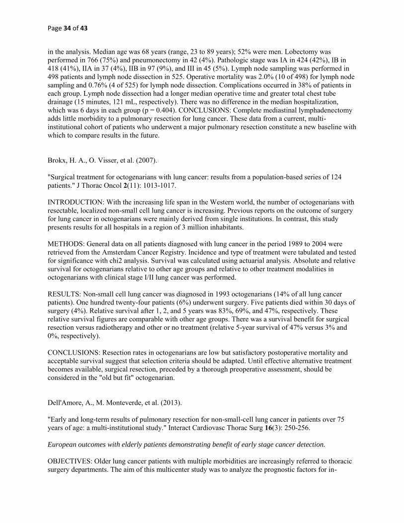
Page 34 of 43
in the analysis. Median age was 68 years (range, 23 to 89 years); 52% were men. Lobectomy was performed in 766 (75%) and pneumonectomy in 42 (4%). Pathologic stage was IA in 424 (42%), IB in 418 (41%), IIA in 37 (4%), IIB in 97 (9%), and III in 45 (5%). Lymph node sampling was performed in 498 patients and lymph node dissection in 525. Operative mortality was 2.0% (10 of 498) for lymph node sampling and 0.76% (4 of 525) for lymph node dissection. Complications occurred in 38% of patients in each group. Lymph node dissection had a longer median operative time and greater total chest tube drainage (15 minutes, 121 mL, respectively). There was no difference in the median hospitalization, which was 6 days in each group (p = 0.404). CONCLUSIONS: Complete mediastinal lymphadenectomy adds little morbidity to a pulmonary resection for lung cancer. These data from a current, multi-institutional cohort of patients who underwent a major pulmonary resection constitute a new baseline with which to compare results in the future. Brokx, H. A., O. Visser, et al. (2007). "Surgical treatment for octogenarians with lung cancer: results from a population-based series of 124 patients." J Thorac Oncol 2(11): 1013-1017. INTRODUCTION: With the increasing life span in the Western world, the number of octogenarians with resectable, localized non-small cell lung cancer is increasing. Previous reports on the outcome of surgery for lung cancer in octogenarians were mainly derived from single institutions. In contrast, this study presents results for all hospitals in a region of 3 million inhabitants. METHODS: General data on all patients diagnosed with lung cancer in the period 1989 to 2004 were retrieved from the Amsterdam Cancer Registry. Incidence and type of treatment were tabulated and tested for significance with chi2 analysis. Survival was calculated using actuarial analysis. Absolute and relative survival for octogenarians relative to other age groups and relative to other treatment modalities in octogenarians with clinical stage I/II lung cancer was performed. RESULTS: Non-small cell lung cancer was diagnosed in 1993 octogenarians (14% of all lung cancer patients). One hundred twenty-four patients (6%) underwent surgery. Five patients died within 30 days of surgery (4%). Relative survival after 1, 2, and 5 years was 83%, 69%, and 47%, respectively. These relative survival figures are comparable with other age groups. There was a survival benefit for surgical resection versus radiotherapy and other or no treatment (relative 5-year survival of 47% versus 3% and 0%, respectively). CONCLUSIONS: Resection rates in octogenarians are low but satisfactory postoperative mortality and acceptable survival suggest that selection criteria should be adapted. Until effective alternative treatment becomes available, surgical resection, preceded by a thorough preoperative assessment, should be considered in the "old but fit" octogenarian. Dell'Amore, A., M. Monteverde, et al. (2013). "Early and long-term results of pulmonary resection for non-small-cell lung cancer in patients over 75 years of age: a multi-institutional study." Interact Cardiovasc Thorac Surg 16(3): 250-256. European outcomes with elderly patients demonstrating benefit of early stage cancer detection. OBJECTIVES: Older lung cancer patients with multiple morbidities are increasingly referred to thoracic surgery departments. The aim of this multicenter study was to analyze the prognostic factors for in-

Page 35 of 43
hospital morbidity and mortality and to elucidate the predictors of long-term survival and oncological outcomes. METHODS: We identified 319 patients aged >/= 75 years who underwent intended curative lung resection for lung cancer in three different thoracic surgery departments between January 2000 and December 2010. RESULTS: Seventy-one patients underwent limited resection, 202 had lobectomy, 16 had bilobectomy and 30 had pneumonectomy. The in-hospital mortality was 6.6%. Chronic renal failure, low respiratory reserve and pneumonectomy were predictors of in-hospital mortality. The mean follow-up time was 3.9 years, ranging from 1 month to 10.4 years. The disease-free survivals at 1, 3 and 5 years were 82, 60 and 47%, respectively. The overall survivals at 1, 3 and 5 years were 86, 59 and 38%, respectively. The long-term overall survival was negatively influenced by pneumonectomy, extended resection, N(1-2) subgroups and pathological TNM stage. CONCLUSIONS: Nowadays, we can consider surgery a safe and justifiable option for elderly patients. Careful preoperative work-up and selection are mandatory to gain satisfactory results. Good long-term results were achieved in elderly patients with early stage who underwent lobar or sublobar lung resection. The role of surgery or other alternative therapies, in patients with advanced stages, extensive nodal involvement and/or requiring extensive surgical resection for curative intent, is still unclear and further studies are certainly needed. Dillman, R. O., D. R. Zusman, et al. (2009). "Surgical resection and long-term survival for octogenarians who undergo surgery for non-small-cell lung cancer." Clin Lung Cancer 10(2): 130-134. US single-institution series showing benefit of surgical treatment for octogenarians with lung cancer. PURPOSE: An increasing proportion of newly diagnosed non-small-cell lung cancer (NSCLC) patients are octogenarians. It has been questioned whether older patients benefit from surgical resection of lung cancer to the same extent as younger patients. PATIENTS AND METHODS: We conducted a single-institution, retrospective analysis of patients newly diagnosed with NSCLC from 2000-2006, who underwent surgical resection of their lung cancer in Hoag Hospital. We compared resection and survival rates for patients who were age 80 years or older to younger cohorts and determined their stage distribution, rates of surgery, and actuarial survival by age-defined cohort. Of 1293 total patients, 17.2% were age 80 years or older; 36.1%, age 70-79 years; 29.2%, age 60-69 years; 12.9%, age 50-59 years; and 4.6%, under age 50. Of these patients, 482 underwent surgical resection. Surgical procedures included 400 lobectomies, 23 pneumonectomies, and 59 wedge resections. RESULTS: The proportion of patients who had local disease at diagnosis was higher for octogenarians compared with younger patients (33.6% vs. 26.6%; P = .021), but the resection rate for octogenarians was lower (64% vs. 83%; P = .0003). For patients determined to have local- or regional-stage disease, resection rates were 52% versus 67.9% (P = .0007). However, survival curves for patients who underwent surgical resection were similar for all five cohorts with 5-year survival rates of 62%, 53%, 63%, 63%, and 79% from oldest to youngest. CONCLUSION: Non-small-cell lung cancer patients < 80 years of age were less likely to undergo potentially curative surgery, but survival for octogenarians who did undergo surgical resection was
Page 36 of 43
comparable to younger age groups. Such patients should not be denied potentially curative surgery simply because of age. Hsie, M., S. Morbidini-Gaffney, et al. (2009). "Definitive treatment of poor-risk patients with stage I lung cancer: a single institution experience." J Thorac Oncol 4(1): 69-73.
US study demonstrating cancer survival in high-risk patients with lung cancer undergoing limited
resection, radiation, or ablative therapies. PURPOSE: Lung cancer remains the leading cause of cancer death in both men and women. A substantial number of patients with early stage non-small cell lung cancer (NSCLC) are unfit for standard surgery due to cardiopulmonary dysfunction and/or other comorbidity. The appropriate management for this population has not been defined. METHODS: Retrospective analysis of patients with clinical stage I NSCLC judged to be unsuitable for lobectomy between 1996 and 2005. RESULTS: Ninety-six patients, representing 23% of all patients treated for clinical stage I NSCLC were included in this analysis. The median age was 73 years and most patients were female. Patients underwent limited resection (LR, n = 45), primary radiotherapy (RT, n = 39) or radiofrequency ablation (n = 12). With median follow-up of 30 months, 61 patients remain alive. Actuarial 3-year survival is 65% following LR and 60% after primary RT. Local tumor relapse and distant metastases were observed with approximate equal probability following either LR or RT. CONCLUSION: Medical inoperability does not necessarily correspond to poor survival in patients with early stage NSCLC. A nihilistic approach is not warranted towards this population, and prospective trials are needed to better define optimal treatment strategies. International Early Lung Cancer Action Program, I., C. I. Henschke, et al. (2006). "Survival of patients with stage I lung cancer detected on CT screening." N Engl J Med 355(17): 1763-1771. BACKGROUND: The outcome among patients with clinical stage I cancer that is detected on annual screening using spiral computed tomography (CT) is unknown. METHODS: In a large collaborative study, we screened 31,567 asymptomatic persons at risk for lung cancer using low-dose CT from 1993 through 2005, and from 1994 through 2005, 27,456 repeated screenings were performed 7 to 18 months after the previous screening. We estimated the 10-year lung-cancer-specific survival rate among participants with clinical stage I lung cancer that was detected on CT screening and diagnosed by biopsy, regardless of the type of treatment received, and among those who underwent surgical resection of clinical stage I cancer within 1 month. A pathology panel reviewed the surgical specimens obtained from participants who underwent resection. RESULTS: Screening resulted in a diagnosis of lung cancer in 484 participants. Of these participants, 412 (85%) had clinical stage I lung cancer, and the estimated 10-year survival rate was 88% in this subgroup (95% confidence interval [CI], 84 to 91). Among the 302 participants with clinical stage I cancer who underwent surgical resection within 1 month after diagnosis, the survival rate was 92% (95% CI, 88 to

Page 37 of 43
95). The 8 participants with clinical stage I cancer who did not receive treatment died within 5 years after diagnosis. CONCLUSIONS: Annual spiral CT screening can detect lung cancer that is curable. Kozower, B. D., S. Sheng, et al. (2010). "STS database risk models: predictors of mortality and major morbidity for lung cancer resection." Ann Thorac Surg 90(3): 875-881; discussion 881-873. BACKGROUND: The aim of this study is to create models for perioperative risk of lung cancer resection using the STS GTDB (Society of Thoracic Surgeons General Thoracic Database). METHODS: The STS GTDB was queried for all patients treated with resection for primary lung cancer between January 1, 2002 and June 30, 2008. Three separate multivariable risk models were constructed (mortality, major morbidity, and composite mortality or major morbidity). RESULTS: There were 18,800 lung cancer resections performed at 111 participating centers. Perioperative mortality was 413 of 18,800 (2.2%). Composite major morbidity or mortality occurred in 1,612 patients (8.6%). Predictors of mortality include the following: pneumonectomy (p < 0.001), bilobectomy (p < 0.001), American Society of Anesthesiology rating (p < 0.018), Zubrod performance status (p < 0.001), renal dysfunction (p = 0.001), induction chemoradiation therapy (p = 0.01), steroids (p = 0.002), age (p < 0.001), urgent procedures (p = 0.015), male gender (p = 0.013), forced expiratory volume in one second (p < 0.001), and body mass index (p = 0.015). CONCLUSIONS: Thoracic surgeons participating in the STS GTDB perform lung cancer resections with a low mortality and morbidity. The risk-adjustment models created have excellent performance characteristics and identify important predictors of mortality and major morbidity for lung cancer resections. These models may be used to inform clinical decisions and to compare risk-adjusted outcomes for quality improvement purposes. Licker, M. J., I. Widikker, et al. (2006). "Operative mortality and respiratory complications after lung resection for cancer: impact of chronic obstructive pulmonary disease and time trends." Ann Thorac Surg 81(5): 1830-1837. BACKGROUND: Smoking is a common risk factor for chronic obstructive pulmonary disease (COPD), cardiovascular disease, and lung cancer. In this observational study, we examined the impact of COPD severity and time-related changes in early outcome after lung cancer resection. METHODS: Over a 15-year period, we analyzed an institutional registry including all consecutive patients undergoing surgery for lung cancer. Using the receiver-operating characteristic (ROC) curve, we analyzed the relationship between forced expiratory volume in 1 second (FEV1) and postoperative mortality and respiratory morbidity. Multiple regression analysis has also been applied to identify other risk factors. RESULTS: A preoperative FEV1 less than 60% was a strong predictor for respiratory complications (odds ratio [OR] = 2.7, confidence interval [CI]: 1.3 to 6.6) and 30-day mortality (OR = 1.9, CI: 1.2 to 3.9), whereas thoracic epidural analgesia was associated with lower mortality (OR = 0.4; CI: 0.2 to 0.8) and respiratory complications (OR = 0.6; CI: 0.3 to 0.9). Mortality was also related to age greater than 70 years, the presence of at least three cardiovascular risk factors, and pneumonectomy. From the period 1990 to 1994, to 2000 to 2004, we observed significant reductions in perioperative mortality (3.7% versus 2.4%) and in the incidence of respiratory complications (18.7% versus 15.2%), that was associated with a

Page 38 of 43
higher rate of lesser resection (from 11% to 17%, p < 0.05) and increasing use of thoracic epidural analgesia (from 65% to 88%, p < 0.05). CONCLUSIONS: Preoperative FEV1 less than 60% is a main predictor of perioperative mortality and respiratory morbidity. Over the last 5-year period, diagnosis of earlier pathologic cancer stages resulting in lesser pulmonary resection as well as provision of continuous thoracic epidural analgesia have contributed to improved surgical outcome. Matsuoka, H., M. Okada, et al. (2005). "Complications and outcomes after pulmonary resection for cancer in patients 80 to 89 years of age." Eur J Cardiothorac Surg 28(3): 380-383. OBJECTIVE: Patients 80 years or older often present with potentially resectable cases of non-small cell lung cancer. Whether such patients should undergo surgical treatment is becoming increasingly important in this rapidly aging society. METHODS: From April 1997 through March 2004, 40 consecutive patients with non-small cell lung cancer who were 80-88 years of age underwent complete resection of their tumors, as confirmed pathologically. We reviewed preoperative data including gender, age, history of smoking, pulmonary function, co-morbidity, and induction/adjuvant therapy. Perioperative data consisted of surgical procedure, operative morbidity and mortality, histopathologic type, pathologic stage, and outcome. RESULTS: The procedures comprised 16 lobectomies (40%), 12 segmentectomies (30%), and 12 wedge resections (30%). The histopathologic diagnosis was adenocarcinoma in 22 patients, squamous cell carcinomas in 11, large cell carcinomas in 4, adenosquamous cell carcinomas in 2, and neuro-endocrine cell carcinoma in 1. The disease stage was IA in 21 patients, IB in 14, IIB in 3, and IIIA in 2. There was no perioperative mortality. Eight patients had non-lethal complications (20%), including five with cardiopulmonary complications (parenchymal air leaks persisting for more than 7 days in two patients, interstitial pneumonia in one, bacterial pneumonia in one, and moderate arrhythmias in one) and three with minor complications (depression or confusion). The actuarial survival rates of the 40 patients, including deaths from all causes, were 92.4, 71.6, and 56.9% at 1, 3, and 5 years, respectively. In patients with stage I disease, the respective survival rates were 94.3, 74.3, and 57.3%. CONCLUSIONS: Advanced age is not a contraindication to curative resection in patients 80-89 years of age with stage I non-small cell lung cancer. Okada, M., T. Koike, et al. (2006). "Radical sublobar resection for small-sized non-small cell lung cancer: a multicenter study." J Thorac Cardiovasc Surg 132(4): 769-775. Japanese study showing increased opportunity and good outcomes of
sub-lobar resections for early stage lung cancer. OBJECTIVE: At present, even when early-stage, small-sized non-small cell lung cancers are being increasingly detected, lesser resection has not become the treatment of choice. We sought to compare sublobar resection (segmentectomy or wedge resection) with lobar resection to test which one is the appropriate procedure for such lesions.

Page 39 of 43
METHODS: From 1992 to 2001, a nonrandomized study was performed in 3 institutes for patients with a peripheral cT1N0M0 non-small cell lung cancer of 2 cm or less who were able to tolerate a lobectomy. The results of the sublobar resection group enrolled preoperatively (n = 305) were compared with those of the lobar resection group (n = 262). RESULTS: Except for distribution of tumor location, there were no significant differences in any variable, patient characteristics, curability, pathologic stage, morbidity, or recurrence rate. Median follow-up was more than 5 years. Disease-free and overall survivals were similar in both groups with 5-year survivals of 85.9% and 89.6% for the sublobar resection group and 83.4% and 89.1% for the lobar resection group, respectively. Multivariate analysis confirmed that the recurrence rate and prognosis associated with sublobar resection were not inferior to those obtained with lobar resection. Postoperative lung function was significantly better in patients who underwent sublobar resection. CONCLUSIONS: Sublobar resection should be considered as an alternative for stage IA non-small cell lung cancers 2 cm or less, even in low-risk patients. These results could lay the foundation for starting randomized controlled trials anew, which would bring great changes of lung cancer surgery in this era of early detection of lung cancer. Pastorino, U., P. Borasio, et al. (2008). "Lung cancer stage is an independent risk factor for surgical mortality." Tumori 94(3): 362-369. AIMS AND BACKGROUND: To study surgical mortality and evaluate major risk factors, with specific focus on the role of pathological stage in patients undergoing lung cancer resection. METHODS AND STUDY DESIGN: Age, gender, comorbidity, resection volume, experience of the hospital and surgical team have been reported as variables related to postoperative morbidity and mortality in lung cancer. The role of pathological tumor stage on postoperative mortality has never been fully evaluated. The study included 1418 consecutive lung cancer resections performed from 1998 to 2002 in two institutions. The effect of age, gender, comorbidity, resection volume, pathological stage and induction therapies on postoperative mortality was assessed by univariable and multivariable logistic regression analysis. RESULTS: Postoperative mortality was 1.8% overall, 3.7% (9/243) for pneumonectomy, 1.7% (17/1016) for lobectomy, and null (0/159) for sublobar resections (P = 0.020). At multivariable analysis, cardiovascular comorbidity (P = 0.008), resection volume (P = 0.036) and pathological stage (P = 0.027) emerged as significant predictors of surgical mortality. CONCLUSIONS: Early stage lung cancer resection has a favorable effect on surgical mortality, not only by preventing the need for pneumonectomy, but also by reducing mortality after lobectomy. Pedersen, J. H., H. Ashraf, et al. (2009). "The Danish randomized lung cancer CT screening trial--overall design and results of the prevalence round." J Thorac Oncol 4(5): 608-614. INTRODUCTION: Lung cancer screening with low dose computed tomography (CT) has not yet been evaluated in randomized clinical trials, although several are underway.

Page 40 of 43
METHODS: In The Danish Lung Cancer Screening Trial, 4104 smokers and previous smokers from 2004 to 2006 were randomized to either screening with annual low dose CT scans for 5 years or no screening. A history of cigarette smoking of at least 20 pack years was required. All participants have annual lung function tests, and questionnaires regarding health status, psychosocial consequences of screening, smoking habits, and smoking cessation. Baseline CT scans were performed in 2052 participants. Pulmonary nodules were classified according to size and morphology: (1) Nodules smaller than 5 mm and calcified (benign) nodules were tabulated, (2) Noncalcified nodules between 5 and 15 mm were rescanned after 3 months. If the nodule increased in size or was larger than 15 mm the participant was referred for diagnostic workup. RESULTS: At baseline 179 persons showed noncalcified nodules larger than 5 mm, and most were rescanned after 3 months: The rate of false-positive diagnoses was 7.9%, and 17 individuals (0.8%) turned out to have lung cancer. Ten of these had stage I disease. Eleven of 17 lung cancers at baseline were treated surgically, eight of these by video assisted thoracic surgery resection. CONCLUSIONS: Screening may facilitate minimal invasive treatment and can be performed with a relatively low rate of false-positive screen results compared with previous studies on lung cancer screening. Petersen, R. H., H. J. Hansen, et al. (2012). "Lung cancer screening and video-assisted thoracic surgery." J Thorac Oncol 7(6): 1026-1031. INTRODUCTION: The objective of this study is to report the impact of computed tomography (CT) screening on the use of Video-Assisted Thoracic Surgery (VATS) in a randomized screening trial. METHODS: The Danish Lung Cancer Screening Trial is a randomized clinically controlled trial of 4104 smokers and previous smokers who were randomized to either screening with five annual low-dose CT scans or no screening in Copenhagen from 2004 to 2006. The major end point is the effect of CT screening on lung cancer mortality and treatment options. All diagnostic and treatment interventions in both groups were monitored prospectively until 1 to 3 years after the last screening round. RESULTS: By February 1, 2011 68 cases of lung cancer were detected in the screening group. Furthermore, seven patients with a benign nodule underwent surgical treatment because of suspicion of malignancy (12%). Fifty-one of the 68 lung cancer patients were eligible for surgical treatment. Eight patients had open thoracotomy. Of the operations for lung cancer, 84% were performed by VATS in the CT-screened arm, significantly higher than the control arm (p < 0.05). Thirty-six patients had a VATS lobectomy. One patient had a VATS segmentectomy, and four patients had a VATS wedge resection. The seven benign nodules were all treated with VATS. CONCLUSIONS: CT screening seems to facilitate the use of VATS in the treatment of lung cancer with an 84% rate in our data. Furthermore, all benign nodules could be removed by VATS. In our view, a basic requirement for a surgical institution to be involved in lung cancer CT screening is a dedicated VATS program.
Page 41 of 43
Rivera, C., M. Dahan, et al. (2011). "Surgical treatment of lung cancer in the octogenarians: results of a nationwide audit." Eur J Cardiothorac Surg 39(6): 981-986. Objective: The elderly is a fast-growing segment of the population and the number of oncogeriatric patients with lung cancer is expected to increase. The purpose of this study was to overview surgical habits for lung cancer in octogenarians. Methods: We used EPITHOR((R)), the French national thoracic database, created in 2002 and including more than 135000 procedures from 93 institutions. We collected prospectively data concerning 622 patients 80 years or older, and 16461 patients younger than 80 years with lung cancer from 1 January 2004 to 31 December 2008. We compared patients' characteristics, lung-cancer presentation, and surgical treatment between these two groups. Results: Patients' characteristics analysis: the distribution by gender, body mass index, and forced expiratory volume was comparable for the two groups. American Society of Anesthesiologists (ASA) score (ASA 1 and 2: 59%, n=363 vs 71%, n=11543, p<0.0001) and performance status (PS) were worse for older patients (PS 0 and 1: 86%, n=470 vs 89%, n=12685, p<0.0001). Mean age (82.0, confidence interval (CI) 95% (81.9; 82.2)) and sex ratio (2.51, n=445 males) were stable for octogenarians across 5 years. Lung-cancer presentation analysis: in the elderly, stages I and II were of 71% (n=361) versus 66% (n=8735) in the younger group (p=0.001). Surgical treatment analysis: resections in octogenarians were pneumonectomy 10% (n=62) versus 15% (n=2409) for patients under 80 years, lobectomy 67% (n=415) versus 65% (n=10734), bilobectomy 4% (n=25) versus 5% (n=809), sub-lobar resection 11% (n=70) versus 8% (n=1355) (p=0.034). They underwent video-assisted thoracic surgery in 7% (n=43) versus 6% (n=917) (p=0.034). No lymph node dissection was more frequent in patients 80 years or older (8%, n=45) than in younger patients (5%, n=738) (p=0.0004). Conclusion: Taken as a whole, octogenarians underwent more sub-lobar resections and less mediastinal lymph node dissections than younger patients. Effective management of lung cancer in older patients should be tailed to individual needs. Surgical treatment should not be denied on chronological age alone anymore. Rogers, S. O., Jr., S. W. Gray, et al. (2010). "Variations in surgeon treatment recommendations for lobectomy in early-stage non-small-cell lung cancer by patient age and comorbidity." Ann Surg Oncol 17(6): 1581-1588. BACKGROUND: Prior research suggests that older patients are less likely to undergo resection of early-stage non-small-cell lung carcinomas (NSCLCs). We surveyed surgeons to understand how their recommendations for lobectomy were influenced by age, the presence and severity of smoking-related lung disease, or by characteristics of the surgeons and their practices. METHODS: We surveyed surgeons caring for NSCLC patients regarding whether they would recommend lobectomy for hypothetical patients with early-stage NSCLC who varied by age (55 vs. 80 years) and comorbid illness (none, moderate, severe chronic obstructive pulmonary disease [COPD]). Ordinal logistic regression was used to identify the importance of patient, surgeon, and practice characteristics on surgery recommendations. RESULTS: Surgeons recommended lobectomy for nearly all patients who were 55 years old with no comorbidity (adjusted proportion 98.6%), 55 years old with moderate COPD (adjusted proportion 97.8%), or 80 years old with no comorbidity (adjusted proportion 98.1%). Fewer recommended lobectomy for 80-year-old patients with moderate COPD (adjusted proportion 82.3%), and far fewer recommended lobectomy for severe COPD, irrespective of age (adjusted rate 18.7% for the 55-year-old

Page 42 of 43
patient and 6.1% for the 80-year-old patient) (P < 0.002). Surgeons who enroll patients onto clinical trials (P = 0.03) were more likely than others to recommend lobectomy, but no other surgeon characteristic predicted recommendations. CONCLUSIONS: Lower rates of lobectomy among older patients do not seem to be explained by age-related biases among surgeons for otherwise healthy patients. Su, S., W. J. Scott, et al. (2014). "Patterns of survival and recurrence after surgical treatment of early stage non-small cell lung carcinoma in the ACOSOG Z0030 (ALLIANCE) trial." J Thorac Cardiovasc Surg 147(2): 747-752: Discussion 752-743. OBJECTIVE: Surgical resection has been the mainstay of curative treatment of early stage lung cancer in selected patients. We evaluated survival and patterns of recurrence after surgical resection for early stage lung cancer from the American College of Surgeons Oncology Group Z0030/Alliance trial. METHODS: One thousand eighteen patients enrolled in the Z0030 trial were analyzed according to clinical T stage. Differences between groups were compared using the 2-sample rank test or chi(2) test. Log rank test and Cox proportional hazards regression were used to compare survival and recurrence. To compare patients who underwent open versus video-assisted thoracoscopic surgery (VATS) resections, propensity-score matched analysis was performed. Seven hundred fifty-two patients (66 undergoing VATS and 686 undergoing open surgery) were classified into 5 equal-sized propensity-score groups. Proportional hazards regression was used to compare these outcomes. RESULTS: There were 578 patients with cT1 tumors and 440 patients with cT2 tumors. Median follow-up was 6.7 years. Median overall survival was 9.1 years (stage T1) and 6.5 years (stage T2). Overall survival at 5 years was 72% (stage T1) and 55% (stage T2). Local recurrence-free survival at 5 years was 95% (stage T1) and 91% (stage T2) (P = .015). Among patients with stage T1 cancer, 4.2% (23 out of 542) had local recurrences, whereas 7.3% (30 out of 409) of those with stage T2 tumors had local failure. There was no difference in the development of new primary tumors between stage T1 and stage T2 groups. In the propensity-score matched analysis of VATS versus open lobectomy patients, there was no difference in overall survival, disease-free survival, and freedom from development of a new primary tumor. CONCLUSIONS: Results of patients with resected early stage non-small cell carcinoma from a large-scale, multicenter trial serve as benchmarks against which to compare nonsurgical therapies for early stage lung cancer. Propensity-score matched analysis shows no difference in survival between patients undergoing VATS and open lobectomy. Tsutani, Y., Y. Miyata, et al. (2013). "Oncologic outcomes of segmentectomy compared with lobectomy for clinical stage IA lung adenocarcinoma: propensity score-matched analysis in a multicenter study." J Thorac Cardiovasc Surg 146(2): 358-364.

Page 43 of 43
OBJECTIVE: Our objective was to compare the oncologic outcomes of lobectomy and segmentectomy for clinical stage IA lung adenocarcinoma. METHODS: We examined 481 of 618 consecutive patients with clinical stage IA lung adenocarcinoma who underwent lobectomy or segmentectomy after preoperative high-resolution computed tomography and F-18-fluorodeoxyglucose positron emission tomography/computed tomography. Patients (n = 137) who underwent wedge resection were excluded. Lobectomy (n = 383) and segmentectomy (n = 98) as well as surgical results were analyzed for all patients and their propensity score-matched pairs. RESULTS: Recurrence-free survival (RFS) and overall survival (OS) were not significantly different between patients undergoing lobectomy (3-year RFS, 87.3%; 3-year OS, 94.1%) and segmentectomy (3-year RFS, 91.4%; hazard ratio [HR], 0.57; 95% confidence interval [CI], 0.27-1.20; P = .14; 3-year OS, 96.9%; HR, 0.49; 95% CI, 0.17-1.38; P = .18). Significant differences in clinical factors such as solid tumor size (P < .001), maximum standardized uptake value (SUVmax) (P < .001), and tumor location (side, P = .005; lobe, P = .001) were observed between both treatment groups. In 81 propensity score-matched pairs including variables such as age, gender, solid tumor size, SUVmax, side, and lobe, RFS and OS were similar between patients undergoing lobectomy (3-year RFS, 92.9%, 3-year OS, 93.2%) and segmentectomy (3-year RFS, 90.9%; 3-year OS, 95.7%). CONCLUSIONS: Segmentectomy is suitable for clinical stage IA lung adenocarcinoma, with survivals equivalent to those of standard lobectomy. Van't Westeinde, S. C., N. Horeweg, et al. (2012). "Complications following lung surgery in the Dutch-Belgian randomized lung cancer screening trial." Eur J Cardiothorac Surg 42(3): 420-429.
OBJECTIVES: To assess the complication rate in participants of the screen arm of the NELSON lung cancer screening trial who underwent surgical resection and to investigate, based on a literature review, whether the complication rate, length of hospital stay, re-thoracotomy and mortality rates after a surgical procedure were different from those of the non-screening series, taking co-morbidity into account.
METHODS: Between April 2004 and December 2008, 198 subjects underwent thoracic surgery. Co-morbid conditions were retrieved from the medical records. Postoperative complications were classified as minor and major. RESULTS: In total, 182 thoracotomies, 5 thoracotomies after video-assisted thoracoscopic surgery (VATS) and 11 VATS procedures were performed. In these patients, 36% had chronic obstructive lung disease, 16% coronary artery disease, 14% diabetes mellitus and 11% peripheral vascular disease. Following thoracotomy, 47% (88/187) had >/=1 minor (7-57% in literature) and 10% (18/187) >/=1 major complication (2-26% in literature); following VATS, 38% (6/16) had >/=1 minor complication, but no major complications. Seventeen per cent (3/18) of major complications and 21% (20/96) of minor complications were seen in subjects operated for benign disease. The re-thoracotomy rate was 3% and there was no 30-day mortality after thoracotomy or VATS (0-8.3% in literature). The mortality rate of 0% after surgical procedures is low when compared with the non-screening series (0-8.3%); the rate of complications (53%) is within range when compared with the non-screening series (8.5-58%).
CONCLUSIONS: In conclusion, mortality rates after surgical procedures are lower in the NELSON lung cancer screening trial than those in the non-screening series. The rate of complications is within the same range as in the non-screening series. Trial registration number: ISR CTN 63545820.