
Sensors and Actuators B: Chemical · Y. Ding, et al. Sensors & Actuators: B. Chemical 297 (2019)...
7
Contents lists available at ScienceDirect Sensors and Actuators B: Chemical journal homepage: www.elsevier.com/locate/snb A dual signal immunochromatographic strip for the detection of imidaclothiz using a recombinant fluorescent-peptide tracer and gold nanoparticles Yuan Ding a , Xiude Hua a, ⁎ , He Chen a , Gualberto Gonzalez-Sapienza b , Bogdan Barnych c , Fengquan Liu a,d , Minghua Wang a , Bruce D. Hammock c a College of Plant Protection, Nanjing Agricultural University, Nanjing 210095, China b Cátedra de Inmunología, Facultad de Química, Instituto de Higiene, Universidad de la República, Montevideo 11600, Uruguay c Department of Entomology and UCD Cancer Center, University of California, Davis, CA 95616, United States d Institute of Plant Protection, Jiangsu Academy of Agricultural Science, Nanjing 210014, China ARTICLE INFO Keywords: Sensitivity Fluorescent peptide tracer Inner filter effect Dual signals Pesticide residue ABSTRACT Immunochromatographic strips (ICS) attract much attention as practical analytical tools, particularly for point of care testing. However, ICS suffer from insufficient sensitivity, especially for small molecules. Competitive assays developed with analyte peptidomimetics obtained from phage display peptide libraries, usually enable to attain lower detection limits than the widely used chemical haptens, but are rarely used in ICS assay because of the shortcomings of the phage particle. In this work, we used a phage-free fluorescent peptide tracer (C2-15-EmGFP) for ICS, which consists of the emerald green fluorescent protein (EmGFP) fused to a peptidomimetic of imida- clothiz (C2-15). C2-15-EmGFP works as fluorescent donor and colloidal gold nanoparticles (AuNPs) coated with an anti-imidaclohtiz antibody as quencher, which allowed to develop a fluorescence quenching-based ICS (FQICS) for the detection of imidaclothiz in agricultural and environmental samples. The colorimetric and fluorescent signals were determined by both naked eye and imageJ software. Quantitative image analysis showed that the sensitivities obtained using the colorimetric (50% inhibitory concentration (IC 50 ): 9.62 ng mL −1 ) and fluorescent (50% saturation concentration (SC 50 ): 10.1 ng mL −1 ) signals were similar. The visual detection by the naked eye showed a sensitivity of 8.00 ng mL −1 with fluorescent readout which was 8- fold lower than that of the colorimetric readout (64.0 ng mL −1 ). The FQICS performed with excellent recoveries and good correlation with high-performance liquid chromatography in different matrices. 1. Introduction Immunochromatographic strips (ICS), as fast on-site detection tools, have an enormous potential for the detection of small compounds, which comprise a vast portion of most common analytes, including drugs, pesticides, biological toxins, antibiotics, etc [1–3]. Although ICS have numerous advantages such as short assay time, use on site, simple operation and low cost, in some cases the sensitivity that can be at- tained is too low, particularly for small analytes where typically require competitive formats [4–6]. This is so, because they cannot generally be recognized simultaneously by two antibodies, which means that a competing tracer or competing coating antigen is required to compete with the analyte for the binding site of the antibody. Most frequently competing tracers in ICS are based on chemical haptens. This requires sophisticated organic synthesis and that multiple haptens are tried in a trial and error procedure. In contrast, analyte peptidomimetics are ea- sily obtained from phage display peptide libraries, and usually allow obtaining better sensitivity than chemical haptens [7–9]. However, their use as phage borne peptides carries the shortcomings of the phage particle, namely their large size, potential aggregation, bad diffusion rates and biological concerns, which represent serious limitation to the use of phage-displayed peptidomimetics in ICS [10]. Recently, we fused a peptidomimetic of imidaclothiz with nano luciferase (NanoLuc) creating phage-free recombinant tracer to develop a bioluminescent ICS, which showed a satisfactory sensitivity for imidaclothiz detection [10]. This tracer also has the remarkable advantage of being well de- fined, eliminating the batch-to-batch variability associated with che- mical conjugation [11–14]. https://doi.org/10.1016/j.snb.2019.126714 Received 2 April 2019; Received in revised form 9 June 2019; Accepted 20 June 2019 ⁎ Corresponding author. E-mail address: [email protected] (X. Hua). Sensors & Actuators: B. Chemical 297 (2019) 126714 Available online 28 June 2019 0925-4005/ © 2019 Elsevier B.V. All rights reserved. T
Transcript of Sensors and Actuators B: Chemical · Y. Ding, et al. Sensors & Actuators: B. Chemical 297 (2019)...

Contents lists available at ScienceDirect
Sensors and Actuators B: Chemical
journal homepage: www.elsevier.com/locate/snb
A dual signal immunochromatographic strip for the detection ofimidaclothiz using a recombinant fluorescent-peptide tracer and goldnanoparticles
Yuan Dinga, Xiude Huaa,⁎, He Chena, Gualberto Gonzalez-Sapienzab, Bogdan Barnychc,Fengquan Liua,d, Minghua Wanga, Bruce D. Hammockc
a College of Plant Protection, Nanjing Agricultural University, Nanjing 210095, Chinab Cátedra de Inmunología, Facultad de Química, Instituto de Higiene, Universidad de la República, Montevideo 11600, Uruguayc Department of Entomology and UCD Cancer Center, University of California, Davis, CA 95616, United Statesd Institute of Plant Protection, Jiangsu Academy of Agricultural Science, Nanjing 210014, China
A R T I C L E I N F O
Keywords:SensitivityFluorescent peptide tracerInner filter effectDual signalsPesticide residue
A B S T R A C T
Immunochromatographic strips (ICS) attract much attention as practical analytical tools, particularly for point ofcare testing. However, ICS suffer from insufficient sensitivity, especially for small molecules. Competitive assaysdeveloped with analyte peptidomimetics obtained from phage display peptide libraries, usually enable to attainlower detection limits than the widely used chemical haptens, but are rarely used in ICS assay because of theshortcomings of the phage particle. In this work, we used a phage-free fluorescent peptide tracer (C2-15-EmGFP)for ICS, which consists of the emerald green fluorescent protein (EmGFP) fused to a peptidomimetic of imida-clothiz (C2-15). C2-15-EmGFP works as fluorescent donor and colloidal gold nanoparticles (AuNPs) coated withan anti-imidaclohtiz antibody as quencher, which allowed to develop a fluorescence quenching-based ICS(FQICS) for the detection of imidaclothiz in agricultural and environmental samples. The colorimetric andfluorescent signals were determined by both naked eye and imageJ software. Quantitative image analysisshowed that the sensitivities obtained using the colorimetric (50% inhibitory concentration (IC50):9.62 ngmL−1) and fluorescent (50% saturation concentration (SC50): 10.1 ngmL−1) signals were similar. Thevisual detection by the naked eye showed a sensitivity of 8.00 ngmL−1 with fluorescent readout which was 8-fold lower than that of the colorimetric readout (64.0 ngmL−1). The FQICS performed with excellent recoveriesand good correlation with high-performance liquid chromatography in different matrices.
1. Introduction
Immunochromatographic strips (ICS), as fast on-site detection tools,have an enormous potential for the detection of small compounds,which comprise a vast portion of most common analytes, includingdrugs, pesticides, biological toxins, antibiotics, etc [1–3]. Although ICShave numerous advantages such as short assay time, use on site, simpleoperation and low cost, in some cases the sensitivity that can be at-tained is too low, particularly for small analytes where typically requirecompetitive formats [4–6]. This is so, because they cannot generally berecognized simultaneously by two antibodies, which means that acompeting tracer or competing coating antigen is required to competewith the analyte for the binding site of the antibody. Most frequentlycompeting tracers in ICS are based on chemical haptens. This requires
sophisticated organic synthesis and that multiple haptens are tried in atrial and error procedure. In contrast, analyte peptidomimetics are ea-sily obtained from phage display peptide libraries, and usually allowobtaining better sensitivity than chemical haptens [7–9]. However,their use as phage borne peptides carries the shortcomings of the phageparticle, namely their large size, potential aggregation, bad diffusionrates and biological concerns, which represent serious limitation to theuse of phage-displayed peptidomimetics in ICS [10]. Recently, we fuseda peptidomimetic of imidaclothiz with nano luciferase (NanoLuc)creating phage-free recombinant tracer to develop a bioluminescentICS, which showed a satisfactory sensitivity for imidaclothiz detection[10]. This tracer also has the remarkable advantage of being well de-fined, eliminating the batch-to-batch variability associated with che-mical conjugation [11–14].
https://doi.org/10.1016/j.snb.2019.126714Received 2 April 2019; Received in revised form 9 June 2019; Accepted 20 June 2019
⁎ Corresponding author.E-mail address: [email protected] (X. Hua).
Sensors & Actuators: B. Chemical 297 (2019) 126714
Available online 28 June 20190925-4005/ © 2019 Elsevier B.V. All rights reserved.
T

Over the past few years, some fluorescent tracers have been appliedin ICS to improve its sensitivity [3–6,15,16]. Among fluorescent ICS,those based on fluorescence quenching have attracted most research[17–20]. In this format, the antibody is labeled with the quencher andthe hapten with the fluorescent tracer, or vice versa. The fluorescentcomponents are commonly dispensed on the nitrocellulose (NC) mem-brane as the test line (T line) and their fluorescence vanishes upon in-teraction with the quenching component in the absence of analyte.With increasing amounts of analyte, the equilibrium shifts toward thedissociation of the hapten-antibody complex, which leads to the in-crease of the fluorescence intensity. If the quenching component iscolored there will be also a decrease in the color depth of the T line.This format produces multiple correlated signals at the same time,which improves detection [21]. Furthermore, the fluorescent “turn-on”mode of the signal is advantageous for the sensitivity of the assay andits visual reading, making the result more intuitive.
Imidaclothiz belongs to neonicotinoid insecticide, which is themajor insecticide in use and the use of this class of pesticides is in-creasing in China currently. Because of concerns for example of toxicityto pollinators there are growing needs for rapid reliable field methods[22,23]. In this work, we used a novel recombinant fluorescent peptidetracer (C2-15-EmGFP) [24], which consists of the peptidomimetic ofimidaclothiz (C2-15, CLPPRMIYEC) fused to emerald green fluorescentprotein (EmGFP) and a His tag, to develop fluorescence quenching-based ICS (FQICS) for the detection of the insecticide. The fluorescentpeptide tracer was captured by an anti-His antibody immobilized on thetest line (T line) to provide the fluorescent signal, while it was properlyoriented to be recognized by the anti-imidaclothiz monoclonal antibody(mAb 1E7) labeled with colloidal gold nanoparticles (AuNPs), whichquenched the fluorescent signal and the same time that generated acolorimetric readout. The recoveries of the FQICS analytical methodwere tested in different matrices and the accuracy of FQICS was vali-dated by high-performance liquid chromatography (HPLC).
2. Material and methods
2.1. Reagents
Chloroauric acid, trisodium citrate and bovine serum albumin (BSA)
were purchased from Sigma Aldrich Co. Ltd. (St Louis, USA). Cys-pro-tein G was purchased from Shanghai Puxin Biotechnology co., Ltd.(Shanghai, China). CH3O-(CH2CH2-O)n-CH2CH2-SH (HS-mPEG, MW ˜5 kDa) was purchased from Shanghai Tuoyang Biotechnology Co., Ltd.(Shanghai, China). Anti-His tag antibody and anti-mouse IgG antibodywere purchased from Tiangen Biochemical Technology Co., Ltd.(Beijing, China). The nitrocellulose (NC) membrane (Hi-flow plus 135)was purchased from Millipore (Bedford, MA, USA). Imidaclothiz(97.82%) was provided by Nantong Jiangshan Agrochemical andChemicals Co., Ltd. (Jiangsu, China), other pesticide standards weresupplied by Dr. Ehrenstorfer GmbH (Germany). The purified C2-15-EmGFP and mAb 1E7 were prepared as described previously [24,25].
2.2. Preparation of mAb-labeled AuNPs
The 20 nm AuNPs were synthesized by the trisodium citrate re-duction method. Briefly, 100mL 0.01% chloroauric acid solution washeated to boiling. Then, 1.7 mL 1% trisodium citrate solution wasquickly added into the boiling solution while stirring. After the color ofthe solution turned to wine red, the solution was boiled for an addi-tional 5min and then cooled to room temperature. The molar con-centration of AuNPs was determined by Beer-Lambert Law [26]. Cys-protein G was mixed with HS-mPEG at the molar ratio of 1:10. Themixture was added to 30mL AuNPs solution (molar ratio of AuNPs: cys-protein G=1:1000) and reacted at room temperature for 5 h undergentle shaking. The excess reagents were removed by centrifugationfollowed by resuspension in 0.14mol L−1 phosphate-buffered salinebuffer (PBS) containing 1mgmL−1 BSA and centrifugation (2 times).Then mAb 1E7 (molar ratio of AuNPs: mAb 1E7= 1:100) was added tothe protein G-labeled AuNPs and reacted for 1 h under gentle shaking.After centrifugation, the mAb-labeled AuNPs were suspended in 1mL0.14mol L−1 PBS containing 1mgmL−1 BSA.
2.3. Assembly of the strip
The anti-His tag antibody (4mgmL−1 in PBS) and anti-mouse IgGantibody (1mgmL-1 in PBS) were dispensed (1 μL cm−1) on the NCmembrane as the T line and control line (C line), respectively. Then, theNC membrane was dried at 37 °C for 1 h. The mAb-labeled AuNPs
Fig. 1. Schematic representation of the FQICS assay. The sample and C2-15-EmGFP are added to the sample pad and, upon flowing across the strip, redissolve thequencher sprayed on the conjugate pad and interact with the antibodies on the test and control lines.
Y. Ding, et al. Sensors & Actuators: B. Chemical 297 (2019) 126714
2

solution was sprayed on the conjugate pad (glass fiber pad) pretreatedwith PBS (containing 1mgmL−1 BSA) at 34 μL cm−1. Treated NCmembrane was assembled with sample pad, conjugate pad and absor-bent pad on a semirigid polyvinyl chloride sheet and then cut into3.5 mm width strips and stored at room temperature (Fig. 1).
2.4. FQICS analytical method
Seventy-five microliters of imidaclothiz standard solutions orsample solutions (containing 10% methanol) was mixed with 75 μL C2-15-EmGFP (0.056mgmL−1, containing 0.5% Tween 20). The mixturewas added to the sample pad to redissolve mAb-labeled AuNPs sprayedon the conjugate pad and was allowed to flow across the NC membraneby capillary action for 15min. Then the strip was exposed to room lightand read on a multifunctional imager (Vilber, Fusion FX7 IR Spectra) torecord the colorimetric signal by a Samsung Note8 camera and thefluorescent signals by the camera of imager (excitation and optical filterwavelengths were chosen as 470 nm and 535 nm), respectively (Fig. 1).
The visual detection was performed based on both the colorimetricand fluorescent signals. The colorimetric signal on strip or image (re-corded by a Samsung Note8 camera) was observed by naked eye, dis-appearance of red T line corresponded to positive sample (+), andnegative otherwise (−). For the fluorescent signal, the image capturedby the camera of imager was observed by naked eye, appearance offluorescence on T line region corresponded to positive sample, andnegative otherwise (Fig. 1). Quantification was carried out by using theimageJ 1.46 r software. Images of the strips were imported into thesoftware to measure the mean gray value (colorimetric signal) andmean optical density (fluorescent signal) of the T line and background,and then the T line values were corrected for background by simple
subtraction.
2.5. Cross-reactivity (CR)
A series of standard solutions of the imidaclothiz analogues wereprepared and tested by FQICS. The concentration producing 50% sa-turation of the fluorescent signal (SC50) was determined for each ana-logue. Then CRs were calculated to assess the specificity of imidaclothizFQICS by the following formula: CR = [SC50 (imidaclothiz)/SC50
(analogue)] × 100%.
2.6. Analysis of spiked samples
The blank soil and wheat samples were collected from a farm inNanjing, China, and were confirmed to be imidaclothiz-free by HPLC(see below). The limit of detection for the HPLC method was 2.00 ngmL−1. The samples (10 g) were homogenized and spiked in a blindfashion with imidaclothiz at the final concentration of 50, 100, 200,400, 800 ng g−1. Then, 20mL 30% methanol-PBS was added to extractthe imidaclothiz. The mixtures were thoroughly shaken for 5min, so-nicated for 10min and shaken for another 5min. Supernatants con-taining imidaclothiz were collected by vacuum filtration. After 3-folddilution, the concentration of imidaclothiz in the solutions was de-termined by FQICS.
2.7. Comparison with HPLC measurement
The accuracy of the FQICS was evaluated by comparing the analy-tical results obtained with HPLC and FQICS in real samples. Samplepretreatment procedures required for FQICS analysis are describedabove. The following sample preparation procedure for HPLC analysiswas used: 10 g homogenized samples (soil and wheat samples) wereaccurately weighed and placed into 250mL triangular flask. Fortymilliliters acetonitrile and 5mL water were added, the flask was shakenfor 1 h at 250 rpm, and then the supernatants were collected by vacuumfiltration. The organic phase was separated from water by adding 5 gNaCl, and 20mL of the organic phase was collected and evaporated todryness. 2 mL of water:methanol= 60:40 (v/v) was used to redissolvethe extracts. The imidaclothiz in the extracts was detected by HPLC(Agilent 1260) with Eclipse pluse-C18 column (250mm×4.6mm,5 μm), the mobile phase was methanol:water= 25:75 (v/v) at 0.9mLmin−1, detection wavelength was 270 nm, injection volume was 20 μL,temperature of column oven was 30 °C.
3. Results and discussions
3.1. Design of FQICS
The schematic of the FQICS is depicted in Fig. 1. The recombinantfluorescent peptide tracer C2-15-EmGFP, consisting of the imidaclothizpeptidomimetic C2-15 at N-terminus, EmGFP in the middle and a6×His tag at the C-terminus (Fig. 1), has been recently described [24].The AuNPs coupled to the mAb 1E7, have an absorption spectrum thatoverlaps well with the emission spectrum of C2-15-EmGFP (Fig. 2a), sothat they will strongly quench the fluorescence of C2-15-EmGFP whenthey are in close proximity [27–29]. The quenching of the fluorescenceis cause by inner filter effect (IFE), which is radiative re-absorption(light is first emitted and then re-absorbed). IFE also can occur if dis-tances between donor and quencher exceed 10 nm [30,31].
The T line contains anti-His antibody that can specifically bind tothe His tag of recombinant fluorescent peptide. The C line contains theanti-mouse-IgG antibody that can bind the mouse-IgG (mAb 1E7 ismouse-IgG). After injecting the mixture of sample solution and re-combinant fluorescent peptide, it dissolved the mAb-labeled AuNPssprayed on the conjugate pad and then flowed across the T line and Cline in turn by capillary action. If there is no imidaclothiz in sample
Fig. 2. Analysis of mAb-labeled AuNPs. (a) The absorption spectrums of AuNPsand mAb-labeled AuNPs, and the emission spectrum of C2-15-EmGFP with485 nm excitation wavelength. (b) SDS-PAGE gel, line 1: protein G-labeledAuNPs; line 2: mAb-labeled AuNPs; line 3: BSA; line 4: protein G; line 5: mAb1E7. (c) Reactivity of the mAb-labelled AuNPs with a imidaclothiz hapten-OVAconjugate immobilized on nitrocellulose strips, tested in the presence of variousconcentration of imidaclothiz (0, 12, 25, 50, 100, 200 ng mL−1).
Y. Ding, et al. Sensors & Actuators: B. Chemical 297 (2019) 126714
3

solution, the recombinant fluorescent peptide would bind to the mAb1E7 labeled on AuNPs and accumulate at the T line. Fluorescence ofrecombinant fluorescent peptide located on T line would be absorbedby AuNPs. If there is imidaclothiz in sample solution, imidaclothizwould compete with recombinant fluorescent peptide for binding tomAb 1E7 labeled on AuNPs so that reduce the binding between therecombinant fluorescent peptide and the mAb 1E7 labeled on AuNPs,which would cause the recovery of fluorescence of recombinant fluor-escent peptide located on T line. Then the unbound mAb 1E7 labeled onAuNPs would accumulate on the C line to indicate the assay is valid.
Recently, the FQICS has attracted more and more attention, becausethe fluorescent “turn-on” mode of the signal is advantageous for thesensitivity of the assay and its visual reading, making the result moreintuitive. In this study, we used the recombinant fluorescent peptideinstead of the commonly used hapten and fluorescent nanoparticles todevelop a FQICS. Although the design of the FQICS is a little compli-cated, the proposed FQICS still has the general advantages of ICS, in-cluding simple operation (one step), rapid reaction (15min) and low-cost (recombinant fluorescent peptide can be efficiently prepared bybacterial fermentation). Importantly, there is no need of chemicalconjugation of the hapten.
3.2. Identification of mAb-labeled AuNPs
Cys-protein G was used instead of the non-specific adsorption of theantibody to reduce its random orientation on the AuNPs surface. To thisend, protein G equipped with a cysteine residue at the N terminus wasattached on the AuNPs surfaces through the coordination bond formedby the thiol group of the cysteine. The HS-mPEG was used as blockingand stabilizing agent. Then, protein G, absorbed on the AuNPs surfaces,specifically bound the Fc region of mAb, which provided an optimalorientation for the mAb. The amount of antibodies on the AuNPs sur-face was adjusted by changing the ratio of protein G and HS-mPEG, toavoid the aggregation of the AuNPs caused by high antibody density.After optimization, the maximum mAb 1E7 immobilization density thatdid not cause aggregation of the AuNPs was achieved at the ratio ofprotein G:HS-mPEG=1:10.
The mAb-labeled AuNPs showed bands of mAb and protein G inSDS-PAGE gel, which confirmed that the mAb had been attached to thesurface of AuNPs (Fig. 2b). Besides, The mAb-labeled AuNPs were fullyfunctional as demonstrated by its reactivity with an imidaclothizhapten-ovalbumin (OVA) conjugate in an immunochromatographictest, which could be inhibited by competition with the pesticide(Fig. 2c). The absorption spectrum of the AuNPs still possesses a goodoverlap with the emission spectrum of C2-15-EmGFP after loading ofthe antibody (Fig. 2a). These results indicated that the mAb-labeledAuNPs were successfully prepared, and could be used as quencher inthe FQICS assay.
3.3. Optimization of FQICS
Initially, the minimal concentration of C2-15-EmGFP(0.028mgmL−1) that produced a readily visible T line was optimizedusing a fix amount of mAb-labeled AuNPs (Fig. S1). Then by usingdifferent amounts of mAb-labeled AuNPs, we found that the highestsensitivity (50% inhibitory concentration (IC50): 9.86 ng mL−1 andSC50: 10.54 ng mL−1) was attained when 12 μL of the gold particleswere used. Since methanol is often used to extract pesticides fromagricultural and environmental samples, its influence on immunoassayperformance was evaluated. FQICSs were run in PBS containing dif-ferent concentrations of methanol, and the result showed the maximumtolerable methanol concentration was 5% (Fig. S2).
3.4. Sensitivity of FQICS
FQICS is used to detect imidaclothiz by a competitive format
(Fig. 1). The increase of imidaclothiz concentration would lead to thedecrease of mAb-labeled AuNPs bound to C2-15-EmGFP, resulting inthe decrease of the colorimetric signal and increase of a fluorescentsignal at the T line. For visual detection, the samples can be identifiedas positive based on colorimetric and fluorescent signals when imida-clothiz concentration is higher than 64.0 ng mL−1 and 8.00 ng mL−1
respectively (Fig. 3a). As expected, the “turn-on” mode of fluorescentsignal increases the sensitivity of the assay 8-fold compared to the“turn-off” mode of decreased colorimetric signal. On the other hand,the two signals have inverse relationship by design. The quantitativestandard curves, established by fitting the corrected mean gray value ormean optical density (y) versus the concentration of imidaclothiz (x)using the logistic equation in OriginPro 8, show that sensitivities andlinear ranges of colorimetric (IC50: 9.62 ng mL−1, IC10–IC90:3.21–35.8 ng mL-1) and fluorescent (SC50: 10.1 ng mL−1, SC10–SC90:2.62–34.6 ng mL−1) signals are roughly the same (Fig. 3b).
In our previous studies, a series of immunoassays were developed todetect imidaclothiz by using the same mAb 1E7 with chemically syn-thesized hapten, phage-displayed peptides and recombinant peptides.The proposed FQICS shows better sensitivity than the indirect compe-titive enzyme-linked immunosorbent assay (ic-ELISA, IC50:87.5 ngmL−1) [25], upconversion immunochromatographic assay(UICA, IC50: 97.2 ngmL−1) [32] and enhanced gold ICA (limit of de-tection: 25 ngmL−1) [33] based on the chemically synthesized hapten.Compared with the magnetic-separation fluorescence immunoassay(MSFIA) that used the same recombinant peptide with this research, theIC50 of MSFIA is 11.0 ng mL−1, which is roughly same with the FQICS[24]. Although the phage ELISA (IC50: 1.45 ng mL−1) and phage che-miluminescent enzyme immunoassay (CLEIA, IC50: 0.86 ng mL−1)based on the phage-displayed peptide (C2-15) [34], and bioluminescent
Fig. 3. Performance of FQICS for the detection of imidaclothiz. (a) Images ofvisual detection based on colorimetric and fluorescent signals. (b) Standardcurves obtained for colorimetric and fluorometric readouts with imageJ soft-ware.
Y. Ding, et al. Sensors & Actuators: B. Chemical 297 (2019) 126714
4
enzyme immunoassay (BLEIA, IC50: 3.3 ng mL−1) and bioluminescencelateral flow immunoassay (BLLFIA, IC50: 6.4 ng mL−1) based on therecombinant peptide (C2-15-NanoLuc) [10] show better sensitivitythan the FQICS in the quantitative detection, they need extra steps(washing and adding substrate to produce measurable signal) andlonger detection time (more than 25min) to complete the assay. Inaddition, the BLLFIA requires 150 ng mL−1 of analyte to inhibit the Tline signal by visual detection. A quantum-dot-based LFIA (IC50: 0.17 ngmL−1, detection time: 25min) for neonicotinoid shows higher sensi-tivity than the FQICS [35]. We speculate that the main reason is the useof different antibodies, because the sensitivity of conventional im-munoassay based on mAb 1E7 is not satisfactory (ic-ELISA, IC50: 87.5 ngmL−1). Fortunately, the FQICS is faster and more intuitive than thequantum-dot-based LFIA. However, the FQICS has the advantages ofsimple operation, short assay time, use on site, and visualization.
3.5. Specificity of FQICS
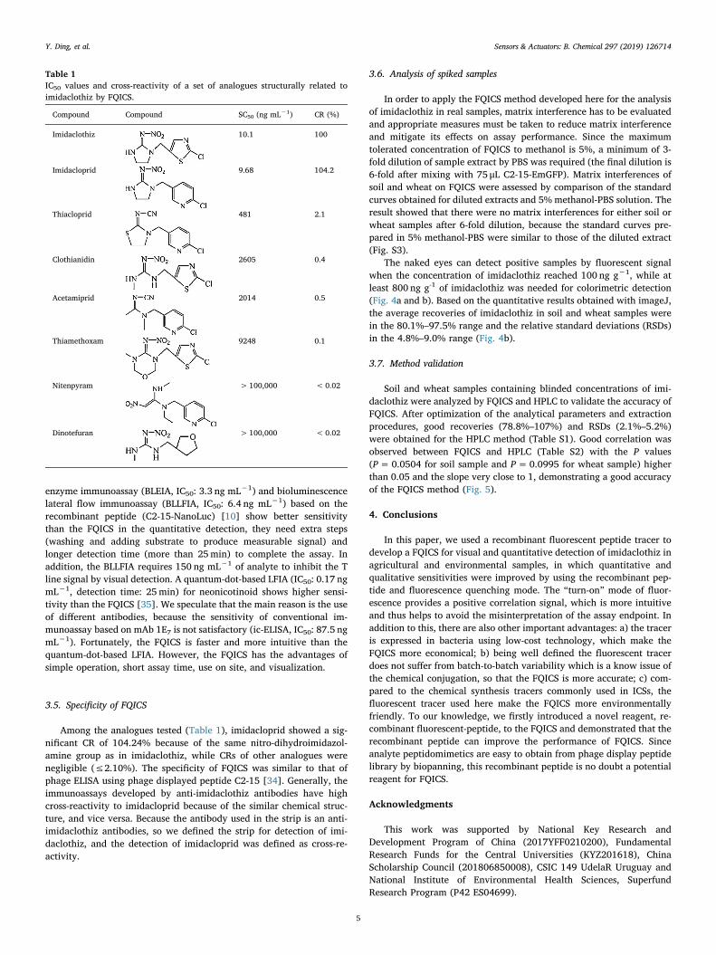
Among the analogues tested (Table 1), imidacloprid showed a sig-nificant CR of 104.24% because of the same nitro-dihydroimidazol-amine group as in imidaclothiz, while CRs of other analogues werenegligible (≤2.10%). The specificity of FQICS was similar to that ofphage ELISA using phage displayed peptide C2-15 [34]. Generally, theimmunoassays developed by anti-imidaclothiz antibodies have highcross-reactivity to imidacloprid because of the similar chemical struc-ture, and vice versa. Because the antibody used in the strip is an anti-imidaclothiz antibodies, so we defined the strip for detection of imi-daclothiz, and the detection of imidacloprid was defined as cross-re-activity.
3.6. Analysis of spiked samples
In order to apply the FQICS method developed here for the analysisof imidaclothiz in real samples, matrix interference has to be evaluatedand appropriate measures must be taken to reduce matrix interferenceand mitigate its effects on assay performance. Since the maximumtolerated concentration of FQICS to methanol is 5%, a minimum of 3-fold dilution of sample extract by PBS was required (the final dilution is6-fold after mixing with 75 μL C2-15-EmGFP). Matrix interferences ofsoil and wheat on FQICS were assessed by comparison of the standardcurves obtained for diluted extracts and 5% methanol-PBS solution. Theresult showed that there were no matrix interferences for either soil orwheat samples after 6-fold dilution, because the standard curves pre-pared in 5% methanol-PBS were similar to those of the diluted extract(Fig. S3).
The naked eyes can detect positive samples by fluorescent signalwhen the concentration of imidaclothiz reached 100 ng g−1, while atleast 800 ng g-1 of imidaclothiz was needed for colorimetric detection(Fig. 4a and b). Based on the quantitative results obtained with imageJ,the average recoveries of imidaclothiz in soil and wheat samples werein the 80.1%–97.5% range and the relative standard deviations (RSDs)in the 4.8%–9.0% range (Fig. 4b).
3.7. Method validation
Soil and wheat samples containing blinded concentrations of imi-daclothiz were analyzed by FQICS and HPLC to validate the accuracy ofFQICS. After optimization of the analytical parameters and extractionprocedures, good recoveries (78.8%–107%) and RSDs (2.1%–5.2%)were obtained for the HPLC method (Table S1). Good correlation wasobserved between FQICS and HPLC (Table S2) with the P values(P=0.0504 for soil sample and P=0.0995 for wheat sample) higherthan 0.05 and the slope very close to 1, demonstrating a good accuracyof the FQICS method (Fig. 5).
4. Conclusions
In this paper, we used a recombinant fluorescent peptide tracer todevelop a FQICS for visual and quantitative detection of imidaclothiz inagricultural and environmental samples, in which quantitative andqualitative sensitivities were improved by using the recombinant pep-tide and fluorescence quenching mode. The “turn-on” mode of fluor-escence provides a positive correlation signal, which is more intuitiveand thus helps to avoid the misinterpretation of the assay endpoint. Inaddition to this, there are also other important advantages: a) the traceris expressed in bacteria using low-cost technology, which make theFQICS more economical; b) being well defined the fluorescent tracerdoes not suffer from batch-to-batch variability which is a know issue ofthe chemical conjugation, so that the FQICS is more accurate; c) com-pared to the chemical synthesis tracers commonly used in ICSs, thefluorescent tracer used here make the FQICS more environmentallyfriendly. To our knowledge, we firstly introduced a novel reagent, re-combinant fluorescent-peptide, to the FQICS and demonstrated that therecombinant peptide can improve the performance of FQICS. Sinceanalyte peptidomimetics are easy to obtain from phage display peptidelibrary by biopanning, this recombinant peptide is no doubt a potentialreagent for FQICS.
Acknowledgments
This work was supported by National Key Research andDevelopment Program of China (2017YFF0210200), FundamentalResearch Funds for the Central Universities (KYZ201618), ChinaScholarship Council (201806850008), CSIC 149 UdelaR Uruguay andNational Institute of Environmental Health Sciences, SuperfundResearch Program (P42 ES04699).
Table 1IC50 values and cross-reactivity of a set of analogues structurally related toimidaclothiz by FQICS.
Compound Compound SC50 (ng mL−1) CR (%)
Imidaclothiz 10.1 100
Imidacloprid 9.68 104.2
Thiacloprid 481 2.1
Clothianidin 2605 0.4
Acetamiprid 2014 0.5
Thiamethoxam 9248 0.1
Nitenpyram >100,000 < 0.02
Dinotefuran > 100,000 < 0.02
Y. Ding, et al. Sensors & Actuators: B. Chemical 297 (2019) 126714
5

Appendix A. Supplementary data
Supplementary material related to this article can be found, in theonline version, at doi:https://doi.org/10.1016/j.snb.2019.126714.
References
[1] W.C. Mak, V. Beni, A.P.F. Turner, Lateral-flow technology: from visual to instru-mental, Trac-Trend. Anal. Chem. 79 (2016) 297–305, https://doi.org/10.1016/j.trac.2015.10.017.
[2] B.X. Zhao, Q. Huang, L.N. Dou, T. Bu, K. Chen, Q.F. Yang, L.Z. Yan, J.L. Wang,D.H. Zhang, Prussian blue nanoparticles based lateral flow assay for high sensitive
Fig. 4. The analysis of spiked samples by FQICS. (a) The images acquired under room light and with multifunctional imager corresponding to the spiked samples inpart b. (b) Analytical results obtained for spiked samples by naked eye detection and quantification with imageJ software. In the visual detection, ‘−’ representsnagative result, ‘+’ represents positive result. In the quantitative detection, the values were calculated by mean optical density of fluorescent signal.
Fig. 5. Correlations between the results obtained with FQICS and HPLC for soil samples (a) and wheat samples (b).
Y. Ding, et al. Sensors & Actuators: B. Chemical 297 (2019) 126714
6
determination of clenbuterol, Sens. Actuat. B-Chem. 275 (2018) 223–229, https://doi.org/10.1016/j.snb.2018.08.029.
[3] B.R. Jin, Y.X. Yang, R.Y. He, Y.I. Park, A. Lee, D. Bai, F. Li, T.J. Lu, F. Xu, M. Lin,Lateral flow aptamer assay integrated smartphone-based portable device for si-multaneous detection of multiple targets using upconversion nanoparticles, Sens.Actuat. B-Chem. 276 (2018) 48–56, https://doi.org/10.1016/j.snb.2018.08.074.
[4] T. Peng, J.Y. Wang, S.J. Zhao, Y.Y. Zeng, P.M. Zheng, D.M. Liang, G.M. Mari,H.Y. Jiang, Highly luminescent green-emitting Au nanocluster-based multiplexlateral flow immunoassay for ultrasensitive detection of clenbuterol and ractopa-mine, Anal. Chim. Acta 1040 (2018) 143–149, https://doi.org/10.1016/j.aca.2018.08.014.
[5] E. Juntunen, R. Arppe, L. Kalliomaki, T. Salminen, S.M. Talha, T. Myyrylainen,T. Soukka, K. Pettersson, Effects of blood sample anticoagulants on lateral flowassays using luminescent photon-upconverting and Eu(III) nanoparticle reporters,Anal. Biochem. 492 (2016) 13–20, https://doi.org/10.1016/j.ab.2015.09.009.
[6] B. Qiao, Y.S. Li, P. Hu, Y. Sun, Z.Z. Si, S.Y. Lu, H.L. Ren, Z.S. Liu, Y. Zhang, L. Meng,Y. Zhou, EuNPs-MAb fluorescent probe based immunochromatographic strip forrapid and sensitive detection of fluorene, Sens. Actuat. B-Chem. 262 (2018)221–227, https://doi.org/10.1016/j.snb.2018.01.231.
[7] S. Cardozo, A. González-Techera, J.A. Last, B.D. Hammock, K. Kramer,G.G. González-Sapienza, Analyte peptidomimetics selected from phage displaypeptide libraries: a systematic strategy for the development of environmental im-munoassays, Environ. Sci. Technol. 39 (2005) 4234–4241, https://doi.org/10.1021/es047931l.
[8] H.J. Kim, A. González-Techera, G.G. González-Sapienza, K.C. Ahn, S.J. Gee,B.D. Hammock, Phage-borne peptidomimetics accelerate the development ofpolyclonal antibody-based heterologous immunoassays for the detection of pesti-cide metabolites, Environ. Sci. Technol. 42 (2008) 2047–2053, https://doi.org/10.1021/es702219a.
[9] Y.R. Wang, H. Wang, P.W. Li, Q. Zhang, H.J. Kim, S.J. Gee, B.D. Hammock, Phage-displayed peptide that mimics aflatoxins and its application in immunoassay, J.Agric. Food Chem. 61 (2013) 2426–2433, https://doi.org/10.1021/jf4004048.
[10] Y. Ding, X.D. Hua, H. Chen, F.Q. Liu, G. Gonzalez-Sapienza, M.H. Wang,Recombinant peptidomimetic-nano luciferase tracers for sensitive single-step im-munodetection of small molecules, Anal. Chem. 90 (2018) 2230–2237, https://doi.org/10.1021/acs.analchem.7b04601.
[11] M. Carlomagno, G. Lassabe, M. Rossotti, A. González-Techera, L. Vanrell,G. González-Sapienza, Recombinant streptavidin nanopeptamer anti-im-munocomplex assay for noncompetitive detection of small analytes, Anal. Chem. 86(2014) 10467–10473, https://doi.org/10.1021/ac503130v.
[12] A. González-Techera, M. Umpiérrez-Failache, S. Cardozo, G. Obal, O. Pritsch,J.A. Last, S.J. Gee, B.D. Hammock, G. González-Sapienza, High-throughput methodfor ranking the affinity of peptide ligands selected from phage display libraries,Bioconjug. Chem. 19 (2008) 993–1000, https://doi.org/10.1021/bc700279y.
[13] G. Lassabe, M. Rossotti, A. González-Techera, G. González-Sapienza, Shiga-liketoxin B subunit of Escherichia coli as scaffold for high avidity display of anti-im-munocomplex peptides, Anal. Chem. 86 (2014) 5541–5546, https://doi.org/10.1021/ac500926f.
[14] R. Peltomaa, F. Amaro-Torres, S. Carrasco, G. Orellana, E. Benito-Pena,M.C. Moreno-Bondi, Homogeneous quenching immunoassay for fumonisin B-1based on gold nanoparticles and an epitope-mimicking yellow fluorescent protein,ACS Nano 12 (2018) 11333–11342, https://doi.org/10.1021/acsnano.8b06094.
[15] J.Z. Liu, D.Y. Ji, H.M. Meng, L. Zhang, J.Y. Wang, Z.M. Huang, J. Chen, J.J. Li,Z.H. Li, A portable fluorescence biosensor for rapid and sensitive glutathione de-tection by using quantum dots-based lateral flow test strip, Sensor. Actuat. B-Chem.272 (2018) 486–492, https://doi.org/10.1016/j.snb.2018.02.040.
[16] V. Borse, R. Srivastava, Fluorescence lateral flow immunoassay based point-of-carenanodiagnostics for orthopedic implant-associated infection, Sens. Actuat. B-Chem.280 (2019) 24–33, https://doi.org/10.1016/j.snb.2018.10.034.
[17] G.S. Hu, W. Sheng, J.M. Li, Y. Zhang, J.P. Wang, S. Wang, Fluorescent quenchingimmune chromatographic strips with quantum dots and upconversion nanoparticlesas fluorescent donors for visual detection of sulfaquinoxaline in foods of animalorigin, Anal. Chim. Acta 982 (2017) 185–192, https://doi.org/10.1016/j.aca.2017.06.013.
[18] B. Zhang, W.J. Ma, F.X. Li, W.C. Gao, Q. Zhao, W.P. Peng, J.F. Piao, X.L. Wu,H.J. Wang, X.Q. Gong, J. Chang, Fluorescence quenching-based signal amplificationon immunochromatography test strips for dual-mode sensing of two biomarkers ofbreast cancer, Nanoscale 9 (2017) 18711–18722, https://doi.org/10.1039/c7nr06781j.
[19] X.Q. Gong, B. Zhang, J.F. Piao, Q. Zhao, W.C. Gao, W.P. Peng, Q. Kang, D.M. Zhou,G.M. Shu, High sensitive and multiple detection of acute myocardial infarctionbiomarkers based on a dual-readout immunochromatography test strip, Nanomed.-Nanotechnol. 14 (2018) 1257–1266, https://doi.org/10.1016/j.nano.2018.02.013.
[20] W. Sheng, Q. Chang, Y.J. Shi, W.X. Duan, Y. Zhang, S. Wang, Visual and fluoro-metric lateral flow immunoassay combined with a dual-functional test mode forrapid determination of tetracycline antibiotics, Microchim. Acta 185 (2018) 404,https://doi.org/10.1007/s00604-018-2945-9.
[21] J.D. Wang, F.J. Cao, S.L. He, Y. Xia, X.Y. Liu, W.X. Jiang, Y.Y. Yu, H.S. Zhang,W.W. Chen, FRET on lateral flow test strip to enhance sensitivity for detectingcancer biomarker, Talanta 176 (2018) 444–449, https://doi.org/10.1016/j.talanta.2017.07.096.
[22] C. Dussaubat, A. Maisonnasse, D. Crauser, S. Tchamitchian, M. Bonnet, M. Cousin,A. Kretzschmar, J.L. Brunet, Y.L. Conte, Combined neonicotinoid pesticide andparasite stress alter honeybee queens’ physiology and survival, Sci. Rep. 6 (2016)31430, https://doi.org/10.1038/srep31430.
[23] M.A. Beketov, B.J. Kefford, R.B. Schafer, M. Liess, Pesticides reduce regional bio-diversity of stream invertebrates, Proc. Natl. Acad. Sci. U. S. A. 110 (2013)11039–11043, https://doi.org/10.1073/pnas.1305618110.
[24] Y. Ding, X.D. Hua, M. Du, Q. Yang, L.N. Hou, L.M. Wang, F.Q. Liu, G. Gonzalez-
Sapienza, M.H. Wang, Recombinant, fluorescent, peptidomimetic tracer for im-munodetection of imidaclothiz, Anal. Chem. 90 (2018) 13996–14002, https://doi.org/10.1021/acs.analchem.8b03685.
[25] S. Fang, B. Zhang, K.W. Ren, M.M. Cao, H.Y. Shi, M.H. Wang, Development of asensitive indirect competitive enzyme-linked immunosorbent assay (ic-ELISA)based on the monoclonal antibody for the detection of the imidaclothiz residue, J.Agric. Food Chem. 59 (2011) 1594–1597, https://doi.org/10.1021/jf104241n.
[26] W. Haiss, N.T.K. Thanh, J. Aveyard, D.G. Fernig, Determination of size and con-centration of gold nanoparticles from UV-Vis spectra, Anal. Chem. 79 (2007)4215–4221, https://doi.org/10.1021/ac0702084.
[27] A.K. Sharma, S. Pandey, M.S. Khan, H.F. Wu, Protein stabilized fluorescent goldnanocubes as selective probe for alkaline phosphatase via inner filter effect, Sensor.Actuat. B-Chem. 259 (2018) 83–89, https://doi.org/10.1016/j.snb.2017.11.190.
[28] A. Barati, M. Shamsipur, H. Abdollahi, Metal-ion-mediated fluorescent carbon dotsfor indirect detection of sulfide ions, Sens. Actuat. B-Chem. 230 (2016) 289–297,https://doi.org/10.1016/j.snb.2016.02.075.
[29] J.J. Liu, Y.L. Chen, W.F. Wang, J. Feng, M.J. Liang, S.D. Ma, X.G. Chen, Switch-on"fluorescent sensing of ascorbic acid in Food samples based on carbon quantum dots-MnO2 probe, J. Agric. Food. Chem. 64 (2016) 371–380, https://doi.org/10.1021/acs.jafc.5b05726.
[30] S. Chen, Y.L. Yu, J.H. Wang, Inner filter effect-based fluorescent sensing systems: areview, Anal. Chim. Acta 999 (2018) 13–26, https://doi.org/10.1016/j.aca.2017.10.026.
[31] F.L. Zu, F.Y. Yan, Z.J. Bai, J.X. Xu, Y.Y. Wang, Y.C. Huang, X.G. Zhou, Thequenching of the fluorescence of carbon dots: a review on mechanisms and appli-cations, Microchim. Acta 184 (2017) 1899–1914, https://doi.org/10.1007/s00604-017-2318-9.
[32] X.D. Hua, H.J. You, J.C. Yang, H.Y. Shi, M.H. Wang, Immunochromatographic assayfor detection of imidaclothiz based on upconversion fluorescence labeling, Chin. J.Anal. Chem. 46 (2018) 413–421, https://doi.org/10.11895/j.issn.0253.3820.171149.
[33] H.Y. Shi, E.Z. Sheng, M. Ma, X.D. Hua, M.H. Wang, Development of an enhancedcolloidal gold immunochromatographic assay for detection of imidaclothiz, Chin. J.Anal. Chem. 45 (2017) 403–408, https://doi.org/10.11895/j.issn.0253.3820.160730.
[34] Y. Ding, X.D. Hua, N.N. Sun, J.C. Yang, J.Q. Deng, H.Y. Shi, M.H. Wang,Development of a phage chemiluminescent enzyme immunoassay with high sensi-tivity for the determination of imidaclothiz in agricultural and environmentalsamples, Sci. Total Environ. 609 (2017) 854–860, https://doi.org/10.1016/j.scitotenv.2017.07.214.
[35] S.J. Wang, Y. Liu, S.S. Jiao, Y. Zhao, Y.R. Guo, M.C. Wang, G.N. Zhu, Quantum-dot-based lateral flow immunoassay for detection of neonicotinoid residues in tealeaves, J. Agric. Food Chem. 65 (2017) 10107–10114, https://doi.org/10.1021/acs.jafc.7b03981.
Yuan Ding received her BS degree from Nanjing Agricultural University in 2015.Currently, she is doctoral student at Nanjing Agricultural University. Her research interestis development of biosensor for small molecule and phage display technology.
Xiude Hua received his Ph.D. degree from Nanjing Agricultural University in 2012.Presently he is associate professor of Nanjing Agricultural University. His research in-terest mainly focuses on development of novel analytical method for small molecule andpreparation of recognition molecule and competitor for small molecule immunoassay.
He Chen received his BS degree from Henan Institute of Science and Technology in 2016.Presently he is doctoral student at Nanjing Agricultural University. His research interest isdevelopment of biosensor for small molecule.
Gualberto Gonzalez-Sapienza received his Ph.D. degree from Universidad de laRepublica in 1997. Presently he is professor of University of Uruguay. He has a long-standing interest in biotechnology, particularly in the field of diagnostic and environ-mental immunoassay.
Bogdan Barnych received his Ph.D. degree from Claude Bernard University Lyon 1. He iscurrently working at University of California, Davis. His research interest includessynthesis of haptens for immunoassays and metabolism and receptor studies.
Fengquan Liu received his Ph.D. degree from Nanjing Agricultural University in 1997.Presently he is the director of Institute of Plant Protection, Jiangsu Academy ofAgricultural Science. His research interests includes a) immunoassay of pesticides re-sidues; b) biological control of plant diseases.
Minghua Wang received his Ph.D. degree from Nanjing Agricultural University in 2004.Presently he is professor of Nanjing Agricultural University. His research interests includea) immunoassay of pesticide residues; b) Environmental toxicology of chiral pesticides.
Bruce D. Hammock received his Ph.D. degree from University of California, Berkeley in1973. Presently he is distinguished professor of University of California, Davis. He is themember of National Academy of Sciences. His research interest mainly focuses on a)Development of immunoassays and biosensors for monitoring human exposure to toxins;b) Development of transition state mimics as enzyme inhibitors; c) Use of oxylipin ana-lysis and epoxide hydrolase inhibitors to investigate the P450 branch of the arachidonicacid cascade.
Y. Ding, et al. Sensors & Actuators: B. Chemical 297 (2019) 126714
7