The Ideal Buttock Size: A Sociodemographic Morphometric ...StKM-Klinikum Bogenhausen. .
Senile Buttock Lifting (One Case)Guzmán YA1*, Olivares J2 and Torres E3
1Centro de Cirugía Ambulatoria 1 de Octubre ISSSTE, Mexico City, Mexico2Department of Aesthetic Surgery, Instituto de Estudios Superiores en Medicina, Mexico City, Mexico3Mexico City, Mexico
*Corresponding author: Guzman YA, Plastic, Reconstructive and Aesthetic Surgeon, Centro de Cirugía Ambulatoria 1 de Octubre ISSSTE,Mexico City, Mexico, Tel: (462) 6233300; E-mail: [email protected]
Citation: Guzman YA, Olivares J, Torres E (2018) Senile Buttock Lifting (One Case). J Aesthet Reconstr Surg Vol 4 No.1: 2
Received date: February 15, 2018; Accepted date: February 26, 2018; Published date: March 05, 2018
Copyright: © 2018 Guzman YA et al. This is an open-access article distributed under the terms of the Creative Commons Attribution License,which permits unrestricted use, distribution, and reproduction in any medium, provided the original author and source are credited.
Abstract
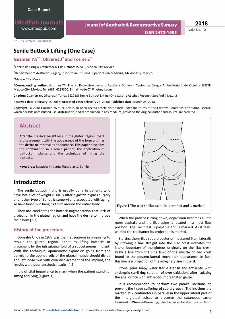
After the massive weight loss, in the gluteal region, thereis disagreement with the appearance of the form and hasthe desire to improve its appearance. This paper describesthe combination in a senile patient, the application ofbuttocks implants and the technique of lifting thebuttocks.
Keywords: Buttock; Implant; Torsoplasty; Senile
IntroductionThe senile buttock lifting is usually done in patients who
have lost a lot of weight (usually after a gastric bypass surgeryor another type of Bariatric surgery) and associated with aging,so have loose skin hanging them around the entire body.
They are candidates for buttock augmentation that lack ofprojection in the gluteal region and have the desire to improvetheir form [1-3].
History of the procedureGonzalez Ulloa in 1977 was the first surgeon in proposing to
rebuild the gluteal region, either by lifting buttocks orplacement by the infragluteal fold of a subcutaneous implant.With this technique, aponeurotic expansion going from thedermis to the aponeurotic of the gluteal muscle should divideand left loose skin with own displacement of the implant, theresults were poor aesthetic results [4,5].
It is of vital importance to mark when the patient standing,sitting and lying (Figure 1).
Figure 1 The part so iliac spine is identified and is marked.
When the patient is lying down, depression becomes a littlemore cephalic and the iliac spine is located in a most flowposition. The iliac crest is palpable and is marked. As it feels,we find the trochanter its projection is marked.
Starting thorn iliac supero posterior measured 5 cm laterallyby drawing a line straight into the iliac crest indicates thelateral boundary of the gluteus originally on the iliac crest.Draw a line from the side limit of the muscle of iliac crestbrand to the postero-lateral trochanter appearance. In fact,this line is a projection of the imaginary line in the skin.
Prone, prior soapy water sterile asepsis and antisepsis withantiseptic sterilizing solution of over-oxidation, after isolatingthe anal orifice with antiseptic-impregnated gauze.
It is recommended to perform two parallel incisions, toprevent the tissue suffering of supra groove. The incisions aremarked at 7 centimeters in parallel in the upper lateral part ofthe intergluteal sulcus to preserve the cutaneous sacralligament. When influencing; the fascia is located 3 cm. from
Case Report
iMedPub Journalswww.imedpub.com
DOI: 10.4172/2472-1905.100036
Journal of Aesthetic & Reconstructive Surgery
ISSN 2472-1905Vol.4 No.1: 2
2018
© Copyright iMedPub | This article is available from: https://aesthetic-reconstructive-surgery.imedpub.com/ 1
the infragluteal fold. After affecting the skin, it is accessedobliquely, leaving a prominence in the subcutaneous tissue.This serves as a barrier of separation and protection of thespaces between the buttocks.
The subcutaneous cellular tissue was dissected with thefibres of the majeure gluteus muscle.
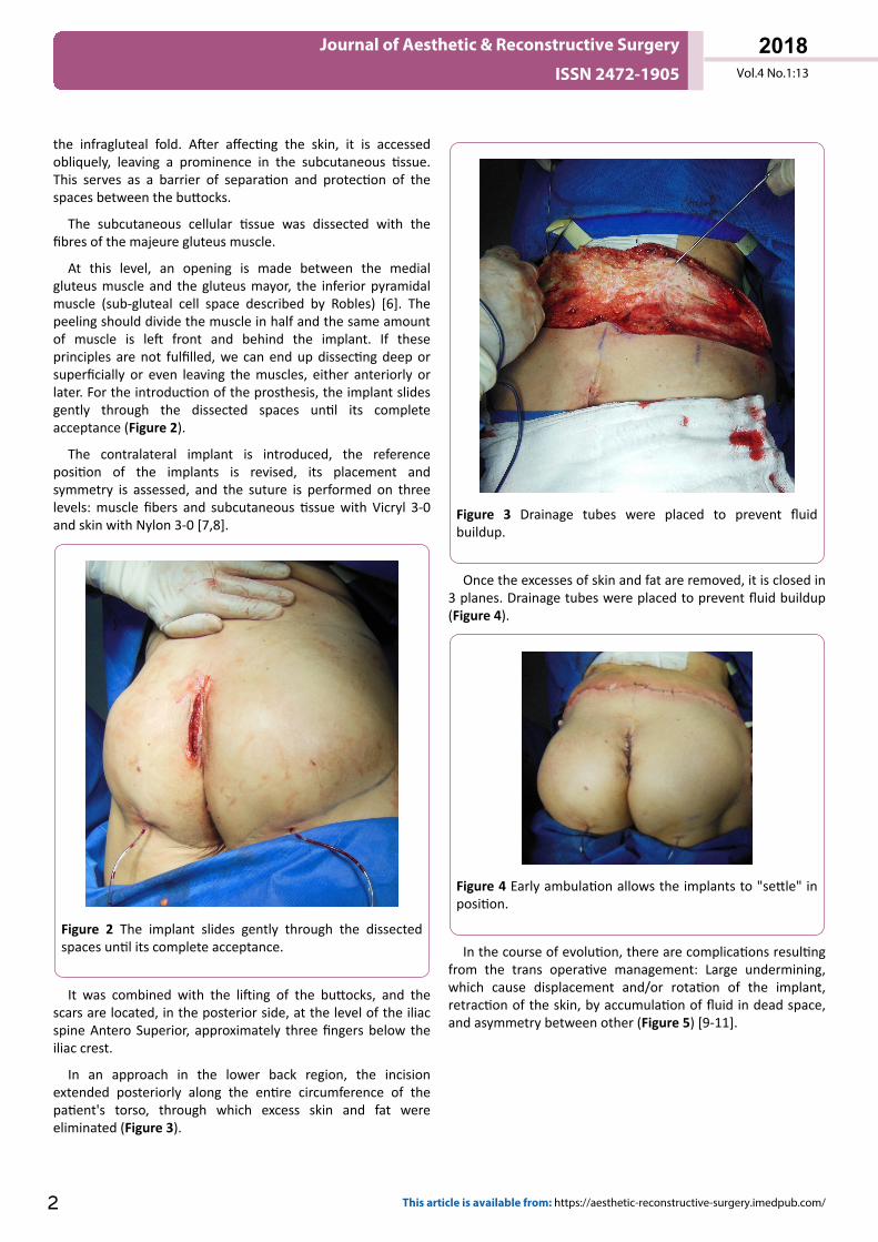
At this level, an opening is made between the medialgluteus muscle and the gluteus mayor, the inferior pyramidalmuscle (sub-gluteal cell space described by Robles) [6]. Thepeeling should divide the muscle in half and the same amountof muscle is left front and behind the implant. If theseprinciples are not fulfilled, we can end up dissecting deep orsuperficially or even leaving the muscles, either anteriorly orlater. For the introduction of the prosthesis, the implant slidesgently through the dissected spaces until its completeacceptance (Figure 2).
The contralateral implant is introduced, the referenceposition of the implants is revised, its placement andsymmetry is assessed, and the suture is performed on threelevels: muscle fibers and subcutaneous tissue with Vicryl 3-0and skin with Nylon 3-0 [7,8].
Figure 2 The implant slides gently through the dissectedspaces until its complete acceptance.
It was combined with the lifting of the buttocks, and thescars are located, in the posterior side, at the level of the iliacspine Antero Superior, approximately three fingers below theiliac crest.
In an approach in the lower back region, the incisionextended posteriorly along the entire circumference of thepatient's torso, through which excess skin and fat wereeliminated (Figure 3).
Figure 3 Drainage tubes were placed to prevent fluidbuildup.
Once the excesses of skin and fat are removed, it is closed in3 planes. Drainage tubes were placed to prevent fluid buildup(Figure 4).
Figure 4 Early ambulation allows the implants to "settle" inposition.
In the course of evolution, there are complications resultingfrom the trans operative management: Large undermining,which cause displacement and/or rotation of the implant,retraction of the skin, by accumulation of fluid in dead space,and asymmetry between other (Figure 5) [9-11].
Journal of Aesthetic & Reconstructive Surgery
ISSN 2472-1905 Vol.4 No.1:13
2018
2 This article is available from: https://aesthetic-reconstructive-surgery.imedpub.com/
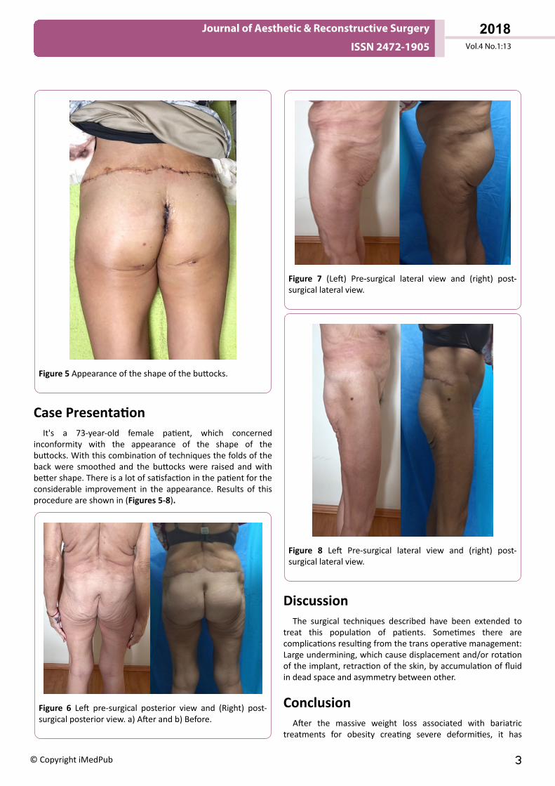
Figure 5 Appearance of the shape of the buttocks.
Case PresentationIt's a 73-year-old female patient, which concerned
inconformity with the appearance of the shape of thebuttocks. With this combination of techniques the folds of theback were smoothed and the buttocks were raised and withbetter shape. There is a lot of satisfaction in the patient for theconsiderable improvement in the appearance. Results of thisprocedure are shown in (Figures 5-8).
Figure 6 Left pre-surgical posterior view and (Right) post-surgical posterior view. a) After and b) Before.
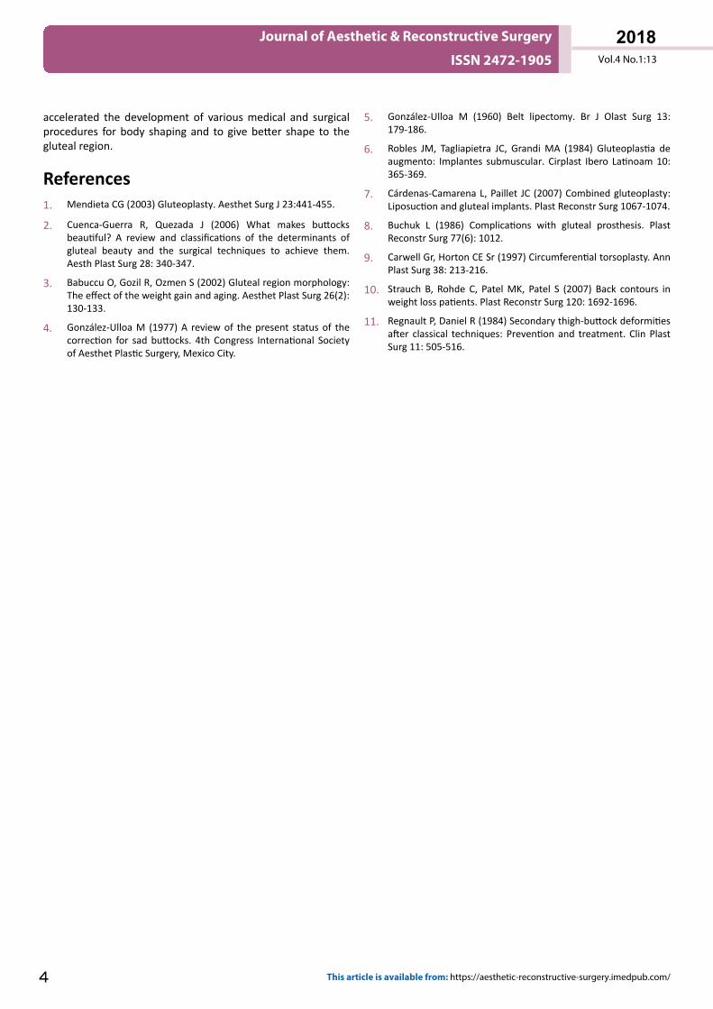
Figure 7 (Left) Pre-surgical lateral view and (right) post-surgical lateral view.
Figure 8 Left Pre-surgical lateral view and (right) post-surgical lateral view.
DiscussionThe surgical techniques described have been extended to
treat this population of patients. Sometimes there arecomplications resulting from the trans operative management:Large undermining, which cause displacement and/or rotationof the implant, retraction of the skin, by accumulation of fluidin dead space and asymmetry between other.
ConclusionAfter the massive weight loss associated with bariatric
treatments for obesity creating severe deformities, it has
Journal of Aesthetic & Reconstructive Surgery
ISSN 2472-1905 Vol.4 No.1:13
2018
© Copyright iMedPub 3
accelerated the development of various medical and surgicalprocedures for body shaping and to give better shape to thegluteal region.
References1. Mendieta CG (2003) Gluteoplasty. Aesthet Surg J 23:441-455.
2. Cuenca-Guerra R, Quezada J (2006) What makes buttocksbeautiful? A review and classifications of the determinants ofgluteal beauty and the surgical techniques to achieve them.Aesth Plast Surg 28: 340-347.
3. Babuccu O, Gozil R, Ozmen S (2002) Gluteal region morphology:The effect of the weight gain and aging. Aesthet Plast Surg 26(2):130-133.
4. González-Ulloa M (1977) A review of the present status of thecorrection for sad buttocks. 4th Congress International Societyof Aesthet Plastic Surgery, Mexico City.
5. González-Ulloa M (1960) Belt lipectomy. Br J Olast Surg 13:179-186.
6. Robles JM, Tagliapietra JC, Grandi MA (1984) Gluteoplastia deaugmento: Implantes submuscular. Cirplast Ibero Latinoam 10:365-369.
7. Cárdenas-Camarena L, Paillet JC (2007) Combined gluteoplasty:Liposuction and gluteal implants. Plast Reconstr Surg 1067-1074.
8. Buchuk L (1986) Complications with gluteal prosthesis. PlastReconstr Surg 77(6): 1012.
9. Carwell Gr, Horton CE Sr (1997) Circumferential torsoplasty. AnnPlast Surg 38: 213-216.
10. Strauch B, Rohde C, Patel MK, Patel S (2007) Back contours inweight loss patients. Plast Reconstr Surg 120: 1692-1696.
11. Regnault P, Daniel R (1984) Secondary thigh-buttock deformitiesafter classical techniques: Prevention and treatment. Clin PlastSurg 11: 505-516.
Journal of Aesthetic & Reconstructive Surgery
ISSN 2472-1905 Vol.4 No.1:13
2018
4 This article is available from: https://aesthetic-reconstructive-surgery.imedpub.com/