J-wave syndromes: Brugada and early repolarization syndromes.
Upload
sucharita-rayCategory
view
467download
0
RARE HEADACHE SYNDROMES
by
SUCHARITA RAY
MODERATOR: DR ROHIT BHATIA

SOURCES AND BIBLIOGRAPHY:
1. Pubmed
2. Handbook in Clinical Neurology Vol. 97 (3rd series); G. Nappi and M.A.
Moskowitz, Headache, ©2011 Elsevier.
3. Atlas of Migraine and other headaches. Second Edition:2005; Stephen
D Silberstein, M Alan Stiles, William B Young.
4. The Green Journal of Neurology Continuum Series focusing on
headache, 2011 and 2005 various editions
5. Neurologic Clinics of North America various editions
6. Bradley’s Neurology in Clinical Practice. Sixth Edition
7. The International Classification of Headache Disorders, 3rd edition (beta
version) available online at www.ihs.org
8. www.google.co.in and wikipedia

TREPANATION 7000-3000
BC
Atlas of Migraine and other headaches;
© 2005 Taylor & Francis

Papyrus from Thebes, Egypt (2500
BC). Now in a British museum and the
accompanying cartoon explaining the
papyrus.
Atlas of Migraine and other
headaches; © 2005 Taylor & Francis

When Should we suspect more in a headache?
• Recent de novo headache
• Unusual headache in a subject with primary
headaches
• Thunderclap headache
• Focal neurological deficits
• Impaired Consciousness /confusion
• Abnormal neurological examination
• Papilledema
• Neck stiffness
• Fever
• Raised blood pressure
• Painful and inflammatory temporal arteries

WORLD STATEMENT ON HEADACHE
Globally, the percentages of the adult population with
an active headache disorder
• 46% for headache in general
• 11% for migraine
• 42% for tension-type headache and
• 3% for chronic daily headache
Stovner Lj et al :The global burden of headache: a documentation of headache prevalence
and disability worldwide. Cephalalgia March 2007 27: 193-210

THE NEED FOR DIAGNOSIS OF RARE HEADACHE
SYNDROMES
• Hitherto uncommon headache syndromes (SUNCT, hemicrania continua,
hypnic headache, and RCVS) are nowadays widely reported .*
• When considered as a group these headaches become sizeable in
number*
• Important also to identify rare manifestations of common headache
disorders**
* Bigal ME. Expert commentary-unusual headache syndromes. Curr Pain Headache
Rep.2012;16:287-8
**Casucci G, d’Onofrio F, Torelli P. Rare primary headaches: Clinical insights. Neurol Sci.
2004;25 (Suppl. 3):S77-S83.

ICHD-3 beta Cephalalgia 2013 © International Headache society 2013/4

CLASSIFICATION
Part 1:
Primary headache disorders
Part 2:
Secondary headache disorders
Part 3:
Painful cranial neuropathies and other facial pains
ICHD-3 beta Cephalalgia 2013 © International Headache society 2013/4

CLASSIFICATION
Part 1: The primary headaches
1. Migraine
2. Tension-type headache
3. Trigeminal autonomic cephalalgias
4. Other primary headache disorders
ICHD-3 beta Cephalalgia 2013 © International Headache society 2013/4

PART 2: THE SECONDARY HEADACHES
5. Headache attributed to trauma or injury to the head and/or
neck
6. Headache attributed to cranial or cervical vascular disorder
7. Headache attributed to non-vascular intracranial disorder
8. Headache attributed to a substance or its withdrawal
9. Headache attributed to infection
10. Headache attributed to disorder of homoeostasis
ICHD-3 beta Cephalalgia 2013 © International Headache society 2013/4

Part 3: Painful cranial neuropathies, other facial
pains and other headaches
13. Painful cranial neuropathies and other facial
pains
14. Other headache disorders
ICHD-3 beta Cephalalgia 2013 © International Headache society 2013/4

1. MIGRAINE
1.1 Migraine without aura
1.2 Migraine with aura
1.3 Chronic migraine
1.4 Complications of migraine
1.5 Probable migraine
1.6 Episodic syndromes that may be associated with
migraine
ICHD-3 beta Cephalalgia 2013 © International Headache society 2013/4

1.4 COMPLICATIONS OF MIGRAINE
1.4.1 Status migrainosus
1.4.2 Persistent aura without infarction
1.4.3 Migrainous infarction
1.4.4 Migraine aura-triggered seizure
ICHD-3 beta Cephalalgia 2013 © International Headache society 2013/4
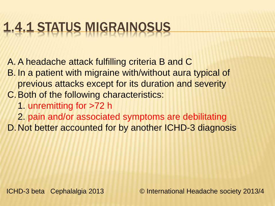
1.4.1 STATUS MIGRAINOSUS
A. A headache attack fulfilling criteria B and C
B. In a patient with migraine with/without aura typical of
previous attacks except for its duration and severity
C.Both of the following characteristics:
1. unremitting for >72 h
2. pain and/or associated symptoms are debilitating
D.Not better accounted for by another ICHD-3 diagnosis
ICHD-3 beta Cephalalgia 2013 © International Headache society 2013/4

Marion Beltramone, Anne Donnet, Cephalalgia;
December 2013,(0)0, 1–5

Background: About the prevalence of Status migrainosus
(SM) and migraine aura status (MAS) in patients with migraine
in one tertiary care center
Methods: 11-year retrospective study
Duration: October 2002 through June 2013, in 8821 patients
enrolled in the participating in the French Observatory of
Migraine and Headaches (OMH)
Five migraine complications were considered:
1. Status migrainosus (>72 hours)
2. Persistent aura without infarction
3. Migrainous infarction
4. Migraine aura-triggered seizure, and
5. Migraine aura status (MAS) (2 auras/day over at least three
consecutive days in the MA patient)

RESULTS FROM THE STUDY:
Among 8821 patients studied:
24 had SM, three had MAS and one had both forms. (<3%,
SM>MAS)
Mean duration: SM---4.8 weeks and MAS---4 weeks.
Stress and menstruation were the main precipitating factors for SM
(68.8% and 31.3%, respectively).
No precipitating factor was found for MAS.
For a majority of patients, the frequency of migraine attack was the
same before and after SM or MAS.
SM and MAS occurred more frequently in patients with initial low-
frequency migraine attacks.
Conclusions: SM and MAS are rare complications of migraine
Similar frequency of migraine attacks occurs before and after SM.

1.2.2 MIGRAINE WITH BRAINSTEM AURA
A. At least 2 attacks fulfilling criteria B and C below, and
criteria C and D for 1.2.1 Migraine with typical aura
B. Aura of fully reversible visual, sensory and/or
speech/language symptoms, but not motor or retinal
C. 2 of the following brainstem symptoms:
1. dysarthria
2. vertigo
3. tinnitus
4. hypacusis
5. diplopia
6. ataxia
7. decreased level of consciousness
ICHD-3 beta Cephalalgia 2013 © International Headache society
2013/4

1.2.2 Migraine with brainstem aura
Terminology and coding changes 1988-2013
1.2.2 Migraine with brainstem aura was previously classified
as
1.2.6 Basilar-type migraine in ICHD-II
1.2.4 Basilar migraine in ICHD-I
There is little evidence that the basilar artery or, basilar-artery
territory is involved.
The name change was done to preclude the involvement of
basilar artery in the pathogenesis of the headache.
ICHD-3 beta Cephalalgia 2013 © International Headache society 2013/4

The differential diagnosis of migraine with brainstem aura:
• Hemiplegic migraine:
• Presence of unilateral weakness/paralysis
• Transient ischemic attack (TIA)/stroke
• Think if similar picture occurs in adults
• Symptoms begin simultaneously ( sequential in
migraine)
• CADASIL (CADASIL-Coma):
• High burden of T2/FLAIR hyperintensity in the
periventricular and deep WM and particularly in the
anterior temporal pole and external capsule
*Basilar-type migraine typically decreaes in frequency and severity
in adulthood and may disappear, (like hemiplegic migraine) by age
40. Often the attacks give way to typical migraine with/without aura.

HEMIPLEGIC MIGRAINE
A. At least 2 attacks fulfilling criteria B and C
B. Aura consisting of both of the following:1. fully reversible motor weakness; 2. fully reversible visual, sensory and/or speech/language symptoms
C. 2 of the following 4 characteristics:1. 1 aura symptom spreads gradually over ≥5 min,
and/or 2 symptoms occur in succession2. each individual non-motor aura symptom lasts 5-60min, and motor symptoms last <72 h3. 1 aura symptom is unilateral4. aura accompanied or followed in <60 min by headache
D. Not better accounted for by another ICHD-3 diagnosis, and TIA excluded
ICHD-3 beta Cephalalgia 2013 © International Headache society 2013/4

FAMILIAL HEMIPLEGIC MIGRAINE
Initially described in 1910
Recurrent headache associated with transient hemiparesis.
AD
Those with family history ( FHM-AD) and without i.e. Sporadic
Hemiplegic migraine
For the familial form:
FHM1: Chr 19p13; CACNA1A. Also seen with Episodic ataxia
type 2, spinocerebellar ataxia type 6, and benign paroxysmal
torticollis of infancy
FHM2: Chr 1q23; ATP1A2 gene which codes for a Na+K+
ATPase pump.
FHM3: Chr 2q24; SCN1A gene coding for a voltage gated
sodium channel

Rohit Bhatia, Soaham Desai, Manjari Tripathi, Ajay Garg ,
M. V. Padma, Kameshwar Prasad, Mamta B. Singh

Case scenario:
30-year-old male with headache and left hemiparesis with
visual aura and systemic signs preceding the headache.
There was no history of fever, trauma, seizure, or
dehydration preceding the episode.
In the preceding 16 years, he had about 50 such episodes,
age of onset at 14 years.
No family history.

Possible differential diagnosis of recurrent hemiplegia
1> Transient ischemic attacks and recurrent strokes
2> Complex partial seizure with and without secondary generalization
(with post-ictal Todd’s palsy)
3> One of the migraine with aura subtypes:
(a) Hemiplegic migraine.(sporadic or familial)
(b) Migraine with brainstem aura (Basilar-type migraine)
(c) Migrainous infarction (complication of migraine)
4> Mitochondrial encephalopathy with lactic acidosis and stroke-like
episodes (MELAS) syndrome
5 >Pseudomigraine with lymphocytic pleocytosis with transient
neurologic deficits
6> Alternating hemiplegia of childhood

(a) T1-weighted axial MRI brain, reveals right cerebral hemispheric
sulcal effacement and cortical thickening which is hyperintense on
T2WI and (b) FLAIR sequence

Followup MR at 4 weeks shows resolution of changes

SPORADIC HEMIPLEGIC MIGRAINE
•Most patients with SHM have ‘typical’ aura symptoms (visual, sensory
and/or aphasic) associated with motor weakness during the acute
attack.
•The auras are more prolonged in sporadic hemiplegic migraine as
compared to a typical migraine with aura.
•Absence of infarction on imaging helps in differentiating it from
migrainous infarction or stroke associated with migraine.
Whitty CWM (1953) Familial hemiplegic migraine. J NNeurosurg Psychiatry 16:172-77
Thomsen LL, Olesen J (2004) Sporadic hemiplegic migraine. Cephalalgia 24:1016–1023
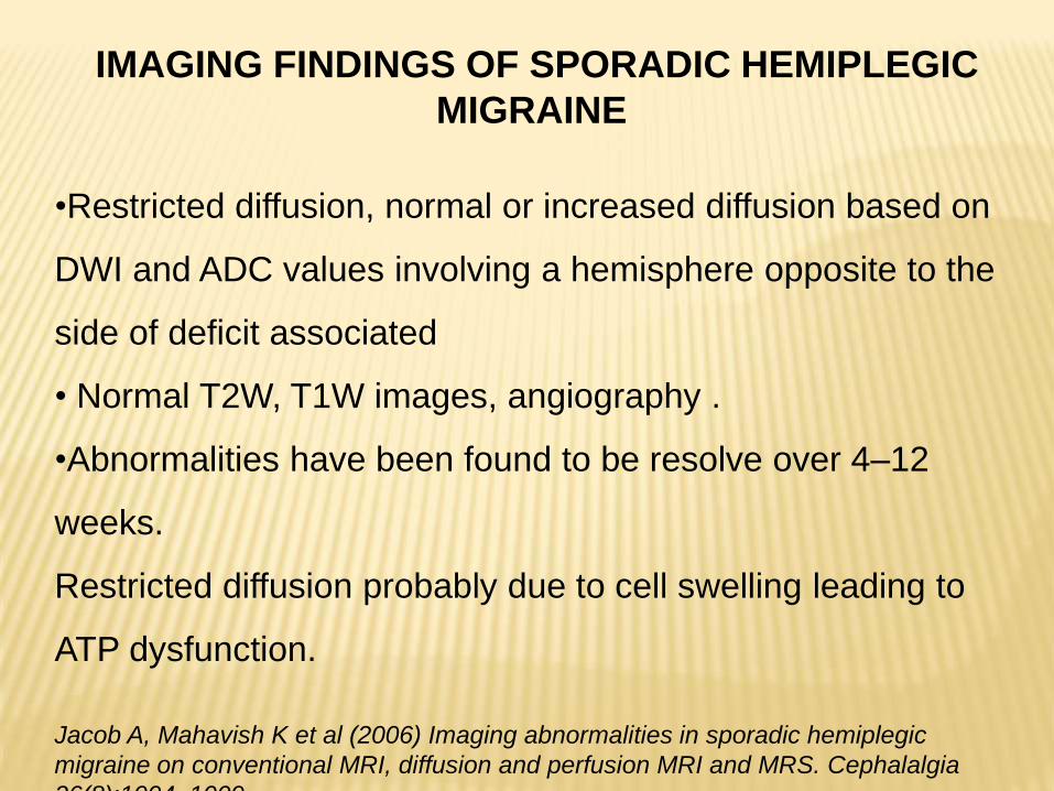
IMAGING FINDINGS OF SPORADIC HEMIPLEGIC
MIGRAINE
•Restricted diffusion, normal or increased diffusion based on
DWI and ADC values involving a hemisphere opposite to the
side of deficit associated
• Normal T2W, T1W images, angiography .
•Abnormalities have been found to be resolve over 4–12
weeks.
Restricted diffusion probably due to cell swelling leading to
ATP dysfunction.
Jacob A, Mahavish K et al (2006) Imaging abnormalities in sporadic hemiplegic
migraine on conventional MRI, diffusion and perfusion MRI and MRS. Cephalalgia
26(8):1004–1009.

MANAGEMENT OF SPORADIC HEMIPLEGIC MIGRAINE
•Treatment strategy : Flunarizine, Naloxone and Verapamil
•Sympathomimetic drugs like ergotamine derivatives and triptans
are avoided in acute attacks (vasospasm may lead to permanent
sequelae)
•Beta-blockers are not used for prophylaxis in patients with
hemiplegic or basilar-type migraine: risk of prolonged aura
/migrainous infarction.
• SHM is a diagnosis of exclusion.
Centonze V, Brucoli C, Macinagrossa G, Attolini E, Campanozzi F, Albano O (1983) Non-
familial hemiplegic migraine responsive to naloxone. Cephalalgia 3:125–127
Yu W, Horowitz SH (2003) Treatment of sporadic hemiplegic migraine with calcium-channel
blocker verapamil. Neurology 60:120–121
Tobita M, Hino M, Ichikawa N, Takase S, Ogawa A (1987) A case of hemiplegic migraine

Pseudomigraine with lymphocytic pleocytosis and
transient neurologic deficits*
Very rare condition with single/multiple attacks of migraine like
headaches, weakness, sensory and visual auras with confusion
and sometimes meningismus lasting minutes to hours.
CSF lymphocytic pleocytosis (10–760 cells/mm3); Normal protein
and sugar and have a normal brain imaging. No infectious etiology
Also known as Headache and Neurological Deficits with
cerebrospinal fluid (CSF) Lymphocytosis (HaNDL) syndrome
Visual auras are very uncommon (12%) as compared to sensory
symptoms (78%). This helps it to differentiate from migraine with
brainstem aura.
* Filina T, Feja K N, Tolan R W: An Adolescent With Pseudomigraine, Transient Headache, Neurological
Deficits, and Lymphocytic Pleocytosis (HaNDL Syndrome). Clinical Pediatrics: 2013 Jun;52(6):496-502.

Cephalalgia; 2010; 30(11) 1284–1289:

Cerebral autosomal dominant arteriopathy with
subcortical infarcts and leukoencephalopathy
(CADASIL)
NOTCH3 gene ( ? Migraine susceptibility gene)Recurrent stroke, cognitive decline, psychiatric disturbances and
migraine.
The prevalence of migraine in CADASIL is slightly higher than in the
general population, The proportion of migraine with aura is much
higher
• increased susceptibility to cortical spreading depression (CSD)
• different expression of CSD
Total of 27 articles on migraine in CADASIL
Prevalence of headache: European studies: 14% to 72%.
Four studies from Asian countries: low migraine prevalence of 5%.
CADASIL and migraine: A narrative review. Liem MK, Oberstein SA, van der grond J, Ferrari

Cerebral autosomal dominant arteriopathy with subcortical
infarcts and leukoencephalopathy (CADASIL)
Pooling of all results: Overall migraine prevalence of migraine is 38%
(151/510) with a 95% CI of 33–42%.
When excluding Asian countries, the pooled prevalence is 43%
(187/434) with a 95% CI of 38–48%.
Cause for low incidence of Migraine in CADASIL patients:
? R544C mutation associated with less headache in Asian patients*
*Headache among CADASIL patients withR544C mutation: Prevalence, characteristics, and
associations. CC Jay et al. Cephalalgia January 2014 vol. 34 no. 1 22-28

3. Trigeminal autonomic cephalalgias (TACs)
3.1 Cluster headache
3.2 Paroxysmal hemicrania
3.3 Short-lasting unilateral neuralgiform headache attacks
3.4 Hemicrania continua
3.5 Probable trigeminal autonomic cephalalgia
ICHD-3 beta Cephalalgia 2013 © International Headache society 2013/4

3.2 Paroxysmal hemicrania
A. At least 20 attacks fulfilling criteria B-E
B. Severe unilateral orbital, supraorbital and/or temporal pain
lasting 2-30 min
C.1 of the following ipsilateral symptoms or signs:
[1.conjunctival injection and/or lacrimation, 2.nasal congestion and/or
rhinorrhoea, 3. eyelid oedema,4.forehead and facial sweating, 5.
forehead and facial flushing, 6.sensation of fullness in the ear, 7.
miosis &/or ptosis]
D. Frequency >5/d for > half the time
E. Prevented absolutely by therapeutic doses of indomethacin
F. Not better accounted for by another ICHD-3 diagnosis
ICHD-3 beta Cephalalgia 2013 © International Headache society 2013/4

3.3 Short-lasting unilateral neuralgiform headache
A. At least 20 attacks fulfilling criteria B-D
B. Moderate/severe unilateral head pain (orbital, supraorbital, temporal
and/or other trigeminal distribution) lasting 1-600 seconds as single
stabs, series of stabs or in a saw-tooth pattern
C. 1 of the following ipsilateral cranial autonomic symptoms or signs ( as in
PH)
D. Frequency 1/d for > half the time when active
E. Not better accounted for by another ICHD-3 diagnosis
First described by Sjaastad et al; Slight higher prevalence in males*
* Sjaastad O, Russell D, Horven I, Bunaes U. Multiple neuralgiform unilateral headache attacks associated with conjunctivalinjection and appearing in clusters. A nosological problem. Proceedings of the Scandinavian Migraine Society 1978; 31

3.3 SHORT-LASTING UNILATERAL NEURALGIFORM HEADACHE
ATTACKS
Two subtypes recognized:
3.3.1 Short-lasting unilateral neuralgiform headache attacks
with conjunctival injection and tearing (SUNCT)
3.3.2 Short lasting unilateral neuralgiform headache attacks
with cranial autonomic symptoms (SUNA)
3.3.1 SUNCT may be a subform of 3.3.2 SUNA
Mean age of onset 50 years (10 to 77 years)
ICHD-3 beta Cephalalgia 2013 © International Headache society 2013/4

•To aid diagnosis of SUNCT/SUNA an indomethacin test and
oxygen trial, which will rule out PH and CH, respectively.
•Intravenous lidocaine is effective for short-term prevention
• Lamotrigine and topiramate are recommended as preventives,
and gabapentin may have a beneficial effect in SUNA.
•Greater occipital nerve injections may play a role.
SUNCT and SUNA: Recognition and Treatment. JA Pareja et al. Current Treatment Options
in Neurology (2013) 15:28–39
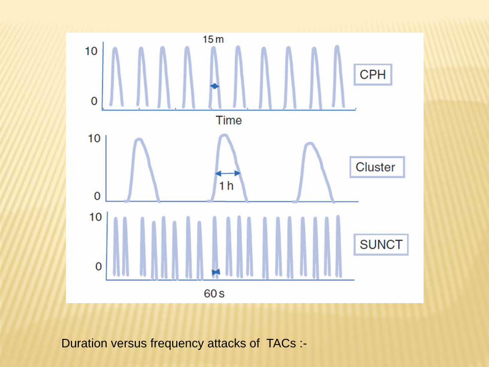
Duration versus frequency attacks of TACs :-

3.4 HEMICRANIA CONTINUA
A. Unilateral headache fulfilling criteria B-D
B. Present >3 mo, with exacerbations of moderate or greater
intensity
C.Either or both of the following:
1. 1 of the following ipsilateral symptoms or signs as in
PH
2. a sense of restlessness or agitation, or aggravation of
pain by movement
D. Responds absolutely to therapeutic doses of
indomethacin
E. Not better accounted for by another ICHD-3 diagnosis
ICHD-3 beta Cephalalgia 2013 © International Headache society 2013/4

4. OTHER PRIMARY HEADACHE DISORDERS
4.1 Primary cough headache
4.2 Primary exercise headache
4.3 Primary headache associated with sexual activity
4.4 Primary thunderclap headache
4.5 Cold-stimulus headache
4.6 External pressure headache
4.7 Primary stabbing headache
4.8 Nummular headache
4.9 Hypnic headache
4.10New daily persistent headache (NDPH)
ICHD-3 beta Cephalalgia 2013 © International Headache society 2013/4

PART 2: THE SECONDARY HEADACHES
5. Headache attributed to trauma or injury to the head and/or neck
6. Headache attributed to cranial or cervical vascular disorder
7. Headache attributed to non-vascular intracranial disorder8. Headache attributed to a substance or its withdrawal9. Headache attributed to infection10. Headache attributed to disorder of homoeostasis 11. Headache or facial pain attributed to disorder of cranium,
neck, eyes, ears, nose, sinuses, teeth, mouth or other facial or cranial structure
12. Headache attributed to psychiatric disorder
ICHD-3 beta Cephalalgia 2013 © International Headache society 2013/4

6.7 Headache due to other acute intracranial arterial
disorder
6.7.1 Headache attributed to an intracranial endovascular
procedure
6.7.2 Angiography headache
6.7.3 Headache attributed to reversible cerebral
vasoconstriction syndrome (RCVS)
6.7.4 Headache attributed to intracranial arterial
dissection
ICHD-3 beta Cephalalgia 2013 © International Headache society 2013/4

6.7.3 Headache attributed to RCVS
A. Any new headache fulfilling criterion C
B. Reversible cerebral vasoconstriction syndrome (RCVS)
diagnosed
C.Evidence of causation demonstrated by ≥1 of:
1. headache, with or without focal deficits and/or seizures,
has led to angiography and diagnosis of RCVS
2. headache has ≥1 of the following characteristics:
a) recurrent during ≤1 mo, and with thunderclap onset
b) triggered by sexual activity, exertion, Valsalva, emotion,
bathing and/or showering
3. No new significant headache occurs >1 m after onset
D.Not better accounted for by another ICHD-3 diagnosis, and
aneurysmal SAH excluded
ICHD-3 beta Cephalalgia 2013 © International Headache society 2013/4
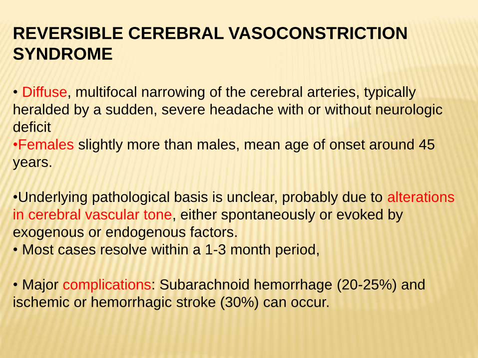
REVERSIBLE CEREBRAL VASOCONSTRICTION
SYNDROME
• Diffuse, multifocal narrowing of the cerebral arteries, typically
heralded by a sudden, severe headache with or without neurologic
deficit
•Females slightly more than males, mean age of onset around 45
years.
•Underlying pathological basis is unclear, probably due to alterations
in cerebral vascular tone, either spontaneously or evoked by
exogenous or endogenous factors.
• Most cases resolve within a 1-3 month period,
• Major complications: Subarachnoid hemorrhage (20-25%) and
ischemic or hemorrhagic stroke (30%) can occur.

Eponyms for reversible cerebral vasoconstriction
syndrome
Isolated benign cerebral vasculitis
Benign acute cerebral angiopathy
Call–Fleming syndrome
Central nervous system pseudovasculitis
Benign angiopathy of the central nervous system (BACNS)
Post-partum angiopathy
Migrainous vasospasm
Fatal vasospasm in migrainous infarction
Migraine angiitis
Thunderclap headache with reversible vasospasm
Idiopathic thunderclap headache
Primary thunderclap headache
Drug-induced cerebral vasculopathy/angiopathy
Reversible Cerebral Vasoconstriction Syndrome. Holly Yancy et al, Headache Currents; 2013;
570-7
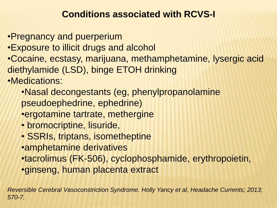
Conditions associated with RCVS-I
•Pregnancy and puerperium
•Exposure to illicit drugs and alcohol
•Cocaine, ecstasy, marijuana, methamphetamine, lysergic acid
diethylamide (LSD), binge ETOH drinking
•Medications:
•Nasal decongestants (eg, phenylpropanolamine
pseudoephedrine, ephedrine)
•ergotamine tartrate, methergine
• bromocriptine, lisuride,
• SSRIs, triptans, isometheptine
•amphetamine derivatives
•tacrolimus (FK-506), cyclophosphamide, erythropoietin,
•ginseng, human placenta extract
Reversible Cerebral Vasoconstriction Syndrome. Holly Yancy et al, Headache Currents; 2013;
570-7.

Conditions associated with RCVS-II
•Exposure to blood products
•Intravenous immune globulin, red blood cell transfusion
•Idiopathic
•Catecholamine secreting tumors:
•Pheochromocytoma, bronchial carcinoid tumor, glomus
tumors
•Vascular disorders
• Unruptured cerebral aneurysm, post-carotid
endarterectomy, cervical dissection, dysplasia
•Miscellaneous:
•Hypercalcemia, porphyria
•Neurosurgical and neurointerventional procedures
•Head trauma
CLINICAL FEATURES OF REVERSIBLE CEREBRAL
VASOCONSTRICTION SYNDROME
•Recurrent thunderclap headaches over several days to weeks are
highly characteristic of RCVS.
•Women aged 20 to 50 and slightly older age than males who present
in third decade.
•Complications: focal or generalized seizures in up to 20%.
•Permanent or transient focal neurologic deficits may represent
transient ischemic attack, infarct, or intracranial hemorrhage
•The vascular abnormalities of RCVS are reversible and this defines
the diagnosis.
• Vasoconstriction typically outlasts the headaches by several weeks
Yancy, H., Lee-Iannotti, J. K., Schwedt, T. J. and Dodick, D. W. (2013), Reversible Cerebral
vasoconstriction Syndrome. Headache: The Journal of Head and Face Pain, 53: 570–576.
doi: 10.1111/head.12040

Cerebral angiography is the gold standard
Characteristic “string of beads” sign
MRA or CTA is recommended to display the diffuse
alternating pattern of vasodilation and vasoconstriction.
Fig 1 and 2: Diagnostic angiogram with multiple areas of vasoconstriction in anterior
circulation.
Yancy, H., Lee-Iannotti, J. K., Schwedt, T. J. and Dodick, D. W. (2013), Reversible Cerebral
vasoconstriction Syndrome. Headache: The Journal of Head and Face Pain, 53: 570–576.
doi: 10.1111/head.12040

7. HEADACHE DUE TO NON-VASCULAR INTRACRANIAL
DISORDER
7.1 Headache attributed to increased cerebrospinal fluid
pressure
7.2 Headache attributed to low cerebrospinal fluid pressure
7.3 Headache attributed to non-infectious inflammatory
intracranial disease
7.4 Headache attributed to intracranial neoplasia
7.5 Headache attributed to intrathecal injection
7.6 Headache attributed to epileptic seizure
7.7 Headache attributed to Chiari malformation type I
7.8 Headache attributed to other non-vascular intracranial
disorder
ICHD-3 beta Cephalalgia 2013 © International Headache society 2013/4

7.2 HEADACHE ATTRIBUTED TO LOW CEREBROSPINAL
FLUID PRESSURE
7.2.1 Post-dural puncture headache
7.2.2 CSF fistula headache
7.2.3 Headache attributed to spontaneous intracranial
hypotension
ICHD-3 beta Cephalalgia 2013 © International Headache society 2013/4
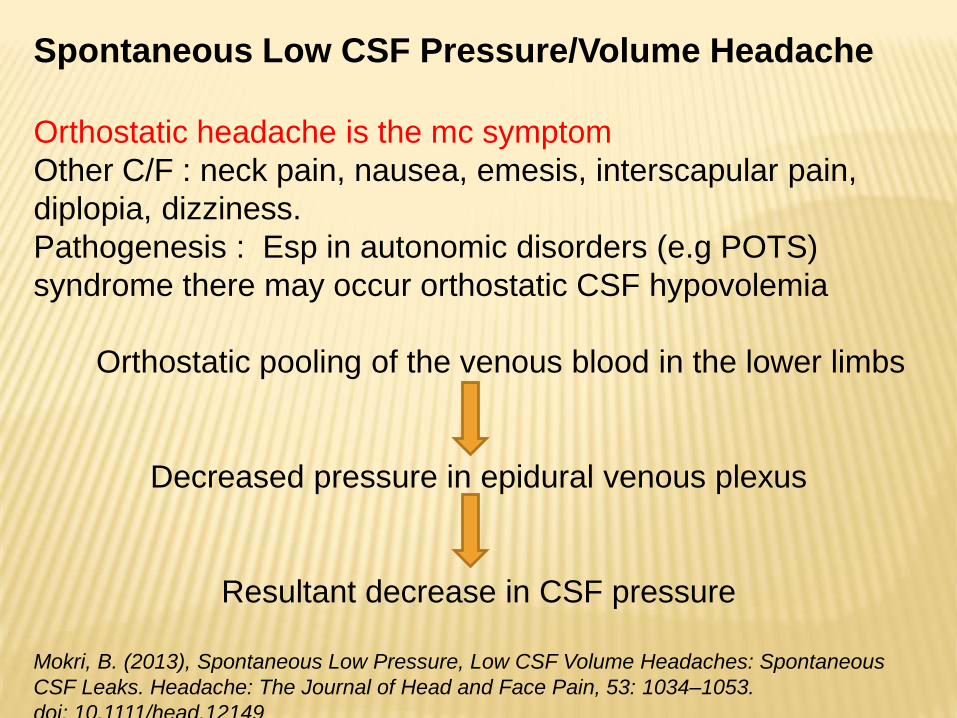
Spontaneous Low CSF Pressure/Volume Headache
Orthostatic headache is the mc symptom
Other C/F : neck pain, nausea, emesis, interscapular pain,
diplopia, dizziness.
Pathogenesis : Esp in autonomic disorders (e.g POTS)
syndrome there may occur orthostatic CSF hypovolemia
Orthostatic pooling of the venous blood in the lower limbs
Decreased pressure in epidural venous plexus
Resultant decrease in CSF pressure
Mokri, B. (2013), Spontaneous Low Pressure, Low CSF Volume Headaches: Spontaneous
CSF Leaks. Headache: The Journal of Head and Face Pain, 53: 1034–1053.
doi: 10.1111/head.12149
Cerebrospinal fluid analyses:
Opening pressure: Low/Negative/normal
Color: Usually transparent, occasionally xanthochromic
Cells: maybe present, upto 50cells/cumm is common
NCCT Head
Limited utility.
Look for subdural fluid collection and/or tentorial herniation
Radioisotope Cisternography
Very useful for detecting csf leaks
M/c abnormality: Absence/paucity of radioactivity over the
cerebral convexities at 24-48 hrs
Site of csf leak: Zone of parathecal activity
Meningeal diverticula: Multiple zones of parathecal activity
Early activity in kidneys and urinnary bladder s/o extradural
csf

Magnetic resonance imaging
Meningeal enhancement: Pachymeningeal (not leptomeningeal)
Supratentorial as well as infratentorial
Linear & non-nodular; uninterrupted & bilateral, usually thick but
maybe subtle and thin.
Ferrante, E., Savino, A., Sances, G. and Nappi, G. (2004), Spontaneous Intracranial
Hypotension Syndrome: Report of Twelve Cases. Headache: The Journal of Head and Face
Pain, 44: 615–622. doi: 10.1111/j.1526-4610.2004.446012.x

•Descent of brain (sagging or sinking of the brain): Descent of
cerebellar tonsils ( like in type I Chiari malformation)
•Decrease in size of prepontine cistern
• Inferior displacement of the optic chiasm
• Effacement of perichiasmatic cisterns
• Crowding of the posterior fossa

Bilateral subdural collection with pachymeningeal thickening
and epidural thickening
Ferrante, E., Savino, A., Sances, G. and Nappi, G. (2004), Spontaneous Intracranial
Hypotension Syndrome: Report of Twelve Cases. Headache: The Journal of Head and Face
Pain, 44: 615–622. doi: 10.1111/j.1526-4610.2004.446012.x

Part 3: Painful cranial neuropathies, other
facial pains and other headaches
13.Painful cranial neuropathies and other facial pains
14.Other headache disorders
ICHD-3 beta Cephalalgia 2013 © International Headache society 2013/4

13. Painful cranial neuropathies and other facial pains
13.1 Trigeminal neuralgia
13.2 Glossopharyngeal neuralgia
13.3 Nervus intermedius (facial nerve) neuralgia
13.4 Occipital neuralgia
13.5 Optic neuritis
13.6 Headache attributed to ischaemic CN III palsy
13.7 Tolosa-Hunt syndrome
13.8 Paratrigeminal oculosympathetic (Raeder’s) syndrome
13.9 Recurrent painful ophthalmoplegic neuropathy
13.10 Burning mouth syndrome (BMS)
13.11 Persistent idiopathic facial pain (PIFP)
13.12 Central neuropathic pain
ICHD-3 beta Cephalalgia 2013 © International Headache society 2013/4

Glossopharyngeal neuralgia
Severe transient stabbing pain in the ear, base of the tongue, tonsillar
fossa, or beneath the angle of the jaw.
Younger patients than TN (40% of patients are under 50 years).**
Female (67%) > Male (33%) patients**
Occasionally vascular compression of the glossopharyngeal nerve by the
posterior inferior cerebellar artery at the root entry zone.
Other causes of Glossopharyngeal Neuralgia:1. Elongated or fractured styloid process
2. Calcified stylohyoid ligament (Eagle’s syndrome)
3. Cerebellopontine angle tumors
4. Parapharyngeal space lesions
5. Carcinoma of the parapharyngeal space, pharynx, nasopharyngeal
carcinoma, posterior fossa arteriovenous malformation
6. Multiple sclerosis
**Patel A, Kassam A, Horowitz M et al. (2002). Microvascular decompression in the
management of glossopharyngeal neuralgia: analysis of 217 cases.Neurosurgery;705-720

CLINICAL FEATURES
Hyperactivity of the CN IX afferents activation of the dorsal motor
nucleus of the vagus nerve vagal efferent response severe
bradycardia /asystole.
•Major difference is in location of pain
•Paroxysmal and lasts for seconds to minutes, with remission.
• Provoked by swallowing sharp foods, talking, or coughing
• Examination is unremarkable.
•The symptomatic forms are often secondary to intra- or extracranial
compressions near the jugular foramen.
•MRI or a panoramic radiograph is useful to exclude Eagle’s
syndrome. An electrocardiogram may be useful to rule out cardiac
abnormalities.

Patel A, Kassam A, Horowitz M et al. (2002). Microvascular decompression in the
management of glossopharyngeal neuralgia: analysis of 217 cases. Neurosurgery
50: 705–710.
Sampson JH, Grossi PM, Asaoka K et al. (2004). Microvascular decompression for
glossopharyngeal neuralgia: long-term effectiveness and complication avoidance.
Neurosurgery 54: 884–889.
MANAGEMENT
The first-line medical treatment: CBZ and all other treatment for
Trigeminal Neuralgia.
Other drugs like Baclofen, Gabapentin and pregabalin have been
seen to be effective in selected cases.
Important to rule out secondary causes.
Surgical treatment : Nerve sectioning to Microvascular
decompression .
NERVUS INTERMEDIUS NEURALGIA
•A rare condition characterized by brief paroxysms of
pain felt deeply in the ear.
•Pain paroxysms are intermittent, last for seconds or
minutes
• Triggered by touching the posterior wall of the auditory
canal.
•Pain is sometimes accompanied by disorders of
lacrimation, salivation, or taste.
•There is a common association with herpes zoster (HZ).

OPHTHALMIC POSTHERPETIC NEURALGIA
Localized infection caused by the VZV.
The main complication of HZ is postherpetic neuralgia (PHN), a
neuropathic pain persisting more than 3 months after skin eruption.
10% of those with HZ will develop PHN
Higher frequency in elderly and diabetics.
Thoracic dermatomal involvement : 50%
Ophthalmic division of the CN V: 22–25%
Pain usually constant or paroxysmal and dynamic allodynia is seen.
Local examination: Scarring and skin changes.
Both large and small sized trigeminal afferents are affected
Pain can be paroxysmal (demyelination of Aβ fibers) and constant
pain(Aδ and C axons)
Truini A, Galeotti F, Haanpaa M et al. Pathophysiology of pain in postherpetic
neuralgia: a clinical and neurophysiological study. 2008; Pain 140: 405–410.

Because of the eye involvement medical management is the
first line of treatment rather than using topical agents.
Recent EFNS guidelines recommend as first-line treatment
with the tricyclic antidepressants, gabapentin/pregabalin, and
topical lidocaine
Attal N, Cruccu G, Haanpaa M et al. (2006). EFNS guidelines on pharmacological
treatment of neuropathic pain. Eur J Neurol 13: 1153–1169.